su2029 medium conditioned with conventionally-activated m1 macrophages inhibits survival of mouse...
TRANSCRIPT
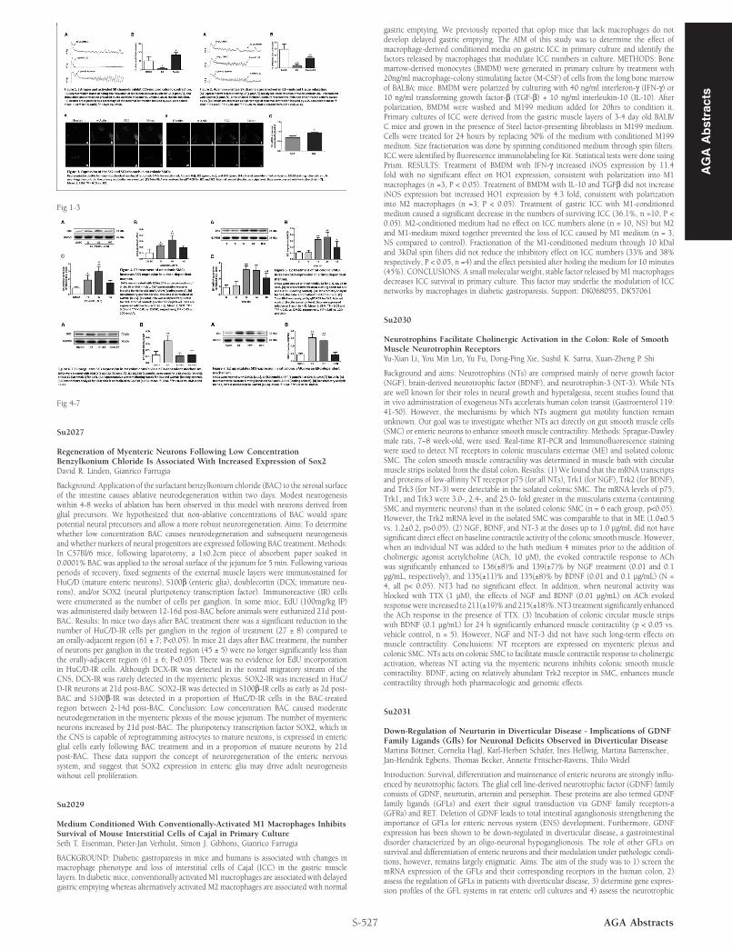
Fig 1-3
Fig 4-7
Su2027
Regeneration of Myenteric Neurons Following Low ConcentrationBenzylkonium Chloride Is Associated With Increased Expression of Sox2David R. Linden, Gianrico Farrugia
Background: Application of the surfactant benzylkonium chloride (BAC) to the serosal surfaceof the intestine causes ablative neurodegeneration within two days. Modest neurogenesiswithin 4-8 weeks of ablation has been observed in this model with neurons derived fromglial precursors. We hypothesized that non-ablative concentrations of BAC would sparepotential neural precursors and allow a more robust neuroregeneration. Aims: To determinewhether low concentration BAC causes neurodegeneration and subsequent neurogenesisand whether markers of neural progenitors are expressed following BAC treatment. Methods:In C57Bl/6 mice, following laparotomy, a 1x0.2cm piece of absorbent paper soaked in0.0001% BAC was applied to the serosal surface of the jejunum for 5 min. Following variousperiods of recovery, fixed segments of the external muscle layers were immunostained forHuC/D (mature enteric neurons), S100β (enteric glia), doublecortin (DCX; immature neu-rons), and/or SOX2 (neural pluripotency transcription factor). Immunoreactive (IR) cellswere enumerated as the number of cells per ganglion. In some mice, EdU (100mg/kg IP)was administered daily between 12-16d post-BAC before animals were euthanized 21d post-BAC. Results: In mice two days after BAC treatment there was a significant reduction in thenumber of HuC/D-IR cells per ganglion in the region of treatment (27 ± 8) compared toan orally-adjacent region (61 ± 7; P<0.05). In mice 21 days after BAC treatment, the numberof neurons per ganglion in the treated region (45 ± 5) were no longer significantly less thanthe orally-adjacent region (61 ± 6; P<0.05). There was no evidence for EdU incorporationin HuC/D-IR cells. Although DCX-IR was detected in the rostral migratory stream of theCNS, DCX-IR was rarely detected in the myenteric plexus. SOX2-IR was increased in HuC/D-IR neurons at 21d post-BAC. SOX2-IR was detected in S100β-IR cells as early as 2d post-BAC and S100β-IR was detected in a proportion of HuC/D-IR cells in the BAC-treatedregion between 2-14d post-BAC. Conclusion: Low concentration BAC caused moderateneurodegeneration in the myenteric plexus of the mouse jejunum. The number of myentericneurons increased by 21d post-BAC. The pluripotency transcription factor SOX2, which inthe CNS is capable of reprogramming astrocytes to mature neurons, is expressed in entericglial cells early following BAC treatment and in a proportion of mature neurons by 21dpost-BAC. These data support the concept of neuroregeneration of the enteric nervoussystem, and suggest that SOX2 expression in enteric glia may drive adult neurogenesiswithout cell proliferation.
Su2029
Medium Conditioned With Conventionally-Activated M1 Macrophages InhibitsSurvival of Mouse Interstitial Cells of Cajal in Primary CultureSeth T. Eisenman, Pieter-Jan Verhulst, Simon J. Gibbons, Gianrico Farrugia
BACKGROUND: Diabetic gastroparesis in mice and humans is associated with changes inmacrophage phenotype and loss of interstitial cells of Cajal (ICC) in the gastric musclelayers. In diabetic mice, conventionally activated M1 macrophages are associated with delayedgastric emptying whereas alternatively activated M2 macrophages are associated with normal
S-527 AGA Abstracts
gastric emptying. We previously reported that op/op mice that lack macrophages do notdevelop delayed gastric emptying. The AIM of this study was to determine the effect ofmacrophage-derived conditioned media on gastric ICC in primary culture and identify thefactors released by macrophages that modulate ICC numbers in culture. METHODS: Bonemarrow-derived monocytes (BMDM) were generated in primary culture by treatment with20ng/ml macrophage-colony stimulating factor (M-CSF) of cells from the long bone marrowof BALB/c mice. BMDM were polarized by culturing with 40 ng/ml interferon-γ (IFN-γ) or10 ng/ml transforming growth factor-β (TGF-β) + 10 ng/ml interleukin-10 (IL-10). Afterpolarization, BMDM were washed and M199 medium added for 20hrs to condition it.Primary cultures of ICC were derived from the gastric muscle layers of 3-4 day old BALB/C mice and grown in the presence of Steel factor-presenting fibroblasts in M199 medium.Cells were treated for 24 hours by replacing 50% of the medium with conditioned M199medium. Size fractionation was done by spinning conditioned medium through spin filters.ICC were identified by fluorescence immunolabeling for Kit. Statistical tests were done usingPrism. RESULTS: Treatment of BMDM with IFN-γ increased iNOS expression by 11.4fold with no significant effect on HO1 expression, consistent with polarization into M1macrophages (n =3, P < 0.05). Treatment of BMDM with IL-10 and TGFβ did not increaseiNOS expression but increased HO1 expression by 4.3 fold, consistent with polarizationinto M2 macrophages (n =3, P < 0.05). Treatment of gastric ICC with M1-conditionedmedium caused a significant decrease in the numbers of surviving ICC (36.1%, n =10, P <0.05). M2-conditioned medium had no effect on ICC numbers alone (n = 10, NS) but M2and M1-medium mixed together prevented the loss of ICC caused by M1 medium (n = 3,NS compared to control). Fractionation of the M1-conditioned medium through 10 kDaland 3kDal spin filters did not reduce the inhibitory effect on ICC numbers (33% and 38%respectively, P < 0.05, n =4) and the effect persisted after boiling the medium for 10 minutes(45%). CONCLUSIONS: A small molecular weight, stable factor released by M1 macrophagesdecreases ICC survival in primary culture. This factor may underlie the modulation of ICCnetworks by macrophages in diabetic gastroparesis. Support: DK068055, DK57061
Su2030
Neurotrophins Facilitate Cholinergic Activation in the Colon: Role of SmoothMuscle Neurotrophin ReceptorsYu-Xian Li, You Min Lin, Yu Fu, Dong-Ping Xie, Sushil K. Sarna, Xuan-Zheng P. Shi
Background and aims: Neurotrophins (NTs) are comprised mainly of nerve growth factor(NGF), brain-derived neurotrophic factor (BDNF), and neurotrophin-3 (NT-3). While NTsare well known for their roles in neural growth and hyperalgesia, recent studies found thatin vivo administration of exogenous NTs accelerats human colon transit (Gastroenterol 119:41-50). However, the mechanisms by which NTs augment gut motility function remainunknown. Our goal was to investigate whether NTs act directly on gut smooth muscle cells(SMC) or enteric neurons to enhance smooth muscle contractility. Methods: Sprague-Dawleymale rats, 7~8 week-old, were used. Real-time RT-PCR and Immunofluorescence stainingwere used to detect NT receptors in colonic muscularis externae (ME) and isolated colonicSMC. The colon smooth muscle contractility was determined in muscle bath with circularmuscle strips isolated from the distal colon. Results: (1) We found that the mRNA transcriptsand proteins of low-affinity NT receptor p75 (for all NTs), Trk1 (for NGF), Trk2 (for BDNF),and Trk3 (for NT-3) were detectable in the isolated colonic SMC. The mRNA levels of p75,Trk1, and Trk3 were 3.0-, 2.4-, and 25.0- fold greater in the muscularis externa (containingSMC and myenteric neurons) than in the isolated colonic SMC (n = 6 each group, p<0.05).However, the Trk2 mRNA level in the isolated SMC was comparable to that in ME (1.0±0.5vs. 1.2±0.2, p>0.05). (2) NGF, BDNF, and NT-3 at the doses up to 1.0 μg/mL did not havesignificant direct effect on baseline contractile activity of the colonic smooth muscle. However,when an individual NT was added to the bath medium 4 minutes prior to the addition ofcholinergic agonist acetylcholine (ACh, 10 μM), the evoked contractile response to AChwas significantly enhanced to 136(±8)% and 139(±7)% by NGF treatment (0.01 and 0.1μg/mL, respectively), and 135(±11)% and 135(±8)% by BDNF (0.01 and 0.1 μg/mL) (N =4, all p< 0.05). NT3 had no significant effect. In addition, when neuronal activity wasblocked with TTX (1 μM), the effects of NGF and BDNF (0.01 μg/mL) on ACh evokedresponse were increased to 211(±19)% and 215(±18)%. NT3 treatment significantly enhancedthe ACh response in the presence of TTX. (3) Incubation of colonic circular muscle stripswith BDNF (0.1 μg/mL) for 24 h significantly enhanced muscle contractility (p < 0.05 vs.vehicle control, n = 5). However, NGF and NT-3 did not have such long-term effects onmuscle contractility. Conclusions: NT receptors are expressed on myenteric plexus andcolonic SMC. NTs acts on colonic SMC to facilitate muscle contractile response to cholinergicactivation, whereas NT acting via the myenteric neurons inhibits colonic smooth musclecontractility. BDNF, acting on relatively abundant Trk2 receptor in SMC, enhances musclecontractility through both pharmacologic and genomic effects.
Su2031
Down-Regulation of Neurturin in Diverticular Disease - Implications of GDNFFamily Ligands (Gfls) for Neuronal Deficits Observed in Diverticular DiseaseMartina Böttner, Cornelia Hagl, Karl-Herbert Schäfer, Ines Hellwig, Martina Barrenschee,Jan-Hendrik Egberts, Thomas Becker, Annette Fritscher-Ravens, Thilo Wedel
Introduction: Survival, differentiation and maintenance of enteric neurons are strongly influ-enced by neurotrophic factors. The glial cell line-derived neurotrophic factor (GDNF) familyconsists of GDNF, neurturin, artemin and persephin. These proteins are also termed GDNFfamily ligands (GFLs) and exert their signal transduction via GDNF family receptors-a(GFRa) and RET. Deletion of GDNF leads to total intestinal aganglionosis strengthening theimportance of GFLs for enteric nervous system (ENS) development. Furthermore, GDNFexpression has been shown to be down-regulated in diverticular disease, a gastrointestinaldisorder characterized by an oligo-neuronal hypoganglionosis. The role of other GFLs onsurvival and differentiation of enteric neurons and their modulation under pathologic condi-tions, however, remains largely enigmatic. Aims: The aim of the study was to 1) screen themRNA expression of the GFLs and their corresponding receptors in the human colon, 2)assess the regulation of GFLs in patients with diverticular disease, 3) determine gene expres-sion profiles of the GFL systems in rat enteric cell cultures and 4) assess the neurotrophic
AG
AA
bst
ract
s