streptomyces subtilisin inhibitor-like proteins are ...aem.asm.org/content/59/12/4338.full.pdf ·...
TRANSCRIPT
Vol. 59, No. 12
Streptomyces Subtilisin Inhibitor-Like Proteins AreDistributed Widely in Streptomycetes
SEIICHI TAGUCHI,1* HIDEKI KIKUCHI,1 MASAYUKI SUZUKI,1SHUICHI KOJIMA,2 MAHITO TERABE,2 KIN-ICHIRO MIURA,2
TAKASHI NAKASE,3 AND HARUO MOMOSE'
Department ofBiological Science and Technology, Science University of Tokyo, Noda, Chiba 278,1Institute for Biomolecular Science, Gakushuin University, Mejiro, Tokyo 171,2 and
The Institute of Physical and Chemical Research, Riken, Wako-shi,Saitama 351-01, Japan
Received 1 June 1993/Accepted 12 September 1993
Streptomyces subtilisin inhibitor-like proteins were found to be distributed widely in streptomycetes by usingthe combination of the convenient, newly developed plate assay system and an established liquid culture assay.Almost all the strains formerly categorized as Streptoverticillium species produced proteins that exhibitedinhibitory activity against both subtilisin BPN' and trypsin. N-terminal regions of three purified proteinsshowed high structural similarity to those of other previously reported SIL inhibitors.
Proteinaceous inhibitors of proteolytic enzymes have beenfound to be produced by a wide variety of animals and plants(5), and they occur also in microorganisms (17). Some haveproved useful for medical and agricultural purposes (1, 16).These inhibitors share several common physical properties:they are rather small proteins with low molecular weights,and they tend to be stable at low pHs and high temperatures(7, 8). Although they are thought to control proteases underphysiological conditions, little is known about their biologi-cal significance. Holzer suggested that the complicated in-teraction between yeast proteases and their respective spe-cific inhibitors plays an important role in the regulation ofspecific proteolytic activation, inactivation, and enzymeactivity modification (3).Most of the extracellular inhibitory proteins discovered so
far have been isolated from Streptomyces spp. and classifiedas members of the Streptomyces subtilisin inhibitor (SSI)family on the basis of their similar structures and proteaseinhibitory specificities (4, 10-12). A typical serine proteaseinhibitor, SSI, produced in large amounts by S. albogniseo-lus, is a stable dimeric molecule, and its structure-functionrelationships have been studied extensively (2).We have already established a highly sensitive method of
using an acid precipitant (liquid culture assay system) forscreening and assaying SSI-like proteins (SIL series) inculture supernatants of various Streptomyces strains (14).By applying this system, we found that production of SILinhibitors is distributed widely among Streptomyces spp.
(13) and investigated their structure and function relation-ships (13, 14; unpublished data). Further discovery of a widevariety of SIL inhibitors would enable us to investigate thephysiological significance and evolutionary process of suchproteins as well as to reach a unified understanding of thebiological role of protease-protease inhibitor systems innature. Furthermore, it would be of interest to ascertainwhether bacteria other than Streptomyces spp. produce SILproteins. In order to realize these aims, we have developeda convenient plate assay system, using Streptomyces strains,for primary screening of such inhibitors.
* Corresponding author.
The protease inhibitory activities of 12 Streptomycesstrains, S. albogriseolus S-3253 and its mutant strain (Ml),S. antifibrinolyticus, S. griseoincarnatus, S. lividans, S.longisporus, S. cacaoi, S. parvulus, S. coelicolor, S. laven-
dulae, S. cellulosae, and S. diastaticus, were determined.The plate assay system for SIL activity we have devel-
oped involves the following steps. (i) Spores of each strainare spread onto 1.5% (wt/vol) agar-containing tryptone soyabroth (3% [wt/vol]) (15). (ii) These agar plates are incubatedat 30°C until distinct colonies appear. (iii) Fully growncolonies are transferred to plates of the same mediumcontaining 2% (wt/vol) skim milk. (iv) After incubation of theplate at 30°C for 2 to 3 days, 3.5 ml of melted soft agar (0.7%[wt/vol] agarose) containing an appropriate concentration ofa target protease (standard amount in 3.5 ml, 500 ,ug) isquickly overlaid onto each plate. (v) The plates are left tostand for at least 2 h, after which a turbid area around eachcolony is clearly visible if the colony has excreted com-
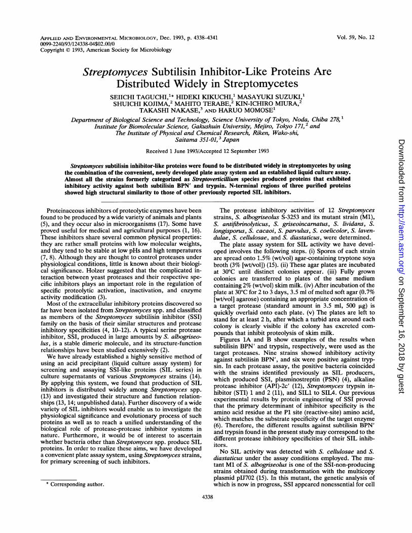
pounds that inhibit proteolysis of skim milk.Figures 1A and B show examples of the results when
subtilisin BPN' and trypsin, respectively, were used as thetarget proteases. Nine strains showed inhibitory activityagainst subtilisin BPN', and six were positive against tryp-sin. In each protease assay, the positive bacteria coincidedwith the strains identified previously as SIL producers,which produced SSI, plasminostreptin (PSN) (4), alkalineprotease inhibitor (API)-2c' (12), Streptomyces trypsin in-hibitor (STI) 1 and 2 (11), and SILl to SIL4. Our previousexperimental results by protein engineering of SSI provedthat the primary determinant of inhibitor specificity is theamino acid residue at the P1 site (reactive-site) amino acid,which matches the substrate specificity of the target enzyme(6). Therefore, the different results against subtilisin BPN'and trypsin found in the present study may correspond to thedifferent protease inhibitory specificities of their SIL inhib-itors.No SIL activity was detected with S. cellulosae and S.
diastaticus under the assay conditions employed. The mu-tant Ml of S. albogriseolus is one of the SSI-non-producingstrains obtained during transformation with the multicopyplasmid pIJ702 (15). In this mutant, the genetic analysis ofwhich is now in progress, SSI appeared nonessential for cell
4338
APPLIED AND ENVIRONMENTAL MICROBIOLOGY, Dec. 1993, p. 4338-43410099-2240/93/124338-04$02.00/0Copyright © 1993, American Society for Microbiology
on Septem
ber 16, 2018 by guesthttp://aem
.asm.org/
Dow
nloaded from
NOTES 4339
AI
ssIS. aibgi
-ssIMl I
SILls. cacaoi KCC-S352
sv-v~~~~I 0
SIL2 SIL3 SIL4S. parnd-lus 2283 S. coclicolor KCC-S006 S. lavendulac KCC-S985 S. cellulosac KCC-S127 S. diastaticus KCC-S128
B
SSIS. altiscolus S-3253
0
PSNS. antifbrinolyticus 7351
SIL2S. pan.rzus 2283
-SSIMl
t a
API-2c'S. giscoincaniatus
KTo-250
SIL3S. welicolor KCC-S006
STh1S. lividas 66
SIL4S. IDlYvendu KCC-S985
STI2S. Iongisporus 4395
S. ceUlosan KCC-S127
SILlS. cecoi KCC-S352
S. diasSttai KCC-S128I £ __ _ _ _ _
FIG. 1. Plate assays of protease inhibitors. The names of 12 Streptomyces strains and 9 SIL inhibitors identified are indicated below eachcolony; A and B denote the turbid areas formning pattems when subtilisin BPN' (A) and trypsin (B), respectively, were used as the targetproteases. Inhibitors produced by nine positive strains against subtilisin (SSI, PSN, API-2c', STI1, STI2, and SILl to SILA) and by sixpositive strains against trypsin (PSN, STI1, STI2, and SIL2 to SIL4).
growth when cultured in tryptone soya broth medium. Thebehavior of Ml, which showed no inhibitory activity againstsubtilisin, indicates that SSI is actually responsible for theformation of the turbid area in S. albogriseolus. The samephenomenon was observed for the mutant of S. griseoincar-natus (data not shown).
On the basis of these results, we considered this simple,sensitive plate assay to be very valuable for the primaryscreening of SIL producers among Streptomyces spp. andother bacteria. This system may also be applicable to thedetection of inhibitors of other types of proteases. In fact, byusing this system, we have observed that some other types
VOL. 59, 1993
on Septem
ber 16, 2018 by guesthttp://aem
.asm.org/
Dow
nloaded from
APPL. ENVIRON. MICROBIOL.
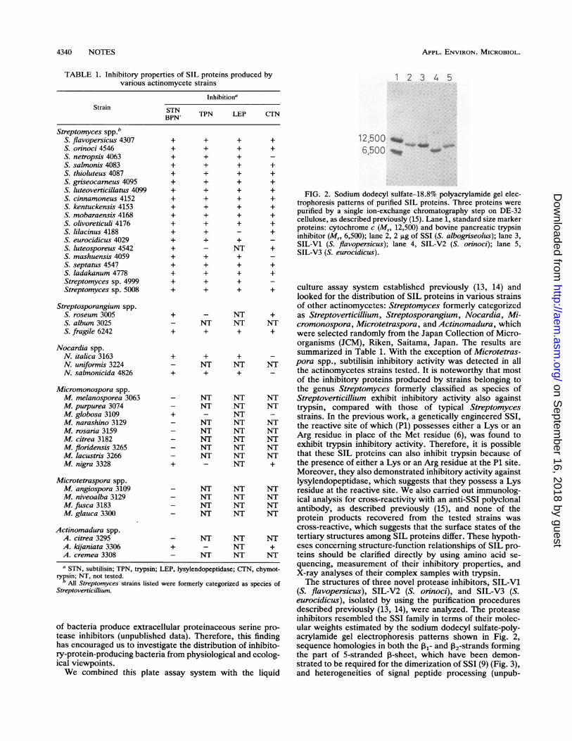
TABLE 1. Inhibitory properties of SIL proteins produced byvarious actinomycete strains
InhibitionaStrain STN
BPN' TPN LEP CITN
Streptomyces Spp.bS. flavopersicus 4307S. orinoci 4546S. netropsis 4063S. salmonis 4083S. thioluteus 4087S. griseocarneus 4095S. luteoverticillatus 4099S. cinnamoneus 4152S. kentuckensis 4153S. mobaraensis 4168S. olivoreticuli 4176S. lilacinus 4188S. eurocidicus 4029S. luteosporeus 4542S. mashuensis 4059S. septatus 4547S. ladakanum 4778Streptomyces sp. 4999Streptomyces sp. 5008
Streptosporangium spp.S. roseum 3005S. album 3025S. fragile 6242
Nocardia spp.N. italica 3163N. uniformis 3224N. salmonicida 4826
Micromonospora spp.M. melanosporea 3063M. purpurea 3074M. globosa 3109M. narashino 3129M. rosaria 3159M. citrea 3182M. floridensis 3265M. lacustris 3266M. nigra 3328
Microtetraspora spp.M. angiospora 3109M. niveoalba 3129M. fusca 3183M. glauca 3300
Actinomadura spp.A. citrea 3295A. kijaniata 3306A. cremea 3308
a STN, subtilisin; TPN, trypsin; LErypsin; NT, not tested.
b All Streptomyces strains listed weStreptoverticillium.
of bacteria produce extracelltease inhibitors (unpublishedhas encouraged us to investigary-protein-producing bacteriaical viewpoints.We combined this plate a
+ + + ++ + + ++ + + _
+ + + ++ + + ++ + + ++ + + ++ + + ++ + + ++ + + ++ + + ++ + - ++ + + _
+ - NT ++ + + _
+ + + ++ + + +
12,500 -65500 -
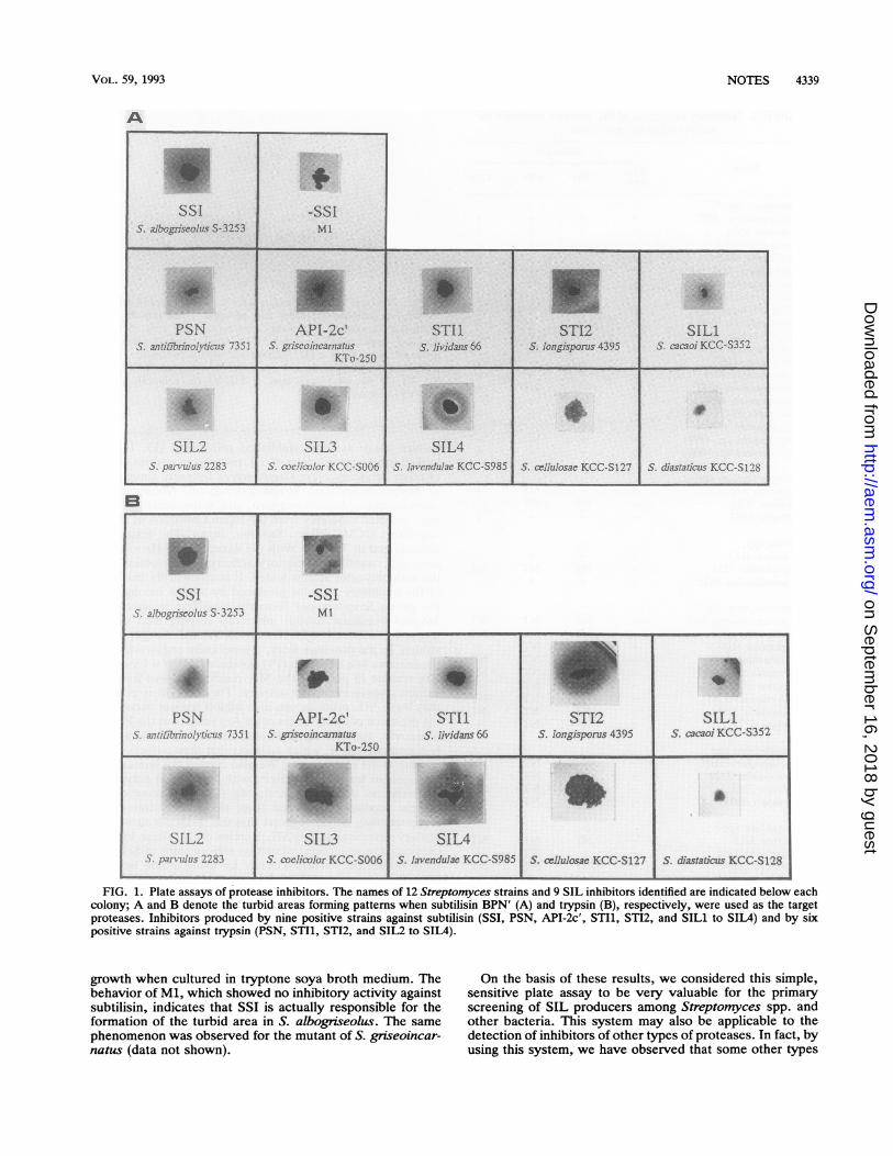
FIG. 2. Sodium dodecyl sulfate-18.8% polyacrylamide gel elec-trophoresis patterns of purified SIL proteins. Three proteins werepurified by a single ion-exchange chromatography step on DE-32cellulose, as described previously (15). Lane 1, standard size markerproteins: cytochrome c (Mr, 12,500) and bovine pancreatic trypsininhibitor (M,. 6,500); lane 2, 2 p.g of SS1 (5. albogrieolus); lane 3,SIL-Vl (S. flavopersicus); lane 4, SIL-2 (S.oyinoci); lane 5,SIL-V3 (S. eurocidicus).
+ + + _
+ + + + culture assay system established previously (13, 14) andlooked for the distribution of SIL proteins in various strainsof other actinomycetes: Streptomyces formerly categorized
+ - NT + as Streptoverticillium, Streptosporangium, Nocardia, Mi-- NT NT NT cromonospora, Microtetraspora, and Actinomadura, which+ + + + were selected randomly from the Japan Collection of Micro-
organisms (JCM), Riken, Saitama, Japan. The results are+ + + _ summarized in Table 1. With the exception of Microtetras-- NT NT NT pora spp., subtilisin inhibitory activity was detected in all+ + + _ the actinomycetes strains tested. It is noteworthy that most
of the inhibitory proteins produced by strains belonging tothe genus Streptomyces formerly classified as species of
- NT NT NT Streptoverticillium exhibit inhibitory activity also against- NT NT NT trypsin, compared with those of typical Streptomyces+ T NTNT strains. In the previous work, a genetically engineered SSI,
- NT NT NT the reactive site of which (P1) possesses either a Lys or an
- NT NT NT Arg residue in place of the Met residue (6), was found to- NT NT NT exhibit trypsin inhibitory activity. Therefore, it is possible- NT NT NT that these SIL proteins can also inhibit trypsin because of+ - NT + the presence of either a Lys or an Arg residue at the P1 site.
Moreover, they also demonstrated inhibitory activity againstlysylendopeptidase, which suggests that they possess a Lys
- NT NT NT residue at the reactive site. We also carried out immunolog-- NT NT NT ical analysis for cross-reactivity with an anti-SSI polyclonal- NT NT NT antibody, as described previously (15), and none of the
protein products recovered from the tested strains wascross-reactive, which suggests that the surface states of the
- NT NT NT tertiary structures among SIL proteins differ. These hypoth-+ - NT + eses concerning structure-function relationships of SIL pro-- NT NT NT teins should be clarified directly by using amino acid se-
quencing, measurement of their inhibitory properties, andP, lysylendopeptidase; CTN, chymot- X-ray analyses of their complex samples with trypsin.ere formerly categorized as species of The structures of three novel protease inhibitors, SIL-Vl
(S. flavopersicus), SIL-V2 (S. orinoci), and SIL-V3 (S.eurocidicus), isolated by using the purification proceduresdescribed previously (13, 14), were analyzed. The proteaseinhibitors resembled the SSI family in terms of their molec-
alar proteinaceous serine pro- ular weights estimated by the sodium dodecyl sulfate-poly-data). Therefore, this finding acrylamide gel electrophoresis patterns shown in Fig. 2,ate the distribution of inhibito- sequence homologies in both the I13- and 2-strands formingfrom physiological and ecolog- the part of 5-stranded 1-sheet, which have been demon-
strated to be required for the dimerization of SSI (9) (Fig. 3),ssay system with the liquid and heterogeneities of signal peptide processing (unpub-
4340 NOTES
on Septem
ber 16, 2018 by guesthttp://aem
.asm.org/
Dow
nloaded from
NOTES 4341
$,-strand 02-strand71J
PSN YAM ALVLTh.GHNsAATVNPER VTNATAS
API-2c' YAM VGKG SAATVTPE PGS
STI Z VGGE STAAPLA TAS
STI2 SAAAATPLR VTLNCAPTAS
1 11 21 31
DAPSA.L ALYItVGK(OV S..TTAAPER .....AVTTAPGPS
SILl VESL AVYISKTE A S' APA-0 ...W..RXLViSIL2 TAPASL tAs IGYIGOGBE STSPLIS IL3 S ATAAPL TCAPTAS
SIL4 APDAAPASL ALLIGHGG AAATATPE1>aVTLTCATSSSIL5 XAP LVLTVAKGE TRTATPLR VLXAP
SIL7 SI. AIYTVHE SAATAVPLA TLXAP1SIL8 SI MAFVOD DVAAPTVVA TVSXAPSIL10 SAIAATPEA TTXAPKAASIL-VSt GEGE SAADSGVO> XTLTTPK_t T.AP,S.,n X ;'
IIG SSA-GI LIMPALYVt~~~~~~~~~~~IG TAAESGVO1~ XT
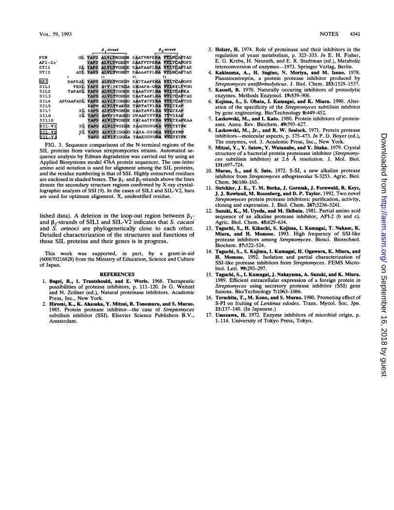
FIG. 3. Sequence comparisons of the N-terminal regions of theSIL proteins from various streptomycetes strains. Automated se-
quence analysis by Edman degradation was carried out by using anApplied Biosystems model 476A protein sequencer. The one-letteramino acid notation is used for alignment among the SIL proteins,and the residue numbering is that of SSI. Highly conserved residuesare enclosed in shaded boxes. The 1- and 12-strands above the linesdenote the secondary structure regions confirmed by X-ray crystal-lographic analysis of SSI (9). In the cases of SILl and SIL-V2, barsare used for optimum alignment. X, unidentified residue.
lished data). A deletion in the loop-out region between Il-and 12-strands of SILl and SIL-V2 indicates that S. cacaoiand S. orinoci are phylogenetically close to each other.Detailed characterization of the structures and functions ofthese SIL proteins and their genes is in progress.
This work was supported, in part, by a grant-in-aid(600670216828) from the Ministry of Education, Science and Cultureof Japan.
REFERENCES1. Bogel, R., I. Trautshould, and E. Werle. 1968. Therapeutic
possibilities of protease inhibitors, p. 111-120. In G. Weitzeland N. Zollner (ed.), Natural proteinase inhibitors. AcademicPress, Inc., New York.
2. Hiromi, K., K. Akasaka, Y. Mitsui, B. Tonomura, and S. Murao.1985. Protein protease inhibitor-the case of Streptomycessubtilisin inhibitor (SSI). Elsevier Science Publishers B.V.,Amsterdam.
3. Holzer, H. 1974. Role of proteinase and their inhibitors in theregulation of yeast metabolism, p. 325-333. In E. H. Fisher,E. G. Krebs, H. Neurath, and E. R. Stadtman (ed.), Metabolicinterconversion of enzymes-1973. Springer Verlag, Berlin.
4. Kakinuma, A., H. Sugino, N. Moriya, and M. Isono. 1978.Plasminostreptin, a protein protease inhibitor produced byStreptomyces antifibrinolyticus. J. Biol. Chem. 253:1529-1537.
5. Kassell, B. 1970. Naturally occuring inhibitors of proteolyticenzymes. Methods Enzymol. 19:839-906.
6. Kojima, S., S. Obata, I. Kumagai, and K. Miura. 1990. Alter-ation of the specificity of the Streptomyces subtilisin inhibitorby gene engineering. Bio/Technology 8:449-452.
7. Laskowski, M., and I. Kato. 1980. Protein inhibitors of protein-ases. Annu. Rev. Biochem. 49:593-627.
8. Laskowski, M., Jr., and R. W. Sealock. 1971. Protein proteaseinhibitors-molecular aspects, p. 375-473. In P. D. Boyer (ed.),The enzymes, vol. 3. Academic Press, Inc., New York.
9. Mitsui, Y., Y. Satow, Y. Watanabe, and Y. Iitaka. 1979. Crystalstructure of a bacterial protein proteinase inhibitor (Streptomy-ces subtilisin inhibitor) at 2.6 A resolution. J. Mol. Biol.131:697-724.
10. Murao, S., and S. Sato. 1972. S-SI, a new alkaline proteaseinhibitor from Streptomyces albogriseolus S-3253. Agric. Biol.Chem. 36:160-163.
11. Strickler, J. E., T. M. Berka, J. Gorniak, J. Fornwald, R. Keys,J. J. Rowland, M. Rosenberg, and D. P. Taylor. 1992. Two novelStreptomyces protein protease inhibitors: purification, activity,cloning and expression. J. Biol. Chem. 267:3236-3241.
12. Suzuki, K., M. Uyeda, and M. Shibata. 1981. Partial amino acidsequence of an alkaline protease inhibitor, API-2 (b and c).Agric. Biol. Chem. 45:629-634.
13. Taguchi, S., H. Kikuchi, S. Kojima, I. Kumagai, T. Nakase, K.Miura, and H. Momose. 1993. High frequency of SSI-likeprotease inhibitors among Streptomyces. Biosci. Biotechnol.Biochem. 57:522-524.
14. Taguchi, S., S. Kojima, I. Kumagai, H. Ogawara, K. Miura, andH. Momose. 1992. Isolation and partial characterization ofSSI-like protease inhibitors from Streptomyces. FEMS Micro-biol. Lett. 99:293-297.
15. Taguchi, S., I. Kumagai, J. Nakayama, A. Suzuki, and K. Miura.1989. Efficient extracellular expression of a foreign protein inStreptomyces using secretory protease inhibitor (SSI) genefusions. Bio/Technology 7:1063-1066.
16. Terashita, T., M. Kono, and S. Murao. 1980. Promoting effect ofS-PI on fruiting of Lentinus edodes. Trans. Mycol. Soc. Jpn.21:137-140. (In Japanese.)
17. Umezawa, H. 1972. Enzyme inhibitors of microbial origin, p.1-114. University of Tokyo Press, Tokyo.
VOL. 59, 1993
on Septem
ber 16, 2018 by guesthttp://aem
.asm.org/
Dow
nloaded from