strandrad treatment protocols paediatrics - slcp.lk · pneumothorax respiratory depression...
TRANSCRIPT
2013
STANDARD TREATMENT PROTOCOLS PAEDIATRICS
[DRAFT ONLY – Please give your feed back to Dr Srilal de Silva, before 14th of April ]
SRI LANKA COLLEGE OF PAEDIATRICIANS
[Pick the date]
2 Standard Treatment Protocols / Paediatrics /Sri Lanka College of Paediatricians / July 06.2013 / Draft
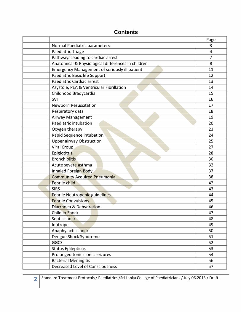
Contents
Page
Normal Paediatric parameters 3
Paediatric Triage 4
Pathways leading to cardiac arrest 7
Anatomical & Physiological differences in children 8
Emergency Management of seriously ill patient 11
Paediatric Basic life Support 12
Paediatric Cardiac arrest 13
Asystole, PEA & Ventricular Fibrillation 14
Childhood Bradycardia 15
SVT 16
Newborn Resuscitation 17
Respiratory data 18
Airway Management 19
Paediatric intubation 20
Oxygen therapy 23
Rapid Sequence intubation 24
Upper airway Obstruction 25
Viral Croup 27
Epiglotittis 28
Bronchiolitis 30
Acute severe asthma 32
Inhaled Foreign Body 37
Community Acquired Pneumonia 38
Febrile child 42
SIRS 43
Febrile Neutropenic guidelines 44
Febrile Convulsions 45
Diarrhoea & Dehydration 46
Child in Shock 47
Septic shock 48
Inotropes 49
Anaphylactic shock 50
Dengue Shock Syndrome 51
GGCS 52
Status Epilepticus 53
Prolonged tonic clonic seizures 54
Bacterial Meningitis 56
Decreased Level of Consciousness 57
3 Standard Treatment Protocols / Paediatrics /Sri Lanka College of Paediatricians / July 06.2013 / Draft
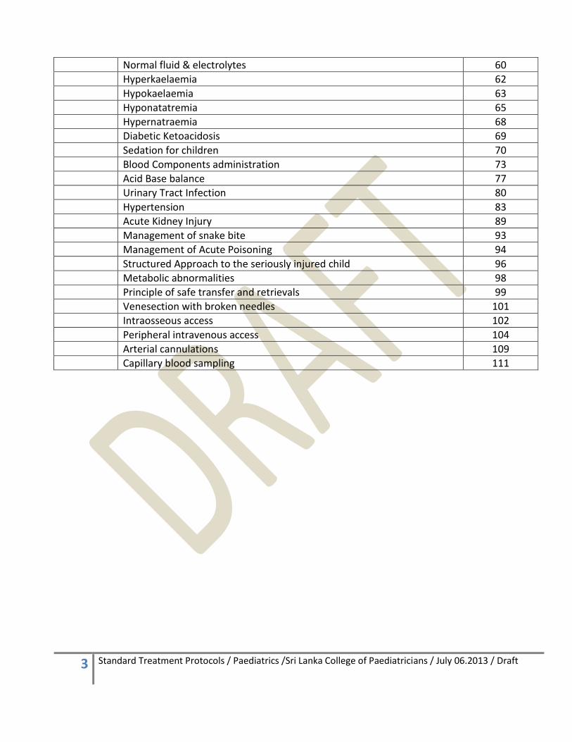
Normal fluid & electrolytes 60
Hyperkaelaemia 62
Hypokaelaemia 63
Hyponatatremia 65
Hypernatraemia 68
Diabetic Ketoacidosis 69
Sedation for children 70
Blood Components administration 73
Acid Base balance 77
Urinary Tract Infection 80
Hypertension 83
Acute Kidney Injury 89
Management of snake bite 93
Management of Acute Poisoning 94
Structured Approach to the seriously injured child 96
Metabolic abnormalities 98
Principle of safe transfer and retrievals 99
Venesection with broken needles 101
Intraosseous access 102
Peripheral intravenous access 104
Arterial cannulations 109
Capillary blood sampling 111

4 Standard Treatment Protocols / Paediatrics /Sri Lanka College of Paediatricians / July 06.2013 / Draft
Normal Paediatric Parameters
Age Weight (kg) Heart rate (per min)
Respiratory rate BP Systolic (mmHg)
Premature 1 145 <40 42 ± 10
Newborn 2-3 125 60 ± 10
1 month 4 120 24-35 80 ± 16
6 month 7 130 89 ± 29
1 year 10 130 20-30 96 ± 30
2-3 years 12-14 120 99 ± 25
4-5 years 16-18 100 99 ± 20
6-8 years 20-26 100 12-25 105 ± 13
10-12 years 32-42 75 112 ± 19
>14 years 50 75 12-18 120 ± 20
5 Standard Treatment Protocols / Paediatrics /Sri Lanka College of Paediatricians / July 06.2013 / Draft
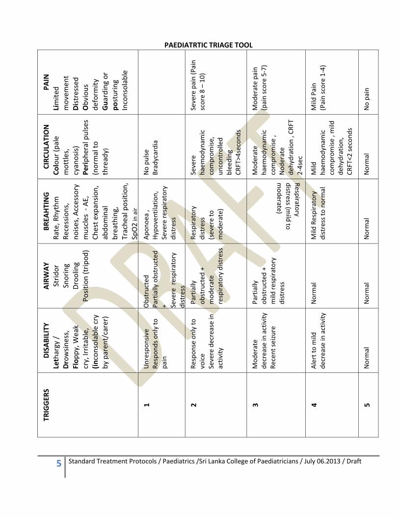
PAEDIATRTIC TRIAGE TOOL P
AIN
Li
mit
ed
mo
vem
ent
Dis
tres
sed
O
bvi
ou
s d
efo
rmit
y
Gu
ard
ing
or
po
stu
rin
g
Inco
nso
lab
le
Seve
re p
ain
(P
ain
sc
ore
8 –
10
)
Mo
der
ate
pai
n
(pai
n s
core
5-7
)
Mild
Pai
n
(Pai
n s
core
1-4
)
No
pai
n
CIR
CU
LATI
ON
C
olo
ur
(pal
e m
ott
les,
cyan
osi
s)
Per
iph
eral
pu
lses
(n
orm
al t
o
thre
ady)
No
pu
lse
B
rad
ycar
dia
Seve
re
hae
mo
dyn
amic
com
pro
mis
e,
un
con
tro
lled
b
leed
ing
CR
FT>4
seco
nd
s
Mo
der
ate
hae
mo
dyn
amic
com
pro
mis
e ,
No
der
ate
deh
ydra
tio
n ,
CR
FT
2-4
sec
Mild
h
aem
od
ynam
ic
com
pro
mis
e , m
ild
deh
ydra
tio
n,
CR
FT<2
se
con
ds
No
rmal
BR
EAH
TIN
G
Rat
e, R
hyt
hm
R
eces
sio
ns,
no
ises
, Acc
esso
ry
mu
scle
s -
AE,
C
he
st e
xpan
sio
n,
abd
om
inal
bre
ath
ing,
Trac
hea
l po
siti
on
, Sp
O2
in a
ir
Ap
on
oea
,
Hyp
ove
nti
lati
on
, Se
vere
res
pir
ato
ry
dis
tres
s
Res
pir
ato
ry
dis
tres
s
(se
vere
to
m
od
erat
e)
Resp
iratory
distress (m
ild to
m
od
erate)
Mild
Re
spir
ato
ry
dis
tres
s to
no
rmal
No
rmal
AIR
WA
Y
Stri
do
r
Sno
rin
g
Dro
olin
g
Po
siti
on
(tr
ipo
d)
Ob
stru
cte
d
Par
tial
ly o
bst
ruct
ed
+
Seve
re r
esp
irat
ory
d
istr
ess
Par
tial
ly
ob
stru
cted
+
mo
der
ate
re
spir
ato
ry d
istr
ess
Par
tial
ly
ob
stru
cted
+
mild
re
spir
ato
ry
dis
tres
s
No
rmal
No
rmal
DIS
AB
ILIT
Y
Leth
argy
/
Dro
wsi
nes
s,
Flo
pp
y, W
eak
cry,
Irri
tab
le,
(in
con
sola
ble
cry
b
y p
aren
t/ca
rer)
Un
resp
on
sive
R
esp
on
ds
on
ly t
o
pai
n
Res
po
nse
on
ly t
o
voic
e
Seve
re d
ecre
ase
in
acti
vity
Mo
der
ate
dec
reas
e in
act
ivit
y
Rec
ent
seiz
ure
Ale
rt t
o m
ild
dec
reas
e in
act
ivit
y
No
rmal
TRIG
GER
S
1
2
3
4
5
6 Standard Treatment Protocols / Paediatrics /Sri Lanka College of Paediatricians / July 06.2013 / Draft
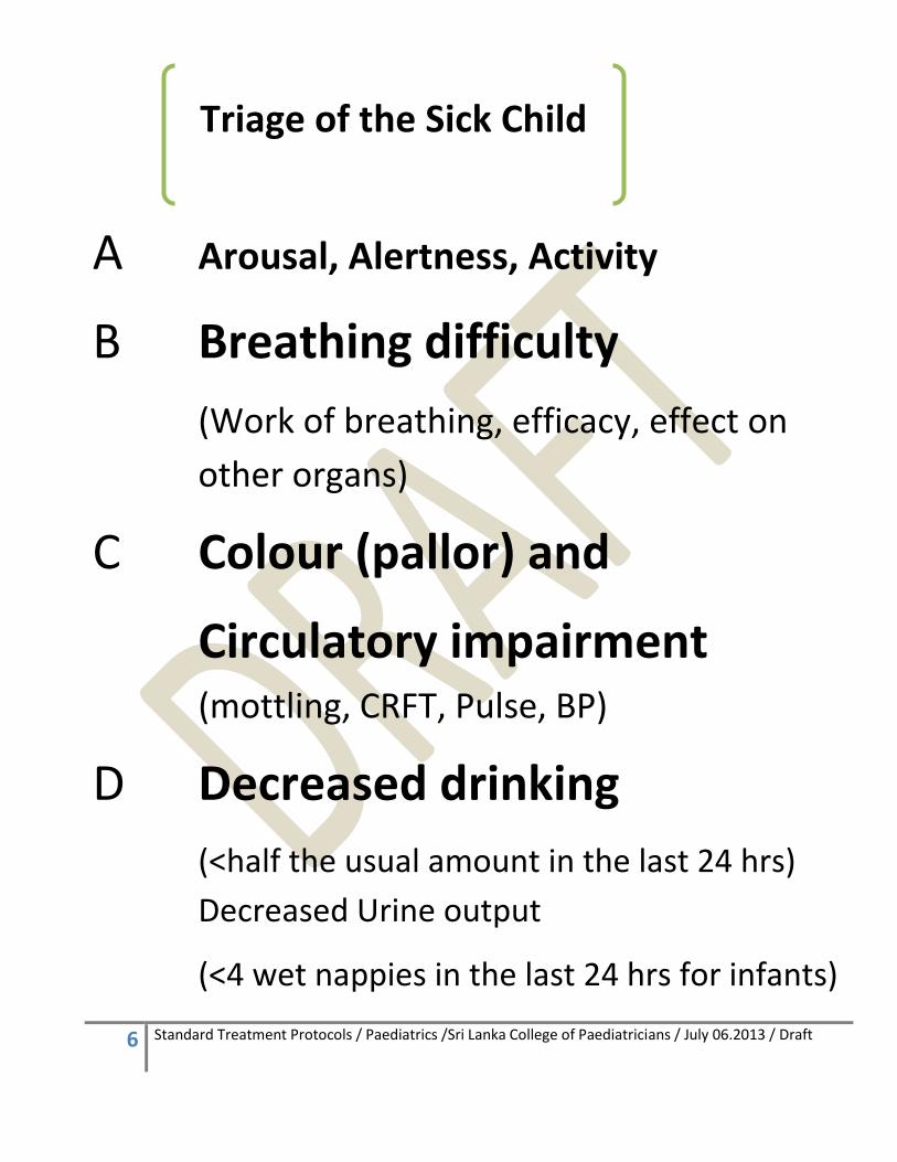
A Arousal, Alertness, Activity
B Breathing difficulty
(Work of breathing, efficacy, effect on
other organs)
C Colour (pallor) and
Circulatory impairment (mottling, CRFT, Pulse, BP)
D Decreased drinking
(<half the usual amount in the last 24 hrs)
Decreased Urine output
(<4 wet nappies in the last 24 hrs for infants)
Triage of the Sick Child
7 Standard Treatment Protocols / Paediatrics /Sri Lanka College of Paediatricians / July 06.2013 / Draft
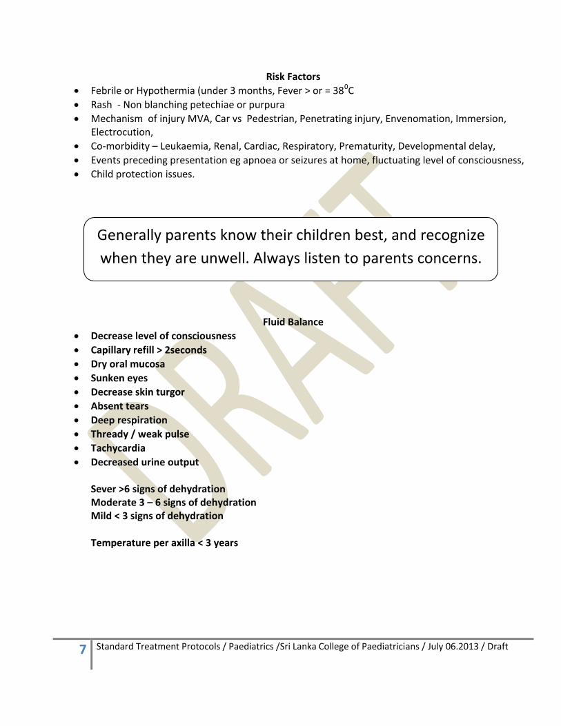
Risk Factors
Febrile or Hypothermia (under 3 months, Fever > or = 380C
Rash - Non blanching petechiae or purpura
Mechanism of injury MVA, Car vs Pedestrian, Penetrating injury, Envenomation, Immersion, Electrocution,
Co-morbidity – Leukaemia, Renal, Cardiac, Respiratory, Prematurity, Developmental delay,
Events preceding presentation eg apnoea or seizures at home, fluctuating level of consciousness,
Child protection issues.
Fluid Balance
Decrease level of consciousness
Capillary refill > 2seconds
Dry oral mucosa
Sunken eyes
Decrease skin turgor
Absent tears
Deep respiration
Thready / weak pulse
Tachycardia
Decreased urine output Sever >6 signs of dehydration Moderate 3 – 6 signs of dehydration Mild < 3 signs of dehydration Temperature per axilla < 3 years
Generally parents know their children best, and recognize
when they are unwell. Always listen to parents concerns.
8 Standard Treatment Protocols / Paediatrics /Sri Lanka College of Paediatricians / July 06.2013 / Draft
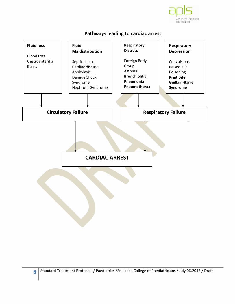
Pathways leading to cardiac arrest
Fluid loss Blood Loss Gastroenteritis Burns
Fluid Maldistribution Septic shock Cardiac disease Anphylaxis Dengue Shock Syndrome Nephrotic Syndrome
Respiratory Distress Foreign Body Croup Asthma Bronchiolitis Pneumonia Pneumothorax
Respiratory Depression Convulsions Raised ICP Poisoning Krait Bite Guillain-Barre Syndrome
Circulatory Failure Respiratory Failure
CARDIAC ARREST
9 Standard Treatment Protocols / Paediatrics /Sri Lanka College of Paediatricians / July 06.2013 / Draft
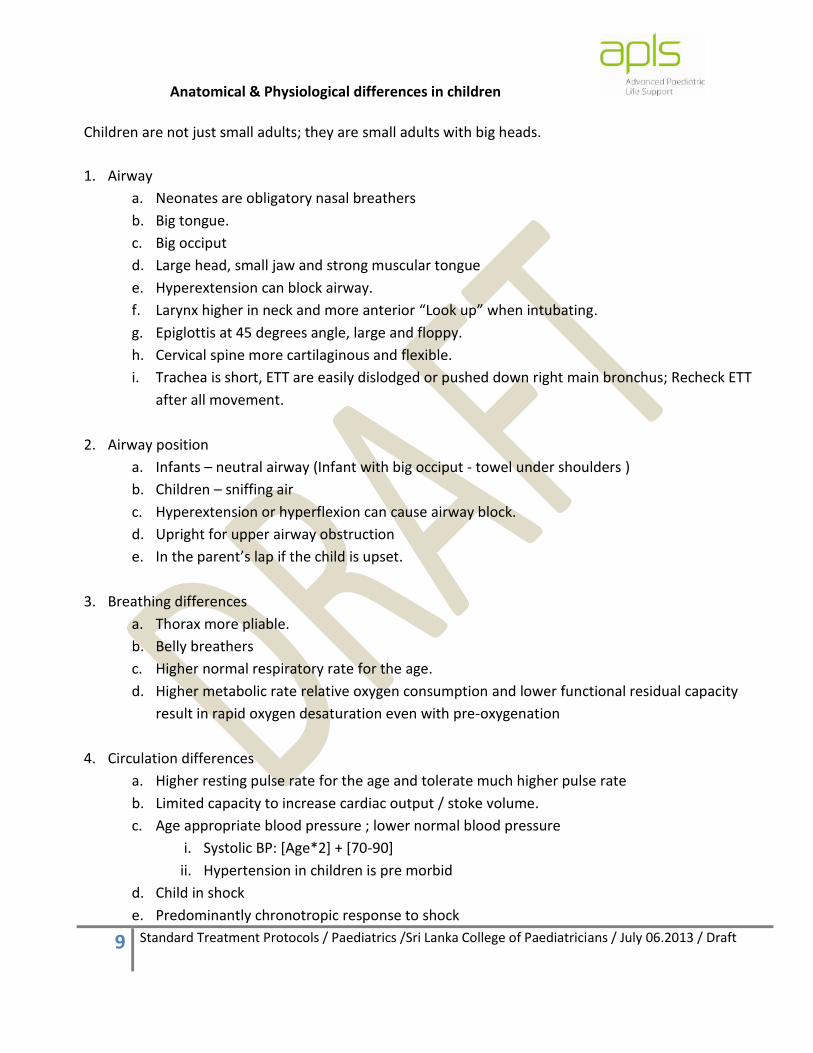
Anatomical & Physiological differences in children
Children are not just small adults; they are small adults with big heads.
1. Airway
a. Neonates are obligatory nasal breathers
b. Big tongue.
c. Big occiput
d. Large head, small jaw and strong muscular tongue
e. Hyperextension can block airway.
f. Larynx higher in neck and more anterior “Look up” when intubating.
g. Epiglottis at 45 degrees angle, large and floppy.
h. Cervical spine more cartilaginous and flexible.
i. Trachea is short, ETT are easily dislodged or pushed down right main bronchus; Recheck ETT
after all movement.
2. Airway position
a. Infants – neutral airway (Infant with big occiput - towel under shoulders )
b. Children – sniffing air
c. Hyperextension or hyperflexion can cause airway block.
d. Upright for upper airway obstruction
e. In the parent’s lap if the child is upset.
3. Breathing differences
a. Thorax more pliable.
b. Belly breathers
c. Higher normal respiratory rate for the age.
d. Higher metabolic rate relative oxygen consumption and lower functional residual capacity
result in rapid oxygen desaturation even with pre-oxygenation
4. Circulation differences
a. Higher resting pulse rate for the age and tolerate much higher pulse rate
b. Limited capacity to increase cardiac output / stoke volume.
c. Age appropriate blood pressure ; lower normal blood pressure
i. Systolic BP: [Age*2] + [70-90]
ii. Hypertension in children is pre morbid
d. Child in shock
e. Predominantly chronotropic response to shock
10 Standard Treatment Protocols / Paediatrics /Sri Lanka College of Paediatricians / July 06.2013 / Draft
f. Volume resuscitation is with isotonic crystalloid solutions
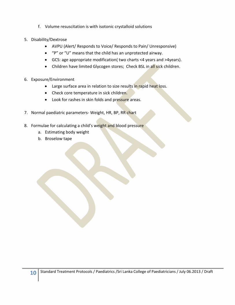
5. Disability/Dextrose
AVPU (Alert/ Responds to Voice/ Responds to Pain/ Unresponsive)
“P” or “U” means that the child has an unprotected airway.
GCS: age appropriate modification( two charts <4 years and >4years).
Children have limited Glycogen stores; Check BSL in all sick children.
6. Exposure/Environment
Large surface area in relation to size results in rapid heat loss.
Check core temperature in sick children.
Look for rashes in skin folds and pressure areas.
7. Normal paediatric parameters- Weight, HR, BP, RR chart
8. Formulae for calculating a child’s weight and blood pressure
a. Estimating body weight
b. Broselow tape
11 Standard Treatment Protocols / Paediatrics /Sri Lanka College of Paediatricians / July 06.2013 / Draft
12 Standard Treatment Protocols / Paediatrics /Sri Lanka College of Paediatricians / July 06.2013 / Draft
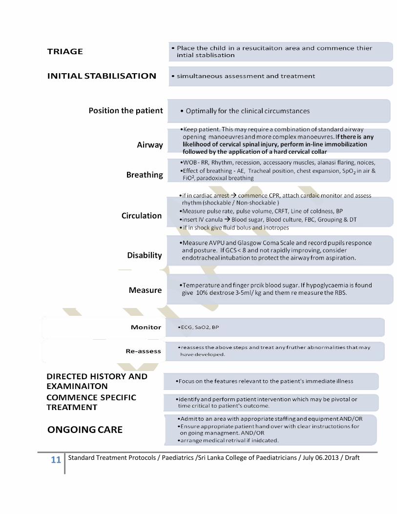
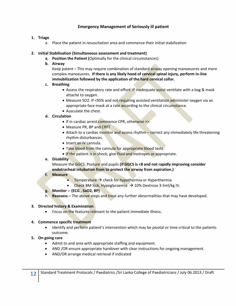
Emergency Management of Seriously ill patient
1. Triage a. Place the patient in resuscitation area and commence their initial stabilization
2. Initial Stabilisation (Simultaneous assessment and treatment)
a. Position the Patient (Optimally for the clinical circumstances) b. Airway
Keep patent – This may require combination of standard airway opening manoeuvres and more complex manoeuvres. If there is any likely hood of cervical spinal injury, perform in-line immobilization followed by the application of the hard cervical collar.
c. Breathing
Assess the respiratory rate and effort. If inadequate assist ventilate with a bag & mask attaché to oxygen.
Measure SO2. If <95% and not requiring assisted ventilation administer oxygen via an appropriate face mask at a rate according to the clinical circumstance.
Ausculate the chest. d. Circulation
If in cardiac arrest commence CPR, otherwise >>
Measure PR, BP and CRFT
Attach to a cardiac monitor and assess rhythm – correct any immediately life threatening rhythm disturbances.
Insert an IV cannula.
Take blood from the cannula for appropriate blood tests.
If the patient is in shock, give fluid and inotropes as appropriate. e. Disability
Measure the GGCS, Posture and pupils (if GGCS is <8 and not rapidly improving consider endotracheal intubation from to protect the airway from aspiration.)
f. Measure
Temperature check for hypothermia or Hyperthermia
Check BM Sick, Hypoglycaemia 10% Dextrose 3-5ml/kg IV. g. Monitor -- (ECG , SaO2, BP) h. Reassess – The above steps and treat any further abnormalities that may have developed.
3. Directed history & Examination
Focus on the features relevant to the patient immediate illness.
4. Commence specific treatment
Identify and perform patient’s intervention which may be pivotal or time critical to the patients outcome.
5. On going care
Admit to and area with appropriate staffing and equipment.
AND /OR ensure appropriate handover with clear instructions for ongoing management.
AND/OR arrange medical retrieval if indicated
13 Standard Treatment Protocols / Paediatrics /Sri Lanka College of Paediatricians / July 06.2013 / Draft
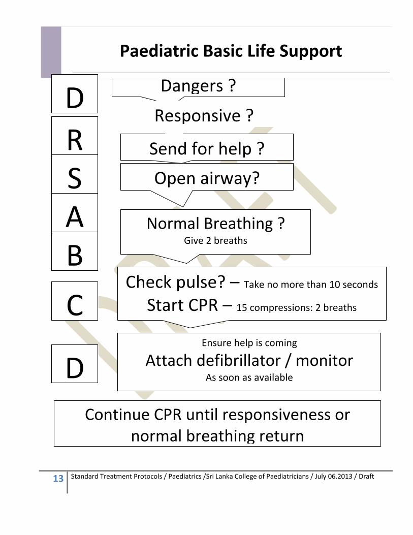
Dangers ?
Paediatric Basic Life Support
D
B A S
W
D
R
D
D
C
Send for help ?
Open airway?
Normal Breathing ? Give 2 breaths
Check pulse? – Take no more than 10 seconds
Start CPR – 15 compressions: 2 breaths
Responsive ?
Ensure help is coming
Attach defibrillator / monitor As soon as available
Continue CPR until responsiveness or normal breathing return
14 Standard Treatment Protocols / Paediatrics /Sri Lanka College of Paediatricians / July 06.2013 / Draft
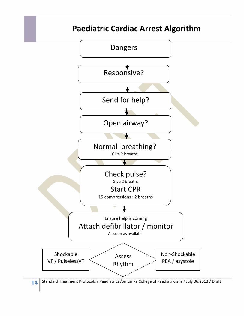
Paediatric Cardiac Arrest Algorithm
Assess Rhythm
Shockable VF / PulselessVT
Non-Shockable PEA / asystole
Dangers?
Send for help?
Responsive?
Open airway?
Check pulse? Give 2 breaths
Start CPR 15 compressions : 2 breaths
Normal breathing? Give 2 breaths
Ensure help is coming
Attach defibrillator / monitor As soon as available
15 Standard Treatment Protocols / Paediatrics /Sri Lanka College of Paediatricians / July 06.2013 / Draft
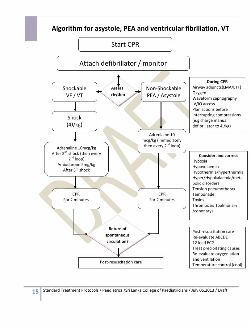
Algorithm for asystole, PEA and ventricular fibrillation, VT
Assess
rhythm
Return of
spontaneous
circulation?
Consider and correct Hypoxia Hypovolaemia Hypothermia/hyperthermia Hyper/Hypokalaemia/metabolic disorders Tension pneumothorax Tamponade Toxins Thrombosis (pulmonary /cononary)
During CPR Airway adjuncts(LMA/ETT) Oxygen Waveform capnography IV/IO access Plan actions before interrupting compressions (e.g charge manual defibrillator to 4j/kg)
Post-resuscitation care
Post resuscitation care Re-evaluate ABCDE 12 lead ECG Treat precipitating causes Re-evaluate oxygen ation and ventilation Temperature control (cool)
Start CPR
Attach defibrillator / monitor
Non-Shockable PEA / Asystole
Adrenlaine 10 mcg/kg (immediately then every 2nd loop)
CPR For 2 minutes
Shockable VF / VT
Shock (4J/kg)
Adrenaline 10mcg/kg After 2nd shock (then every
2nd loop) Amiodarone 5mg/kg
After 3rd shock
CPR For 2 minutes
16 Standard Treatment Protocols / Paediatrics /Sri Lanka College of Paediatricians / July 06.2013 / Draft
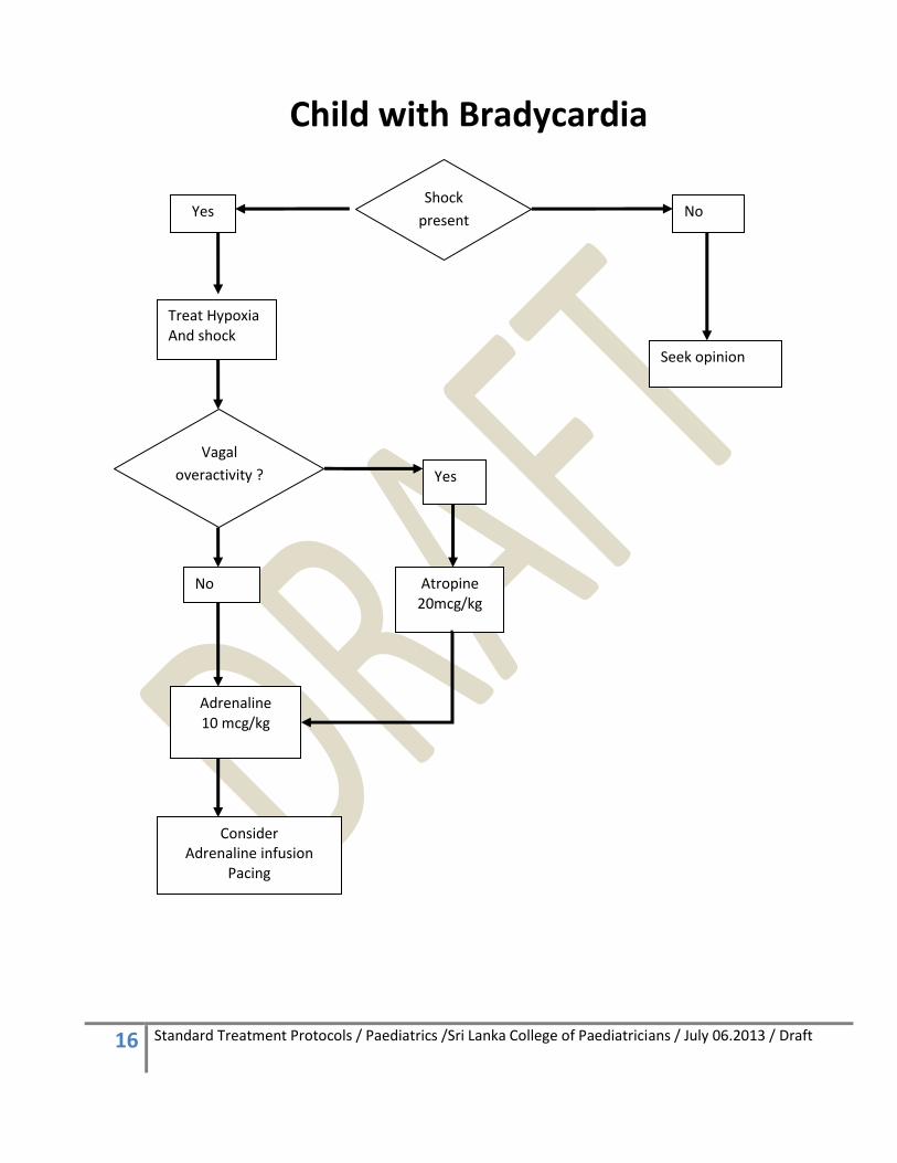
Child with Bradycardia
Shock
present Yes No
Seek opinion
Treat Hypoxia And shock
Vagal
overactivity ?
No
Adrenaline 10 mcg/kg
Consider Adrenaline infusion
Pacing
Yes
Atropine 20mcg/kg
17 Standard Treatment Protocols / Paediatrics /Sri Lanka College of Paediatricians / July 06.2013 / Draft
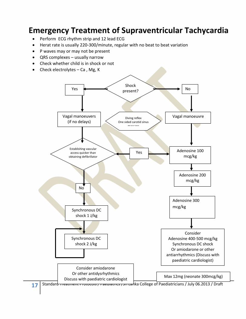
Emergency Treatment of Supraventricular Tachycardia Perform ECG rhythm strip and 12 lead ECG
Herat rate is usually 220-300/minute, regular with no beat to beat variation
P waves may or may not be present
QRS complexes – usually narrow
Check whether child is in shock or not
Check electrolytes – Ca , Mg, K
Shock present? Yes No
Vagal manoeuvers (if no delays)
Establishing vascular access quicker than
obtaining defibrillator
No
Synchronous DC shock 1 J/kg
Synchronous DC shock 2 J/kg
Consider amiodarone Or other antidysrhythmics
Discuss with paediatric cardiologist
Vagal manoeuvre
Adenosine 100 mcg/kg
Adenosine 200 mcg/kg
Adenosine 300
mcg/kg
Consider Adenosine 400-500 mcg/kg
Synchronous DC shock Or amiodarone or other
antiarrhythmics (Discuss with paediatric cardiologist)
Max 12mg (neonate 300mcg/kg)
Diving reflex One sided carotid sinus
massage Valsalva manoeuvre in
older child
Yes
18 Standard Treatment Protocols / Paediatrics /Sri Lanka College of Paediatricians / July 06.2013 / Draft
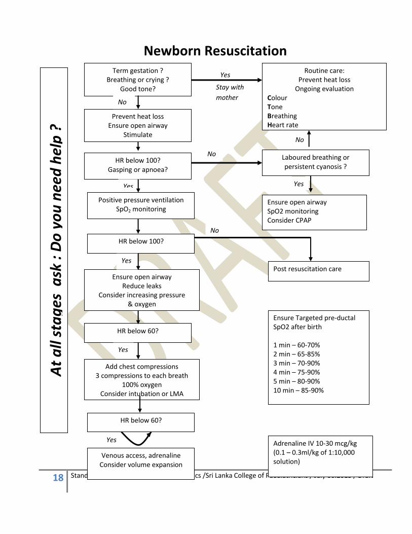
Newborn Resuscitation
Term gestation ? Breathing or crying ?
Good tone?
HR below 100? Gasping or apnoea?
Prevent heat loss Ensure open airway
Stimulate
HR below 100?
Positive pressure ventilation SpO2 monitoring
HR below 60?
Ensure open airway Reduce leaks
Consider increasing pressure & oxygen
Add chest compressions 3 compressions to each breath
100% oxygen Consider intubation or LMA
HR below 60?
Venous access, adrenaline Consider volume expansion
Routine care: Prevent heat loss
Ongoing evaluation Colour Tone Breathing Heart rate
Laboured breathing or persistent cyanosis ?
Ensure open airway SpO2 monitoring Consider CPAP
Post resuscitation care
Ensure Targeted pre-ductal SpO2 after birth 1 min – 60-70% 2 min – 65-85% 3 min – 70-90% 4 min – 75-90% 5 min – 80-90% 10 min – 85-90%
Adrenaline IV 10-30 mcg/kg (0.1 – 0.3ml/kg of 1:10,000 solution)
At
all
sta
ges
ask
: D
o y
ou
nee
d h
elp
?
Yes
Yes
Yes
Yes
Yes
Stay with
mother
Yes
No
No
No
No
19 Standard Treatment Protocols / Paediatrics /Sri Lanka College of Paediatricians / July 06.2013 / Draft
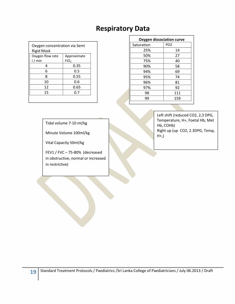
Respiratory Data
Oxygen concentration via Semi Rigid Mask Oxygen flow rate l / min
Approximate FiO2
4 0.35
6 0.5
8 0.55
10 0.6
12 0.65
15 0.7
Oxygen dissociation curve
Saturation PO2
25% 14
50% 27
75% 40
90% 58
94% 69
95% 74
96% 81
97% 92
98 111
99 159
Left shift (reduced CO2, 2,3 DPG, Temperature, H+, Foetal Hb, Met Hb, COHb) Right up (up CO2, 2.3DPG, Temp, H+,)
Tidal volume 7-10 ml/kg
Minute Volume 100ml/kg
Vital Capacity 50ml/kg
FEV1 / FVC – 75-80% (decreased
in obstructive, normal or increased
in restrictive)
20 Standard Treatment Protocols / Paediatrics /Sri Lanka College of Paediatricians / July 06.2013 / Draft
Airway Management
1. Basic airway opening manoeuvres and clearance 2. Oxygen delivery 3. Oropharyngeal airway insertion 4. nasopharyngeal airway insertion 5. mouth-to-mask ventilation including chin lift and jaw thrust manoeuvres with mask application 6. bag-mask ventilation 7. orotracheal intubation of an infant or small child 8. orotracheal intubation of an older child 9. ventilation with bag through tracheal tube 10. discussion of the procedure for Rapid Sequence Induction of anaesthesia and their role as an assistant
Basic airway station &
1) Airway opening maneuvers
a) Head tilt / chin lift
b) Jaw thrust
2) Airway adjuncts
a) Oral and nasal airways
21 Standard Treatment Protocols / Paediatrics /Sri Lanka College of Paediatricians / July 06.2013 / Draft
Rapid Sequence Intubation 3) Intubation
a) Golden Rules
You don’t need an ETT in place to keep an airway open to oxygenate a patient.
Don’t give paralytic agent to a patient unless you are sure you can ventilate and oxygenate.
All intubations are difficult for inexperienced operator.
Have a back up plan ready for the failed intubation before attempting any intubation.
b) Essential Skills- Intubation using Rapid Sequence Induction( RSI) : the 4 Ps
Purpose
Why intubate
Why RSI
Prerequisites
(a) Full stomach- No bagging
(b) Predict that you can intubate (Skill station 2= difficult intubation)
(c) Predict that you can ventilate (If you are unsuccessful intubating)
c) 6 “P”s for the Rapid Sequence Intubation
1.Preparation
Staff
Patient
Equipment
Non-invasive monitoring
Drugs
2.Preoxygenation
3.Paralysis after induction
4.Protection and positioning
Cricoid pressure
5.Placement and proof
EtCO2 and clinically
6.Post intubation management
Sedation
Paralysis
Ventilation
Monitoring
22 Standard Treatment Protocols / Paediatrics /Sri Lanka College of Paediatricians / July 06.2013 / Draft
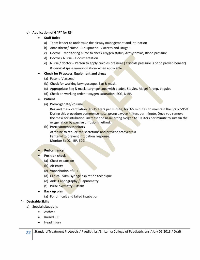
d) Application of 6 “P” for RSI
Staff Roles
a) Team leader to undertake the airway management and intubation
b) Anaesthetic/ Nurse – Equipment, IV access and Drugs –
c) Doctor – Monitoring nurse to check Oxygen status, Arrhythmias, Blood pressure
d) Doctor / Nurse – Documentation
e) Nurse / doctor – Person to apply cricoids pressure ( Cricoids pressure is of no proven benefit)
& Cervical spine immobilization- when applicable
Check for IV access, Equipment and drugs
(a) Patent IV access
(b) Check for working laryngoscope, Bag & mask,
(c) Appropriate Bag & mask, Laryngoscope with blades, Steylet, Maggi forcep, boguies
(d) Check on working order – oxygen saturation, ECG, NIBP.
Patient
(a) Preoxygenate/Volume
Bag and mask ventilation (10-15 liters per minute) for 3-5 minutes to maintain the SpO2 >95% During this procedure commence nasal prong oxygen 4 liters per minute. Once you remove the mask for intubation, increase the nasal prong oxygen to 10 liters per minute to sustain the oxygenation by passive diffusion method.
(b) Pretreatment/Monitors
Atropine to reduce the secretions and prevent bradycardia Fentanyl to prevent intubation response. Monitor SpO2 , BP, ECG
Performance
Position check
(a) Chest expansion
(b) Air entry
(c) Vaporization of ETT
(d) Clinical: 50ml syringe aspiration technique
(e) Aids: Capnography / Capnometry
(f) Pulse oxymetry- Pitfalls
Back up plan
(a) For difficult and failed intubation
4) Desirable Skills
a) Special situations
Asthma
Raised ICP
Head injury
23 Standard Treatment Protocols / Paediatrics /Sri Lanka College of Paediatricians / July 06.2013 / Draft
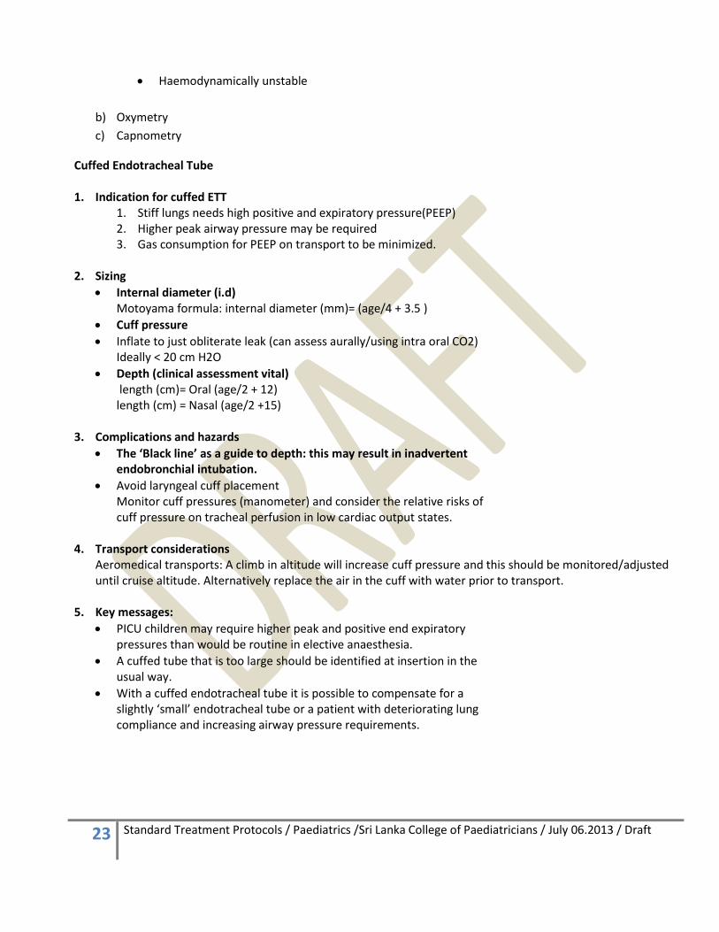
Haemodynamically unstable
b) Oxymetry
c) Capnometry
Cuffed Endotracheal Tube 1. Indication for cuffed ETT
1. Stiff lungs needs high positive and expiratory pressure(PEEP) 2. Higher peak airway pressure may be required 3. Gas consumption for PEEP on transport to be minimized.
2. Sizing
Internal diameter (i.d) Motoyama formula: internal diameter (mm)= (age/4 + 3.5 )
Cuff pressure
Inflate to just obliterate leak (can assess aurally/using intra oral CO2) Ideally < 20 cm H2O
Depth (clinical assessment vital) length (cm)= Oral (age/2 + 12) length (cm) = Nasal (age/2 +15)
3. Complications and hazards
The ‘Black line’ as a guide to depth: this may result in inadvertent endobronchial intubation.
Avoid laryngeal cuff placement Monitor cuff pressures (manometer) and consider the relative risks of cuff pressure on tracheal perfusion in low cardiac output states.
4. Transport considerations Aeromedical transports: A climb in altitude will increase cuff pressure and this should be monitored/adjusted until cruise altitude. Alternatively replace the air in the cuff with water prior to transport.
5. Key messages:
PICU children may require higher peak and positive end expiratory pressures than would be routine in elective anaesthesia.
A cuffed tube that is too large should be identified at insertion in the usual way.
With a cuffed endotracheal tube it is possible to compensate for a slightly ‘small’ endotracheal tube or a patient with deteriorating lung compliance and increasing airway pressure requirements.
24 Standard Treatment Protocols / Paediatrics /Sri Lanka College of Paediatricians / July 06.2013 / Draft
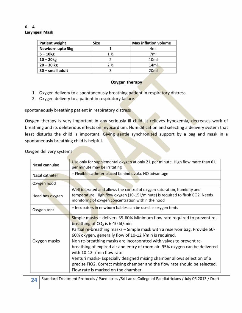
6. A Laryngeal Mask
Patient weight Size Max inflation volume
Newborn upto 5kg 1 4ml
5 – 10kg 1 ½ 7ml
10 – 20kg 2 10ml
20 – 30 kg 2 ½ 14ml
30 – small adult 3 20ml
Oxygen therapy
1. Oxygen delivery to a spontaneously breathing patient in respiratory distress. 2. Oxygen delivery to a patient in respiratory failure.
spontaneously breathing patient in respiratory distress
Oxygen therapy is very important in any seriously ill child. It relieves hypoxemia, decreases work of
breathing and its deleterious effects on myocardium. Humidification and selecting a delivery system that
least disturbs the child is important. Giving gentle synchronized support by a bag and mask in a
spontaneously breathing child is helpful.
Oxygen delivery systems
Nasal cannulae Use only for supplemental oxygen at only 2 L per minute. High flow more than 6 L per minute may be irritating
Nasal catheter – Flexible catheter placed behind uvula. NO advantage
Oxygen hood
Head box oxygen
Well tolerated and allows the control of oxygen saturation, humidity and temperature. High flow oxygen (10-15 l/minute) is required to flush CO2. Needs monitoring of oxygen concentration within the hood
Oxygen tent – Incubators in newborn babies can be used as oxygen tents
Oxygen masks
Simple masks – delivers 35-60% Minimum flow rate required to prevent re-breathing of CO2 is 6-10 lit/min Partial re-breathing masks – Simple mask with a reservoir bag. Provide 50-60% oxygen, generally flow of 10-12 l/min is required. Non re-breathing masks are incorporated with valves to prevent re-breathing of expired air and entry of room air. 95% oxygen can be delivered with 10-12 l/min flow rate. Venturi masks- Especially designed mixing chamber allows selection of a precise FiO2. Correct mixing chamber and the flow rate should be selected. Flow rate is marked on the chamber.
25 Standard Treatment Protocols / Paediatrics /Sri Lanka College of Paediatricians / July 06.2013 / Draft
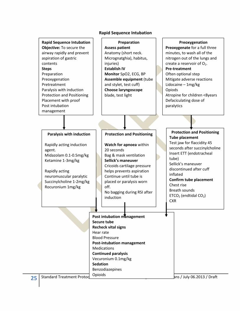
Rapid Sequence Intubation
Rapid Sequence Intubation Objective: To secure the airway rapidly and prevent aspiration of gastric contents Steps Preparation Preoxygenation Pretreatment Paralysis with induction Protection and Positioning Placement with proof Post intubation management
Preparation Assess patient Anatomy (short neck. Micrognatghia), habitus, injuries) Establish IV Monitor SpO2, ECG, BP Assemble equipment (tube and stylet, test cuff) Choose laryngoscope blade, test light
Preoxygenation Preoxygenate for a full three minutes, to wash all of the nitrogen out of the lungs and create a reservoir of O2. Pre-treatment Often optional step Mitigate adverse reactions Lidocaine – 1mg/kg Opiods Atropine for children <8years Defaciculating dose of paralytics
Paralysis with induction Rapidly acting induction agent. Midazolam 0.1-0.5mg/kg Ketamine 1-3mg/kg Rapidly acting neuromuscular paralytic Succinylcholine 1-2mg/kg Rocuronium 1mg/kg
Protection and Positioning Watch for apnoea within 20 seconds Bag & mask ventilation Sellick’s maneuver Cricoids cartilage pressure helps prevents aspiration Continue until tube is placed or paralysis worn off. No bagging during RSI after induction
Protection and Positioning Tube placement Test jaw for flaccidity 45 seconds after succinylcholine Insert ETT (endotracheal tube) Sellick’s maneuver discontinued after cuff inflated Confirm tube placement Chest rise Breath sounds ETCO2 (endtidal CO2) CXR
Post intubation management Secure tube Recheck vital signs Hear rate Blood Pressure Post-intubation management Medications Continued paralysis Vecuronium 0.1mg/kg Sedation Benzodiazepines Opioids
26 Standard Treatment Protocols / Paediatrics /Sri Lanka College of Paediatricians / July 06.2013 / Draft
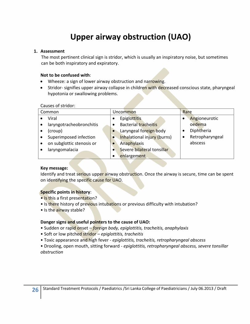
Upper airway obstruction (UAO)
1. Assessment The most pertinent clinical sign is stridor, which is usually an inspiratory noise, but sometimes can be both inspiratory and expiratory. Not to be confused with:
Wheeze: a sign of lower airway obstruction and narrowing.
Stridor- signifies upper airway collapse in children with decreased conscious state, pharyngeal hypotonia or swallowing problems.
Causes of stridor:
Common Uncommon Rare
Viral
laryngotracheobronchitis
(croup)
Superimposed infection
on subglottic stenosis or
laryngomalacia
Epiglottitis
Bacterial tracheitis
Laryngeal foreign body
Inhalational injury (burns)
Anaphylaxis
Severe bilateral tonsillar
enlargement
Angioneurotic oedema
Diphtheria
Retropharyngeal abscess
Key message: Identify and treat serious upper airway obstruction. Once the airway is secure, time can be spent on identifying the specific cause for UAO. Specific points in history: • Is this a first presentation? • Is there history of previous intubations or previous difficulty with intubation? • Is the airway stable? Danger signs and useful pointers to the cause of UAO: • Sudden or rapid onset – foreign body, epiglottitis, tracheitis, anaphylaxis • Soft or low pitched stridor – epiglottitis, tracheitis • Toxic appearance and high fever - epiglottitis, tracheitis, retropharyngeal abscess • Drooling, open mouth, sitting forward - epiglottitis, retropharyngeal abscess, severe tonsillar obstruction
27 Standard Treatment Protocols / Paediatrics /Sri Lanka College of Paediatricians / July 06.2013 / Draft
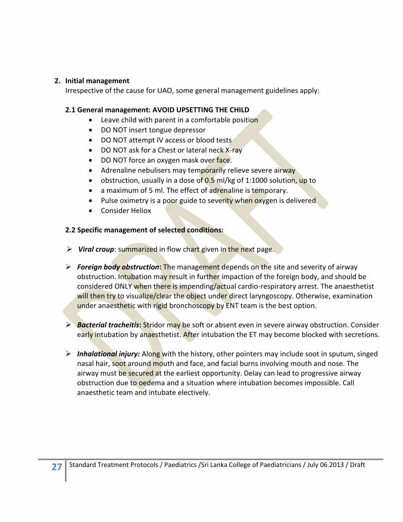
2. Initial management Irrespective of the cause for UAO, some general management guidelines apply: 2.1 General management: AVOID UPSETTING THE CHILD
Leave child with parent in a comfortable position
DO NOT insert tongue depressor
DO NOT attempt IV access or blood tests
DO NOT ask for a Chest or lateral neck X-ray
DO NOT force an oxygen mask over face.
Adrenaline nebulisers may temporarily relieve severe airway
obstruction, usually in a dose of 0.5 ml/kg of 1:1000 solution, up to
a maximum of 5 ml. The effect of adrenaline is temporary.
Pulse oximetry is a poor guide to severity when oxygen is delivered
Consider Heliox 2.2 Specific management of selected conditions: Viral croup: summarized in flow chart given in the next page.
Foreign body obstruction: The management depends on the site and severity of airway obstruction. Intubation may result in further impaction of the foreign body, and should be considered ONLY when there is impending/actual cardio-respiratory arrest. The anaesthetist will then try to visualize/clear the object under direct laryngoscopy. Otherwise, examination under anaesthetic with rigid bronchoscopy by ENT team is the best option.
Bacterial tracheitis: Stridor may be soft or absent even in severe airway obstruction. Consider early intubation by anaesthetist. After intubation the ET may become blocked with secretions.
Inhalational injury: Along with the history, other pointers may include soot in sputum, singed nasal hair, soot around mouth and face, and facial burns involving mouth and nose. The airway must be secured at the earliest opportunity. Delay can lead to progressive airway obstruction due to oedema and a situation where intubation becomes impossible. Call anaesthetic team and intubate electively.
28 Standard Treatment Protocols / Paediatrics /Sri Lanka College of Paediatricians / July 06.2013 / Draft
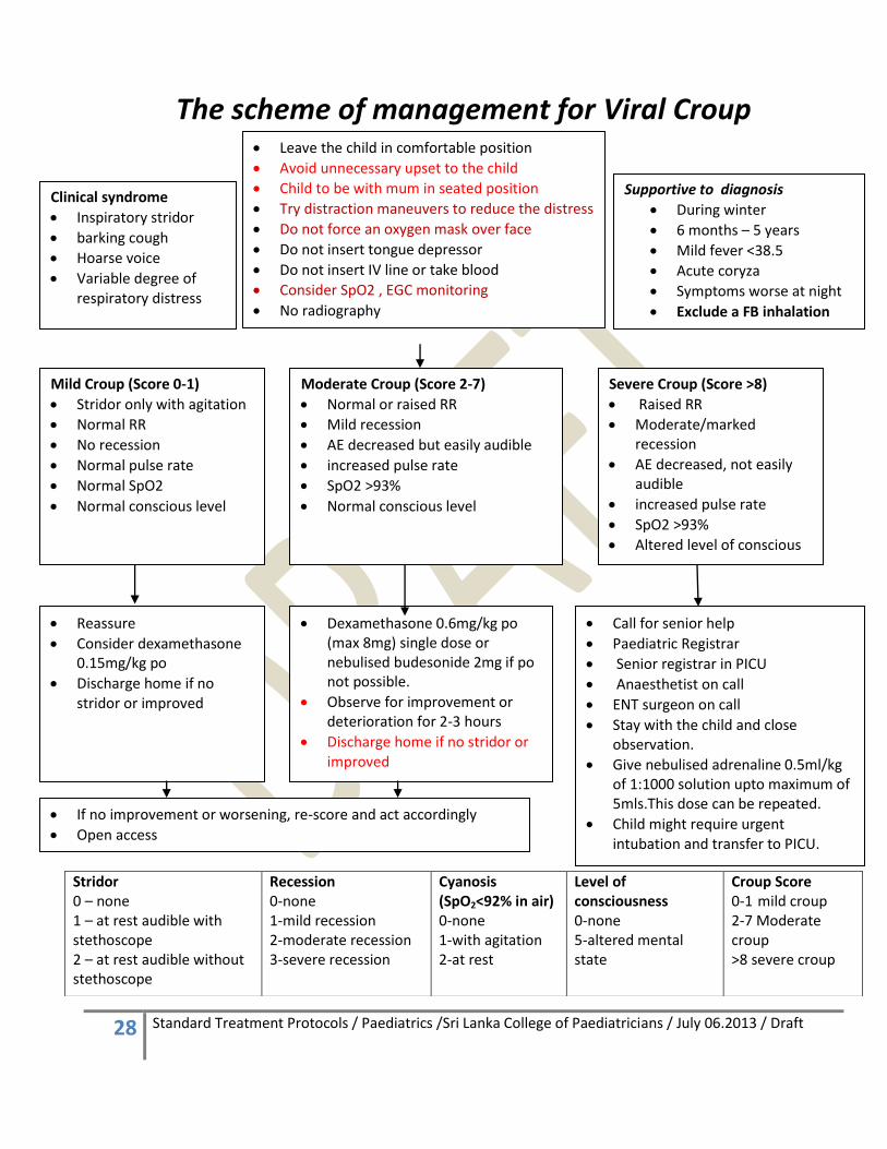
The scheme of management for Viral Croup
Stridor 0 – none 1 – at rest audible with stethoscope 2 – at rest audible without stethoscope
Recession 0-none 1-mild recession 2-moderate recession 3-severe recession
Cyanosis (SpO2<92% in air) 0-none 1-with agitation 2-at rest
Level of consciousness 0-none 5-altered mental state
Croup Score 0-1 mild croup 2-7 Moderate croup >8 severe croup
Supportive to diagnosis
During winter
6 months – 5 years
Mild fever <38.5
Acute coryza
Symptoms worse at night
Exclude a FB inhalation
Clinical syndrome
Inspiratory stridor
barking cough
Hoarse voice
Variable degree of respiratory distress
Leave the child in comfortable position
Avoid unnecessary upset to the child
Child to be with mum in seated position
Try distraction maneuvers to reduce the distress
Do not force an oxygen mask over face
Do not insert tongue depressor
Do not insert IV line or take blood
Consider SpO2 , EGC monitoring
No radiography
Mild Croup (Score 0-1)
Stridor only with agitation
Normal RR
No recession
Normal pulse rate
Normal SpO2
Normal conscious level
Reassure
Consider dexamethasone 0.15mg/kg po
Discharge home if no stridor or improved
Moderate Croup (Score 2-7)
Normal or raised RR
Mild recession
AE decreased but easily audible
increased pulse rate
SpO2 >93%
Normal conscious level
Severe Croup (Score >8)
Raised RR
Moderate/marked recession
AE decreased, not easily audible
increased pulse rate
SpO2 >93%
Altered level of conscious
Dexamethasone 0.6mg/kg po (max 8mg) single dose or nebulised budesonide 2mg if po not possible.
Observe for improvement or deterioration for 2-3 hours
Discharge home if no stridor or improved
Call for senior help
Paediatric Registrar
Senior registrar in PICU
Anaesthetist on call
ENT surgeon on call
Stay with the child and close observation.
Give nebulised adrenaline 0.5ml/kg of 1:1000 solution upto maximum of 5mls.This dose can be repeated.
Child might require urgent intubation and transfer to PICU.
If no improvement or worsening, re-score and act accordingly
Open access
29 Standard Treatment Protocols / Paediatrics /Sri Lanka College of Paediatricians / July 06.2013 / Draft
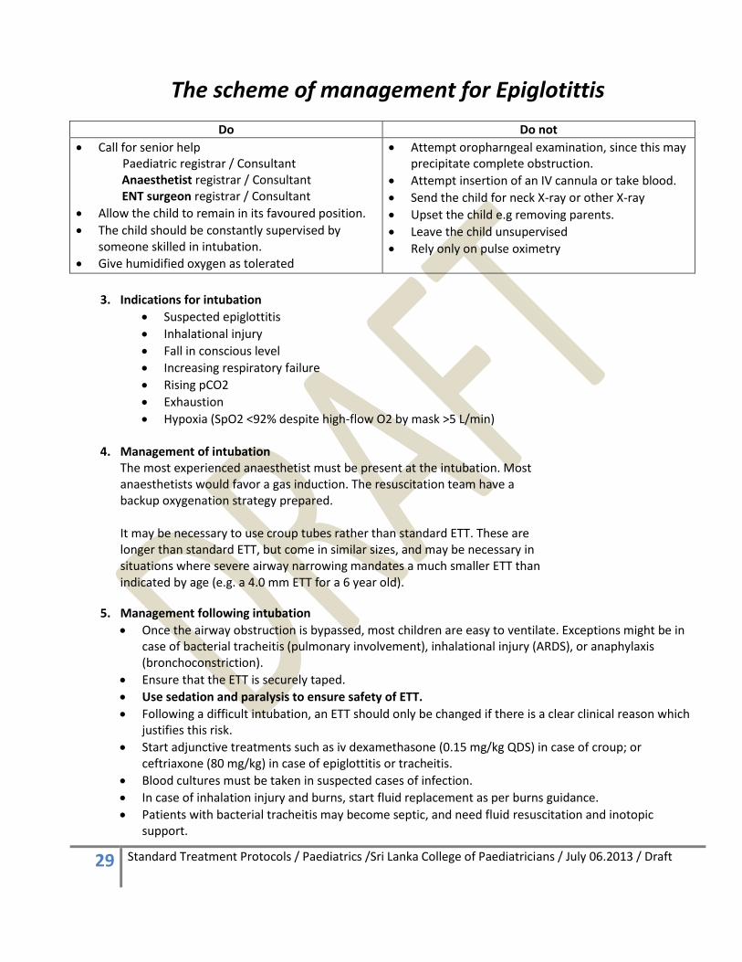
The scheme of management for Epiglotittis
Do Do not
Call for senior help Paediatric registrar / Consultant
Anaesthetist registrar / Consultant ENT surgeon registrar / Consultant
Allow the child to remain in its favoured position.
The child should be constantly supervised by someone skilled in intubation.
Give humidified oxygen as tolerated
Attempt oropharngeal examination, since this may precipitate complete obstruction.
Attempt insertion of an IV cannula or take blood.
Send the child for neck X-ray or other X-ray
Upset the child e.g removing parents.
Leave the child unsupervised
Rely only on pulse oximetry
3. Indications for intubation
Suspected epiglottitis
Inhalational injury
Fall in conscious level
Increasing respiratory failure
Rising pCO2
Exhaustion
Hypoxia (SpO2 <92% despite high-flow O2 by mask >5 L/min)
4. Management of intubation The most experienced anaesthetist must be present at the intubation. Most anaesthetists would favor a gas induction. The resuscitation team have a backup oxygenation strategy prepared. It may be necessary to use croup tubes rather than standard ETT. These are longer than standard ETT, but come in similar sizes, and may be necessary in situations where severe airway narrowing mandates a much smaller ETT than indicated by age (e.g. a 4.0 mm ETT for a 6 year old).
5. Management following intubation
Once the airway obstruction is bypassed, most children are easy to ventilate. Exceptions might be in case of bacterial tracheitis (pulmonary involvement), inhalational injury (ARDS), or anaphylaxis (bronchoconstriction).
Ensure that the ETT is securely taped.
Use sedation and paralysis to ensure safety of ETT.
Following a difficult intubation, an ETT should only be changed if there is a clear clinical reason which justifies this risk.
Start adjunctive treatments such as iv dexamethasone (0.15 mg/kg QDS) in case of croup; or ceftriaxone (80 mg/kg) in case of epiglottitis or tracheitis.
Blood cultures must be taken in suspected cases of infection.
In case of inhalation injury and burns, start fluid replacement as per burns guidance.
Patients with bacterial tracheitis may become septic, and need fluid resuscitation and inotopic support.
30 Standard Treatment Protocols / Paediatrics /Sri Lanka College of Paediatricians / July 06.2013 / Draft
6. Transport considerations
Children with an unstable airway should not be transported without detailed discussion with on call consultant.
ETCO2 monitoring is mandatory during transfer to maintain continuous correct ETT placement.
Use continuous muscle relaxation during retrieval to ensure safety of ETT.
If transporting an un-intubated child with suspected foreign body obstruction, avoid unnecessary delay and transfer immediately to ENT centre of Teaching or Provincial hospital (directly to theatres if necessary). The team must have a strategy to manage unexpected obstruction or hypoxia.
7. a
31 Standard Treatment Protocols / Paediatrics /Sri Lanka College of Paediatricians / July 06.2013 / Draft
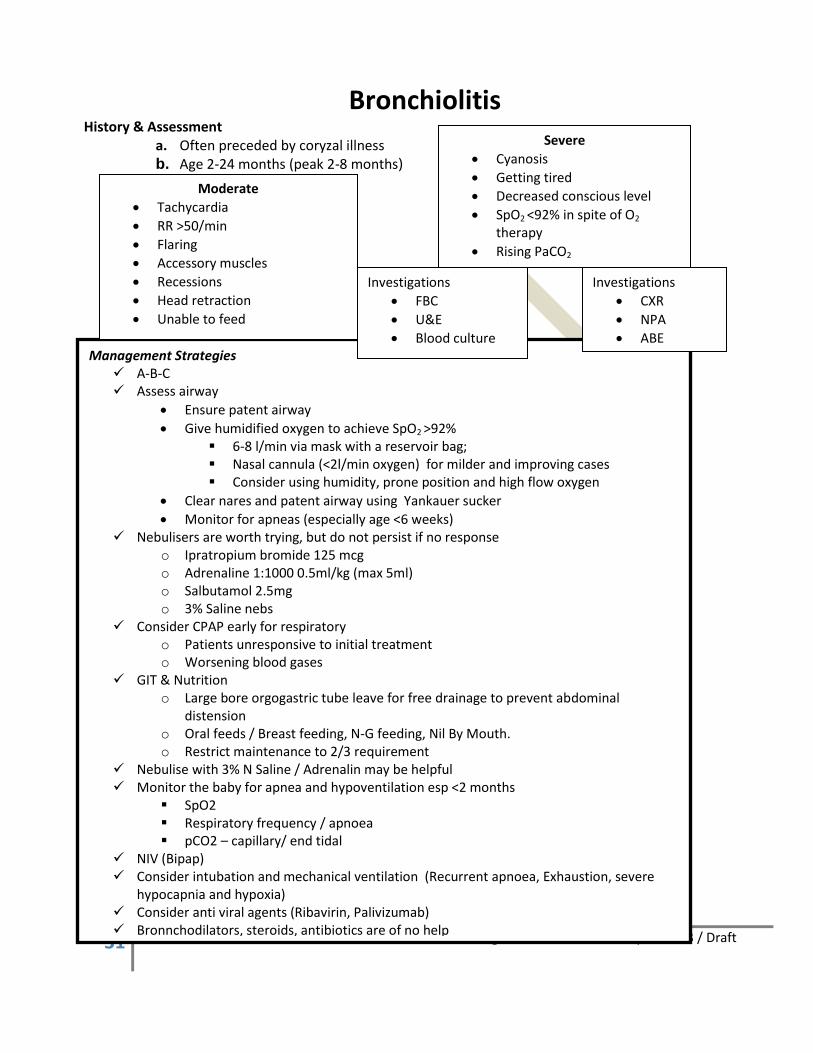
Management Strategies A-B-C Assess airway
Ensure patent airway
Give humidified oxygen to achieve SpO2 >92% 6-8 l/min via mask with a reservoir bag; Nasal cannula (<2l/min oxygen) for milder and improving cases Consider using humidity, prone position and high flow oxygen
Clear nares and patent airway using Yankauer sucker
Monitor for apneas (especially age <6 weeks) Nebulisers are worth trying, but do not persist if no response
o Ipratropium bromide 125 mcg o Adrenaline 1:1000 0.5ml/kg (max 5ml) o Salbutamol 2.5mg o 3% Saline nebs
Consider CPAP early for respiratory o Patients unresponsive to initial treatment o Worsening blood gases
GIT & Nutrition o Large bore orgogastric tube leave for free drainage to prevent abdominal
distension o Oral feeds / Breast feeding, N-G feeding, Nil By Mouth. o Restrict maintenance to 2/3 requirement
Nebulise with 3% N Saline / Adrenalin may be helpful Monitor the baby for apnea and hypoventilation esp <2 months
SpO2 Respiratory frequency / apnoea pCO2 – capillary/ end tidal
NIV (Bipap) Consider intubation and mechanical ventilation (Recurrent apnoea, Exhaustion, severe
hypocapnia and hypoxia) Consider anti viral agents (Ribavirin, Palivizumab) Bronnchodilators, steroids, antibiotics are of no help
Bronchiolitis History & Assessment
a. Often preceded by coryzal illness b. Age 2-24 months (peak 2-8 months)
Moderate
Tachycardia
RR >50/min
Flaring
Accessory muscles
Recessions
Head retraction
Unable to feed
Severe
Cyanosis
Getting tired
Decreased conscious level
SpO2 <92% in spite of O2 therapy
Rising PaCO2
Investigations
FBC
U&E
Blood culture
Investigations
CXR
NPA
ABE
32 Standard Treatment Protocols / Paediatrics /Sri Lanka College of Paediatricians / July 06.2013 / Draft
Indications for intubation
Exhausted
Recurrent apnoeas
Reduce conscious level
Worsening hypoxemia
Worsening hypercarbia Intubation
Pre-oxygenation
Fluid boluses and resuscitation drugs
Consider modified rapid sequence induction with ketamine 1-2mg/kg (bronchodilator activity)
CXR 9 post intubation) Management following intubation
Sedation for ventilation
Permissive hypercapnia strategy (limit PIP<35cmH2O, TV 5-8ml/kg, Rate <30bpm, Higher rates may leads to air trapping, I:E 1:2,
PEEP 5-7 is often necessary to counteract intrinsic PEEP. Failure to apply extrinsic PEEP at 85-100% of intrinsic PEEP will result in progressive over inflation and haemodynamic compromise.
Regular chest physiotherapy and suctioning for mucus plugging.
Check CXR for ETT position and to exclude pneumothorax. Transport Consideration
EtCO2 is mandatory to ensure ETT position
If having problems with CO2 --. Minimize dead space
Adrenaline can be administered down the ETT tube in severe air trapping.
If ventilation deteriorates – hand ventilate, auscultate, suction, perform manual decompression, treat pneumothorax.
33 Standard Treatment Protocols / Paediatrics /Sri Lanka College of Paediatricians / July 06.2013 / Draft
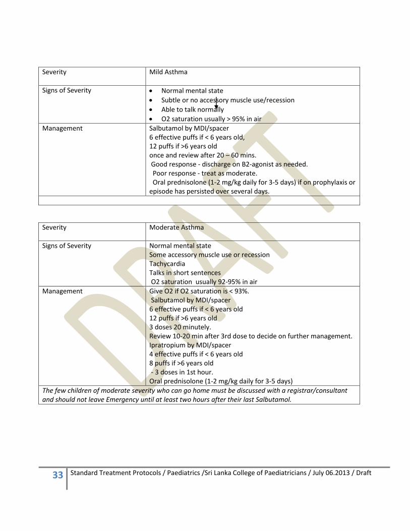
Severity Mild Asthma
Signs of Severity
Normal mental state
Subtle or no accessory muscle use/recession
Able to talk normally
O2 saturation usually > 95% in air
Management
Salbutamol by MDI/spacer 6 effective puffs if < 6 years old, 12 puffs if >6 years old once and review after 20 – 60 mins. Good response - discharge on B2-agonist as needed. Poor response - treat as moderate. Oral prednisolone (1-2 mg/kg daily for 3-5 days) if on prophylaxis or episode has persisted over several days.
Severity Moderate Asthma
Signs of Severity
Normal mental state Some accessory muscle use or recession Tachycardia Talks in short sentences O2 saturation usually 92-95% in air
Management
Give O2 if O2 saturation is < 93%. Salbutamol by MDI/spacer 6 effective puffs if < 6 years old 12 puffs if >6 years old 3 doses 20 minutely. Review 10-20 min after 3rd dose to decide on further management. Ipratropium by MDI/spacer 4 effective puffs if < 6 years old 8 puffs if >6 years old - 3 doses in 1st hour. Oral prednisolone (1-2 mg/kg daily for 3-5 days)
The few children of moderate severity who can go home must be discussed with a registrar/consultant and should not leave Emergency until at least two hours after their last Salbutamol.
34 Standard Treatment Protocols / Paediatrics /Sri Lanka College of Paediatricians / July 06.2013 / Draft
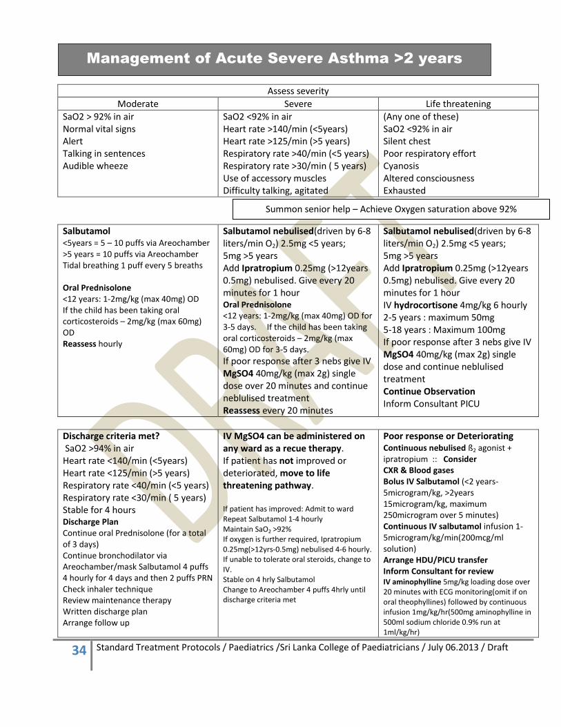
Assess severity
Moderate Severe Life threatening
SaO2 > 92% in air Normal vital signs Alert Talking in sentences Audible wheeze
SaO2 <92% in air Heart rate >140/min (<5years) Heart rate >125/min (>5 years) Respiratory rate >40/min (<5 years) Respiratory rate >30/min ( 5 years) Use of accessory muscles Difficulty talking, agitated
(Any one of these) SaO2 <92% in air Silent chest Poor respiratory effort Cyanosis Altered consciousness Exhausted
Salbutamol <5years = 5 – 10 puffs via Areochamber >5 years = 10 puffs via Areochamber Tidal breathing 1 puff every 5 breaths Oral Prednisolone <12 years: 1-2mg/kg (max 40mg) OD If the child has been taking oral corticosteroids – 2mg/kg (max 60mg) OD Reassess hourly
Salbutamol nebulised(driven by 6-8 liters/min O2) 2.5mg <5 years; 5mg >5 years Add Ipratropium 0.25mg (>12years 0.5mg) nebulised. Give every 20 minutes for 1 hour Oral Prednisolone <12 years: 1-2mg/kg (max 40mg) OD for 3-5 days. If the child has been taking oral corticosteroids – 2mg/kg (max 60mg) OD for 3-5 days.
If poor response after 3 nebs give IV MgSO4 40mg/kg (max 2g) single dose over 20 minutes and continue neblulised treatment Reassess every 20 minutes
Salbutamol nebulised(driven by 6-8 liters/min O2) 2.5mg <5 years; 5mg >5 years Add Ipratropium 0.25mg (>12years 0.5mg) nebulised. Give every 20 minutes for 1 hour IV hydrocortisone 4mg/kg 6 hourly 2-5 years : maximum 50mg 5-18 years : Maximum 100mg If poor response after 3 nebs give IV MgSO4 40mg/kg (max 2g) single dose and continue neblulised treatment Continue Observation Inform Consultant PICU
Discharge criteria met? SaO2 >94% in air Heart rate <140/min (<5years) Heart rate <125/min (>5 years) Respiratory rate <40/min (<5 years) Respiratory rate <30/min ( 5 years) Stable for 4 hours Discharge Plan Continue oral Prednisolone (for a total of 3 days) Continue bronchodilator via Areochamber/mask Salbutamol 4 puffs 4 hourly for 4 days and then 2 puffs PRN Check inhaler technique Review maintenance therapy Written discharge plan Arrange follow up
IV MgSO4 can be administered on any ward as a recue therapy. If patient has not improved or deteriorated, move to life threatening pathway. If patient has improved: Admit to ward Repeat Salbutamol 1-4 hourly Maintain SaO2 >92% If oxygen is further required, Ipratropium 0.25mg(>12yrs-0.5mg) nebulised 4-6 hourly. If unable to tolerate oral steroids, change to IV. Stable on 4 hrly Salbutamol Change to Areochamber 4 puffs 4hrly until discharge criteria met
Poor response or Deteriorating Continuous nebulised ß2 agonist + ipratropium :: Consider CXR & Blood gases Bolus IV Salbutamol (<2 years-5microgram/kg, >2years 15microgram/kg, maximum 250microgram over 5 minutes) Continuous IV salbutamol infusion 1-5microgram/kg/min(200mcg/ml solution) Arrange HDU/PICU transfer Inform Consultant for review IV aminophylline 5mg/kg loading dose over 20 minutes with ECG monitoring(omit if on oral theophyllines) followed by continuous infusion 1mg/kg/hr(500mg aminophylline in 500ml sodium chloride 0.9% run at 1ml/kg/hr)
Management of Acute Severe Asthma >2 years
Summon senior help – Achieve Oxygen saturation above 92%
35 Standard Treatment Protocols / Paediatrics /Sri Lanka College of Paediatricians / July 06.2013 / Draft
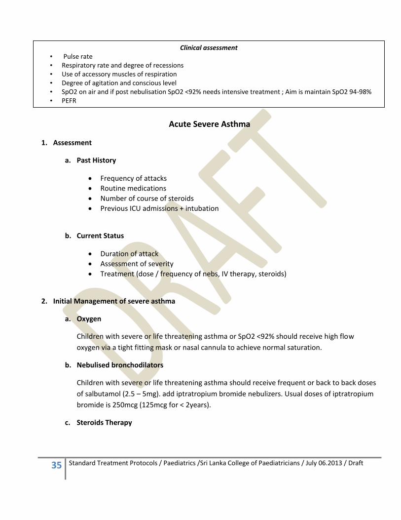
Acute Severe Asthma
1. Assessment
a. Past History
Frequency of attacks
Routine medications
Number of course of steroids
Previous ICU admissions + intubation
b. Current Status
Duration of attack
Assessment of severity
Treatment (dose / frequency of nebs, IV therapy, steroids)
2. Initial Management of severe asthma
a. Oxygen
Children with severe or life threatening asthma or SpO2 <92% should receive high flow
oxygen via a tight fitting mask or nasal cannula to achieve normal saturation.
b. Nebulised bronchodilators
Children with severe or life threatening asthma should receive frequent or back to back doses
of salbutamol (2.5 – 5mg). add iptratropium bromide nebulizers. Usual doses of iptratropium
bromide is 250mcg (125mcg for < 2years).
c. Steroids Therapy
Clinical assessment • Pulse rate • Respiratory rate and degree of recessions • Use of accessory muscles of respiration • Degree of agitation and conscious level • SpO2 on air and if post nebulisation SpO2 <92% needs intensive treatment ; Aim is maintain SpO2 94-98% • PEFR
36 Standard Treatment Protocols / Paediatrics /Sri Lanka College of Paediatricians / July 06.2013 / Draft
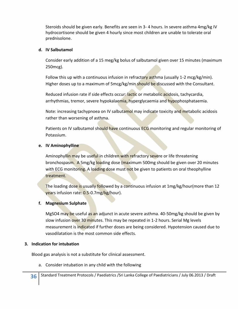
Steroids should be given early. Benefits are seen in 3- 4 hours. In severe asthma 4mg/kg IV hydrocortisone should be given 4 hourly since most children are unable to tolerate oral prednisolone.
d. IV Salbutamol
Consider early addition of a 15 meg/kg bolus of salbutamol given over 15 minutes (maximum
250mcg).
Follow this up with a continuous infusion in refractory asthma (usually 1-2 mcg/kg/min).
Higher doses up to a maximum of 5mcg/kg/min should be discussed with the Consultant.
Reduced infusion rate if side effects occur: lactic or metabolic acidosis, tachycardia,
arrhythmias, tremor, severe hypokalaemia, hyperglycaemia and hypophosphataemia.
Note: increasing tachypnoea on IV salbutamol may indicate toxicity and metabolic acidosis
rather than worsening of asthma.
Patients on IV salbutamol should have continuous ECG monitoring and regular monitoring of
Potassium.
e. IV Aminophylline
Aminophyllin may be useful in children with refractory severe or life threatening
bronchospasm. A 5mg/kg loading dose (maximum 500mg should be given over 20 minutes
with ECG monitoring. A loading dose must not be given to patients on oral theophylline
treatment.
The loading dose is usually followed by a continuous infusion at 1mg/kg/hour(more than 12
years infusion rate: 0.5-0.7mg/kg/hour).
f. Magnesium Sulphate
MgSO4 may be useful as an adjunct in acute severe asthma. 40-50mg/kg should be given by
slow infusion over 30 minutes. This may be repeated in 1-2 hours. Serial Mg levels
measurement is indicated if further doses are being considered. Hypotension caused due to
vasodilatation is the most common side effects.
3. Indication for intubation
Blood gas analysis is not a substitute for clinical assessment.
a. Consider intubation in any child with the following
37 Standard Treatment Protocols / Paediatrics /Sri Lanka College of Paediatricians / July 06.2013 / Draft
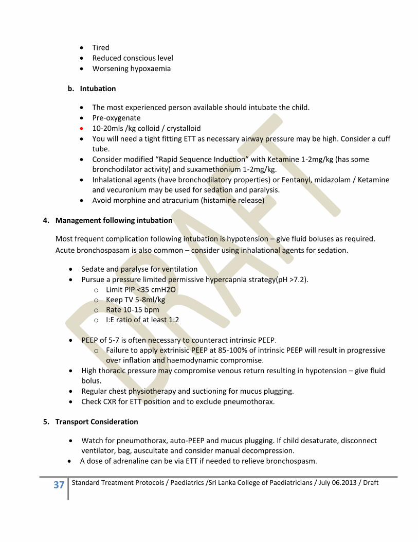
Tired
Reduced conscious level
Worsening hypoxaemia
b. Intubation
The most experienced person available should intubate the child.
Pre-oxygenate
10-20mls /kg colloid / crystalloid
You will need a tight fitting ETT as necessary airway pressure may be high. Consider a cuff tube.
Consider modified “Rapid Sequence Induction” with Ketamine 1-2mg/kg (has some bronchodilator activity) and suxamethonium 1-2mg/kg.
Inhalational agents (have bronchodilatory properties) or Fentanyl, midazolam / Ketamine and vecuronium may be used for sedation and paralysis.
Avoid morphine and atracurium (histamine release) 4. Management following intubation
Most frequent complication following intubation is hypotension – give fluid boluses as required.
Acute bronchospasam is also common – consider using inhalational agents for sedation.
Sedate and paralyse for ventilation
Pursue a pressure limited permissive hypercapnia strategy(pH >7.2). o Limit PIP <35 cmH2O o Keep TV 5-8ml/kg o Rate 10-15 bpm o I:E ratio of at least 1:2
PEEP of 5-7 is often necessary to counteract intrinsic PEEP. o Failure to apply extrinisic PEEP at 85-100% of intrinsic PEEP will result in progressive
over inflation and haemodynamic compromise.
High thoracic pressure may compromise venous return resulting in hypotension – give fluid bolus.
Regular chest physiotherapy and suctioning for mucus plugging.
Check CXR for ETT position and to exclude pneumothorax.
5. Transport Consideration
Watch for pneumothorax, auto-PEEP and mucus plugging. If child desaturate, disconnect ventilator, bag, auscultate and consider manual decompression.
A dose of adrenaline can be via ETT if needed to relieve bronchospasm.
38 Standard Treatment Protocols / Paediatrics /Sri Lanka College of Paediatricians / July 06.2013 / Draft
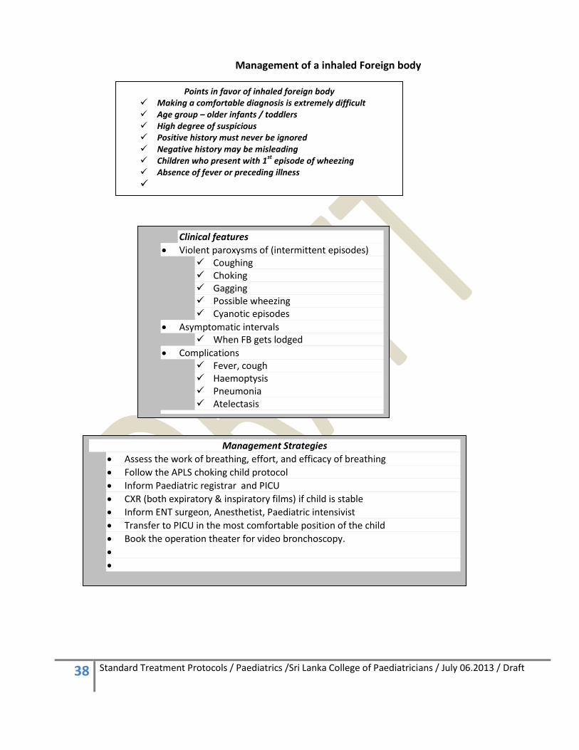
Points in favor of inhaled foreign body Making a comfortable diagnosis is extremely difficult Age group – older infants / toddlers High degree of suspicious Positive history must never be ignored Negative history may be misleading Children who present with 1
st episode of wheezing
Absence of fever or preceding illness
Management Strategies
Assess the work of breathing, effort, and efficacy of breathing
Follow the APLS choking child protocol
Inform Paediatric registrar and PICU
CXR (both expiratory & inspiratory films) if child is stable
Inform ENT surgeon, Anesthetist, Paediatric intensivist
Transfer to PICU in the most comfortable position of the child
Book the operation theater for video bronchoscopy.
Management of a inhaled Foreign body
Clinical features
Violent paroxysms of (intermittent episodes) Coughing Choking Gagging Possible wheezing Cyanotic episodes
Asymptomatic intervals When FB gets lodged
Complications Fever, cough Haemoptysis Pneumonia Atelectasis
39 Standard Treatment Protocols / Paediatrics /Sri Lanka College of Paediatricians / July 06.2013 / Draft
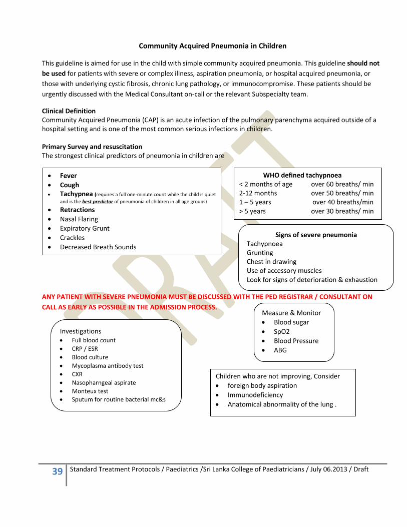
Community Acquired Pneumonia in Children
This guideline is aimed for use in the child with simple community acquired pneumonia. This guideline should not
be used for patients with severe or complex illness, aspiration pneumonia, or hospital acquired pneumonia, or
those with underlying cystic fibrosis, chronic lung pathology, or immunocompromise. These patients should be
urgently discussed with the Medical Consultant on-call or the relevant Subspecialty team.
Clinical Definition Community Acquired Pneumonia (CAP) is an acute infection of the pulmonary parenchyma acquired outside of a hospital setting and is one of the most common serious infections in children. Primary Survey and resuscitation The strongest clinical predictors of pneumonia in children are
ANY PATIENT WITH SEVERE PNEUMONIA MUST BE DISCUSSED WITH THE PED REGISTRAR / CONSULTANT ON
CALL AS EARLY AS POSSIBLE IN THE ADMISSION PROCESS.
WHO defined tachypnoea
< 2 months of age over 60 breaths/ min 2-12 months over 50 breaths/ min 1 – 5 years over 40 breaths/min > 5 years over 30 breaths/ min
Fever
Cough Tachypnea (requires a full one-minute count while the child is quiet
and is the best predictor of pneumonia of children in all age groups)
Retractions
Nasal Flaring
Expiratory Grunt
Crackles
Decreased Breath Sounds
Signs of severe pneumonia Tachypnoea Grunting Chest in drawing Use of accessory muscles Look for signs of deterioration & exhaustion
Investigations Full blood count
CRP / ESR
Blood culture
Mycoplasma antibody test
CXR
Nasopharngeal aspirate
Monteux test
Sputum for routine bacterial mc&s
Children who are not improving, Consider
foreign body aspiration
Immunodeficiency
Anatomical abnormality of the lung .
Measure & Monitor
Blood sugar
SpO2
Blood Pressure
ABG
40 Standard Treatment Protocols / Paediatrics /Sri Lanka College of Paediatricians / July 06.2013 / Draft
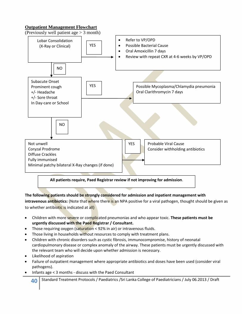
Outpatient Management Flowchart
(Previously well patient age > 3 month)
The following patients should be strongly considered for admission and inpatient management with
intravenous antibiotics: (Note that where there is an NPA positive for a viral pathogen, thought should be given as
to whether antibiotic is indicated at all)
Children with more severe or complicated pneumonias and who appear toxic. These patients must be urgently discussed with the Paed Registrar / Consultant.
Those requiring oxygen (saturation < 92% in air) or intravenous fluids.
Those living in households without resources to comply with treatment plans.
Children with chronic disorders such as cystic fibrosis, immunocompromise, history of neonatal cardiopulmonary disease or complex anomaly of the airway. These patients must be urgently discussed with the relevant team who will decide upon whether admission is necessary.
Likelihood of aspiration
Failure of outpatient management where appropriate antibiotics and doses have been used (consider viral pathogens).
Infants age < 3 months - discuss with the Paed Consultant
Lobar Consolidation (X-Ray or Clinical)
Refer to VP/OPD
Possible Bacterial Cause
Oral Amoxicillin 7 days
Review with repeat CXR at 4-6 weeks by VP/OPD
Subacute Onset Prominent cough +/- Headache +/- Sore throat In Day-care or School
Possible Mycoplasma/Chlamydia pneumonia Oral Clarithromycin 7 days
Not unwell Coryzal Prodrome Diffuse Crackles Fully immunised Minimal patchy bilateral X-Ray changes (if done)
Probable Viral Cause Consider withholding antibiotics
All patients require, Paed Registrar review if not improving for admission.
YES
YES
YES
NO
NO
41 Standard Treatment Protocols / Paediatrics /Sri Lanka College of Paediatricians / July 06.2013 / Draft
Intravenous Antibiotics 1) Simple CAP: Benzylpenicillin – 30 mg/kg/dose (max 1.2g) 6 hourly 2) Age < 3 months: ADD Gentamicin 8 mg/kg iv daily (if age < 4weeks, consult neonatal dosing guidelines). 3) If clinical suspicion of Mycoplasma pneumonia or Chlamydophila pneumoniae AND patient over 1 month age:
a) ADD Roxithromycin orally 4 mg/kg/dose (max 150mg) 12 hourly. b) If patient unable to tolerate oral antibiotics then use intravenous Azithromycin 10mg/kg/dose (maximum
500mg) daily. 4) For severe/toxic patient (systemic toxicity, increasing 02 dependence) and/or suspicion of Staphylococcus
aureus (cavitations, multiple lesions, effusions, empyema, increased incidence in Indigenous and Pacific Island Children)
1. These patients must be urgently discussed with the PED and General Medical Consultant on-call. Following admission, ID consultation may be considered.
2. Ceftriaxone intravenous 50 mg/kg/dose (max 1g) once daily PLUS 3. Flucloxacillin intravenous 50 mg/kg/dose (max 2g) 6 hourly 4. If community acquired MRSA is suspected or proven, consult ID for approval of intravenous
Vancomycin 25 mg/kg/dose up to maximum 1g 12 hourly until sensitivity results known.
Discharge Plan (if improved at 24-48 hours)
Antibiotics should be directed towards pathogen identified.
1) Amoxicillin orally 25mg/kg/dose (maximum dose up to 1g) 8 hourly. Total duration of antibiotic therapy for 7 days
2) Roxithromycin oral dose as above for total 7 days if Mycoplasma pneumonia or Chlamydophila pneumonia identified or strongly suspected
3) Choice and duration of oral therapy for Staphylococcus aureus pneumonia should be discussed with ID 4) Early clinic review at 1 week 5) If lobar consolidation, then needs repeat CXR at 4-6 weeks
a) If normal – nil further action b) If abnormal – urgent referral for General Medical OPD review
42 Standard Treatment Protocols / Paediatrics /Sri Lanka College of Paediatricians / July 06.2013 / Draft
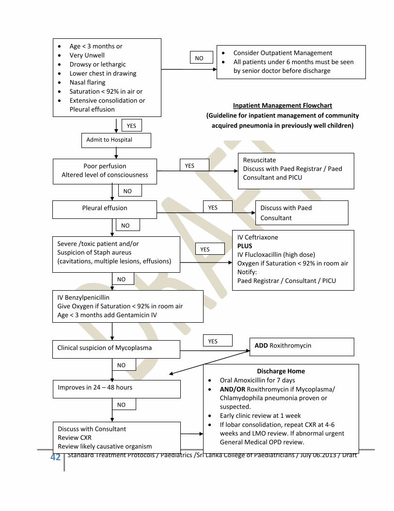
YES
ADD Roxithromycin
Admit to Hospital
IV Ceftriaxone PLUS IV Flucloxacillin (high dose) Oxygen if Saturation < 92% in room air Notify: Paed Registrar / Consultant / PICU
Discuss with Paed
Consultant
NO
Resuscitate Discuss with Paed Registrar / Paed Consultant and PICU
Consider Outpatient Management
All patients under 6 months must be seen by senior doctor before discharge
Poor perfusion Altered level of consciousness
YES
NO
Pleural effusion YES
Severe /toxic patient and/or Suspicion of Staph aureus (cavitations, multiple lesions, effusions)
NO
YES
NO
Clinical suspicion of Mycoplasma YES
IV Benzylpenicillin Give Oxygen if Saturation < 92% in room air Age < 3 months add Gentamicin IV
Improves in 24 – 48 hours
YES
NO
NO
Discharge Home
Oral Amoxicillin for 7 days
AND/OR Roxithromycin if Mycoplasma/ Chlamydophila pneumonia proven or suspected.
Early clinic review at 1 week
If lobar consolidation, repeat CXR at 4-6 weeks and LMO review. If abnormal urgent General Medical OPD review.
Discuss with Consultant Review CXR Review likely causative organism
Inpatient Management Flowchart
(Guideline for inpatient management of community
acquired pneumonia in previously well children)
Age < 3 months or
Very Unwell
Drowsy or lethargic
Lower chest in drawing
Nasal flaring
Saturation < 92% in air or
Extensive consolidation or Pleural effusion
43 Standard Treatment Protocols / Paediatrics /Sri Lanka College of Paediatricians / July 06.2013 / Draft
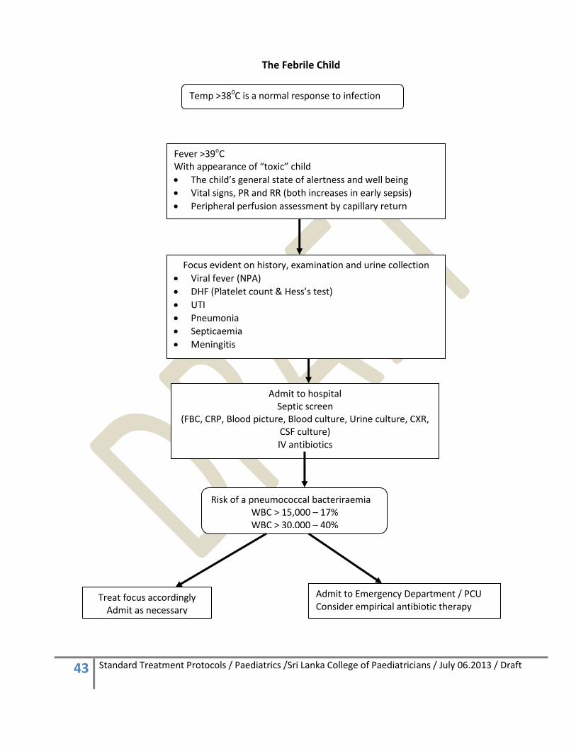
The Febrile Child
Temp >380C is a normal response to infection
Fever >39oC With appearance of “toxic” child
The child’s general state of alertness and well being
Vital signs, PR and RR (both increases in early sepsis)
Peripheral perfusion assessment by capillary return
Focus evident on history, examination and urine collection
Viral fever (NPA)
DHF (Platelet count & Hess’s test)
UTI
Pneumonia
Septicaemia
Meningitis
Admit to hospital Septic screen
(FBC, CRP, Blood picture, Blood culture, Urine culture, CXR, CSF culture) IV antibiotics
Treat focus accordingly Admit as necessary
Risk of a pneumococcal bacteriraemia WBC > 15,000 – 17% WBC > 30,000 – 40%
Admit to Emergency Department / PCU Consider empirical antibiotic therapy
44 Standard Treatment Protocols / Paediatrics /Sri Lanka College of Paediatricians / July 06.2013 / Draft
Systaemic Inflammatory Response Signs (SIRS)
The inflammatory triad of fever, tachycardia & abnormal perfusion is very common in children with benign infections. Septic shock should be considered in children who manifest this triad with additional features such as tachypnoea, reduced urine output , irritability & lethargy / drowsiness.
1. Systemic Inflammatory Response Syndrome (SIRS) Suspected infection • Hypo or hyperthermia (temp <36° or >38.5°) • Tachycardia • Tachypnoea • Altered mental status • Decreased urine output (<1 ml/kg/min) • Other end organ dysfunction • Signs of either cold or warm shock
2. Signs of shock
3. Types of shock
4. Compensated vs Uncompensated shock
45 Standard Treatment Protocols / Paediatrics /Sri Lanka College of Paediatricians / July 06.2013 / Draft
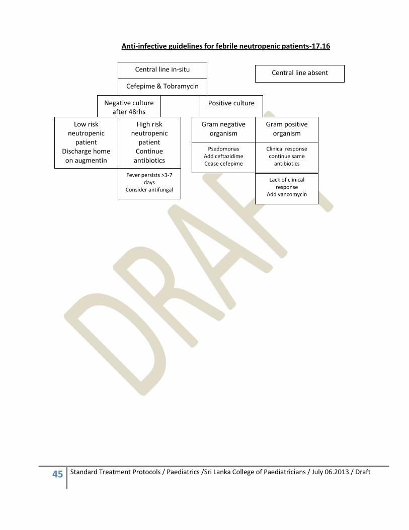
Anti-infective guidelines for febrile neutropenic patients-17.16
Central line in-situ Central line absent
Cefepime & Tobramycin
Negative culture after 48rhs
Positive culture
Low risk neutropenic
patient Discharge home on augmentin
High risk neutropenic
patient Continue
antibiotics
Gram negative organism
Gram positive organism
Fever persists >3-7 days
Consider antifungal
Psedomonas Add ceftazidime Cease cefepime
Clinical response continue same
antibiotics
Lack of clinical response
Add vancomycin
46 Standard Treatment Protocols / Paediatrics /Sri Lanka College of Paediatricians / July 06.2013 / Draft
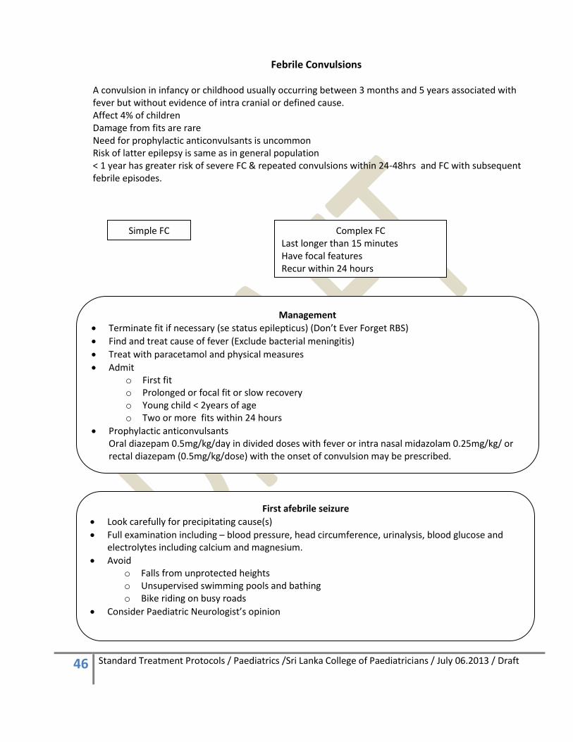
Febrile Convulsions A convulsion in infancy or childhood usually occurring between 3 months and 5 years associated with fever but without evidence of intra cranial or defined cause. Affect 4% of children Damage from fits are rare Need for prophylactic anticonvulsants is uncommon Risk of latter epilepsy is same as in general population < 1 year has greater risk of severe FC & repeated convulsions within 24-48hrs and FC with subsequent febrile episodes.
Simple FC Complex FC Last longer than 15 minutes Have focal features Recur within 24 hours
Management
Terminate fit if necessary (se status epilepticus) (Don’t Ever Forget RBS)
Find and treat cause of fever (Exclude bacterial meningitis)
Treat with paracetamol and physical measures
Admit o First fit o Prolonged or focal fit or slow recovery o Young child < 2years of age o Two or more fits within 24 hours
Prophylactic anticonvulsants Oral diazepam 0.5mg/kg/day in divided doses with fever or intra nasal midazolam 0.25mg/kg/ or rectal diazepam (0.5mg/kg/dose) with the onset of convulsion may be prescribed.
First afebrile seizure
Look carefully for precipitating cause(s)
Full examination including – blood pressure, head circumference, urinalysis, blood glucose and electrolytes including calcium and magnesium.
Avoid o Falls from unprotected heights o Unsupervised swimming pools and bathing o Bike riding on busy roads
Consider Paediatric Neurologist’s opinion
47 Standard Treatment Protocols / Paediatrics /Sri Lanka College of Paediatricians / July 06.2013 / Draft
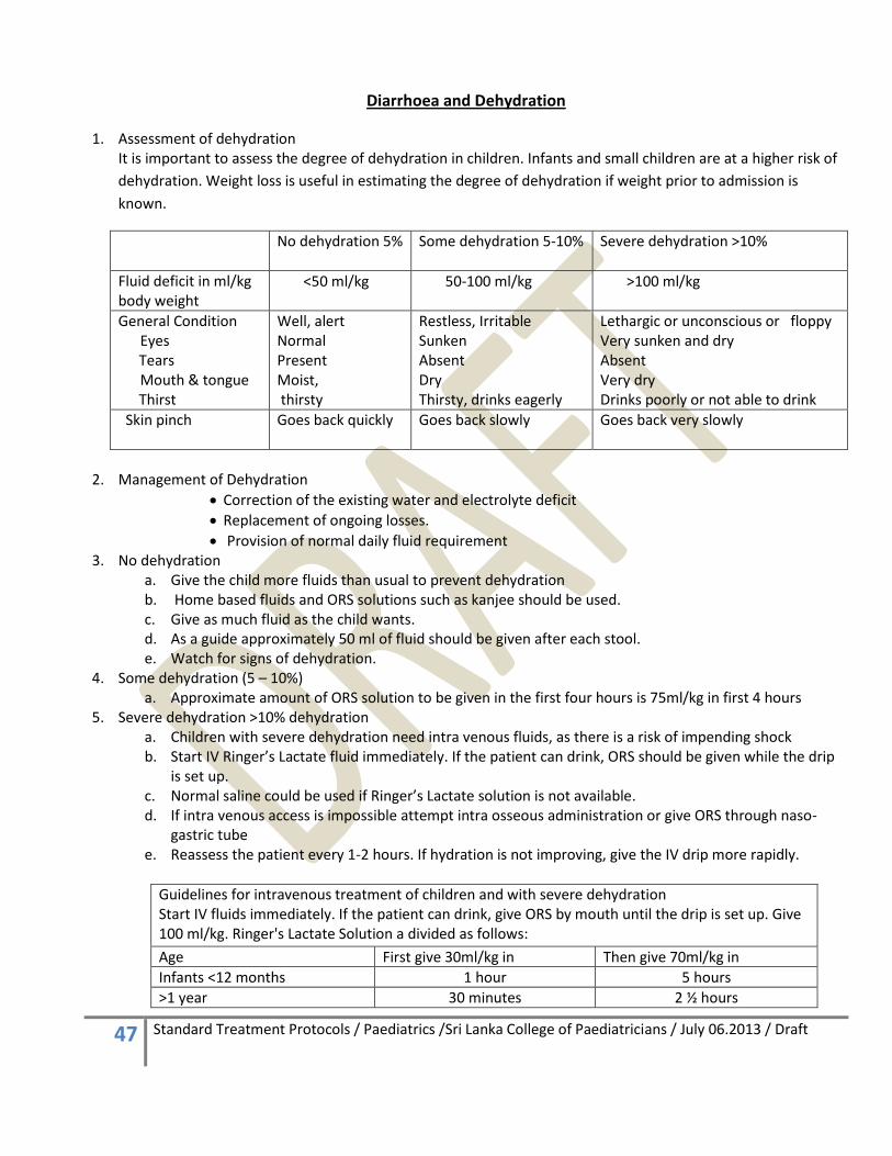
Diarrhoea and Dehydration
1. Assessment of dehydration It is important to assess the degree of dehydration in children. Infants and small children are at a higher risk of
dehydration. Weight loss is useful in estimating the degree of dehydration if weight prior to admission is
known.
No dehydration 5% Some dehydration 5-10% Severe dehydration >10%
Fluid deficit in ml/kg body weight
<50 ml/kg 50-100 ml/kg >100 ml/kg
General Condition Eyes
Tears Mouth & tongue
Thirst
Well, alert Normal Present Moist, thirsty
Restless, Irritable Sunken Absent Dry Thirsty, drinks eagerly
Lethargic or unconscious or floppy Very sunken and dry Absent Very dry Drinks poorly or not able to drink
Skin pinch Goes back quickly Goes back slowly
Goes back very slowly
2. Management of Dehydration
Correction of the existing water and electrolyte deficit
Replacement of ongoing losses.
Provision of normal daily fluid requirement 3. No dehydration
a. Give the child more fluids than usual to prevent dehydration b. Home based fluids and ORS solutions such as kanjee should be used. c. Give as much fluid as the child wants. d. As a guide approximately 50 ml of fluid should be given after each stool. e. Watch for signs of dehydration.
4. Some dehydration (5 – 10%) a. Approximate amount of ORS solution to be given in the first four hours is 75ml/kg in first 4 hours
5. Severe dehydration >10% dehydration a. Children with severe dehydration need intra venous fluids, as there is a risk of impending shock b. Start IV Ringer’s Lactate fluid immediately. If the patient can drink, ORS should be given while the drip
is set up. c. Normal saline could be used if Ringer’s Lactate solution is not available. d. If intra venous access is impossible attempt intra osseous administration or give ORS through naso-
gastric tube e. Reassess the patient every 1-2 hours. If hydration is not improving, give the IV drip more rapidly.
Guidelines for intravenous treatment of children and with severe dehydration Start IV fluids immediately. If the patient can drink, give ORS by mouth until the drip is set up. Give 100 ml/kg. Ringer's Lactate Solution a divided as follows:
Age First give 30ml/kg in Then give 70ml/kg in
Infants <12 months 1 hour 5 hours
>1 year 30 minutes 2 ½ hours
48 Standard Treatment Protocols / Paediatrics /Sri Lanka College of Paediatricians / July 06.2013 / Draft
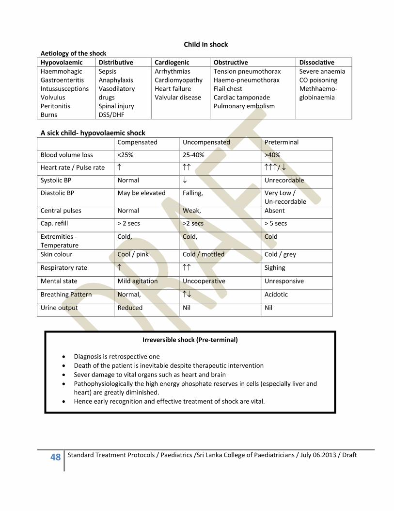
Child in shock Aetiology of the shock
Hypovolaemic Distributive Cardiogenic Obstructive Dissociative
Haemmohagic Gastroenteritis Intussusceptions Volvulus Peritonitis Burns
Sepsis Anaphylaxis Vasodilatory drugs Spinal injury DSS/DHF
Arrhythmias Cardiomyopathy Heart failure Valvular disease
Tension pneumothorax Haemo-pneumothorax Flail chest Cardiac tamponade Pulmonary embolism
Severe anaemia CO poisoning Methhaemo- globinaemia
A sick child- hypovolaemic shock Compensated Uncompensated Preterminal
Blood volume loss <25% 25-40% >40%
Heart rate / Pulse rate /
Systolic BP Normal Unrecordable
Diastolic BP May be elevated Falling, Very Low / Un-recordable
Central pulses Normal Weak, Absent
Cap. refill > 2 secs >2 secs > 5 secs
Extremities - Temperature
Cold, Cold, Cold
Skin colour Cool / pink Cold / mottled Cold / grey
Respiratory rate Sighing
Mental state Mild agitation Uncooperative Unresponsive
Breathing Pattern Normal, Acidotic
Urine output Reduced Nil Nil
Irreversible shock (Pre-terminal)
Diagnosis is retrospective one
Death of the patient is inevitable despite therapeutic intervention
Sever damage to vital organs such as heart and brain
Pathophysiologically the high energy phosphate reserves in cells (especially liver and heart) are greatly diminished.
Hence early recognition and effective treatment of shock are vital.
49 Standard Treatment Protocols / Paediatrics /Sri Lanka College of Paediatricians / July 06.2013 / Draft
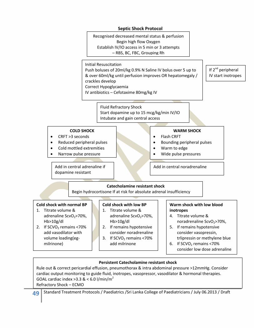
Septic Shock Protocol
Recognised decreased mental status & perfusion Begin high flow Oxygen
Establish IV/IO access in 5 min or 3 attempts – RBS, BC, FBC, Grouping Rh
Initial Resuscitation Push boluses of 20ml/kg 0.9% N Saline IV bolus over 5 up to & over 60ml/kg until perfusion improves OR hepatomegaly / crackles develop Correct Hypoglycaemia IV antibiotics – Cefotaxime 80mg/kg IV
COLD SHOCK
CRFT >3 seconds
Reduced peripheral pulses
Cold mottled extremities
Narrow pulse pressure
WARM SHOCK
Flash CRFT
Bounding peripheral pulses
Warm to edge
Wide pulse pressures
Catecholamine resistant shock Begin hydrocortisone If at risk for absolute adrenal insufficiency
Add in central noradrenaline Add in central adrenaline if dopamine resistant
Fluid Refractory Shock Start dopamine up to 15 mcg/kg/min IV/IO Intubate and gain central access
If 2nd peripheral IV start inotropes
Cold shock with normal BP 1. Titrate volume &
adrenaline ScvO2>70%, Hb>10g/dl
2. If SCVO2 remains <70% add vasodilator with volume loading(eg-milrinone)
Cold shock with low BP 1. Titrate volume &
adrenaline ScvO2>70%, Hb>10g/dl
2. If remains hypotensive consider noradrenaline
3. If SCVO2 remains <70% add milrinone
Warm shock with low blood inotropes 4. Titrate volume &
noradrenaline ScvO2>70%, 5. If remains hypotensive
consider vasopressin, trlipressin or methylene blue
6. If SCVO2 remains <70% consider low dose adrenaline
Persistent Catecholamine resistant shock Rule out & correct pericardial effusion, pneumothorax & intra abdominal pressure >12mmHg. Consider cardiac output monitoring to guide fluid, inotropes, vasopressor, vasodilator & hormonal therapies. GOAL cardiac index >3.3 & < 6.0 l/min/m2
Refractory Shock – ECMO
50 Standard Treatment Protocols / Paediatrics /Sri Lanka College of Paediatricians / July 06.2013 / Draft
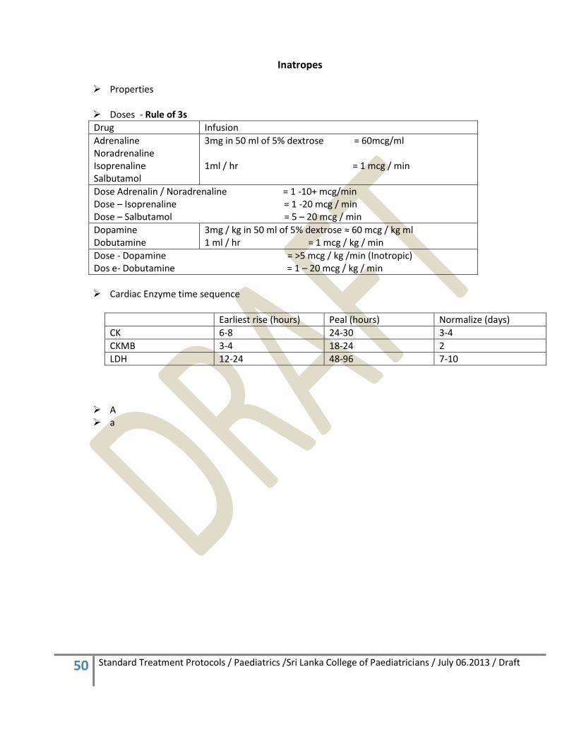
Inatropes
Properties
Doses - Rule of 3s
Drug Infusion
Adrenaline Noradrenaline Isoprenaline Salbutamol
3mg in 50 ml of 5% dextrose = 60mcg/ml 1ml / hr = 1 mcg / min
Dose Adrenalin / Noradrenaline = 1 -10+ mcg/min Dose – Isoprenaline = 1 -20 mcg / min Dose – Salbutamol = 5 – 20 mcg / min
Dopamine Dobutamine
3mg / kg in 50 ml of 5% dextrose ≈ 60 mcg / kg ml 1 ml / hr = 1 mcg / kg / min
Dose - Dopamine = >5 mcg / kg /min (Inotropic) Dos e- Dobutamine = 1 – 20 mcg / kg / min
Cardiac Enzyme time sequence
Earliest rise (hours) Peal (hours) Normalize (days)
CK 6-8 24-30 3-4
CKMB 3-4 18-24 2
LDH 12-24 48-96 7-10
A a
51 Standard Treatment Protocols / Paediatrics /Sri Lanka College of Paediatricians / July 06.2013 / Draft
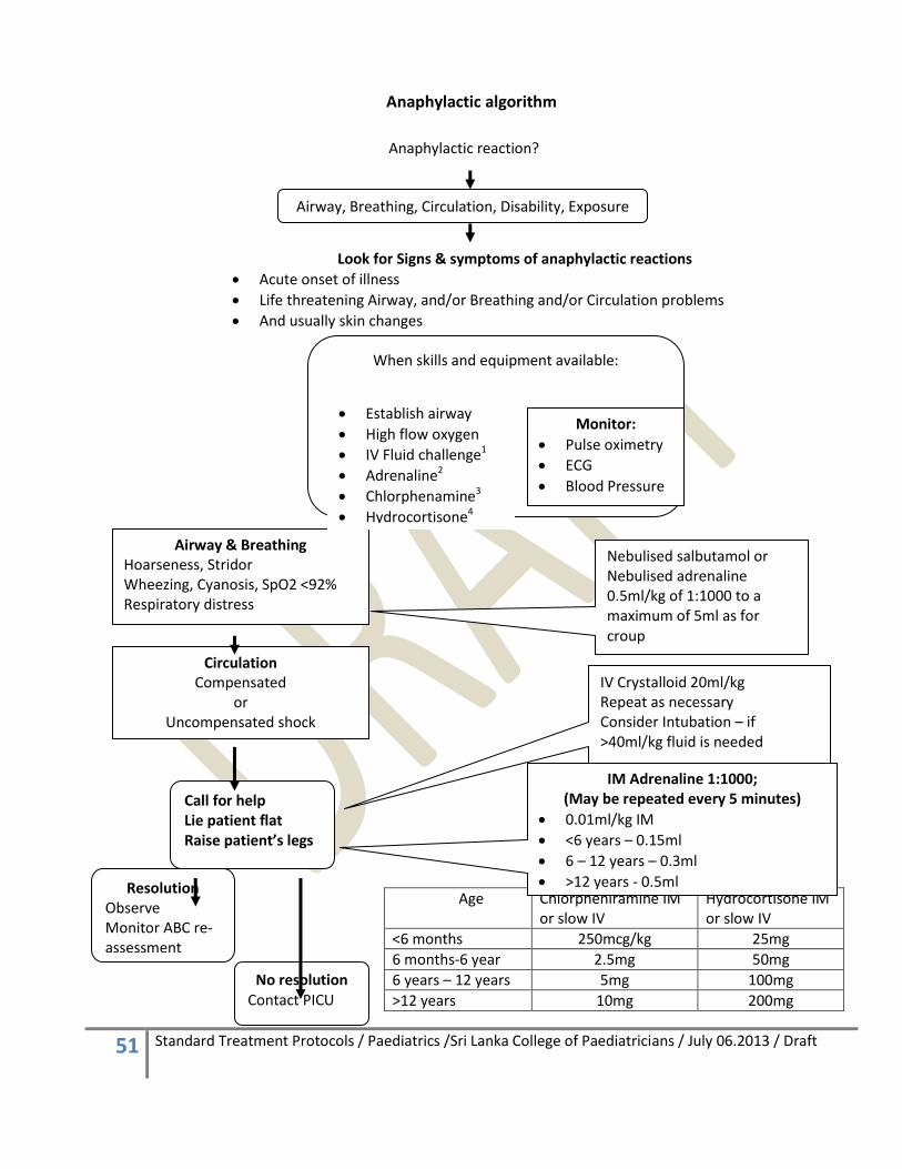
Anaphylactic algorithm
Age Chlorpheniramine IM or slow IV
Hydrocortisone IM or slow IV
<6 months 250mcg/kg 25mg
6 months-6 year 2.5mg 50mg
6 years – 12 years 5mg 100mg
>12 years 10mg 200mg
Look for Signs & symptoms of anaphylactic reactions
Acute onset of illness
Life threatening Airway, and/or Breathing and/or Circulation problems
And usually skin changes
Airway & Breathing Hoarseness, Stridor Wheezing, Cyanosis, SpO2 <92% Respiratory distress
Call for help Lie patient flat Raise patient’s legs
IV Crystalloid 20ml/kg Repeat as necessary Consider Intubation – if >40ml/kg fluid is needed
IM Adrenaline 1:1000; (May be repeated every 5 minutes)
0.01ml/kg IM
<6 years – 0.15ml
6 – 12 years – 0.3ml
>12 years - 0.5ml
Nebulised salbutamol or Nebulised adrenaline 0.5ml/kg of 1:1000 to a maximum of 5ml as for croup
Anaphylactic reaction?
Airway, Breathing, Circulation, Disability, Exposure
When skills and equipment available:
Monitor:
Pulse oximetry
ECG
Blood Pressure
Establish airway
High flow oxygen
IV Fluid challenge1
Adrenaline2
Chlorphenamine3
Hydrocortisone4
Circulation Compensated
or Uncompensated shock
No resolution Contact PICU
Resolution Observe Monitor ABC re-assessment
52 Standard Treatment Protocols / Paediatrics /Sri Lanka College of Paediatricians / July 06.2013 / Draft
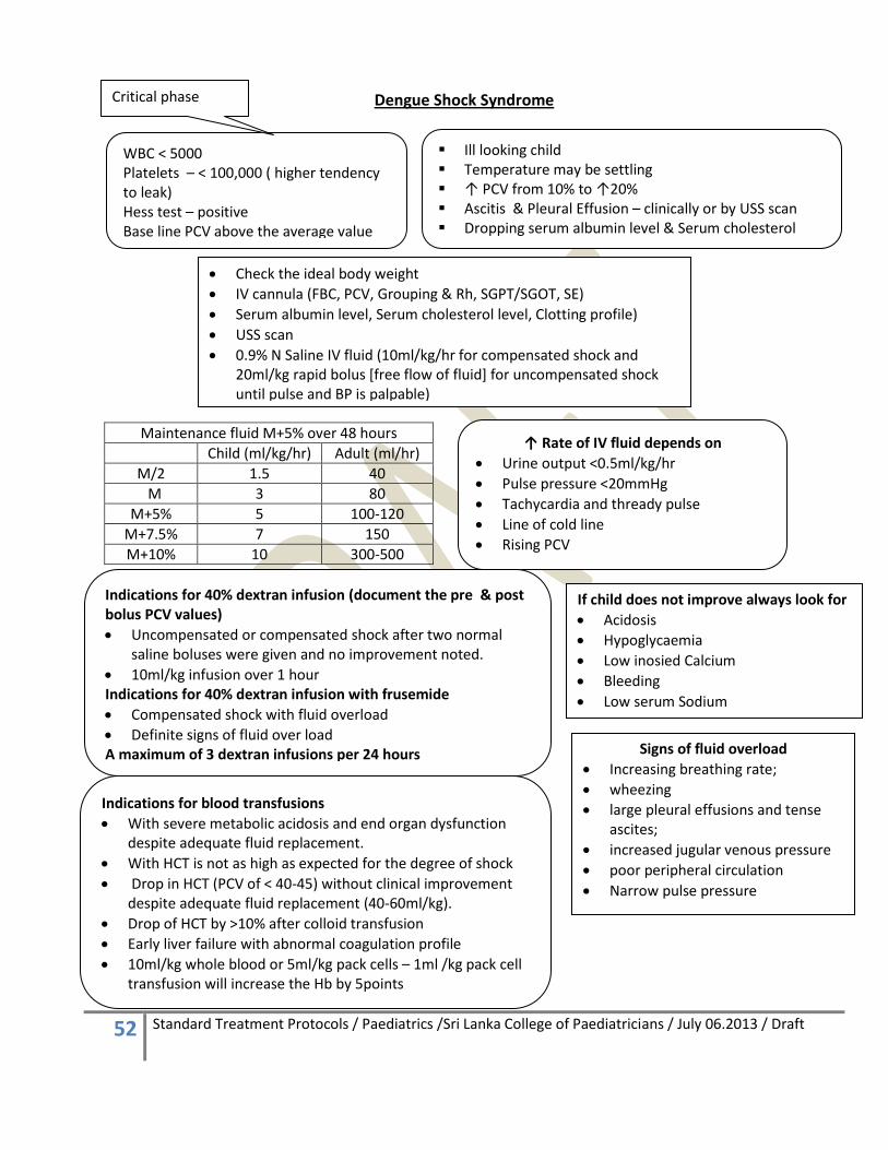
Dengue Shock Syndrome
Maintenance fluid M+5% over 48 hours
Child (ml/kg/hr) Adult (ml/hr)
M/2 1.5 40
M 3 80
M+5% 5 100-120
M+7.5% 7 150
M+10% 10 300-500
WBC < 5000 Platelets – < 100,000 ( higher tendency to leak) Hess test – positive Base line PCV above the average value
Critical phase
Ill looking child Temperature may be settling ↑ PCV from 10% to ↑20% Ascitis & Pleural Effusion – clinically or by USS scan Dropping serum albumin level & Serum cholesterol
level
Check the ideal body weight
IV cannula (FBC, PCV, Grouping & Rh, SGPT/SGOT, SE)
Serum albumin level, Serum cholesterol level, Clotting profile)
USS scan
0.9% N Saline IV fluid (10ml/kg/hr for compensated shock and 20ml/kg rapid bolus [free flow of fluid] for uncompensated shock until pulse and BP is palpable)
Catheterize the child
↑ Rate of IV fluid depends on
Urine output <0.5ml/kg/hr
Pulse pressure <20mmHg
Tachycardia and thready pulse
Line of cold line
Rising PCV
Indications for 40% dextran infusion (document the pre & post bolus PCV values)
Uncompensated or compensated shock after two normal saline boluses were given and no improvement noted.
10ml/kg infusion over 1 hour Indications for 40% dextran infusion with frusemide
Compensated shock with fluid overload
Definite signs of fluid over load A maximum of 3 dextran infusions per 24 hours
Indications for blood transfusions
With severe metabolic acidosis and end organ dysfunction despite adequate fluid replacement.
With HCT is not as high as expected for the degree of shock
Drop in HCT (PCV of < 40-45) without clinical improvement despite adequate fluid replacement (40-60ml/kg).
Drop of HCT by >10% after colloid transfusion
Early liver failure with abnormal coagulation profile
10ml/kg whole blood or 5ml/kg pack cells – 1ml /kg pack cell transfusion will increase the Hb by 5points
If child does not improve always look for
Acidosis
Hypoglycaemia
Low inosied Calcium
Bleeding
Low serum Sodium
Signs of fluid overload
Increasing breathing rate;
wheezing
large pleural effusions and tense ascites;
increased jugular venous pressure
poor peripheral circulation
Narrow pulse pressure
53 Standard Treatment Protocols / Paediatrics /Sri Lanka College of Paediatricians / July 06.2013 / Draft
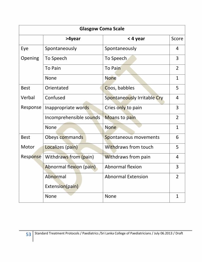
Glasgow Coma Scale
>4year < 4 year Score
Eye
Opening
Spontaneously Spontaneously 4
To Speech To Speech 3
To Pain To Pain 2
None None 1
Best
Verbal
Response
Orientated Coos, babbles 5
Confused Spontaneously Irritable Cry 4
Inappropriate words Cries only to pain 3
Incomprehensible sounds Moans to pain 2
None None 1
Best
Motor
Response
Obeys commands Spontaneous movements 6
Localizes (pain) Withdraws from touch 5
Withdraws from (pain) Withdraws from pain 4
Abnormal flexion (pain) Abnormal flexion 3
Abnormal
Extension(pain)
Abnormal Extension 2
None None 1
54 Standard Treatment Protocols / Paediatrics /Sri Lanka College of Paediatricians / July 06.2013 / Draft
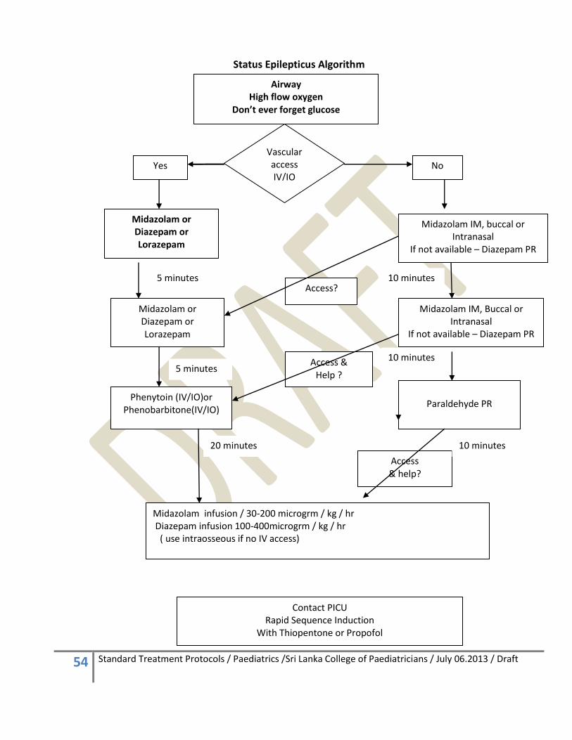
Status Epilepticus Algorithm
Airway High flow oxygen
Don’t ever forget glucose
Vascular access IV/IO
Yes No
Midazolam or Diazepam or Lorazepam
Midazolam or Diazepam or Lorazepam
Phenytoin (IV/IO)or Phenobarbitone(IV/IO)
Midazolam IM, buccal or Intranasal
If not available – Diazepam PR
Midazolam IM, Buccal or Intranasal
If not available – Diazepam PR
Paraldehyde PR
Contact PICU Rapid Sequence Induction
With Thiopentone or Propofol
Access?
Access & Help ?
Access & help?
Midazolam infusion / 30-200 microgrm / kg / hr Diazepam infusion 100-400microgrm / kg / hr ( use intraosseous if no IV access)
10 minutes
5 minutes
5 minutes
10 minutes
10 minutes 20 minutes
55 Standard Treatment Protocols / Paediatrics /Sri Lanka College of Paediatricians / July 06.2013 / Draft
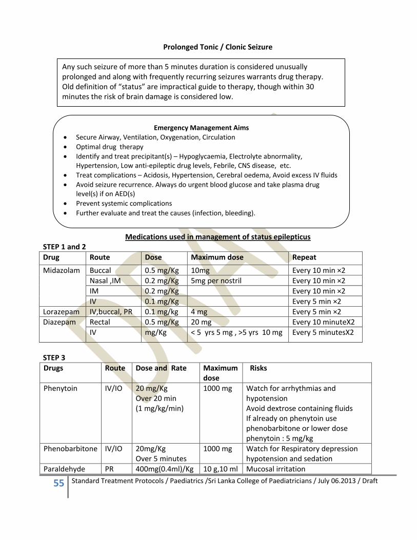
Prolonged Tonic / Clonic Seizure
Medications used in management of status epilepticus STEP 1 and 2
Drug Route Dose Maximum dose Repeat
Midazolam Buccal 0.5 mg/Kg 10mg Every 10 min ×2
Nasal ,IM 0.2 mg/Kg 5mg per nostril Every 10 min ×2
IM 0.2 mg/Kg Every 10 min ×2
IV 0.1 mg/Kg Every 5 min ×2
Lorazepam IV,buccal, PR 0.1 mg/kg 4 mg Every 5 min ×2
Diazepam Rectal 0.5 mg/Kg 20 mg Every 10 minuteX2
IV mg/Kg < 5 yrs 5 mg , >5 yrs 10 mg Every 5 minutesX2
STEP 3
Drugs Route Dose and Rate Maximum dose
Risks
Phenytoin
IV/IO 20 mg/Kg Over 20 min (1 mg/kg/min)
1000 mg Watch for arrhythmias and hypotension Avoid dextrose containing fluids If already on phenytoin use phenobarbitone or lower dose phenytoin : 5 mg/kg
Phenobarbitone IV/IO 20mg/Kg Over 5 minutes
1000 mg Watch for Respiratory depression hypotension and sedation
Paraldehyde PR 400mg(0.4ml)/Kg 10 g,10 ml Mucosal irritation
Emergency Management Aims
Secure Airway, Ventilation, Oxygenation, Circulation
Optimal drug therapy
Identify and treat precipitant(s) – Hypoglycaemia, Electrolyte abnormality, Hypertension, Low anti-epileptic drug levels, Febrile, CNS disease, etc.
Treat complications – Acidosis, Hypertension, Cerebral oedema, Avoid excess IV fluids
Avoid seizure recurrence. Always do urgent blood glucose and take plasma drug level(s) if on AED(s)
Prevent systemic complications
Further evaluate and treat the causes (infection, bleeding).
Any such seizure of more than 5 minutes duration is considered unusually prolonged and along with frequently recurring seizures warrants drug therapy. Old definition of “status” are impractical guide to therapy, though within 30 minutes the risk of brain damage is considered low.
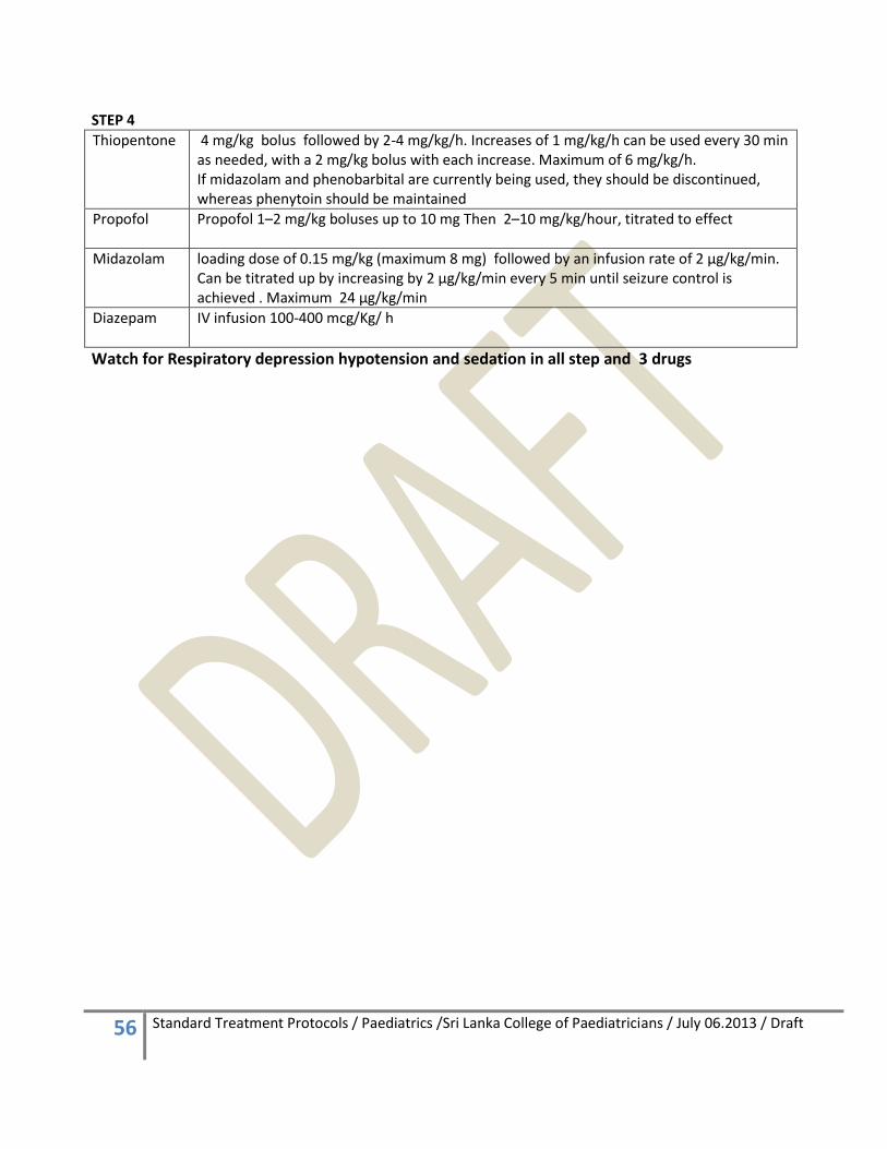
56 Standard Treatment Protocols / Paediatrics /Sri Lanka College of Paediatricians / July 06.2013 / Draft
STEP 4
Thiopentone 4 mg/kg bolus followed by 2-4 mg/kg/h. Increases of 1 mg/kg/h can be used every 30 min as needed, with a 2 mg/kg bolus with each increase. Maximum of 6 mg/kg/h. If midazolam and phenobarbital are currently being used, they should be discontinued, whereas phenytoin should be maintained
Propofol Propofol 1–2 mg/kg boluses up to 10 mg Then 2–10 mg/kg/hour, titrated to effect
Midazolam
loading dose of 0.15 mg/kg (maximum 8 mg) followed by an infusion rate of 2 µg/kg/min. Can be titrated up by increasing by 2 µg/kg/min every 5 min until seizure control is achieved . Maximum 24 µg/kg/min
Diazepam
IV infusion 100-400 mcg/Kg/ h
Watch for Respiratory depression hypotension and sedation in all step and 3 drugs
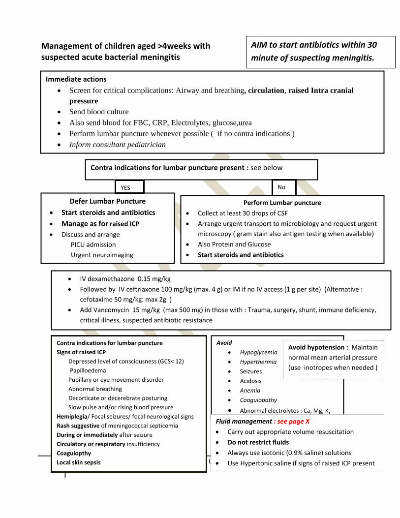
57 Standard Treatment Protocols / Paediatrics /Sri Lanka College of Paediatricians / July 06.2013 / Draft
Management of children aged >4weeks with suspected acute bacterial meningitis
AIM to start antibiotics within 30
minute of suspecting meningitis.
Immediate actions
Screen for critical complications: Airway and breathing, circulation, raised Intra cranial
pressure
Send blood culture
Also send blood for FBC, CRP, Electrolytes, glucose,urea
Perform lumbar puncture whenever possible ( if no contra indications )
Inform consultant pediatrician
Contra indications for lumbar puncture present : see below
Defer Lumbar Puncture
Start steroids and antibiotics
Manage as for raised ICP
Discuss and arrange
PICU admission
Urgent neuroimaging
Perform Lumbar puncture
Collect at least 30 drops of CSF
Arrange urgent transport to microbiology and request urgent
microscopy ( gram stain also antigen testing when available)
Also Protein and Glucose
Start steroids and antibiotics
YES No
IV dexamethazone 0.15 mg/kg
Followed by IV ceftriaxone 100 mg/kg (max. 4 g) or IM if no IV access (1 g per site) (Alternative :
cefotaxime 50 mg/kg: max 2g )
Add Vancomycin 15 mg/kg (max 500 mg) in those with : Trauma, surgery, shunt, immune deficiency,
critical illness, suspected antibiotic resistance
Contra indications for lumbar puncture
Signs of raised ICP
Depressed level of consciousness (GCS< 12)
Papilloedema
Pupillary or eye movement disorder
Abnormal breathing
Decorticate or decerebrate posturing
Slow pulse and/or rising blood pressure
Hemiplegia/ Focal seizures/ focal neurological signs
Rash suggestive of meningococcal septicemia
During or immediately after seizure
Circulatory or respiratory insufficiency
Coagulopthy
Local skin sepsis
Avoid
Hypoglycemia
Hyperthermia
Seizures
Acidosis
Anemia
Coagulopathy
Abnormal electrolytes : Ca, Mg, K,
Avoid hypotension : Maintain
normal mean arterial pressure
(use inotropes when needed )
Fluid management : see page X
Carry out appropriate volume resuscitation
Do not restrict fluids
Always use isotonic (0.9% saline) solutions
Use Hypertonic saline if signs of raised ICP present
ICP
Features of Raised ICP
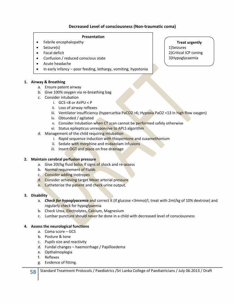
58 Standard Treatment Protocols / Paediatrics /Sri Lanka College of Paediatricians / July 06.2013 / Draft
Decreased Level of consciousness (Non-traumatic coma)
1. Airway & Breathing
a. Ensure patent airway b. Give 100% oxygen via re-breathing bag c. Consider intubation
i. GCS <8 or AVPU < P ii. Loss of airway reflexes
iii. Ventilator insufficiency (hypercarbia PaCO2 >6; Hypoxia PaO2 <13 in high flow oxygen) iv. Obtunded / agitated v. Consider intubation when CT scan cannot be performed safely otherwise
vi. Status epilepticus unresponsive to APLS algorithm d. Management of the child requiring intubation
i. Rapid sequence induction with thiopentone and suxamethonium ii. Sedate with morphine and midazolam infusions
iii. Insert OGT and place on free drainage
2. Maintain cerebral perfusion pressure a. Give 20l/kg fluid bolus if signs of shock and re-assess b. Normal requirement of Fluids c. Consider adding inotropes d. Consider achieving target Mean arterial pressure e. Catheterize the patient and check urine output.
3. Disability
a. Check for hypoglyacemia and correct it (if glucose <3mmol/l, treat with 2ml/kg of 10% dextrose) and regularly check for hypoglyaemia
b. Check Urea, Electrolytes, Calcium, Magnesium c. Lumbar puncture should never be done in a child with decreased level of consciousness
4. Assess the neurological functions
a. Coma score – GCS b. Posture & tone c. Pupils size and reactivity d. Fundal changes – haemorrhage / Papilloedema e. Opthalmoplegia f. Reflexes g. Evidence of fitting.
Presentation
Febrile encephalopathy
Seizure(s)
Focal deficit
Confusion / reduced conscious state
Acute headache
In early infancy – poor feeding, lethargy, vomiting, hypotonia
Treat urgently 1) Seizures 2) Critical ICP coning 3) Hypoglycaemia
59 Standard Treatment Protocols / Paediatrics /Sri Lanka College of Paediatricians / July 06.2013 / Draft
5. Assess for signs of raised ICP a. Pupillary dilatation or asymmetry b. Abnormal posture c. Abnormal breathing pattern d. Bradycardia & Hypertension
6. Critically raised ICP (“coning”) – signs imply brain distortion or shift with or without reduced cerebral blood
floor especially to brainstem. Can be mistaken for seizure (coma before posturing and bradycardia suggest coning)
Rapid decrease in conscious state
Dilated poorly reactive pupil(s)
Eye deviation (especially downward) and no doll’s eye movements
Abnormal respiration especially depression
Extensor posturing
Bradycardia ± raised BP
Any combination of above signs 7. Management of raised intra cranial pressure
a. Consider mannitol 0.5g/kg IV b. 3-5ml of 3% Normal saline (aim Na 145mmol/l) c. Urgent CT scan (+/- Contrast)
8. Urgent Treatment (without waiting for CT scan)
Mild hyperventilation (usually after intubation). Effect on ICP is immediate. Do this first if any doubt about respiration
IV Mannitol 0.25 – 0.5g /kg (2-4ml/kg of 12.5% solution ) as rapid infusion. Effect in 10-15 minutes and for 3 – 5 hours.
Subdural, shunt or ventricular taps or emergency craniotomy rarely required – consult an neurosurgeon.
9. Assess for treatable causes a. Bacterial Meningitis – Cefotaxime 50mg/kg/dose x tds b. Meningoencephalitis (Herpes ) – Acyclovir c. Meningoencephalitis (Mycoplasma) – Clarythromycine d. Krait bite – signs of envenomation e. Poisoning – serum and urine for toxicology screen f. Cerebral malaria – IV quinine
10. Other causes a. Trauma (including neglectful or inflicted injury in infancy [shaking, direct trauma, asphyxiation]) b. Infectious / Inflammatory CNS disease – refer above c. Severe seizure(s) d. Hypoxia / Hypotension – asphyxia, drowning etc. e. Metabolic – hypoglycaemia, electrolyte abnormality, genetic disease (especially infancy) f. Neurotoxic i.e drug, venom, toxin (Refer above) g. Systemic diseases h. Vascular i.e Hypertension, ICH, CVA, migraine i. Acute hydrocephalus i.e shunt blockage
60 Standard Treatment Protocols / Paediatrics /Sri Lanka College of Paediatricians / July 06.2013 / Draft
11. Transport Consideration
a. Manage as for raised ICP (ventilate to normocarbia, maintain CPP, normothermia, nurse head up and midline, etc)
b. Monitor pupils c. Consider mannitol or 3% saline if signs of raised ICP (take pre-prepared bolus) d. Monitor glucose e. Take copies of CT scan if these have been done
12. A
Traumatic Brain Injury (TBI) 1. Assessment 2. Initial Management 3. Indication for intubation 4. Management of post intubation
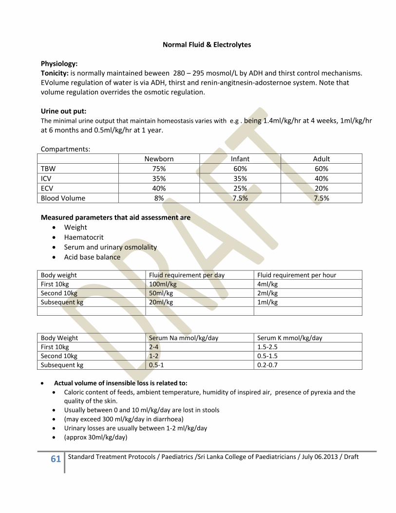
61 Standard Treatment Protocols / Paediatrics /Sri Lanka College of Paediatricians / July 06.2013 / Draft
Normal Fluid & Electrolytes Physiology: Tonicity: is normally maintained beween 280 – 295 mosmol/L by ADH and thirst control mechanisms. EVolume regulation of water is via ADH, thirst and renin-angitnesin-adosternoe system. Note that volume regulation overrides the osmotic regulation. Urine out put: The minimal urine output that maintain homeostasis varies with e.g . being 1.4ml/kg/hr at 4 weeks, 1ml/kg/hr at 6 months and 0.5ml/kg/hr at 1 year. Compartments:
Newborn Infant Adult
TBW 75% 60% 60%
ICV 35% 35% 40%
ECV 40% 25% 20%
Blood Volume 8% 7.5% 7.5%
Measured parameters that aid assessment are
Weight
Haematocrit
Serum and urinary osmolality
Acid base balance
Body weight Fluid requirement per day Fluid requirement per hour
First 10kg 100ml/kg 4ml/kg
Second 10kg 50ml/kg 2ml/kg
Subsequent kg 20ml/kg 1ml/kg
Body Weight Serum Na mmol/kg/day Serum K mmol/kg/day
First 10kg 2-4 1.5-2.5
Second 10kg 1-2 0.5-1.5
Subsequent kg 0.5-1 0.2-0.7
Actual volume of insensible loss is related to:
Caloric content of feeds, ambient temperature, humidity of inspired air, presence of pyrexia and the quality of the skin.
Usually between 0 and 10 ml/kg/day are lost in stools
(may exceed 300 ml/kg/day in diarrhoea)
Urinary losses are usually between 1-2 ml/kg/day
(approx 30ml/kg/day)
62 Standard Treatment Protocols / Paediatrics /Sri Lanka College of Paediatricians / July 06.2013 / Draft
How to caluculate the percentage of dehydration o Percentage dehydration x weight x 10 o Percentage dehydration means the number of grams of fluid lost per 100 gm of body weight. o Percentage x 10 converts this volume into ml/kg
A Shock occurs as a result of rapid loss of 20ml/kg from the intravascular space. If the intravascular volume is maintained, clinical dehydration is only evident after losses > 25ml/kg of total body water.
Shock occurs as a result of rapid loss of 20ml/kg from the intravascular space. If the intravascular volume is maintained, clinical dehydration is only evident after losses > 25ml/kg of total body water.
It is possible to be shocked and not dehydrated, dehydrated and not shocked, or dehydrated and shocked
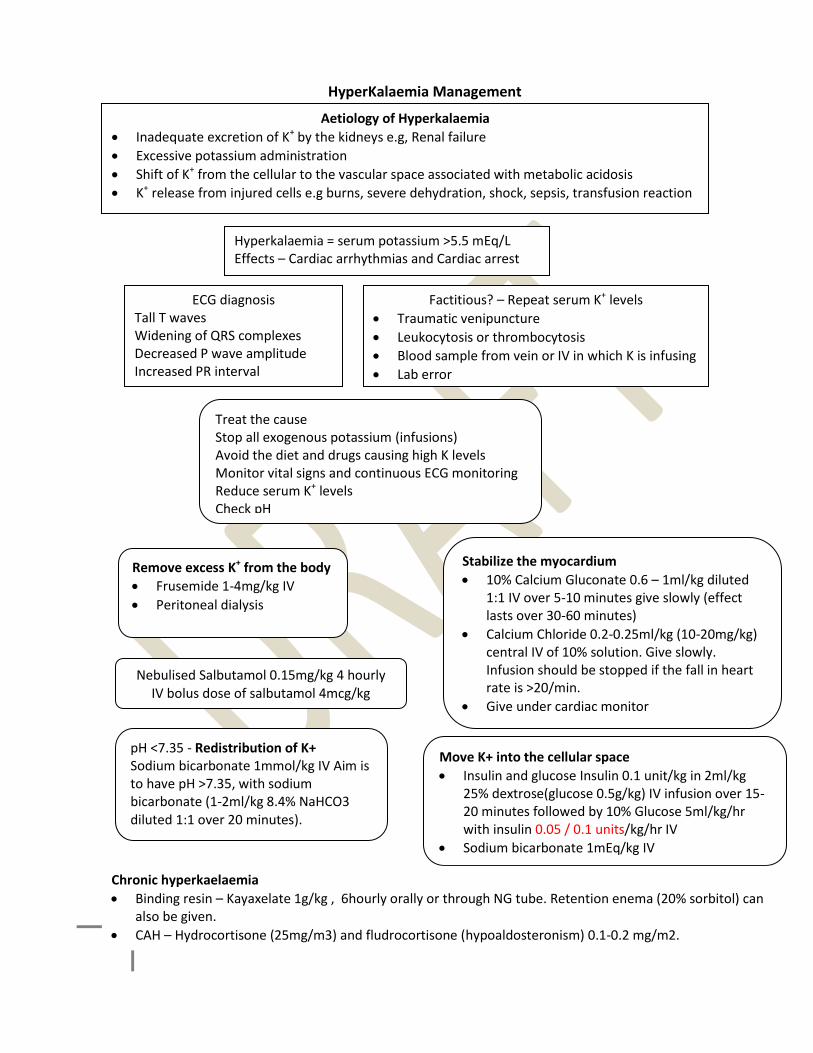
63 Standard Treatment Protocols / Paediatrics /Sri Lanka College of Paediatricians / July 06.2013 / Draft
HyperKalaemia Management
Nebulised Salbutamol 0.15mg/kg 4 hourly IV bolus dose of salbutamol 4mcg/kg
pH <7.35 - Redistribution of K+ Sodium bicarbonate 1mmol/kg IV Aim is to have pH >7.35, with sodium bicarbonate (1-2ml/kg 8.4% NaHCO3 diluted 1:1 over 20 minutes).
ECG diagnosis Tall T waves Widening of QRS complexes Decreased P wave amplitude Increased PR interval
Factitious? – Repeat serum K+ levels
Traumatic venipuncture
Leukocytosis or thrombocytosis
Blood sample from vein or IV in which K is infusing
Lab error
Hyperkalaemia = serum potassium >5.5 mEq/L Effects – Cardiac arrhythmias and Cardiac arrest
Aetiology of Hyperkalaemia
Inadequate excretion of K+ by the kidneys e.g, Renal failure
Excessive potassium administration
Shift of K+ from the cellular to the vascular space associated with metabolic acidosis
K+ release from injured cells e.g burns, severe dehydration, shock, sepsis, transfusion reaction
Treat the cause Stop all exogenous potassium (infusions) Avoid the diet and drugs causing high K levels Monitor vital signs and continuous ECG monitoring Reduce serum K+ levels Check pH
Move K+ into the cellular space
Insulin and glucose Insulin 0.1 unit/kg in 2ml/kg 25% dextrose(glucose 0.5g/kg) IV infusion over 15-20 minutes followed by 10% Glucose 5ml/kg/hr with insulin 0.05 / 0.1 units/kg/hr IV
Sodium bicarbonate 1mEq/kg IV
Remove excess K+ from the body
Frusemide 1-4mg/kg IV
Peritoneal dialysis
Stabilize the myocardium
10% Calcium Gluconate 0.6 – 1ml/kg diluted 1:1 IV over 5-10 minutes give slowly (effect lasts over 30-60 minutes)
Calcium Chloride 0.2-0.25ml/kg (10-20mg/kg) central IV of 10% solution. Give slowly. Infusion should be stopped if the fall in heart rate is >20/min.
Give under cardiac monitor
Chronic hyperkaelaemia
Binding resin – Kayaxelate 1g/kg , 6hourly orally or through NG tube. Retention enema (20% sorbitol) can also be given.
CAH – Hydrocortisone (25mg/m3) and fludrocortisone (hypoaldosteronism) 0.1-0.2 mg/m2.
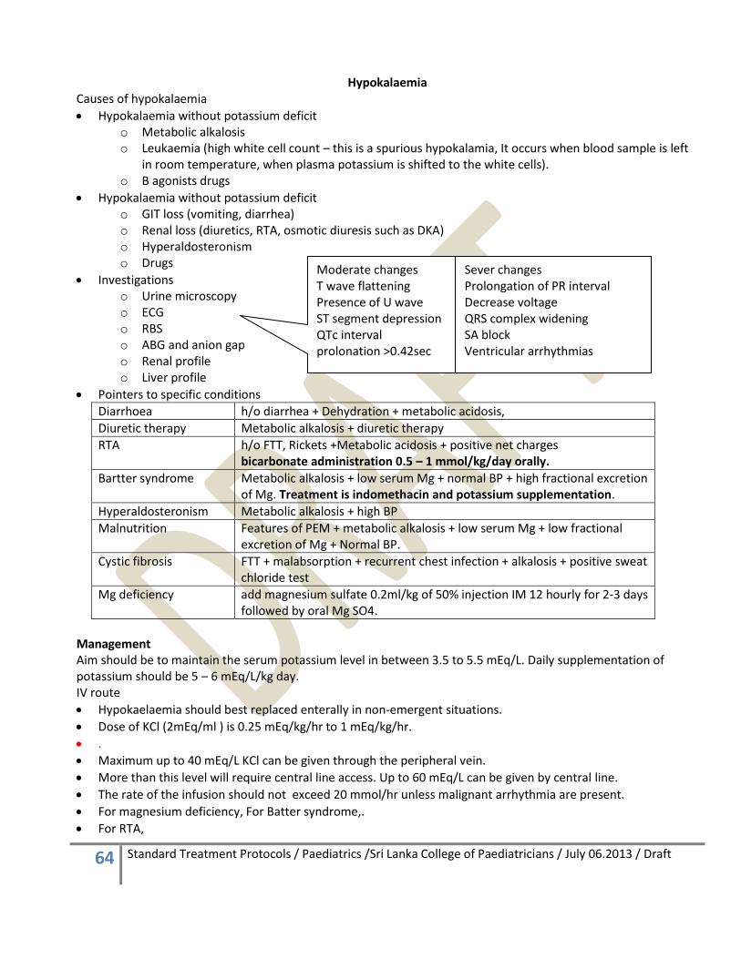
64 Standard Treatment Protocols / Paediatrics /Sri Lanka College of Paediatricians / July 06.2013 / Draft
Hypokalaemia Causes of hypokalaemia
Hypokalaemia without potassium deficit o Metabolic alkalosis o Leukaemia (high white cell count – this is a spurious hypokalamia, It occurs when blood sample is left
in room temperature, when plasma potassium is shifted to the white cells). o Β agonists drugs
Hypokalaemia without potassium deficit o GIT loss (vomiting, diarrhea) o Renal loss (diuretics, RTA, osmotic diuresis such as DKA) o Hyperaldosteronism o Drugs
Investigations o Urine microscopy o ECG o RBS o ABG and anion gap o Renal profile o Liver profile
Pointers to specific conditions
Diarrhoea h/o diarrhea + Dehydration + metabolic acidosis,
Diuretic therapy Metabolic alkalosis + diuretic therapy
RTA h/o FTT, Rickets +Metabolic acidosis + positive net charges bicarbonate administration 0.5 – 1 mmol/kg/day orally.
Bartter syndrome Metabolic alkalosis + low serum Mg + normal BP + high fractional excretion of Mg. Treatment is indomethacin and potassium supplementation.
Hyperaldosteronism Metabolic alkalosis + high BP
Malnutrition Features of PEM + metabolic alkalosis + low serum Mg + low fractional excretion of Mg + Normal BP.
Cystic fibrosis FTT + malabsorption + recurrent chest infection + alkalosis + positive sweat chloride test
Mg deficiency add magnesium sulfate 0.2ml/kg of 50% injection IM 12 hourly for 2-3 days followed by oral Mg SO4.
Management Aim should be to maintain the serum potassium level in between 3.5 to 5.5 mEq/L. Daily supplementation of potassium should be 5 – 6 mEq/L/kg day. IV route
Hypokaelaemia should best replaced enterally in non-emergent situations.
Dose of KCl (2mEq/ml ) is 0.25 mEq/kg/hr to 1 mEq/kg/hr.
.
Maximum up to 40 mEq/L KCl can be given through the peripheral vein.
More than this level will require central line access. Up to 60 mEq/L can be given by central line.
The rate of the infusion should not exceed 20 mmol/hr unless malignant arrhythmia are present.
For magnesium deficiency, For Batter syndrome,.
For RTA,
Moderate changes T wave flattening Presence of U wave ST segment depression QTc interval prolonation >0.42sec
Sever changes Prolongation of PR interval Decrease voltage QRS complex widening SA block Ventricular arrhythmias
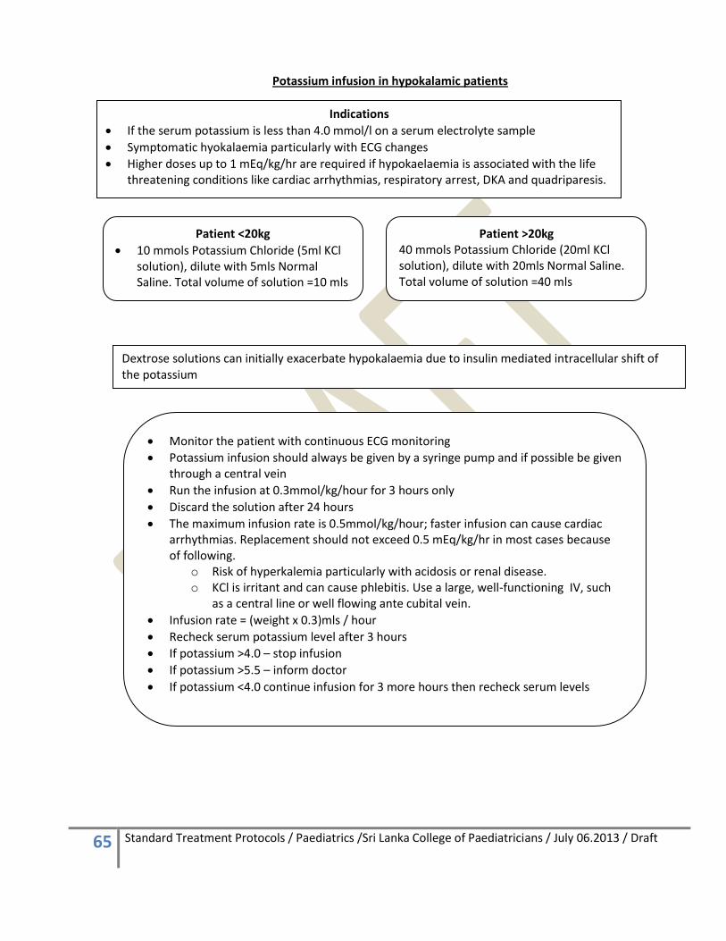
65 Standard Treatment Protocols / Paediatrics /Sri Lanka College of Paediatricians / July 06.2013 / Draft
Potassium infusion in hypokalamic patients
Indications
If the serum potassium is less than 4.0 mmol/l on a serum electrolyte sample
Symptomatic hyokalaemia particularly with ECG changes
Higher doses up to 1 mEq/kg/hr are required if hypokaelaemia is associated with the life threatening conditions like cardiac arrhythmias, respiratory arrest, DKA and quadriparesis.
Patient <20kg
10 mmols Potassium Chloride (5ml KCl solution), dilute with 5mls Normal Saline. Total volume of solution =10 mls
Patient >20kg 40 mmols Potassium Chloride (20ml KCl solution), dilute with 20mls Normal Saline. Total volume of solution =40 mls
Monitor the patient with continuous ECG monitoring
Potassium infusion should always be given by a syringe pump and if possible be given through a central vein
Run the infusion at 0.3mmol/kg/hour for 3 hours only
Discard the solution after 24 hours
The maximum infusion rate is 0.5mmol/kg/hour; faster infusion can cause cardiac arrhythmias. Replacement should not exceed 0.5 mEq/kg/hr in most cases because of following.
o Risk of hyperkalemia particularly with acidosis or renal disease. o KCl is irritant and can cause phlebitis. Use a large, well-functioning IV, such
as a central line or well flowing ante cubital vein.
Infusion rate = (weight x 0.3)mls / hour
Recheck serum potassium level after 3 hours
If potassium >4.0 – stop infusion
If potassium >5.5 – inform doctor
If potassium <4.0 continue infusion for 3 more hours then recheck serum levels
Dextrose solutions can initially exacerbate hypokalaemia due to insulin mediated intracellular shift of the potassium
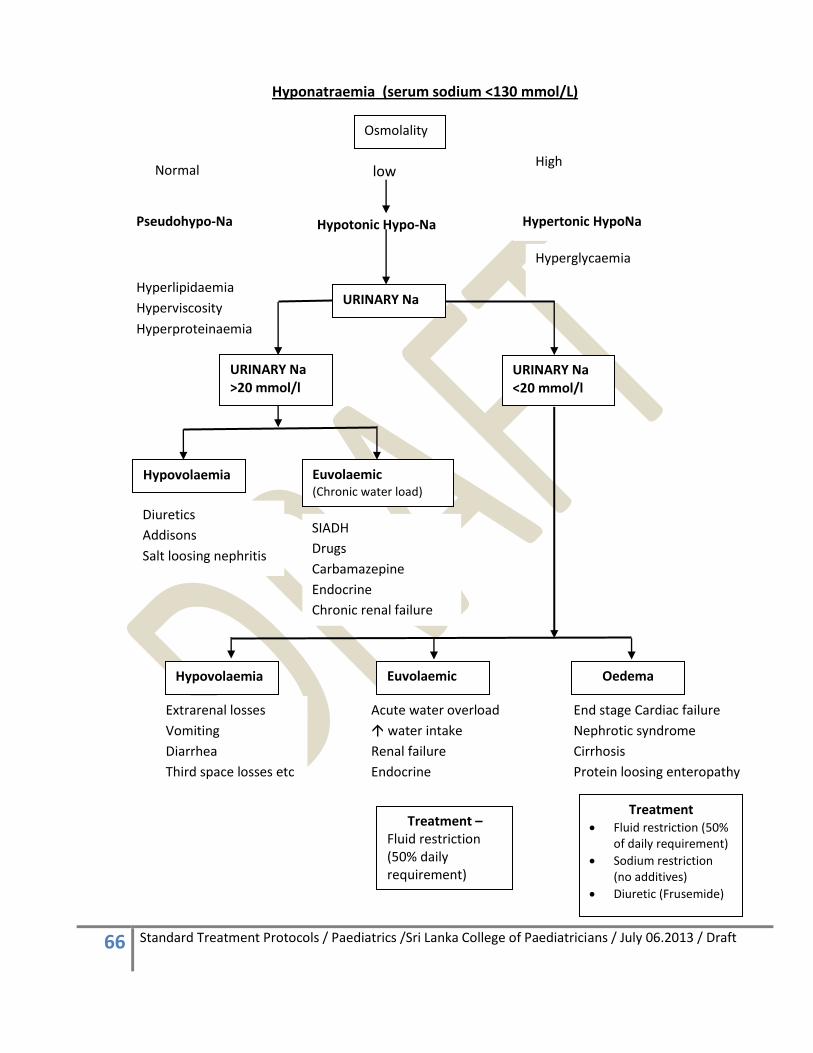
66 Standard Treatment Protocols / Paediatrics /Sri Lanka College of Paediatricians / July 06.2013 / Draft
Hyponatraemia (serum sodium <130 mmol/L)
Osmolality
Normal low High
Pseudohypo-Na Hypotonic Hypo-Na Hypertonic HypoNa
Hyperlipidaemia
Hyperviscosity
Hyperproteinaemia
Hyperglycaemia
URINARY Na
URINARY Na >20 mmol/l
URINARY Na <20 mmol/l
Hypovolaemia Euvolaemic (Chronic water load)
Diuretics
Addisons
Salt loosing nephritis
SIADH
Drugs
Carbamazepine
Endocrine
Chronic renal failure
Hypovolaemia Euvolaemic
Oedema
Extrarenal losses
Vomiting
Diarrhea
Third space losses etc
Acute water overload
water intake
Renal failure
Endocrine
End stage Cardiac failure
Nephrotic syndrome
Cirrhosis
Protein loosing enteropathy
Treatment Fluid restriction (50%
of daily requirement)
Sodium restriction (no additives)
Diuretic (Frusemide)
Treatment – Fluid restriction (50% daily requirement)
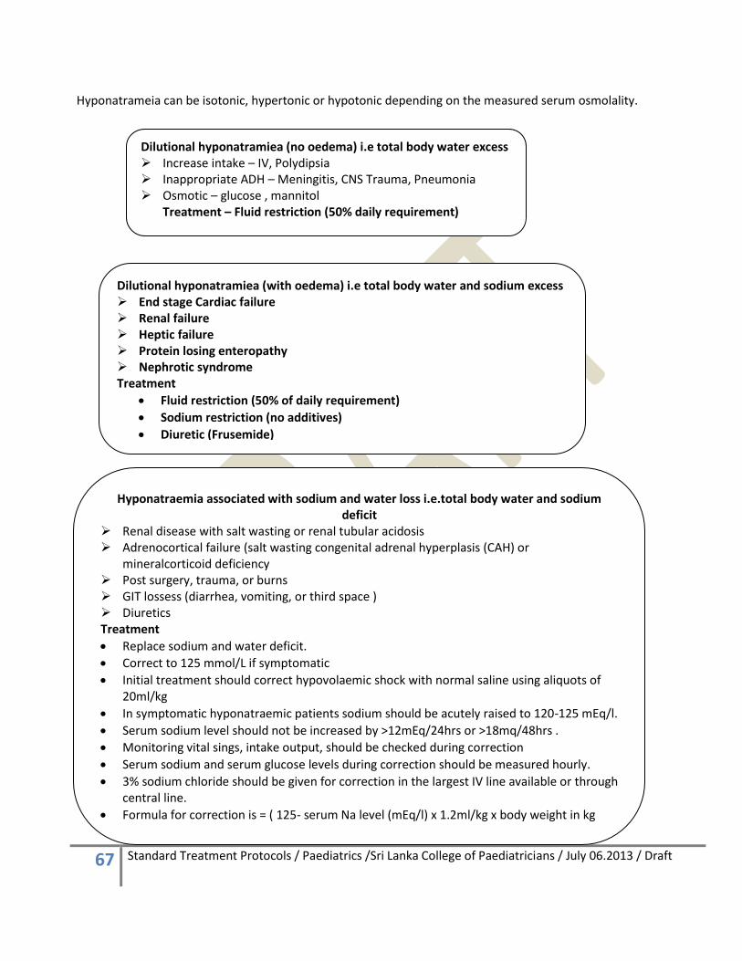
67 Standard Treatment Protocols / Paediatrics /Sri Lanka College of Paediatricians / July 06.2013 / Draft
Hyponatrameia can be isotonic, hypertonic or hypotonic depending on the measured serum osmolality.
Dilutional hyponatramiea (no oedema) i.e total body water excess Increase intake – IV, Polydipsia Inappropriate ADH – Meningitis, CNS Trauma, Pneumonia Osmotic – glucose , mannitol
Treatment – Fluid restriction (50% daily requirement)
Dilutional hyponatramiea (with oedema) i.e total body water and sodium excess End stage Cardiac failure Renal failure Heptic failure Protein losing enteropathy Nephrotic syndrome Treatment
Fluid restriction (50% of daily requirement)
Sodium restriction (no additives)
Diuretic (Frusemide)
Hyponatraemia associated with sodium and water loss i.e.total body water and sodium deficit
Renal disease with salt wasting or renal tubular acidosis Adrenocortical failure (salt wasting congenital adrenal hyperplasis (CAH) or
mineralcorticoid deficiency Post surgery, trauma, or burns GIT lossess (diarrhea, vomiting, or third space ) Diuretics Treatment
Replace sodium and water deficit.
Correct to 125 mmol/L if symptomatic
Initial treatment should correct hypovolaemic shock with normal saline using aliquots of 20ml/kg
In symptomatic hyponatraemic patients sodium should be acutely raised to 120-125 mEq/l.
Serum sodium level should not be increased by >12mEq/24hrs or >18mq/48hrs .
Monitoring vital sings, intake output, should be checked during correction
Serum sodium and serum glucose levels during correction should be measured hourly.
3% sodium chloride should be given for correction in the largest IV line available or through central line.
Formula for correction is = ( 125- serum Na level (mEq/l) x 1.2ml/kg x body weight in kg
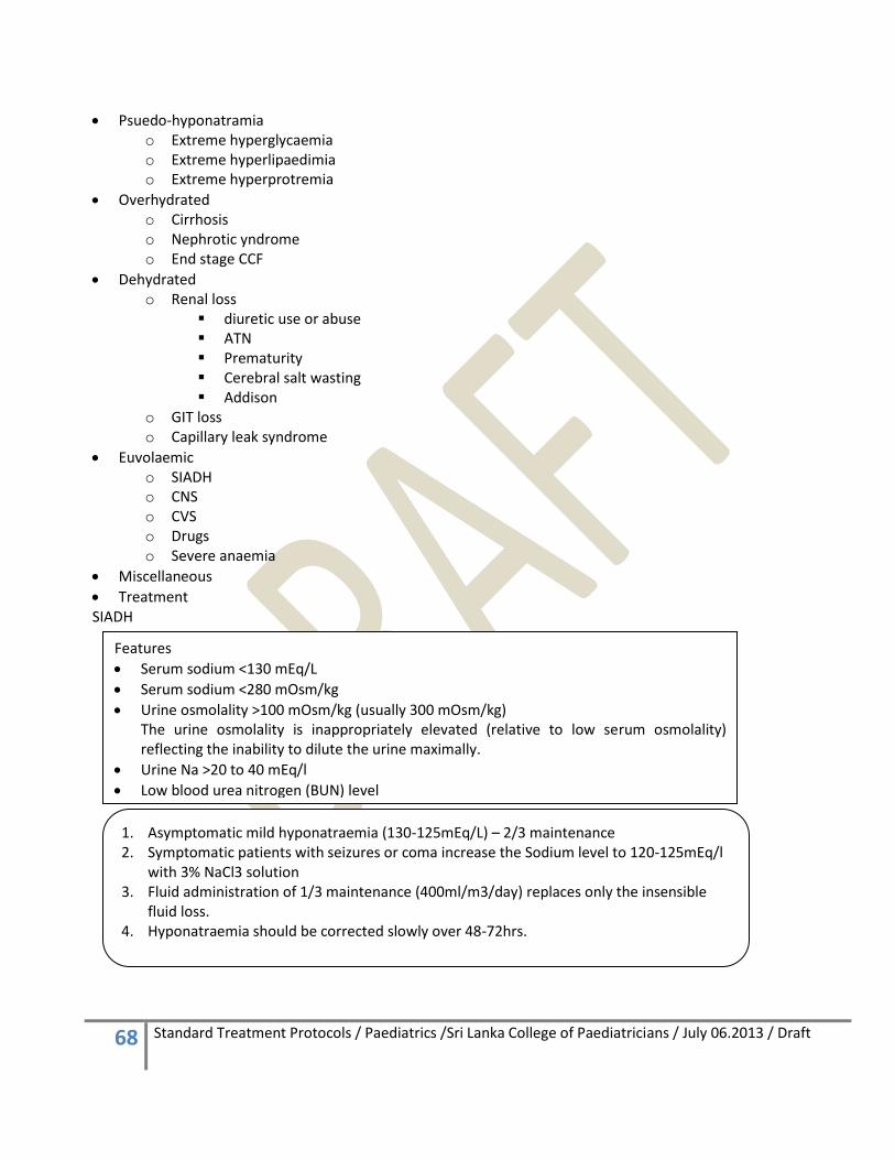
68 Standard Treatment Protocols / Paediatrics /Sri Lanka College of Paediatricians / July 06.2013 / Draft
Psuedo-hyponatramia o Extreme hyperglycaemia o Extreme hyperlipaedimia o Extreme hyperprotremia
Overhydrated o Cirrhosis o Nephrotic yndrome o End stage CCF
Dehydrated o Renal loss
diuretic use or abuse ATN Prematurity Cerebral salt wasting Addison
o GIT loss o Capillary leak syndrome
Euvolaemic o SIADH o CNS o CVS o Drugs o Severe anaemia
Miscellaneous
Treatment SIADH
Features
Serum sodium <130 mEq/L
Serum sodium <280 mOsm/kg
Urine osmolality >100 mOsm/kg (usually 300 mOsm/kg) The urine osmolality is inappropriately elevated (relative to low serum osmolality) reflecting the inability to dilute the urine maximally.
Urine Na >20 to 40 mEq/l
Low blood urea nitrogen (BUN) level
1. Asymptomatic mild hyponatraemia (130-125mEq/L) – 2/3 maintenance 2. Symptomatic patients with seizures or coma increase the Sodium level to 120-125mEq/l
with 3% NaCl3 solution 3. Fluid administration of 1/3 maintenance (400ml/m3/day) replaces only the insensible
fluid loss. 4. Hyponatraemia should be corrected slowly over 48-72hrs.
69 Standard Treatment Protocols / Paediatrics /Sri Lanka College of Paediatricians / July 06.2013 / Draft
Hypernatraemia (serum sodium > 150mmol/L) Hypernatraemia is always associated with hypertonicity
Hypocalcaemia
DHF / DSS Results in leaking of albumin into pleural and peritoneal cavity
10% Calcium Gluconate 0.5-1ml/kg (maximum dose 10ml) diluted 1:1 IV over 5-10 minutes give slowly (effect lasts over 30-60 minutes) x 6 hourly
No indication for Ca in asymptomatic and uncomplicated DHF cases grade 1, 111, 11 patients.
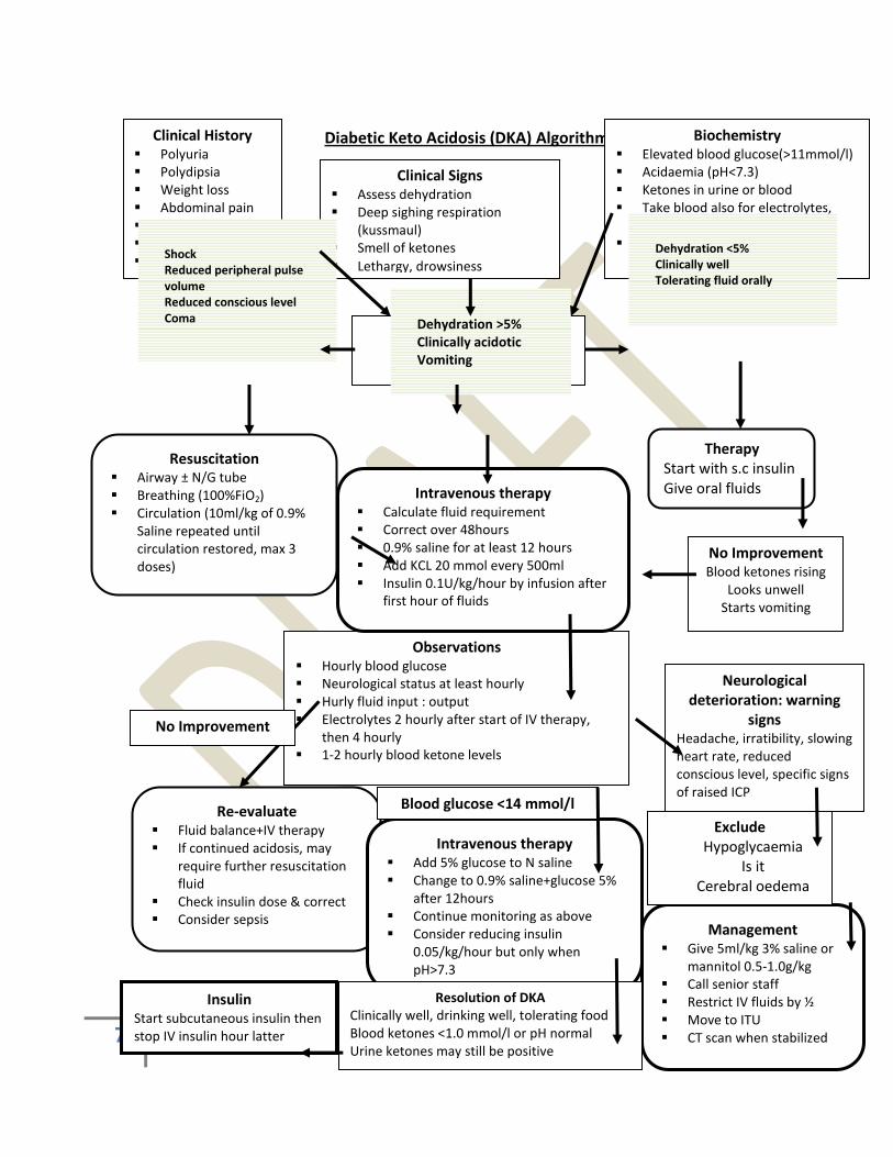
70 Standard Treatment Protocols / Paediatrics /Sri Lanka College of Paediatricians / July 06.2013 / Draft
Diabetic Keto Acidosis (DKA) Algorithm
Clinical History Polyuria Polydipsia Weight loss Abdominal pain Weakness Vomiting Confusion
Observations Hourly blood glucose Neurological status at least hourly Hurly fluid input : output Electrolytes 2 hourly after start of IV therapy,
then 4 hourly 1-2 hourly blood ketone levels
Confirm Diagnosis Diabetic Ketoacidosis
Inform your consultant
Insulin Start subcutaneous insulin then stop IV insulin hour latter
Clinical Signs Assess dehydration Deep sighing respiration
(kussmaul) Smell of ketones
Lethargy, drowsiness
Biochemistry Elevated blood glucose(>11mmol/l) Acidaemia (pH<7.3) Ketones in urine or blood Take blood also for electrolytes,
urea Perform other investigations if
indicated
Resuscitation Airway ± N/G tube Breathing (100%FiO2) Circulation (10ml/kg of 0.9%
Saline repeated until circulation restored, max 3 doses)
Intravenous therapy Calculate fluid requirement Correct over 48hours 0.9% saline for at least 12 hours Add KCL 20 mmol every 500ml Insulin 0.1U/kg/hour by infusion after
first hour of fluids
Therapy Start with s.c insulin Give oral fluids
Shock Reduced peripheral pulse volume Reduced conscious level Coma
Re-evaluate Fluid balance+IV therapy If continued acidosis, may
require further resuscitation fluid
Check insulin dose & correct Consider sepsis
Intravenous therapy Add 5% glucose to N saline Change to 0.9% saline+glucose 5%
after 12hours Continue monitoring as above Consider reducing insulin
0.05/kg/hour but only when pH>7.3
Management Give 5ml/kg 3% saline or
mannitol 0.5-1.0g/kg Call senior staff Restrict IV fluids by ½ Move to ITU CT scan when stabilized
Exclude Hypoglycaemia
Is it Cerebral oedema
Dehydration <5% Clinically well Tolerating fluid orally
Dehydration >5% Clinically acidotic Vomiting
Neurological deterioration: warning
signs Headache, irratibility, slowing heart rate, reduced conscious level, specific signs of raised ICP
No Improvement Blood ketones rising
Looks unwell Starts vomiting
Blood glucose <14 mmol/l
Resolution of DKA Clinically well, drinking well, tolerating food Blood ketones <1.0 mmol/l or pH normal Urine ketones may still be positive
No Improvement
71 Standard Treatment Protocols / Paediatrics /Sri Lanka College of Paediatricians / July 06.2013 / Draft
SEDATION FOR CRITICALLY ILL CHILDREN
Aims of sedation Reduced anxiety and distress Facilitation of mechanical ventilation a nd avoidance of inadvertent self-extubation Reduced metabolic rate and oxygen demand Enhanced analgesia Less disrupted sleep Reduced patient recall To allow tolerance of diagnostic and therapeutic procedures Usual intravenous sedative/analgesic combination in children is midazolam and morphine MIDAZOLAM Intravenous infusion; 2-8 micrograms/kg/min Intravenous bolus; 100-200 micrograms/kg/min (Adult dose 5mg) Repeat bolus doses as necessary For patients >50kg give 5-15mg/hr (not per kg) as infusion Do not use routinely in infants below 6 months Consider reduced dose in renal and hepatic impairment Morphine Intravenous infusion; 10-60micrograms/kg/hr Intravenous bolus; 100-200 micrograms/kg/dose (Adult dose 5-10mg) Start neonates at 5–20micrograms/kg/hr as infusion For patients >50 kg give 0.8-3mg/hr (not per kg) as infusion Consider reduced dose in renal and hepatic impairment Use with caution in asthmatic patients COMPLICATIONS OF SEDATION Tolerance Withdrawal phenomenon Inadvertent hypotension Increased protein breakdown Immunosuppression Renal dysfunction Hepatotoxicity Increased financial cost Prolonged duration of mechanical ventilation and intensive care admission Where possible use ENTERAL SEDATION; chloral hydrate with or without a sedative antihistamine such as alimemazine CHLORAL HYDRATE NG; 25-50mg/kg/dose 4-6 hrly
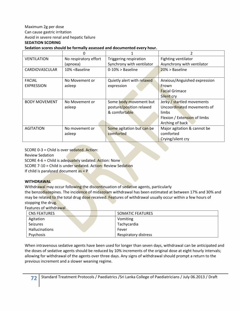
72 Standard Treatment Protocols / Paediatrics /Sri Lanka College of Paediatricians / July 06.2013 / Draft
Maximum 2g per dose Can cause gastric irritation Avoid in severe renal and hepatic failure SEDATION SCORING Sedation scores should be formally assessed and documented every hour.
0 1 2
VENTILATION No respiratory effort (apnoea)
Triggering respiration Synchrony with ventilator
Fighting ventilator Asynchrony with ventilator
CARDIOVASCULAR
10% <Baseline 0-10% > Baseline 20% > Baseline
FACIAL EXPRESSION
No Movement or asleep
Quietly alert with relaxed expression
Anxious/Anguished expression Frown Facial Grimace Silent cry
BODY MOVEMENT
No Movement or asleep
Some body movement but posture/position relaxed & comfortable
Jerky / startled movements Uncoordinated movements of limbs Flexion / Extension of limbs Arching of back
AGITATION
No movement or asleep
Some agitation but can be comforted
Major agitation & cannot be comforted Crying/silent cry
SCORE 0-3 = Child is over sedated. Action: Review Sedation SCORE 4-6 = Child is adequately sedated. Action: None SCORE 7-10 = Child is under sedated. Action: Review Sedation If child is paralysed document as = P WITHDRAWAL Withdrawal may occur following the discontinuation of sedative agents, particularly the benzodiazepines. The incidence of midazolam withdrawal has been estimated at between 17% and 30% and may be related to the total drug dose received. Features of withdrawal usually occur within a few hours of stopping the drug. Features of withdrawal
CNS FEATURES SOMATIC FEATURES
Agitation Seizures Hallucinations Psychosis
Vomiting Tachycardia Fever Respiratory distress
When intravenous sedative agents have been used for longer than seven days, withdrawal can be anticipated and the doses of sedative agents should be reduced by 10% increments of the original dose at eight hourly intervals; allowing for withdrawal of the agents over three days. Any signs of withdrawal should prompt a return to the previous increment and a slower weaning regime.
73 Standard Treatment Protocols / Paediatrics /Sri Lanka College of Paediatricians / July 06.2013 / Draft
Additional agents that may be useful inthe management of withdrawal include clonidine, methadone and lorazepam. CLONIDINE Intravenous infusion; 0.1-2 micrograms/kg/hr NG; 1-5 micrograms/kg/dose 6 hrly Start infusions at 1microgram/kg/hr LORAZEPAM Intravenous bolus; 50-100 micrograms/kg/dose (max 4mg) NG; 20-60micrograms/kg/dose 8-24 hrly (max 1-3mg) Reduce doses in severe renal and hepatic impairment IV preparation may be given via NGT METHADONE Divide total daily dose of morphine by 5 for conversion to methadone dose and administer in two divided doses enterally OTHER AGENTS KETAMINE Intravenous infusion; 5-20 micrograms/kg/min Intravenous bolus; 1-2mg/kg/dose May be useful in status asthmaticus Use with caution in systemic hypertension Avoid in raised intracranial pressure Concentrations exceeding 10mg/ml should be infused centrally FENTANYL Intravenous infusion; 5-10 micrograms/kg/hr Intravenous bolus; 1-2 micrograms/kg/dose May be more effective than morphine Less histamine response so potential benefit in status asthmaticus Reduce dose in hepatic impairment Beware hypotension and bradycardia Transdermal patches may be of use in treating withdrawal PROPOFOL PROPOFOL SHOULD NOT BEUSED FOR LONG-TERM SEDATION IN CHILDREN UNDER THE AGE OF 16 YEARS Intravenous bolus dose; 1-3mg/kg/dose
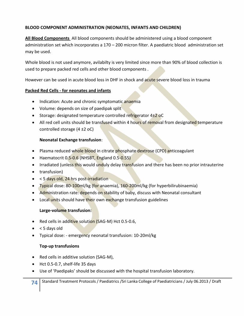
74 Standard Treatment Protocols / Paediatrics /Sri Lanka College of Paediatricians / July 06.2013 / Draft
BLOOD COMPONENT ADMINISTRATION (NEONATES, INFANTS AND CHILDREN)
All Blood Components All blood components should be administered using a blood component
administration set which incorporates a 170 – 200 micron filter. A paediatric blood administration set
may be used.
Whole blood is not used anymore, avilabilty is very limited since more than 90% of blood collection is
used to prepare packed red cells and other blood components .
However can be used in acute blood loss in DHF in shock and acute severe blood loss in trauma
Packed Red Cells - for neonates and infants
Indication: Acute and chronic symptomatic anaemia
Volume: depends on size of paedipak split
Storage: designated temperature controlled refrigerator 4±2 oC
All red cell units should be transfused within 4 hours of removal from designated temperature
controlled storage (4 ±2 oC)
Neonatal Exchange transfusion:
Plasma reduced whole blood in citrate phosphate dextrose (CPD) anticoagulant
Haematocrit 0.5-0.6 (NHSBT, England 0.5-0.55)
Irradiated (unless this would unduly delay transfusion and there has been no prior intrauterine
transfusion)
< 5 days old, 24 hrs post-irradiation
Typical dose: 80-100ml/kg (for anaemia), 160-200ml/kg (for hyperbilirubinaemia)
Administration rate: depends on stability of baby, discuss with Neonatal consultant
Local units should have their own exchange transfusion guidelines
Large-volume transfusion:
Red cells in additive solution (SAG-M) Hct 0.5-0.6,
< 5 days old
Typical dose: - emergency neonatal transfusion: 10-20ml/kg
Top-up transfusions
Red cells in additive solution (SAG-M),
Hct 0.5-0.7, shelf-life 35 days
Use of ‘Paedipaks’ should be discussed with the hospital transfusion laboratory.
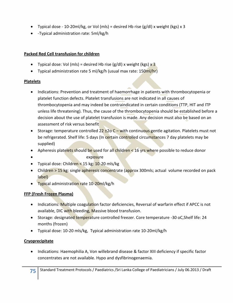
75 Standard Treatment Protocols / Paediatrics /Sri Lanka College of Paediatricians / July 06.2013 / Draft
Typical dose - 10-20ml/kg, or Vol (mls) = desired Hb rise (g/dl) x weight (kgs) x 3
-Typical administration rate: 5ml/kg/h
Packed Red Cell transfusion for children
Typical dose: Vol (mls) = desired Hb rise (g/dl) x weight (kgs) x 3
Typical administration rate 5 ml/kg/h (usual max rate: 150ml/hr)
Platelets
Indications: Prevention and treatment of haemorrhage in patients with thrombocytopenia or
platelet function defects. Platelet transfusions are not indicated in all causes of
thrombocytopenia and may indeed be contraindicated in certain conditions (TTP, HIT and ITP
unless life threatening). Thus, the cause of the thrombocytopenia should be established before a
decision about the use of platelet transfusion is made. Any decision must also be based on an
assessment of risk versus benefit
Storage: temperature controlled 22 ±2o C – with continuous gentle agitation. Platelets must not
be refrigerated. Shelf life: 5 days (In certain controlled circumstances 7 day platelets may be
supplied)
Apheresis platelets should be used for all children < 16 yrs where possible to reduce donor
exposure
Typical dose: Children < 15 kg: 10-20 mls/kg
Children > 15 kg: single apheresis concentrate (approx 300mls; actual volume recorded on pack
label)
Typical administration rate 10-20ml/kg/h
FFP (Fresh Frozen Plasma)
Indications: Multiple coagulation factor deficiencies, Reversal of warfarin effect if APCC is not
available, DIC with bleeding, Massive blood transfusion.
Storage: designated temperature controlled freezer. Core temperature -30 oC,Shelf life: 24
months (frozen)
Typical dose: 10-20 mls/kg, Typical administration rate 10-20ml/kg/h
Cryoprecipitate
Indications: Haemophilia A, Von willebrand disease & factor XIII deficiency if specific factor
concentrates are not available. Hypo and dysfibrinogenaemia.
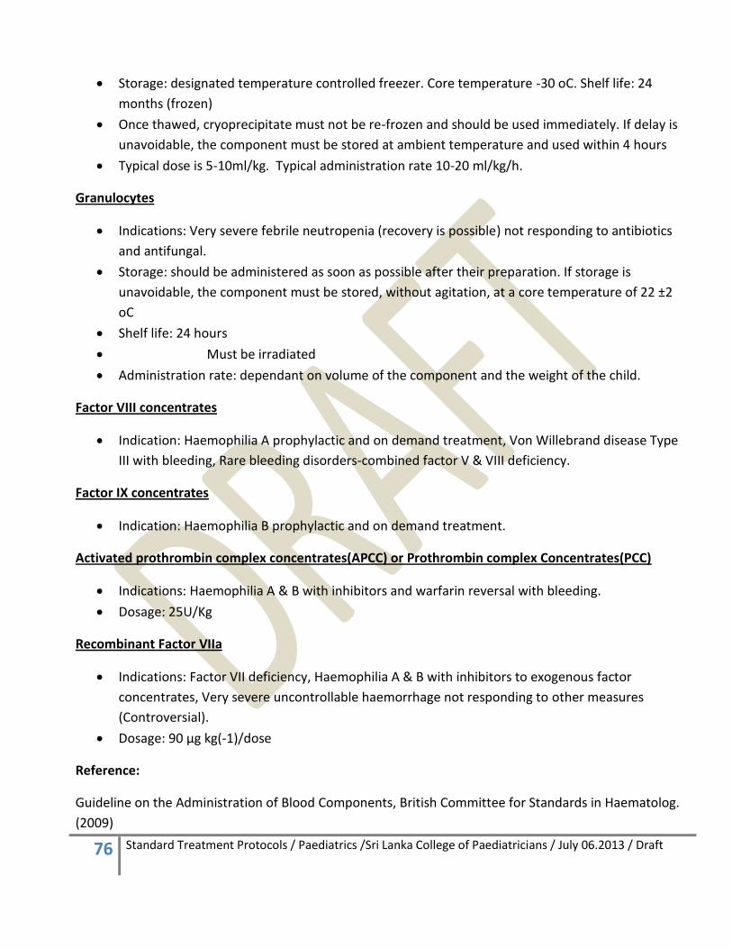
76 Standard Treatment Protocols / Paediatrics /Sri Lanka College of Paediatricians / July 06.2013 / Draft
Storage: designated temperature controlled freezer. Core temperature -30 oC. Shelf life: 24
months (frozen)
Once thawed, cryoprecipitate must not be re-frozen and should be used immediately. If delay is
unavoidable, the component must be stored at ambient temperature and used within 4 hours
Typical dose is 5-10ml/kg. Typical administration rate 10-20 ml/kg/h.
Granulocytes
Indications: Very severe febrile neutropenia (recovery is possible) not responding to antibiotics
and antifungal.
Storage: should be administered as soon as possible after their preparation. If storage is
unavoidable, the component must be stored, without agitation, at a core temperature of 22 ±2
oC
Shelf life: 24 hours
Must be irradiated
Administration rate: dependant on volume of the component and the weight of the child.
Factor VIII concentrates
Indication: Haemophilia A prophylactic and on demand treatment, Von Willebrand disease Type
III with bleeding, Rare bleeding disorders-combined factor V & VIII deficiency.
Factor IX concentrates
Indication: Haemophilia B prophylactic and on demand treatment.
Activated prothrombin complex concentrates(APCC) or Prothrombin complex Concentrates(PCC)
Indications: Haemophilia A & B with inhibitors and warfarin reversal with bleeding.
Dosage: 25U/Kg
Recombinant Factor VIIa
Indications: Factor VII deficiency, Haemophilia A & B with inhibitors to exogenous factor
concentrates, Very severe uncontrollable haemorrhage not responding to other measures
(Controversial).
Dosage: 90 μg kg(-1)/dose
Reference:
Guideline on the Administration of Blood Components, British Committee for Standards in Haematolog.
(2009)
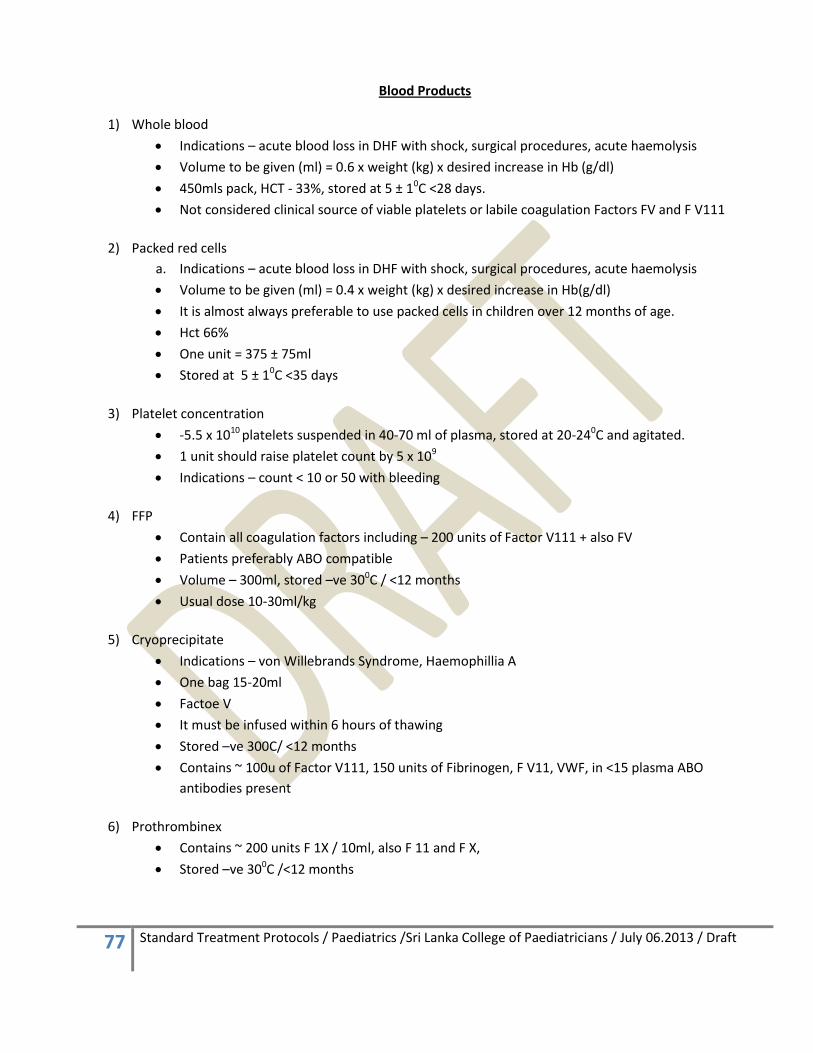
77 Standard Treatment Protocols / Paediatrics /Sri Lanka College of Paediatricians / July 06.2013 / Draft
Blood Products
1) Whole blood
Indications – acute blood loss in DHF with shock, surgical procedures, acute haemolysis
Volume to be given (ml) = 0.6 x weight (kg) x desired increase in Hb (g/dl)
450mls pack, HCT - 33%, stored at 5 ± 10C <28 days.
Not considered clinical source of viable platelets or labile coagulation Factors FV and F V111
2) Packed red cells
a. Indications – acute blood loss in DHF with shock, surgical procedures, acute haemolysis
Volume to be given (ml) = 0.4 x weight (kg) x desired increase in Hb(g/dl)
It is almost always preferable to use packed cells in children over 12 months of age.
Hct 66%
One unit = 375 ± 75ml
Stored at 5 ± 10C <35 days
3) Platelet concentration
-5.5 x 10¹⁰ platelets suspended in 40-70 ml of plasma, stored at 20-240C and agitated.
1 unit should raise platelet count by 5 x 10⁹
Indications – count < 10 or 50 with bleeding
4) FFP
Contain all coagulation factors including – 200 units of Factor V111 + also FV
Patients preferably ABO compatible
Volume – 300ml, stored –ve 300C / <12 months
Usual dose 10-30ml/kg
5) Cryoprecipitate
Indications – von Willebrands Syndrome, Haemophillia A
One bag 15-20ml
Factoe V
It must be infused within 6 hours of thawing
Stored –ve 300C/ <12 months
Contains ~ 100u of Factor V111, 150 units of Fibrinogen, F V11, VWF, in <15 plasma ABO
antibodies present
6) Prothrombinex
Contains ~ 200 units F 1X / 10ml, also F 11 and F X,
Stored –ve 300C /<12 months
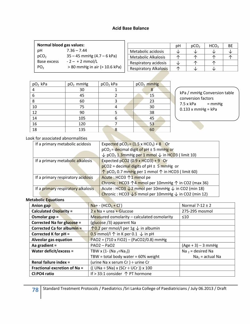
78 Standard Treatment Protocols / Paediatrics /Sri Lanka College of Paediatricians / July 06.2013 / Draft
Acid Base Balance
Look for associated abnormalities
If a primary metabolic acidosis Expected pCO2 = (1.5 x HCO3) + 8 Or pCO2 = decimal digit of pH ± 5 mmHg or ↓ pCO2 1.3mmHg per 1 mmol ↓ in HCO3 ( limit 10)
If a primary metabolic alkalosis Expected pCO2 (0.9 x HCO3) + 9 Or pCO2 = decimal digits of pH ± 5 mmHg or ↑ pCO2 0.7 mmHg per 1 mmol ↑ in HCO3 ( limit 60)
If a primary respiratory acidosis Acute : HCO3 ↑1 mmol pe Chronic : HCO3 ↑4 mmol per 10mmHg ↑ in CO2 (max 36)
If a primary respiratory alkalosis Acute : HCO3 ↓2 mmol per 10mmHg ↓ in CO2 (min 18) Chronic : HCO3 ↓5 mmol per 10mmHg ↓ in CO2 (min 12)
Metabolic Equations
Anion gap Na+ - (HCO3 + Cl-) Normal 7-12 ± 2
Calculated Osolarity = 2 x Na + urea + Glucose 275-295 mosmol
Osmolar gap = Measured osmolarity – calculated osmolarity ≤10
Corrected Na for glucose = (glucose /3) apparent Na
Corrected Ca for albumin = ↑0.2 per mmol/l per 1g ↓ in albumin
Corrected K for pH = 0.5 mmol/l ↑ in K per 0.1 ↓ in pH
Alveolar gas equation PAO2 = (710 x FiO2) – (PaCO2/0.8) mmHg
Aa gradient = PAO2 – PaO2 (Age + 3) – 3 mmHg
Water deficit/excess = TBW x (1- (Na 2÷Na1)) TBW = total body water = 60% weight
Na 2 = desired Na Na1 = actual Na
Renal failure index = (urine Na x serum Cr ) ÷ urine Cr
Fractional excretion of Na = (( UNa ÷ SNa) x (SCr ÷ UCr )) x 100
Cl:PO4 ratio If > 33:1 consider ↑ PT hormone
pH pCO2 HCO3 BE
Metabolic acidosis ↓ ↓ ↓ ↓
Metabolic Alkalosis ↑ ↑ ↑ ↑
Respiratory acidosis ↓ ↑ ↑
Respiratory Alkalosis ↑ ↓ ↓
pO2 kPa pO2 mmHg pCO2 kPa pCO2 mmHg
4 30 1 8
6 45 2 15
8 60 3 23
10 75 4 30
12 90 5 38
14 105 6 45
16 120 7 53
18 135 8 60
Normal blood gas values: pH 7.36 – 7.44 pCO2 35 – 45 mmHg (4.7 – 6 kPa) Base excess - 2 – + 2 mmol/L PO2 > 80 mmHg in air (> 10.6 kPa)
kPa / mmHg Conversion table conversion factors 7.5 x kPa = mmHg 0.133 x mmHg = kPa
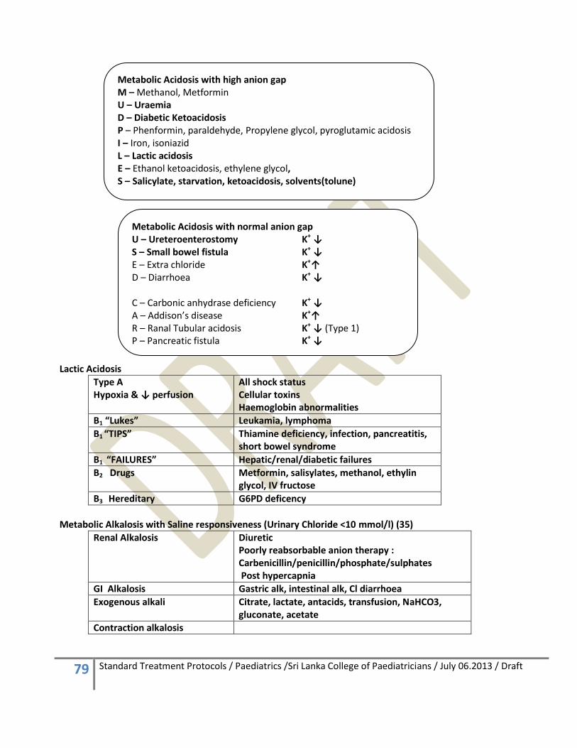
79 Standard Treatment Protocols / Paediatrics /Sri Lanka College of Paediatricians / July 06.2013 / Draft
Lactic Acidosis
Type A Hypoxia & ↓ perfusion
All shock status Cellular toxins Haemoglobin abnormalities
B1 “Lukes” Leukamia, lymphoma
B1 “TIPS” Thiamine deficiency, infection, pancreatitis, short bowel syndrome
B1 “FAILURES” Hepatic/renal/diabetic failures
B2 Drugs Metformin, salisylates, methanol, ethylin glycol, IV fructose
B3 Hereditary G6PD deficency
Metabolic Alkalosis with Saline responsiveness (Urinary Chloride <10 mmol/l) (35)
Renal Alkalosis Diuretic Poorly reabsorbable anion therapy : Carbenicillin/penicillin/phosphate/sulphates Post hypercapnia
GI Alkalosis Gastric alk, intestinal alk, Cl diarrhoea
Exogenous alkali Citrate, lactate, antacids, transfusion, NaHCO3, gluconate, acetate
Contraction alkalosis
Metabolic Acidosis with high anion gap M – Methanol, Metformin U – Uraemia D – Diabetic Ketoacidosis P – Phenformin, paraldehyde, Propylene glycol, pyroglutamic acidosis I – Iron, isoniazid L – Lactic acidosis E – Ethanol ketoacidosis, ethylene glycol, S – Salicylate, starvation, ketoacidosis, solvents(tolune)
Metabolic Acidosis with normal anion gap U – Ureteroenterostomy K+ ↓ S – Small bowel fistula K+ ↓ E – Extra chloride K+↑ D – Diarrhoea K+ ↓ C – Carbonic anhydrase deficiency K+ ↓ A – Addison’s disease K+↑ R – Ranal Tubular acidosis K+ ↓ (Type 1) P – Pancreatic fistula K+ ↓
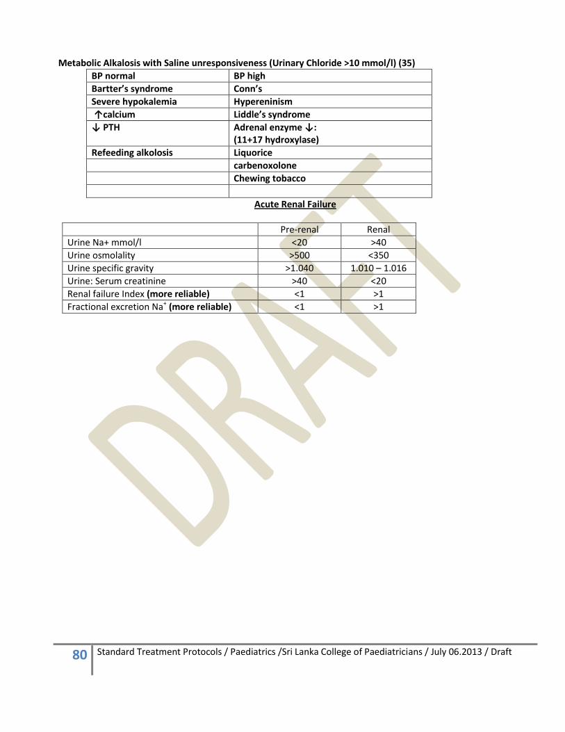
80 Standard Treatment Protocols / Paediatrics /Sri Lanka College of Paediatricians / July 06.2013 / Draft
Metabolic Alkalosis with Saline unresponsiveness (Urinary Chloride >10 mmol/l) (35)
BP normal BP high
Bartter’s syndrome Conn’s
Severe hypokalemia Hypereninism
↑calcium Liddle’s syndrome
↓ PTH Adrenal enzyme ↓: (11+17 hydroxylase)
Refeeding alkolosis Liquorice
carbenoxolone
Chewing tobacco
Acute Renal Failure
Pre-renal Renal
Urine Na+ mmol/l <20 >40
Urine osmolality >500 <350
Urine specific gravity >1.040 1.010 – 1.016
Urine: Serum creatinine >40 <20
Renal failure Index (more reliable) <1 >1
Fractional excretion Na+ (more reliable) <1 >1
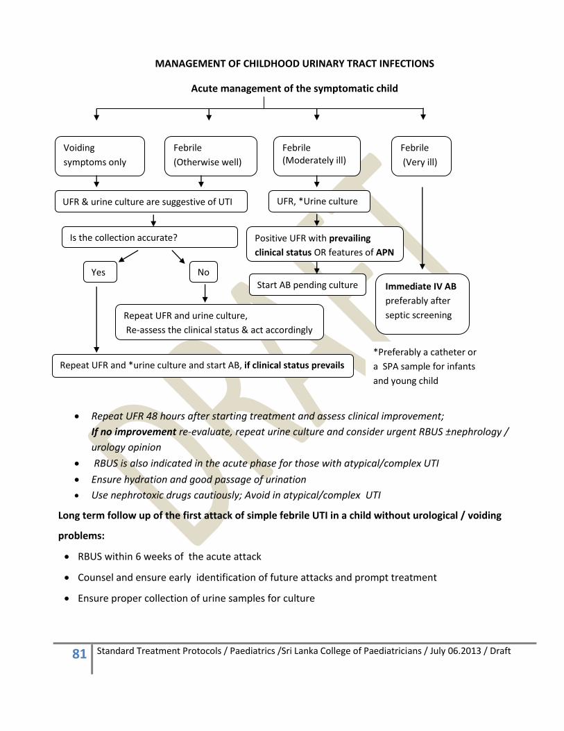
81 Standard Treatment Protocols / Paediatrics /Sri Lanka College of Paediatricians / July 06.2013 / Draft
MANAGEMENT OF CHILDHOOD URINARY TRACT INFECTIONS
Acute management of the symptomatic child
Repeat UFR 48 hours after starting treatment and assess clinical improvement;
If no improvement re-evaluate, repeat urine culture and consider urgent RBUS ±nephrology /
urology opinion
RBUS is also indicated in the acute phase for those with atypical/complex UTI
Ensure hydration and good passage of urination
Use nephrotoxic drugs cautiously; Avoid in atypical/complex UTI
Long term follow up of the first attack of simple febrile UTI in a child without urological / voiding
problems:
RBUS within 6 weeks of the acute attack
Counsel and ensure early identification of future attacks and prompt treatment
Ensure proper collection of urine samples for culture
Voiding
symptoms only
Febrile
(Otherwise well)
Febrile (Moderately ill)
Febrile
(Very ill)
UFR, *Urine culture
Positive UFR with prevailing
clinical status OR features of APN
Repeat UFR and urine culture,
Re-assess the clinical status & act accordingly
Repeat UFR and *urine culture and start AB, if clinical status prevails
Yes
UFR & urine culture are suggestive of UTI
Immediate IV AB
preferably after
septic screening
Is the collection accurate?
No
Start AB pending culture
*Preferably a catheter or
a SPA sample for infants
and young child
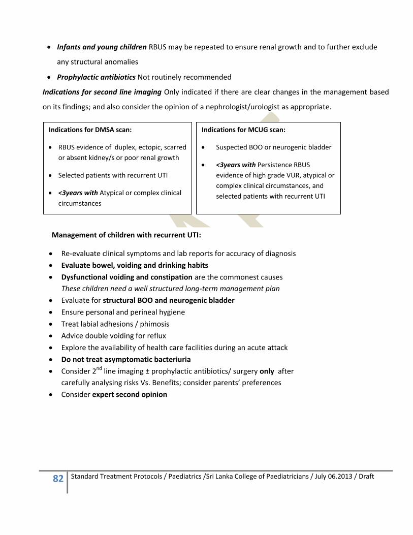
82 Standard Treatment Protocols / Paediatrics /Sri Lanka College of Paediatricians / July 06.2013 / Draft
Infants and young children RBUS may be repeated to ensure renal growth and to further exclude
any structural anomalies
Prophylactic antibiotics Not routinely recommended
Indications for second line imaging Only indicated if there are clear changes in the management based
on its findings; and also consider the opinion of a nephrologist/urologist as appropriate.
Management of children with recurrent UTI:
Re-evaluate clinical symptoms and lab reports for accuracy of diagnosis
Evaluate bowel, voiding and drinking habits
Dysfunctional voiding and constipation are the commonest causes
These children need a well structured long-term management plan
Evaluate for structural BOO and neurogenic bladder
Ensure personal and perineal hygiene
Treat labial adhesions / phimosis
Advice double voiding for reflux
Explore the availability of health care facilities during an acute attack
Do not treat asymptomatic bacteriuria
Consider 2nd line imaging ± prophylactic antibiotics/ surgery only after
carefully analysing risks Vs. Benefits; consider parents’ preferences
Consider expert second opinion
Indications for DMSA scan:
RBUS evidence of duplex, ectopic, scarred
or absent kidney/s or poor renal growth
Selected patients with recurrent UTI
<3years with Atypical or complex clinical
circumstances
Indications for MCUG scan:
Suspected BOO or neurogenic bladder
<3years with Persistence RBUS
evidence of high grade VUR, atypical or
complex clinical circumstances, and
selected patients with recurrent UTI
83 Standard Treatment Protocols / Paediatrics /Sri Lanka College of Paediatricians / July 06.2013 / Draft
References
1. Urinary Tract Infection: Clinical Practice Guideline for the Diagnosis and Management of the
Initial UTI in Febrile Infants and Children 2 to 24 Months: Subcommittee on Urinary Tract
Infection, Steering Committee on Quality Improvement and Management. Pediatrics
2011;128;595-610
2. National Institute for Health and Clinical Excellence: Urinary tract infection in children London:
NICE,2007
3. Mathews R, Carpenter M, Chesney R, Hoberman A et al. Controversies in the management of
vesicoureteral reflux: The rationale for the RIVUR study. J Pediatr Urol. 2009; 5: 336 –341.
4. Abdulaziz Kari J, Tullus K. Controversy in Urinary Tract Infection Management in Children: A
Review of New Data and Subseq
Abbreviations
UFR = urine full report
AB = antibiotics
APN= acute pyelo-nephritis
SPA = supra pubic aspiration
RBUS = renal bladder ultra sound scan
BOO = bladder outlet obstruction
Atypical UTI (NICE 2007)
1. Seriously ill or septic child
2. Evidence of obstructive uropathy
3. Rising serum creatinine
4. Failure to respond to appropriate AB therapy with in 48 hrs
5. Non E-coli UTI
6. Any prenatal urinary tract finding
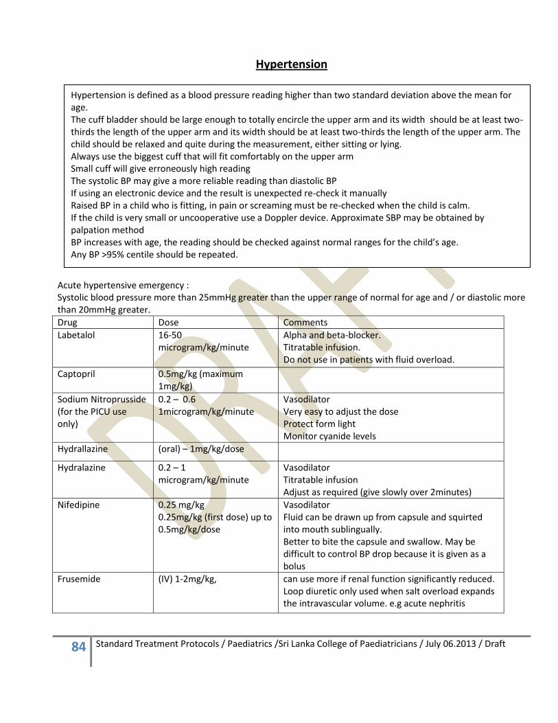
84 Standard Treatment Protocols / Paediatrics /Sri Lanka College of Paediatricians / July 06.2013 / Draft
Hypertension
Acute hypertensive emergency : Systolic blood pressure more than 25mmHg greater than the upper range of normal for age and / or diastolic more than 20mmHg greater.
Drug Dose Comments
Labetalol 16-50 microgram/kg/minute
Alpha and beta-blocker. Titratable infusion. Do not use in patients with fluid overload.
Captopril 0.5mg/kg (maximum 1mg/kg)
Sodium Nitroprusside (for the PICU use only)
0.2 – 0.6 1microgram/kg/minute
Vasodilator Very easy to adjust the dose Protect form light Monitor cyanide levels
Hydrallazine (oral) – 1mg/kg/dose
Hydralazine 0.2 – 1 microgram/kg/minute
Vasodilator Titratable infusion Adjust as required (give slowly over 2minutes)
Nifedipine 0.25 mg/kg 0.25mg/kg (first dose) up to 0.5mg/kg/dose
Vasodilator Fluid can be drawn up from capsule and squirted into mouth sublingually. Better to bite the capsule and swallow. May be difficult to control BP drop because it is given as a bolus
Frusemide (IV) 1-2mg/kg, can use more if renal function significantly reduced. Loop diuretic only used when salt overload expands the intravascular volume. e.g acute nephritis
Hypertension is defined as a blood pressure reading higher than two standard deviation above the mean for age. The cuff bladder should be large enough to totally encircle the upper arm and its width should be at least two-thirds the length of the upper arm and its width should be at least two-thirds the length of the upper arm. The child should be relaxed and quite during the measurement, either sitting or lying. Always use the biggest cuff that will fit comfortably on the upper arm Small cuff will give erroneously high reading The systolic BP may give a more reliable reading than diastolic BP If using an electronic device and the result is unexpected re-check it manually Raised BP in a child who is fitting, in pain or screaming must be re-checked when the child is calm. If the child is very small or uncooperative use a Doppler device. Approximate SBP may be obtained by palpation method BP increases with age, the reading should be checked against normal ranges for the child’s age. Any BP >95% centile should be repeated.
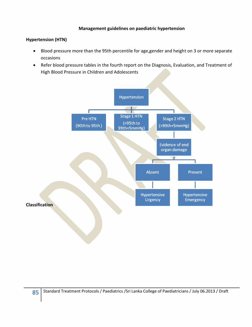
85 Standard Treatment Protocols / Paediatrics /Sri Lanka College of Paediatricians / July 06.2013 / Draft
Management guidelines on paediatric hypertension
Hypertension (HTN)
Blood pressure more than the 95th percentile for age,gender and height on 3 or more separate
occasions
Refer blood pressure tables in the fourth report on the Diagnosis, Evaluation, and Treatment of
High Blood Pressure in Children and Adolescents
Classification
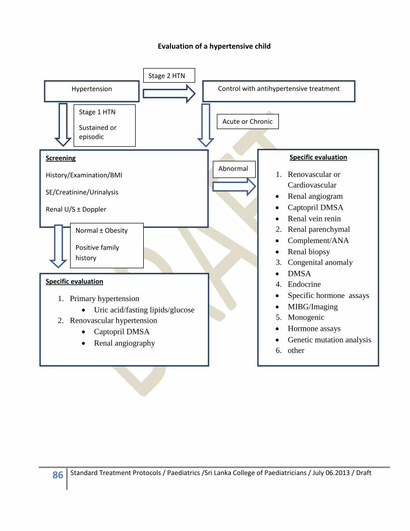
86 Standard Treatment Protocols / Paediatrics /Sri Lanka College of Paediatricians / July 06.2013 / Draft
Evaluation of a hypertensive child
Hypertension Control with antihypertensive treatment
Stage 2 HTN
Stage 1 HTN
Sustained or episodic
Screening
History/Examination/BMI
SE/Creatinine/Urinalysis
Renal U/S ± Doppler
Specific evaluation
1. Renovascular or
Cardiovascular
Renal angiogram
Captopril DMSA
Renal vein renin
2. Renal parenchymal
Complement/ANA
Renal biopsy
3. Congenital anomaly
DMSA
4. Endocrine
Specific hormone assays
MIBG/Imaging
5. Monogenic
Hormone assays
Genetic mutation analysis
6. other
Abnormal
Normal ± Obesity
Positive family
history
Specific evaluation
1. Primary hypertension
Uric acid/fasting lipids/glucose
2. Renovascular hypertension
Captopril DMSA
Renal angiography
Acute or Chronic
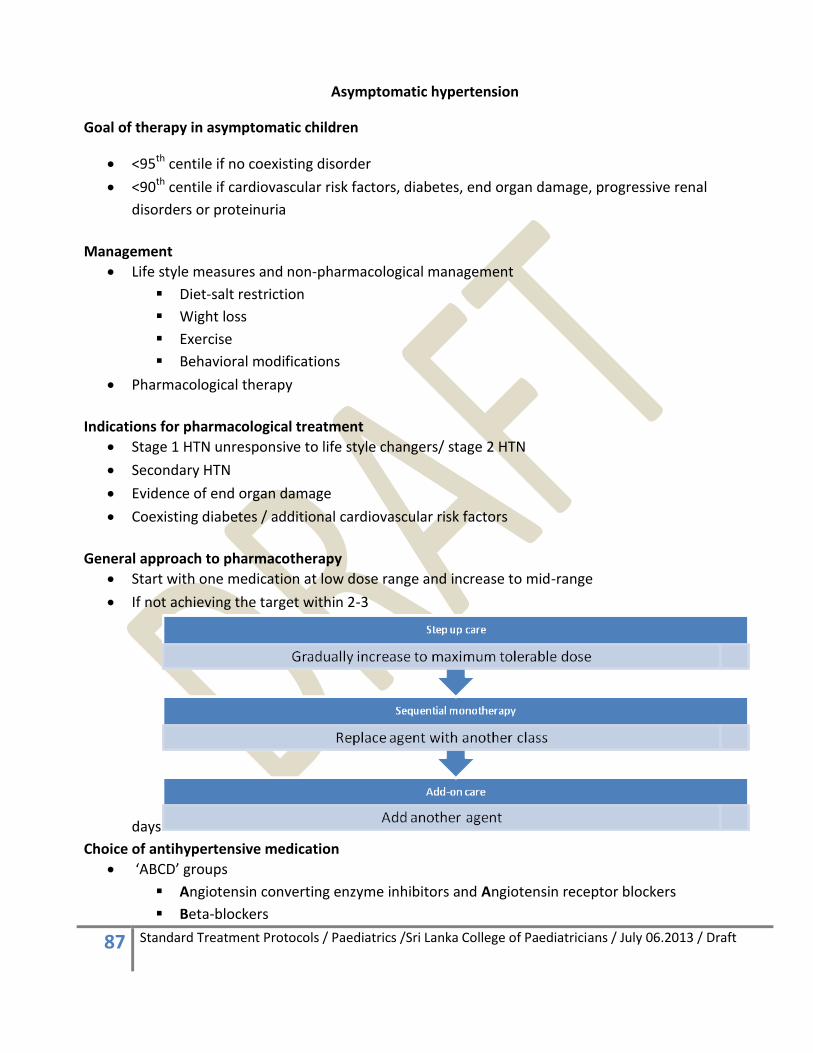
87 Standard Treatment Protocols / Paediatrics /Sri Lanka College of Paediatricians / July 06.2013 / Draft
Asymptomatic hypertension
Goal of therapy in asymptomatic children
<95th centile if no coexisting disorder
<90th centile if cardiovascular risk factors, diabetes, end organ damage, progressive renal
disorders or proteinuria
Management
Life style measures and non-pharmacological management
Diet-salt restriction
Wight loss
Exercise
Behavioral modifications
Pharmacological therapy
Indications for pharmacological treatment
Stage 1 HTN unresponsive to life style changers/ stage 2 HTN
Secondary HTN
Evidence of end organ damage
Coexisting diabetes / additional cardiovascular risk factors
General approach to pharmacotherapy
Start with one medication at low dose range and increase to mid-range
If not achieving the target within 2-3
days
Choice of antihypertensive medication
‘ABCD’ groups
Angiotensin converting enzyme inhibitors and Angiotensin receptor blockers
Beta-blockers
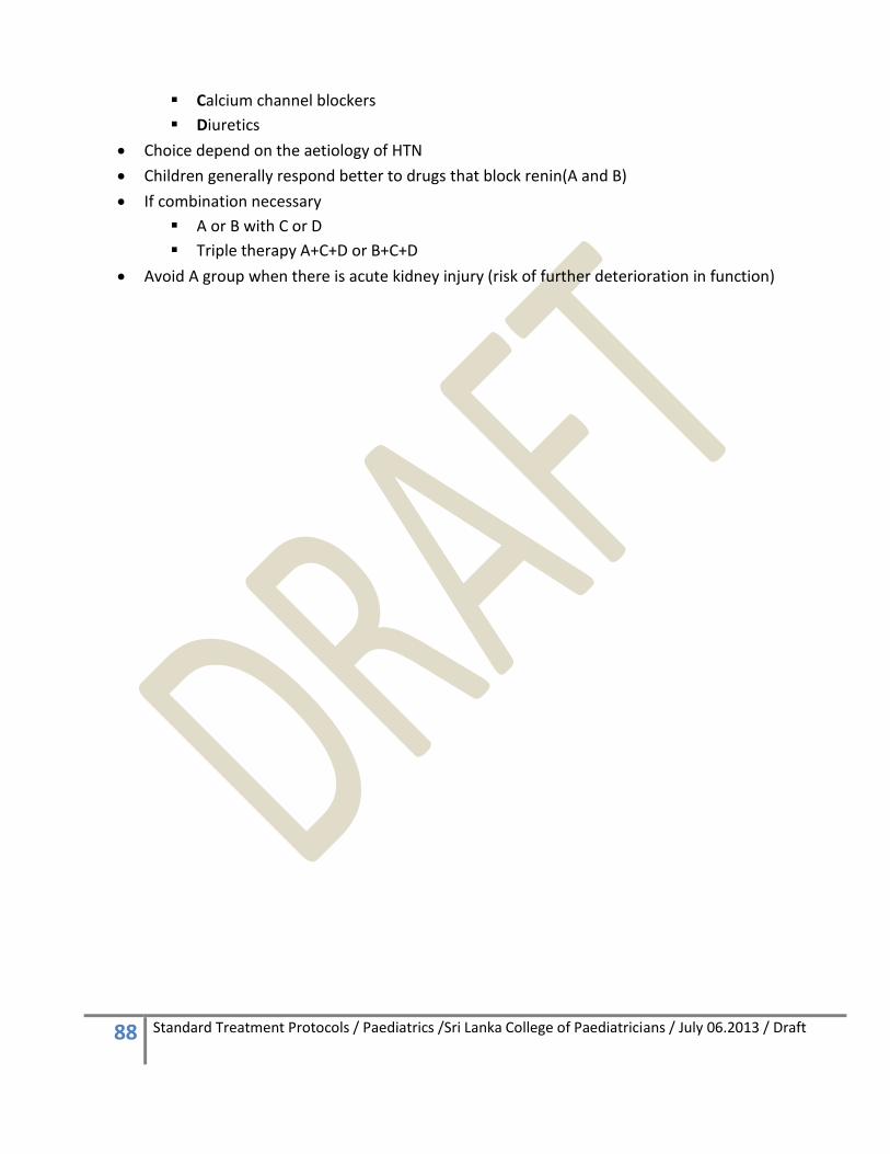
88 Standard Treatment Protocols / Paediatrics /Sri Lanka College of Paediatricians / July 06.2013 / Draft
Calcium channel blockers
Diuretics
Choice depend on the aetiology of HTN
Children generally respond better to drugs that block renin(A and B)
If combination necessary
A or B with C or D
Triple therapy A+C+D or B+C+D
Avoid A group when there is acute kidney injury (risk of further deterioration in function)
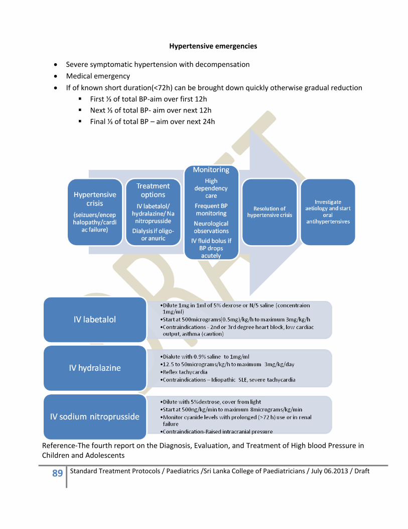
89 Standard Treatment Protocols / Paediatrics /Sri Lanka College of Paediatricians / July 06.2013 / Draft
Hypertensive emergencies
Severe symptomatic hypertension with decompensation
Medical emergency
If of known short duration(<72h) can be brought down quickly otherwise gradual reduction
First ⅓ of total BP-aim over first 12h
Next ⅓ of total BP- aim over next 12h
Final ⅓ of total BP – aim over next 24h
Reference-The fourth report on the Diagnosis, Evaluation, and Treatment of High blood Pressure in Children and Adolescents
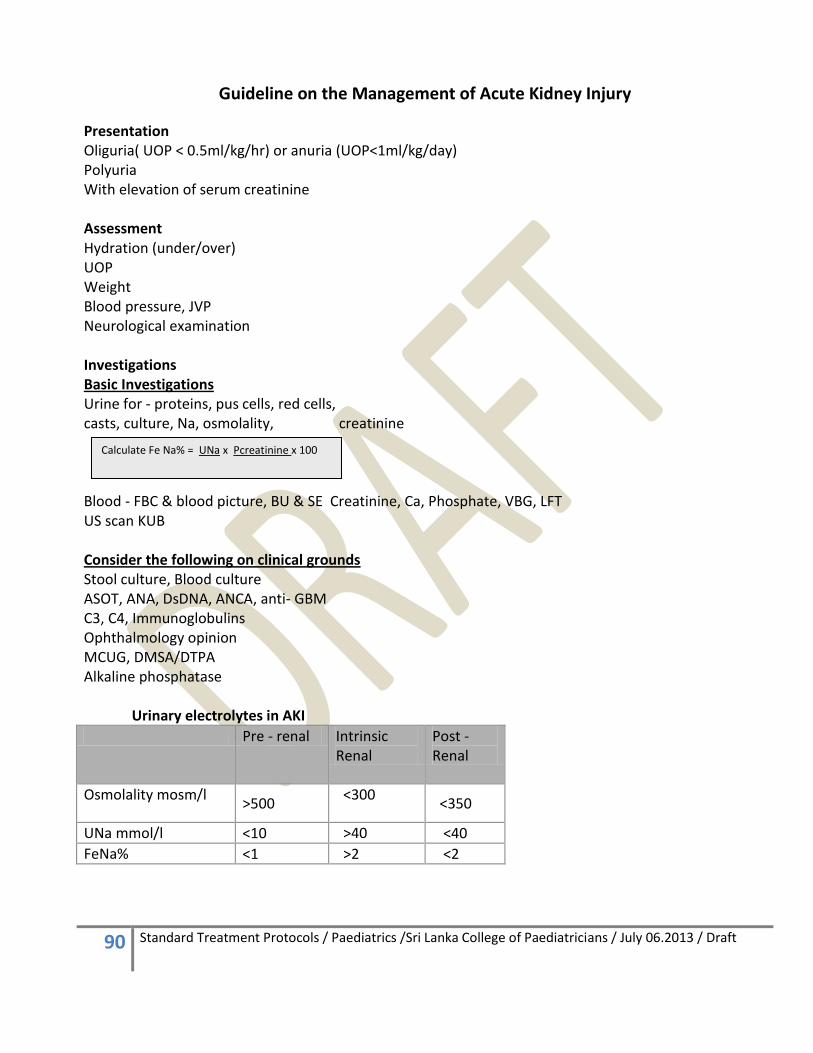
90 Standard Treatment Protocols / Paediatrics /Sri Lanka College of Paediatricians / July 06.2013 / Draft
Guideline on the Management of Acute Kidney Injury
Presentation Oliguria( UOP < 0.5ml/kg/hr) or anuria (UOP<1ml/kg/day) Polyuria With elevation of serum creatinine Assessment Hydration (under/over) UOP Weight Blood pressure, JVP Neurological examination Investigations Basic Investigations Urine for - proteins, pus cells, red cells, casts, culture, Na, osmolality, creatinine Blood - FBC & blood picture, BU & SE Creatinine, Ca, Phosphate, VBG, LFT US scan KUB Consider the following on clinical grounds Stool culture, Blood culture ASOT, ANA, DsDNA, ANCA, anti- GBM C3, C4, Immunoglobulins Ophthalmology opinion MCUG, DMSA/DTPA Alkaline phosphatase Urinary electrolytes in AKI
Pre - renal Intrinsic Renal
Post - Renal
Osmolality mosm/l >500
<300 <350
UNa mmol/l <10 >40 <40
FeNa% <1 >2 <2
Calculate Fe Na% = UNa x Pcreatinine x 100
PNa Ucreatinine
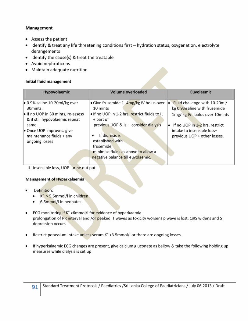
91 Standard Treatment Protocols / Paediatrics /Sri Lanka College of Paediatricians / July 06.2013 / Draft
Management
Assess the patient
Identify & treat any life threatening conditions first – hydration status, oxygenation, electrolyte derangements
Identify the cause(s) & treat the treatable
Avoid nephrotoxins
Maintain adequate nutrition Initial fluid management
Hypovolaemic Volume overloaded Euvolaemic
0.9% saline 10-20ml/kg over 30mints.
If no UOP in 30 mints, re-assess & if still hypovolaemic repeat same.
Once UOP improves. give maintenance fluids + any ongoing losses
Give frusemide 1- 4mg/kg IV bolus over 10 mints
If no UOP in 1-2 hrs, restrict fluids to IL + part of
If previous UOP & is. consider dialysis
If diuresis is established with frusemide, minimise fluids as above to allow a negative balance till euvolaemic.
Fluid challenge with 10-20ml/ kg 0.9%saline with frusemide
1mg/ kg IV bolus over 10mints
If no UOP in 1-2 hrs, restrict intake to insensible loss+ previous UOP + other losses.
IL- insensible loss, UOP- urine out put
Management of Hyperkalaemia
Definition:
K+ > 5.5mmol/l in children
6.5mmol/l in neonates
ECG monitoring if K+ >6mmol/l for evidence of hyperkaemia . prolongation of PR interval and /or peaked T waves as toxicity worsens p wave is lost, QRS widens and ST depression occurs
Restrict potassium intake unless serum K+ <3.5mmol/l or there are ongoing losses.
If hyperkalaemic ECG changes are present, give calcium gluconate as bellow & take the following holding up measures while dialysis is set up
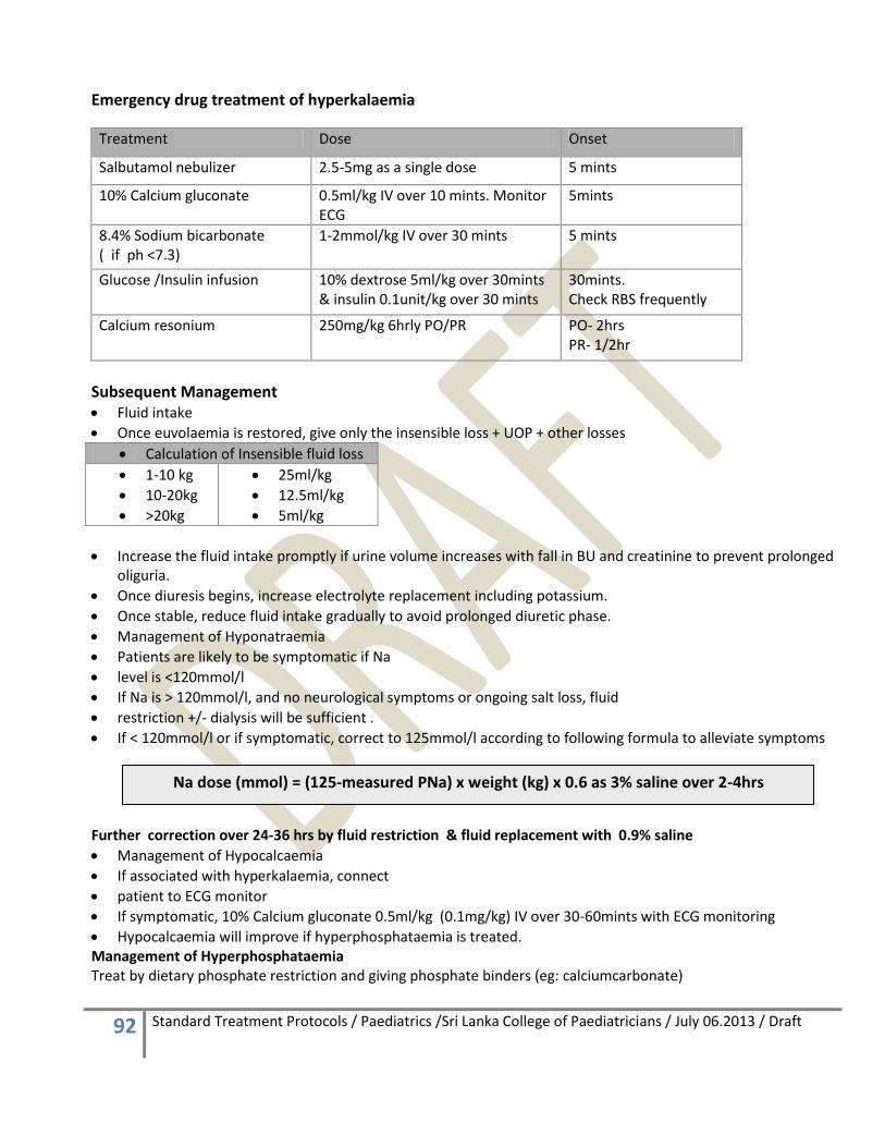
92 Standard Treatment Protocols / Paediatrics /Sri Lanka College of Paediatricians / July 06.2013 / Draft
Emergency drug treatment of hyperkalaemia
Treatment Dose Onset
Salbutamol nebulizer 2.5-5mg as a single dose 5 mints
10% Calcium gluconate 0.5ml/kg IV over 10 mints. Monitor ECG
5mints
8.4% Sodium bicarbonate ( if ph <7.3)
1-2mmol/kg IV over 30 mints 5 mints
Glucose /Insulin infusion 10% dextrose 5ml/kg over 30mints & insulin 0.1unit/kg over 30 mints
30mints. Check RBS frequently
Calcium resonium 250mg/kg 6hrly PO/PR PO- 2hrs PR- 1/2hr
Subsequent Management Fluid intake
Once euvolaemia is restored, give only the insensible loss + UOP + other losses
Calculation of Insensible fluid loss
1-10 kg
10-20kg
>20kg
25ml/kg
12.5ml/kg
5ml/kg
Increase the fluid intake promptly if urine volume increases with fall in BU and creatinine to prevent prolonged oliguria.
Once diuresis begins, increase electrolyte replacement including potassium.
Once stable, reduce fluid intake gradually to avoid prolonged diuretic phase.
Management of Hyponatraemia
Patients are likely to be symptomatic if Na
level is <120mmol/l
If Na is > 120mmol/l, and no neurological symptoms or ongoing salt loss, fluid
restriction +/- dialysis will be sufficient .
If < 120mmol/l or if symptomatic, correct to 125mmol/l according to following formula to alleviate symptoms Further correction over 24-36 hrs by fluid restriction & fluid replacement with 0.9% saline
Management of Hypocalcaemia
If associated with hyperkalaemia, connect
patient to ECG monitor
If symptomatic, 10% Calcium gluconate 0.5ml/kg (0.1mg/kg) IV over 30-60mints with ECG monitoring
Hypocalcaemia will improve if hyperphosphataemia is treated. Management of Hyperphosphataemia Treat by dietary phosphate restriction and giving phosphate binders (eg: calciumcarbonate)
Na dose (mmol) = (125-measured PNa) x weight (kg) x 0.6 as 3% saline over 2-4hrs
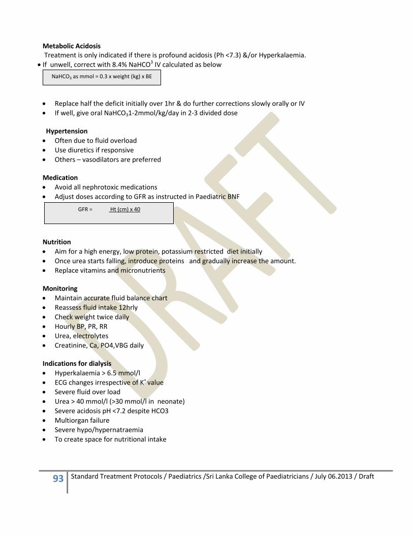
93 Standard Treatment Protocols / Paediatrics /Sri Lanka College of Paediatricians / July 06.2013 / Draft
Metabolic Acidosis Treatment is only indicated if there is profound acidosis (Ph <7.3) &/or Hyperkalaemia.
If unwell, correct with 8.4% NaHCO3 IV calculated as below
Replace half the deficit initially over 1hr & do further corrections slowly orally or IV
If well, give oral NaHCO31-2mmol/kg/day in 2-3 divided dose Hypertension
Often due to fluid overload
Use diuretics if responsive
Others – vasodilators are preferred Medication
Avoid all nephrotoxic medications
Adjust doses according to GFR as instructed in Paediatric BNF Nutrition
Aim for a high energy, low protein, potassium restricted diet initially
Once urea starts falling, introduce proteins and gradually increase the amount.
Replace vitamins and micronutrients Monitoring
Maintain accurate fluid balance chart
Reassess fluid intake 12hrly
Check weight twice daily
Hourly BP, PR, RR
Urea, electrolytes
Creatinine, Ca, PO4,VBG daily Indications for dialysis
Hyperkalaemia > 6.5 mmol/l
ECG changes irrespective of K+ value
Severe fluid over load
Urea > 40 mmol/l (>30 mmol/l in neonate)
Severe acidosis pH <7.2 despite HCO3
Multiorgan failure
Severe hypo/hypernatraemia
To create space for nutritional intake
NaHCO3 as mmol = 0.3 x weight (kg) x BE
GFR = Ht (cm) x 40
S. Creatinine ( micromol/l)
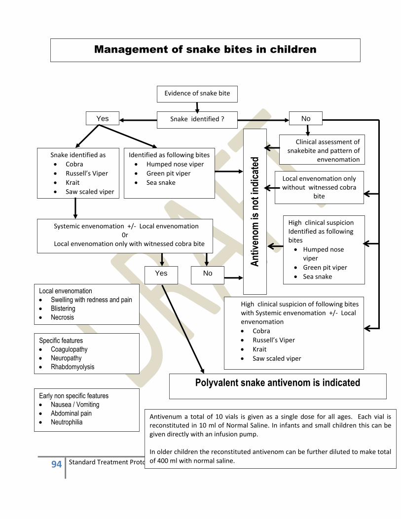
94 Standard Treatment Protocols / Paediatrics /Sri Lanka College of Paediatricians / July 06.2013 / Draft
Management of snake bites in children
Evidence of snake bite
Snake identified ? Yes
Snake identified as
Cobra
Russell’s Viper
Krait
Saw scaled viper
Identified as following bites
Humped nose viper
Green pit viper
Sea snake
Systemic envenomation +/- Local envenomation 0r
Local envenomation only with witnessed cobra bite
Yes No
Local envenomation
Swelling with redness and pain
Blistering
Necrosis
Specific features
Coagulopathy
Neuropathy
Rhabdomyolysis
Early non specific features
Nausea / Vomiting
Abdominal pain
Neutrophilia
An
tive
no
m is
no
t in
dic
ated
No
Clinical assessment of snakebite and pattern of
envenomation
Local envenomation only without witnessed cobra
bite
High clinical suspicion Identified as following bites
Humped nose viper
Green pit viper
Sea snake
High clinical suspicion of following bites
with Systemic envenomation +/- Local envenomation
Cobra
Russell’s Viper
Krait
Saw scaled viper
Polyvalent snake antivenom is indicated
Antivenum a total of 10 vials is given as a single dose for all ages. Each vial is reconstituted in 10 ml of Normal Saline. In infants and small children this can be given directly with an infusion pump. In older children the reconstituted antivenom can be further diluted to make total of 400 ml with normal saline.
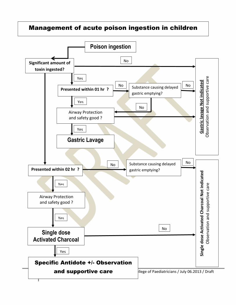
95 Standard Treatment Protocols / Paediatrics /Sri Lanka College of Paediatricians / July 06.2013 / Draft
Management of acute poison ingestion in children
Poison ingestion
Significant amount of
toxin ingested?
Presented within 01 hr ?
Airway Protection and safety good ?
Gastric Lavage
Presented within 02 hr ?
Airway Protection and safety good ?
Single dose Activated Charcoal
Gas
tric
lava
ge N
ot
ind
icat
ed
O
bse
rvat
ion
an
d s
up
po
rtiv
e ca
re
Sin
gle
do
se A
ctiv
ate
d C
har
coal
No
t in
dic
ate
d
Ob
serv
atio
n a
nd
su
pp
ort
ive
care
Specific Antidote +/- Observation
and supportive care
No
Yes
Yes
Yes
Substance causing delayed
gastric emptying?
No No
No
Substance causing delayed
gastric emptying? No
No
Yes
Yes
Yes
No
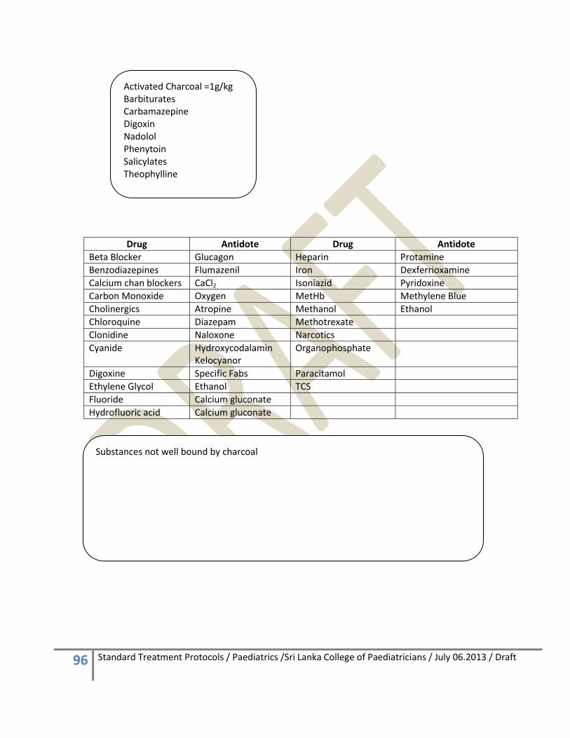
96 Standard Treatment Protocols / Paediatrics /Sri Lanka College of Paediatricians / July 06.2013 / Draft
Drug Antidote Drug Antidote
Beta Blocker Glucagon Heparin Protamine
Benzodiazepines Flumazenil Iron Dexferrioxamine
Calcium chan blockers CaCl2 Isoniazid Pyridoxine
Carbon Monoxide Oxygen MetHb Methylene Blue
Cholinergics Atropine Methanol Ethanol
Chloroquine Diazepam Methotrexate
Clonidine Naloxone Narcotics
Cyanide Hydroxycodalamin Kelocyanor
Organophosphate
Digoxine Specific Fabs Paracitamol
Ethylene Glycol Ethanol TCS
Fluoride Calcium gluconate
Hydrofluoric acid Calcium gluconate
Activated Charcoal =1g/kg Barbiturates Carbamazepine Digoxin Nadolol Phenytoin Salicylates Theophylline
Substances not well bound by charcoal
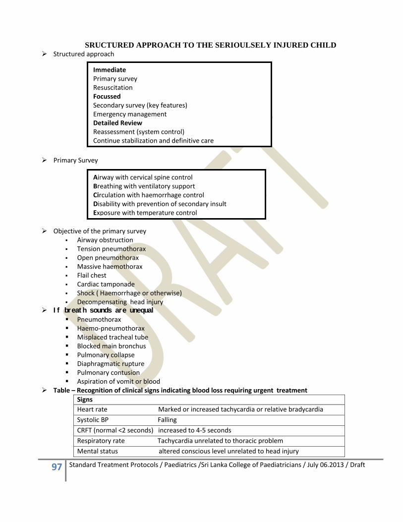
97 Standard Treatment Protocols / Paediatrics /Sri Lanka College of Paediatricians / July 06.2013 / Draft
SRUCTURED APPROACH TO THE SERIOULSELY INJURED CHILD Structured approach
Primary Survey
Objective of the primary survey Airway obstruction Tension pneumothorax Open pneumothorax Massive haemothorax Flail chest Cardiac tamponade Shock ( Haemorrhage or otherwise) Decompensating head injury
I f breat h sounds are unequal
Pneumothorax Haemo-pneumothorax Misplaced tracheal tube Blocked main bronchus Pulmonary collapse Diaphragmatic rupture Pulmonary contusion Aspiration of vomit or blood
Table – Recognition of clinical signs indicating blood loss requiring urgent treatment
Signs
Heart rate Marked or increased tachycardia or relative bradycardia
Systolic BP Falling
CRFT (normal <2 seconds) increased to 4-5 seconds
Respiratory rate Tachycardia unrelated to thoracic problem
Mental status altered conscious level unrelated to head injury
Immediate Primary survey Resuscitation Focussed Secondary survey (key features) Emergency management Detailed Review Reassessment (system control) Continue stabilization and definitive care
Airway with cervical spine control Breathing with ventilatory support Circulation with haemorrhage control Disability with prevention of secondary insult Exposure with temperature control
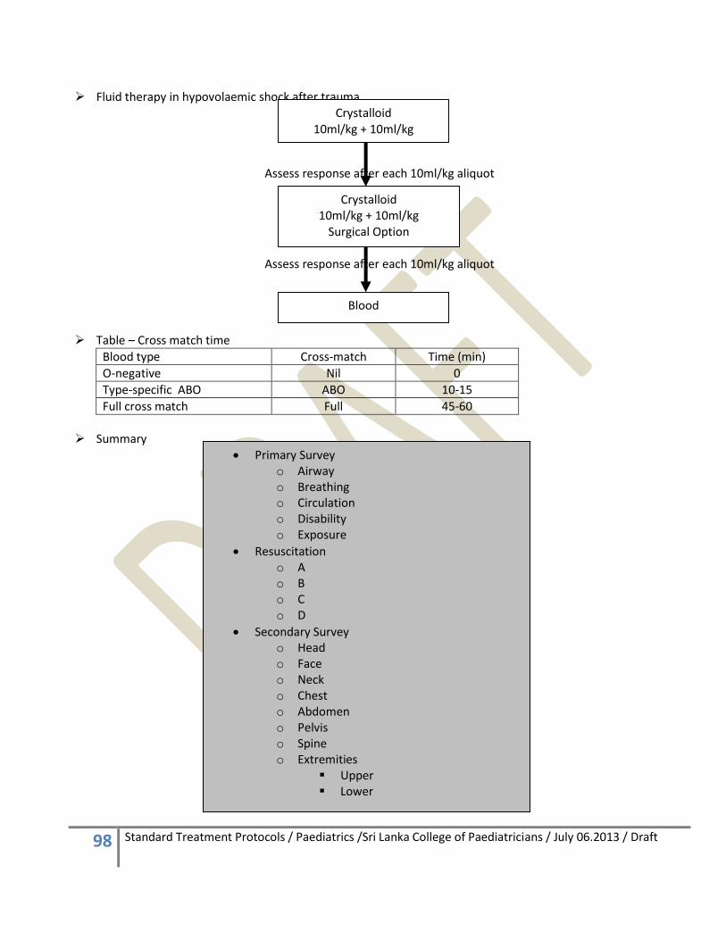
98 Standard Treatment Protocols / Paediatrics /Sri Lanka College of Paediatricians / July 06.2013 / Draft
Fluid therapy in hypovolaemic shock after trauma.
Assess response after each 10ml/kg aliquot
Assess response after each 10ml/kg aliquot
Table – Cross match time
Blood type Cross-match Time (min)
O-negative Nil 0
Type-specific ABO ABO 10-15
Full cross match Full 45-60
Summary
Crystalloid 10ml/kg + 10ml/kg
Crystalloid 10ml/kg + 10ml/kg
Surgical Option
Blood
Primary Survey o Airway o Breathing o Circulation o Disability o Exposure
Resuscitation o A o B o C o D
Secondary Survey o Head o Face o Neck o Chest o Abdomen o Pelvis o Spine o Extremities
Upper Lower
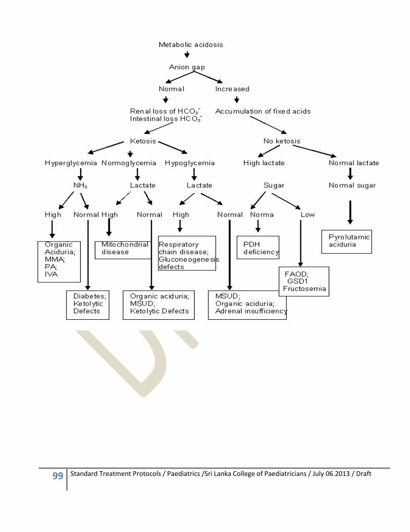
99 Standard Treatment Protocols / Paediatrics /Sri Lanka College of Paediatricians / July 06.2013 / Draft
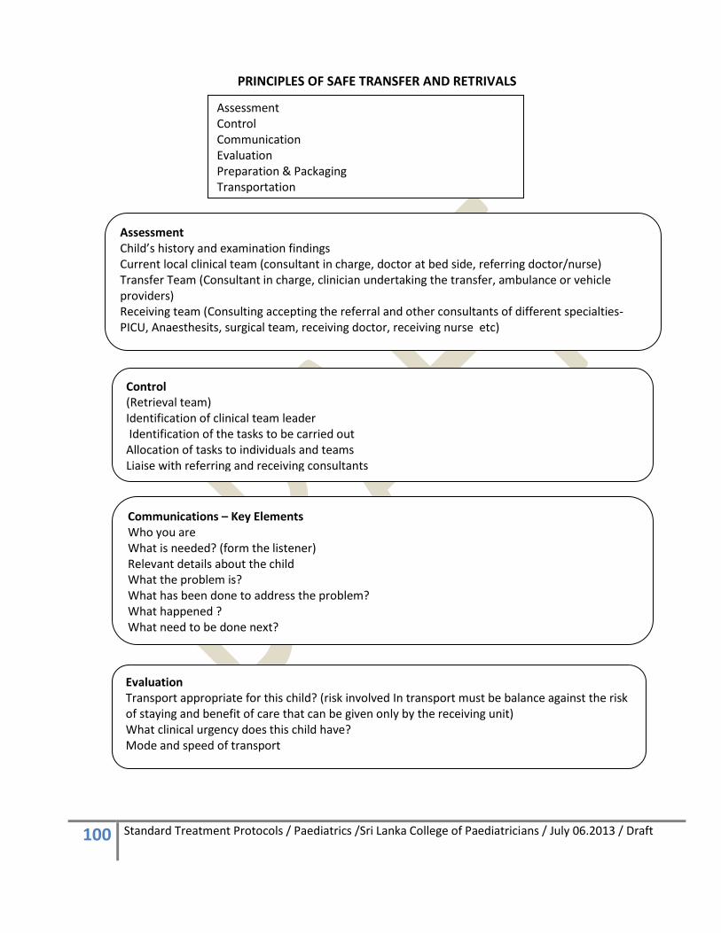
100 Standard Treatment Protocols / Paediatrics /Sri Lanka College of Paediatricians / July 06.2013 / Draft
PRINCIPLES OF SAFE TRANSFER AND RETRIVALS
Assessment Control Communication Evaluation Preparation & Packaging Transportation
Assessment Child’s history and examination findings Current local clinical team (consultant in charge, doctor at bed side, referring doctor/nurse) Transfer Team (Consultant in charge, clinician undertaking the transfer, ambulance or vehicle providers) Receiving team (Consulting accepting the referral and other consultants of different specialties-PICU, Anaesthesits, surgical team, receiving doctor, receiving nurse etc)
Control (Retrieval team) Identification of clinical team leader Identification of the tasks to be carried out Allocation of tasks to individuals and teams Liaise with referring and receiving consultants
Communications – Key Elements Who you are What is needed? (form the listener) Relevant details about the child What the problem is? What has been done to address the problem? What happened ? What need to be done next? Evaluation Transport appropriate for this child? (risk involved In transport must be balance against the risk of staying and benefit of care that can be given only by the receiving unit) What clinical urgency does this child have? Mode and speed of transport
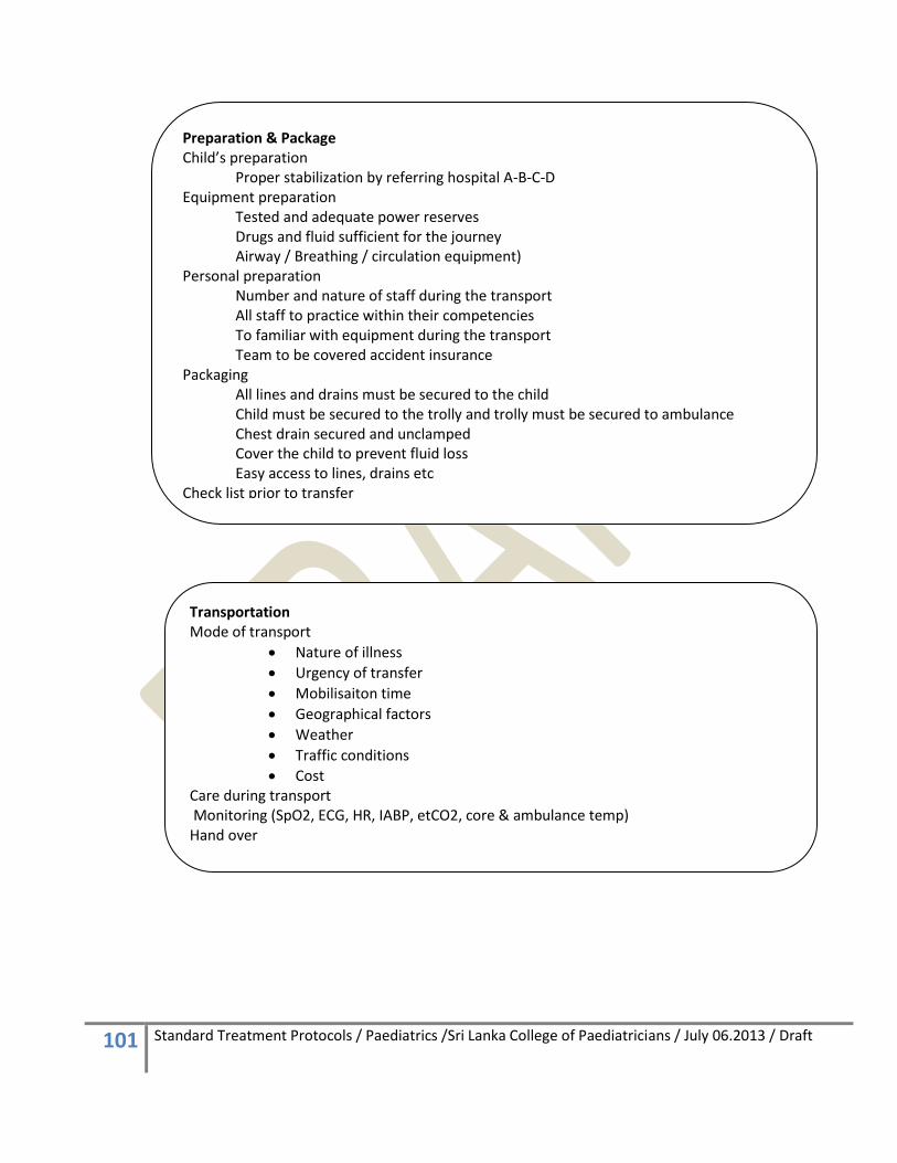
101 Standard Treatment Protocols / Paediatrics /Sri Lanka College of Paediatricians / July 06.2013 / Draft
Preparation & Package Child’s preparation
Proper stabilization by referring hospital A-B-C-D Equipment preparation Tested and adequate power reserves Drugs and fluid sufficient for the journey Airway / Breathing / circulation equipment) Personal preparation Number and nature of staff during the transport All staff to practice within their competencies To familiar with equipment during the transport Team to be covered accident insurance Packaging All lines and drains must be secured to the child
Child must be secured to the trolly and trolly must be secured to ambulance Chest drain secured and unclamped Cover the child to prevent fluid loss Easy access to lines, drains etc
Check list prior to transfer
Transportation Mode of transport
Nature of illness
Urgency of transfer
Mobilisaiton time
Geographical factors
Weather
Traffic conditions
Cost Care during transport Monitoring (SpO2, ECG, HR, IABP, etCO2, core & ambulance temp) Hand over
102 Standard Treatment Protocols / Paediatrics /Sri Lanka College of Paediatricians / July 06.2013 / Draft
VENESECTION WITH BROKEN NEEDLES
Indications: Neonates for investigations Infants for investigations Sometimes in Children
Procedure: o You should have a good picture of the venous plexus of the hands / feet o IV needle (black or Blue colour) o Take the needle out o Break the needle cleanly o Hold the hand or feet with your grip o Get the veins distended o Attacks the small veins in the distal venous plexus o Sharp end in to the vein o Blunt end to the bottle o Gradually squeeze and release the grip o If needle gets dislodged – re site the needle o Collect the U&E first because EDTA contaminate K
Advantages o No wasting of bigger veins which are suitable for IV cannulation o Puncture the veins which are not suitable for cannulations o With good skills one should be able to collect necessary volume of blood. o Minimal damage to veins
Complications: o Whole of the needle can get lost inside the vein o Tight squeezing can cause fracture of the metacarpals o Osteomylitis
103 Standard Treatment Protocols / Paediatrics /Sri Lanka College of Paediatricians / July 06.2013 / Draft
INTRAOSSEOUS ACCESS
Why choose this route: o Largely a life saving resuscitative measure for a short term as gaining peripheral venous access for
initial resuscitation fluids and drugs can waste valuable minutes or hours. o The venous drainage from the marrow rapidly accesses the main venous system. o Refilling the circulation via this route may make peripheral cannulation more possible.
Indications: o Patients less than 6 years of age. o For administration of fluids, drugs o Can be inserted through burnt skin if no alternative.
Sites: o Proximal tibia
Flat anterior-medial surface 1cm inferior to and 1 cm medial to the tibial tubercle
o Femur Distal femur – anterolateral surface 3cm above lateral condyle. Avoid fractured limb or those with proximal fractures.
Equipment o Alcohol solutions o Cleaning solutions o 18 gauge with a trocha at least 1.5cm in length o Bone Marrow needle with stylet o Blood needle o A 5ml syringe o A 20ml Syringe o Infusion fluids
Procedure: o Prepare the skin sites with antiseptic o Administer local Anaesthesia o Use bone narrow needle with a stylet gauge 13 or 18 o Introduce the needle with a rotating or screwing motion in the bone with pressure at 90 degrees
to skin. o Needle should be pointing a way from the joint o Advance the needle until a give is felt – this may take one minute of patience. o Needle has now entered the marrow cavity
104 Standard Treatment Protocols / Paediatrics /Sri Lanka College of Paediatricians / July 06.2013 / Draft
Confirmation of the site of entry: o Then connect a 5cc empty syringe and withdraw the marrow o Ability to aspirate marrow OR o Ability to inject without extravasations o Marrow contains should be sent for cultures CRP and Full Blood Count.
Fixation: o Use line extension and 3-way tap (not direct syringe application onto the needle) or the wiggling
of the syringe enlarges entry hole into bone and extravasations will occur. o Splint the limb securely.
Rate of fluid entry: o Start giving bolus solutions o 10mls / minute when under gravity o 40mls / minute when syringed in.
Type of fluid: o Anything that can be given intravenously
Complications: o Incorrect placement leading to extravasations and skin infiltration around the needle (needle is
too superficial or too deep) o The limb should be observed and not covered in dressings. Excess extravasated fluid could lead to
a compartment syndrome. o Cellulitis / Osteomyelitis o Damage to growth plate. o Rarely fractures
Contraindications o Fractures of the bone o Bone diseases like bone tumour or osteogenesis imperfecta
105 Standard Treatment Protocols / Paediatrics /Sri Lanka College of Paediatricians / July 06.2013 / Draft
PERIPHERAL INTRAVENOUS ACCESS
Clinical indicators of Peripheral cannulations are:
To maintain hydration and or correct dehydration in children who are unable to tolerate sufficient volumes of oral fluids
To maintain hydration / transfusion of IV fluids
Transfusion of blood or blood products
Administration of drugs/medication
1. Use with caution: a. Administration of irritant drugs
o Sodium bicarbonate therapy o Dopamine o Some of the antibiotics as boluses e.g. Vancomycin / Clarithromycin, Cloxacillin o Thiopentone
b. Large blood transfusions c. A high flow rates
i. Peripheral Cannulation a) Preparation b) Equipments c) The Patient d) Operator
ii. Selecting a suitable vein iii. Selecting appropriate cannula iv. Prepare the puncture site v. Getting the vein prominent
vi. Insertion and advancement of the cannula vii. How to make sure the cannula is in situ.
viii. Common problems with IV cannulation ix. Securing the IV cannula x. Care of IV cannula
XI. Complications
PRINCIPLES OF IV CANNULATIONS Preparation of IV cannulation Equipment
Correct cannula size 20 –pink, 22- blue, 24- yellow
Cleaning agents – alcohol swab
Sterile Cotton wool/ guaze-1 packet
Flushing solution – 0.9% normal saline in 2cc syringe
Fresh syringe – 5cc-to withdraw blood for essential investigations
Blood collecting bottles – FBC, U&E, Capillary tubes
Splints, bandages, Sticking tapes / tegederms, Scissors
Connection for cannula –L.S-connection- [T]
106 Standard Treatment Protocols / Paediatrics /Sri Lanka College of Paediatricians / July 06.2013 / Draft
The Patient o Explain in simple terms the procedure to the patient and parents and make the child comfortable. o Check patient’s identification o Get the patient to inhale deeply during the actual venepunctures to relieve possible anxiety
Operator
Organize correct lightening
Wash hands thoroughly with antiseptic soap according to Hand washing policy
This prevents
-Nosocomial infection
-Cross contamination
Wearing protective gloves is optional – Remember in high risk patients that gloves will not protect against needle stick injuries
Selecting a suitable vein o Have a good knowledge of venous plexus of both hands and feet o Avoid bigger veins as far as possible. E.g. veins in antecubital fossa and greater saphenous vein. o Use a patient’s non-dominant side o First use distal veins of both dorsum of hands and feet o Subsequently moves proximally o Always allow adequate time on inspection and palpation of veins in the dorsum of hands and feet. o Avoid very small veins –rarely successful. o Cannula must never totally occlude a vein. o The cannula length should correspond approximately to the length of straight vein to be used. o If in doubt always call a more experienced colleague or a senior colleague Avoid:
Areas of joint flexion
Veins close to arteries and deeper lying vessels.
Median cubital vein – Reserved for difficult blood sampling
Small visible but impalpable veins – this is rarely successful.
Veins that may be irritated from previous attempts.
Distal veins where proximal vein have been punctured Selecting appropriate cannula
Expiry date is not lapsed
Be Easy to insert
Permit optimal flow rates
Causes minimum discomfort to patient
Always should give high consistent and reliable performance
Should have a side injection port
Have minimal complications
Type of the infusion – Potent drugs and or irritant solution will require a large blood flow to assist haemodilution.
Cannula life – a smaller size cannula will minimize venous irritation.
Make sure package and its content are intact
107 Standard Treatment Protocols / Paediatrics /Sri Lanka College of Paediatricians / July 06.2013 / Draft
Prepare the puncture site
Apply local anaesthetic cream
E.g.; Emla or Ametop and leave it for ½ hour
Clean the puncture site and surrounding area thoroughly with iodine or alcohol swabs.
Allow antiseptic to dry
Do not re-palpate the area – it can be done if your hands are sterile.
Getting the vein prominent
Create an adequate venous pressure by applying a tight tourniquet or with your handgrip avoid using a tourniquet in neonates as much as possible.
Apply enough pressure to obstruct venous flow but not arterial flow
To encourage venous filling adopt one or more of the following man oeuvres.
Instruct the patient to clench or pump fist.
Tap vein lightly
Warm the extremities. (by wrapping with warm towels / gauze or dipping in warm water)
Allow the arm to hang over bedside.
Insertion and advancement of the cannula
Put on protective gloves- optional
Fold down the wings
Hold the Veinflown firmly with a three point grip-using-thumb & index finger
This minimizes the risk of touch contaminating the lure connection.
Ensure the correct positioning between the needle point and the catheter tip
Insertion & advancement of the Cannula
Select the straight section of the vein close to or at a confluence of two tributaries
Insert the cannula at 20-30 degrees or very low angle to the skin…
Puncture the vein few mm away from the vein (cannulation through the skin layers act as a barrier and prevent leakage)
Do not puncture the vein from the top.
Maintain the grip to make the veins prominent
Avoid if vein is moving or sliding.
Entry of needlepoint to vein is indicated by the presence of blood in the flash back chamber.
Insert the cannula slightly in order to make sure cannula tip is well within the vein. Make sure needle tip has not pierced the posterior wall of the vein.
Release the tourniquet and advance the cannula
Connect a 2cc syringe filled with normal saline and push the fluid in. make sure the cannula is well within the vein.
It is essential to record the time and date of insertion and person who did it in the nurses’ flow chart and over the sticking plaster.
Check the peripheral circulation after splinting.
108 Standard Treatment Protocols / Paediatrics /Sri Lanka College of Paediatricians / July 06.2013 / Draft
Advancing the Catheter
Advance the cannula a few millimetres further into the vein. This ensures that the catheter tip also enters the vein.
Entry of the needle tip in to the vein is indicated by the presence of blood in the flash back chamber
Withdraw the needle partially (about 5mm) to avoid exit through the posterior vein wall.
Advance the catheter of the cannula into the vein gently.
Release the tourniquet.
The needle must never be reinserted while the catheter is in the vein.
This may severe the catheter.
Avoid blood spillage by pressing a finger on the vein on or above the catheter tip
withdraw the needle completely.
Fix 2cc syringe, which is filled with N-saline, and inject a small amount to make sure that there is no extravasations/resistance.
How to make sure the cannula is in situ. If there is a resistance to the flushing solution, flush it again and see whether cannula is in situ. If the resistance is still there consider resiting. Common problems with IV cannulation
Initially blood flow now stopped
Blood flow stopped while advancing
No blood flowing to flash back chamber
Not piercing the vein
Try deep veins
Securing the IV cannula
The method you use should fix the cannula securely so that it can be neither pulled nor kinked
With your cannula is in place, have your assistance pass a piece of tape {¼”-2”} under the hub, stick side up.
Take one side of the tape & bend it over the hub to wrap around it to stick to the skin at an angle of 45 degree to the line of the catheter.
Do the same with opposite wing of tape.
Take your other piece of1/4”x2” tape & lay out it across the hub of the cannula close to the end.
Attach the extension set – so that weight of the IV lines is not put on the cannula.
Place a soft splint beneath the area to be fixed.
Wrap one piece of tape around the distal end of the cannula, securing the limb to the splint below.
The injection port of the cannula should be left exposed and a small piece of cotton wool can be placed underneath it to protect the skin below.
Do not put the plasters right round the limb so that proximal vein may be totally or partially occluded.
Wrap a second piece of tape around the proximal end of the splint, catching the extension tube below it.
This piece of the tape should not be too tight, as it may occlude the venous flow.
Allow the thumb to be free in case of cannula in the dorsum of the hand.
Leave the fingertips free. So that you can detect early cyanosis.
109 Standard Treatment Protocols / Paediatrics /Sri Lanka College of Paediatricians / July 06.2013 / Draft
Care of the cannula:
Idea is to prevent infections
Site inspection - daily basis for signs of infection
Dressing Change – Wet or soiled dressing should be changed, this predisposed to bacterial colonization.
Venous tenderness (Pain)
Local venous reaction
Redness, Tenderness, Swelling
Syringe pumps— 3 indicator pressure bulbs to show the level of resistance to the flow are present in new pumps.
o P1 – Very minimal resistance to the flow o P2 – Moderate resistance o P3 – Severe resistance
If one of the bulbs is indicated, flush the line with heparinized solution to make sure the line is blocked or not
If the line is not working, resite the cannula.
Careful handling
Do not contaminate. Wash your hand before handling the cannula. Leave a sterile towel under the IV cannula.
Cannula Change o Record time & Date of insertion o Re-site the cannula after 48-72 hours o Change IV giving sets every 24-48 hours
Complications:
Phlebitis and infection.
Haematoma.
Venospasm.
Embolization of veins with blood clots during forced flushing.
Air embolism. – Common in neonates with open foreman ovale.
Infiltration of subcutaneous tissues with IV solutions.
Accidental injection or infusion into an artery.
Ischaemia or gangrene of the distal extremity
EXTERNAL JUGULAR VEIN
A very useful site
Position the child as for internal jugular access.
Assistant can stretch the skin and position their finger to occlude the vein, just above the clavicle.
Puncture the vein few millimetres away from the vein. (Do not puncture the vein from the top).
Anchor the vein well.

110 Standard Treatment Protocols / Paediatrics /Sri Lanka College of Paediatricians / July 06.2013 / Draft
ARTERIAL CANNULATIONS
Indications: o Patients needing continuous intra arterial blood pressure monitoring o Frequent blood gas determinations o Frequent blood sampliing
Sites: o Radial o Posterior Tibial o Dorsalis Pedis o Femoral o Axillary artery o Neonates:
Umbilical arterial cannulation Radial arterial cannulaitons
o Avoid Brachial artery cannulation – poor collateral circulation
Procedure for Radial artery cannulation: o IV cannula 22 /24 G o Perform Allen’s test
Occlude both ulnar and Radial artery for 30 seconds Ulnar artery site is released In less than 3-4 seconds palm should show signs of circulation by the return of pink colour If palms remain pale – ulnar collateral circulation insufficient and radial artery cannulation
should not be performed
o Clean the site o Administer local anaesthetic o Extend the wrist o Catheter inserted at 30 degrees angle at the site of maximum radial pulsation with wrist extended o Catheter is advanced with the needle until blood return is obtained o Needle is removed half way through and catheter is advanced in the artery o If needle is difficult to advance although there is blood spurting at the end of catheter (artery has
gone in to spasm) o Do not push the catheter
Connect the flush and while flushing the line, advance the catheter
Problems with procedure o Arterial spasm
Occur after repeated unsuccessful attempts Arterial Pulse can become weak Leave for 10-15 minutes Spasm should ease out
111 Standard Treatment Protocols / Paediatrics /Sri Lanka College of Paediatricians / July 06.2013 / Draft
o Failed arterial; cannulation After multiple attempts Do a arterial cut down similar to venous cut down
Arterial Line fluid o Heparin saline 1 unit / 1 cc at a rate of 2cc / hour – continuous flush via transducer
Complications o Infections – Handle the line carefully o Distal ischaemia and necrosis
Monitor – distal to the line placement o Capillary circulation CRFT o Peripheral pulses o Temperature o Look for blanching / Ischaemia
Peripheral Arterial Puncture
Procedure
Get the correct size butter fly needle Leave the distal end open Clean the area well with antiseptic and dry with sterile gauze Get the light source from below
o Pen torch o Fibro-optic light source
Judge the size and the depth of the artery Puncture the artery Moment it puncture blood will flow backwards Connect a 1cc empty pre-heparinised syringe and collect the blood Remove the butterfly needle Apply pressure with gauze until bleeding stops Use hypoallergenic plasters Uses
Blood gas
Fungal blood cultures
Complications o Haematoma formation o Damage to deeper structures
112 Standard Treatment Protocols / Paediatrics /Sri Lanka College of Paediatricians / July 06.2013 / Draft
CAPILLARY BLOOD SAMPLING
CATEGORAY o Neonates / Infants
INDICATIONS Microsample for laboratory analysis eg: SBR, Capillary blood-gas analysis Blood glucose Micro – haematocrit
CONTRAINDICATIONS ● Infant in shock ● Compromised peripheral blood flow ● Local oedema ● Local infection
● Severe polycythaemia (haematocrit > 70%) ● Monitoring oxygen concentration
PRECAUTIONS Avoid anteromedial surface of the heel
The heel should be warm – do not use temperatures >40 C to warm it
Use a lancet specifically designed for the purpose with a Pint no longer than 2.5mm and preferably shorter
Do not apply an adhesive dressing to the wound after
sampling – it will allow the tissue to become damp and may lead to maceration
Venous stasis in the hours after birth may give misleading values for both gases and haematocrit.
Use of alcohol skin wipes may alter blood glucose measurements made with Dextrostix using an optical electrical method
PO2 does not correlate with arterial oxygen concentrations.
EQUIPMENT Lancet – tip no longer than 2.5 mm IV Needle (Pink or Brown) Alcohol swab Containers for blood samples Capillary tube Cotton wool ball Warm compress if heel needs warming Vaseline / paraffin jelly
TECHNIQUE
Warm heel if necessary by wrapping in wet towel a 39C to 40C
Select puncture site. Do repeatedly use the same sites.
Cleanse site with alcohol and allow to dry
Wipe a small amount of paraffin jelly over the area to be punctured – this will allow the blood to accumulate as smooth droplets, which are easier to collect.
113 Standard Treatment Protocols / Paediatrics /Sri Lanka College of Paediatricians / July 06.2013 / Draft
Grasp ankle firmly with the index finger and thumb forming a circle through which the heel protrudes
Puncture the heel with a single continuous motion
introducing the lancet perpendicular to the skin and to a depth of no more than 2.5 mm
Apply gentle pressure as far away from the site as possible by squeezing the ring made by your thumb and index finger
Draw blood into the appropriate collection pots with a scooper or into capillary tube
Stop bleeding by gentle pressure with a cotton wool ball.
Do not apply an adhesive dressing
COMPLICATIONS
Inaccuracy of blood gas estimation
Cellulitis
Osteomyelitis of the calcaneus
Abscess formation
Calcified nodules
Tissue loss and scarring of the heel
Erroneously high Dextrostix values
Over estimating potassium concentration
Underestimating pO2