stem cell-containing hyaluronic acid-based spongy...
TRANSCRIPT
ORIGINAL ARTICLE
Stem Cell-Containing HyaluronicAcid-Based Spongy Hydrogels forIntegrated Diabetic Wound Healing
Lucılia Pereira da Silva1,2, Tırcia Carlos Santos1,2, Daniel Barreira Rodrigues1,2,Rogerio Pedro Pirraco1,2, Mariana Teixeira Cerqueira1,2, Rui Luıs Reis1,2, Vitor Manuel Correlo1,2 andAlexandra Pinto Marques1,2The detailed pathophysiology of diabetic foot ulcers is yet to be established and improved treatments are stillrequired. We propose a strategy that directs inflammation, neovascularization, and neoinnervation of diabeticwounds. Aiming to potentiate a relevant secretome for nerve regeneration, stem cells were precultured inhyaluronic acid-based spongy hydrogels under neurogenic/standard media before transplantation into diabeticmice full-thickness wounds. Acellular spongy hydrogels and empty wounds were used as controls. Re-epithelialization was attained 4 weeks after transplantation independently of the test groups, whereas athicker and more differentiated epidermis was observed for the cellular spongy hydrogels. A switch from theinflammatory to the proliferative phase of wound healing was revealed for all the experimental groups 2 weeksafter injury, but a significantly higher M2(CD163þ)/M1(CD86þ) subtype ratio was observed in the neurogenicpreconditioned group that also failed to promote neoinnervation. A higher number of intraepidermal nervefibers were observed for the unconditioned group probably due to a more controlled transition from the in-flammatory to the proliferative phase. Overall, stem cell-containing spongy hydrogels represent a promisingapproach to enhance diabetic wound healing by positively impacting re-epithelialization and by modulatingthe inflammatory response to promote a successful neoinnervation.
Journal of Investigative Dermatology (2017) 137, 1541e1551; doi:10.1016/j.jid.2017.02.976
INTRODUCTIONDiabetic foot ulcerations (DFUs) are the major cause ofnontraumatic foot amputation worldwide (Edwards et al.,2008). Many of these DFUs have associated diabeticperipheral neuropathy that is responsible for an absent senseof touch, pain, and/or temperature due to injured nerves(Laverdet et al., 2015). Diabetic peripheral neuropathy incombination with other factors such as persistentinflammation, impaired re-epithelialization, misbalancedmetalloproteinases and tissue inhibitor metalloproteinaseslevels, and reduced angiogenesis and blood flow have beenimplicated in the hindered progression of diabetic woundhealing (Blakytny and Jude, 2009). Treatment of DFUs with
13B’s Research Group—Biomaterials, Biodegradables and Biomimetics,University of Minho, Headquarters of the European Institute of Excellenceon Tissue Engineering and Regenerative Medicine, AvePark-Parque daCiencia e Tecnologia, Barco, Taipas, Guimaraes, Portugal; and 2ICVS/3B’s—PT Government Associate Laboratory, Braga/Guimaraes, Portugal
Correspondence: Alexandra Pinto Marques, 3B’s Research Group—Biomaterials, Biodegradables and Biomimetics, University of Minho,Headquarters of the European Institute of Excellence on Tissue Engineeringand Regenerative Medicine, AvePark—Parque da Ciencia e Tecnologia, ZonaIndustrial da Gandra, 4805-017 Barco, Taipas, Guimaraes, Portugal. E-mail:[email protected]
Abbreviations: BME, b-mercaptoethanol; condAhASCs, human adipose-derived stem cells conditioned to neurogenic medium A; DFU, diabetic footulceration; GG-HA, gellan gum-hyaluronic acid; hASC, human adipose-derived stem cell; RA, retinoic acid
Received 1 September 2016; revised 31 January 2017; accepted 6 February2017; accepted manuscript published online 1 March 2017
ª 2017 The Authors. Published by Elsevier, Inc. on behalf of the Society for Inv
advanced dressings composed of extracellular matrix (e.g.,Integra, Integra LifeSciences) or comprising cells and growthfactors (e.g., Graftskin/Apligraf, Organogenesis; or Derma-graft, Advanced BioHealing) has been shown to acceleratewound closure (Mulder et al., 2014). Nonetheless, themechanisms that aid wound healing progression are notclearly understood.
A correlation between DFU healing and diabetic periph-eral neuropathy is yet to be established. This is potentially thereason why neuropathy has not been directly targeted in thedevelopment of new DFU therapies. So far some works (Kantet al., 2015; Leal et al., 2015; Moura et al., 2014) have tar-geted the reduced levels of neuromediators in diabeticwounds by delivering neuromediators to the wounds, inorder to improve wound re-epithelialization. Others (Blaiset al., 2009; Caissie et al., 2006; Gingras et al., 2003) haveincorporated Schwann cells into skin tissue-engineered sub-stitutes showing the increased number of nerve fibers,enhanced nerve migration, nerve growth, and myelin sheathformation in the grafted tissues. Despite these interesting re-sults, the isolation and expansion of autologous humanSchwann cells for clinical application can be a challenge.Stem cells have been posed as a potential alternative.Nonhuman origin stem cells cultured with a neurogeniccocktail or in coculture with dorsal root ganglia neuronsdifferentiated toward a Schwann cell phenotype displaytypical functional characteristics of Schwann cells (Caddicket al., 2006; Kingham et al., 2007; Mahay et al., 2008a,2008b; Tomita et al., 2012). However, the actual
estigative Dermatology. www.jidonline.org 1541
LP da Silva et al.Stem Cell-Containing HA-Based Spongy Hydrogel
1542
differentiation of human-origin stem cells is still controversialdue to the coexpression of glial cell markers and the pro-duction of neurotrophic factors by undifferentiated stem cells(Brohlin et al., 2009; Park et al., 2010; Tomita et al., 2013;Tondreau et al., 2004).
Gellan gum spongy hydrogels obtained from gellan gumhydrogels have distinctive properties, including intrinsic celladhesive characteristics, that have proven beneficial in skintissue engineering (Cerqueira et al., 2014b; da Silva et al.,2014a, 2014b). When combined with hyaluronic acid,spongy hydrogels also exhibited hyaluronidase-mediatedbiodegradation that resulted in hyaluronic acid content-dependent neovascularization of ischemic mice hind limb(da Silva et al., 2016). Moreover, an enhanced effect over theneovascularization of excisional skin wound was observedwhen microvascular endothelial cells and human adiposestem cells (hASCs) were entrapped in gellan gum-hyaluronicacid (GG-HA) spongy hydrogels (Cerqueira et al., 2014b).
In this context, we hypothesized that hASCs precondi-tioned in neurogenic medium would acquire a Schwanncell-like phenotype with the ability to release neurotrophicfactors, which are essential for nerve repair. Thus, hASCscultured within GG-HA spongy hydrogels in the selectedneurogenic conditioning medium would function as a neu-rotrophic factor-producing platform, ultimately promotingnerve formation in diabetic skin wounds. It was also envi-sioned that the angiogenic HA fragments released from thespongy hydrogels, associated with the hASCs’ capacity to actas a modulator of inflammation, would have an extendedeffect on balancing the inflammatory state of the wounds.For this purpose, the neurotrophic secretome of hASCscultured in the standard and neurogenic media wasevaluated before entrapment within GG-HA spongy hydro-gels. Stem cell-containing GG-HA spongy hydrogels pre-cultured in standard and selected neurogenic conditioningmedia were implanted in diabetic mice excisional skinwounds, and the capacity of the generated constructs tomodulate inflammation and angiogenesis and promote neo-innervation was assessed to confirm the potential of theproposed approach to treat DFUs.
RESULTShASCs phenotype and secretome before and afterconditioning
In standard culture conditions, hASCs displayed the charac-teristic mesenchymal stem cell phenotype; more than 90% ofthe population expressed CD105, CD90, and CD73, and wasnegative for the hematopoietic markers CD31, CD34, andCD45 (Supplementary Table S1 online). In addition, morethan 95% of the hASCs expressed immature (nestin) andmature (glial fibrillary acidic protein, p75, and S100B) glialcell markers (Supplementary Table S1 and Figure 1a), char-acteristic of neurogenic lineages.
hASCs were preinduced to the neurogenic lineage with b-mercaptoethanol (BME) followed by retinoic acid (RA)exposure, and then induced to a Schwann cell-like pheno-type with specific differentiation components and mitogenssuch as glial growth factor-2, forskolin, platelet-derivedgrowth factor-AA, and basic fibroblast growth factor,following a previously described method (Brohlin et al.,
Journal of Investigative Dermatology (2017), Volume 137
2009), described as neurogenic conditioning medium B.Although this was our reference medium, cells kept prolif-erating after the preinduction with BME and RA. Thus, toachieve a cell population similarly exposed to BME and RA,these factors were maintained during culture in the pres-ence (medium A) and absence (medium C) of basic fibro-blast growth factor, which is responsible for cellproliferation (Figure 1b). Cells exposed to neurogenic con-ditioning media A and C showed a more polarizedmorphology in relation to cells cultured in standard andconditioned medium B (Figure 1c). Moreover, whencultured in neurogenic conditioning medium A, the numberof cells expressing CD105 and CD73 decreased, respec-tively, to 75e85% and to 93% (Supplementary Table S1).Independently of the neurogenic conditioning media, nosignificant changes were observed regarding the expressionof the Schwann cell-associated markers nestin, glial fibril-lary acidic protein, p75, and S100B (SupplementaryTable S1), which were already highly expressed by hASCs.No significant alterations in the secretome of hASCs weredetected after the neurogenic conditioning (Figure 1d).However, a tendency for a higher release of brain-derivedneurotrophic factor (55.56 � 30.00 to 31.33 � 18.85 pg/ml) and ciliary neurotrophic factor (1.67 � 0.23 to e1.17 �0.31 ng/ml) from hASCs cultured in conditioning medium B,and of glial cell-derived neurotrophic factor (47.50 �9.92e24.58 � 3.82 pg/ml) from hASCs in conditioningmedium A, relative to unconditioned hASCs was observed(Figure 1d).
The majority of the hASCs entrapped within GG-HAspongy hydrogels were alive and distributed throughoutit, colonizing the whole structure along the culture(Figure 1e). Moreover, in the 3D spongy hydrogels, thesecretome of the hASCs cultured in the selected condi-tioned medium A did not change in relation to uncondi-tioned hASCs (Figure 1d).
Wound closure and re-epithelialization
Although wounds were still open 2 weeks after trans-plantation (Figure 2a), within this period of time, thepercentage of wound closure for the GG-HAþhASCS-conditioned to neurogenic medium A (condAhASCs)condition was higher (83.7 � 11.2%) than for the GG-HA(56.1 � 28.2%), GG-HAþhASCs (56.4 � 23.4%), andcontrol (48.6 � 19.6%) groups (Figure 2b). As the majorityof the wounds were closed 4 weeks after transplantation,wound closure quantification based on the macroscopicimages was not performed to avoid inaccurate measure-ments. Wound re-epithelialization assessment 2 weeks afterinjury revealed a thin layer of k5 expressing keratinocytesmigrating toward the wound center and a thick layer of k10expressing keratinocytes in the margins for all the conditions(Figure 2c), characteristic of re-epithelialization. At 4 weeks,a thin and fragile epidermis consisting of one to two layersof keratinocytes was observed in the control and GG-HAconditions. In contrast, the wounds treated with cell-containing GG-HA spongy hydrogels presented a well-organized and thicker epidermis with a high number ofproliferative keratinocytes in the basal layer, as determinedby ki-67 staining (Figure 2c and d).
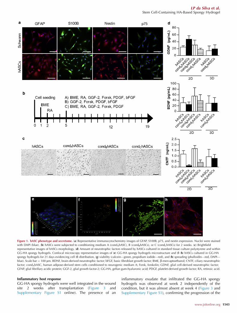
Figure 1. hASC phenotype and secretome. (a) Representative immunocytochemistry images of GFAP, S100B, p75, and nestin expression. Nuclei were stained
with DAPI (blue). (b) hASCs were subjected to conditioning medium A (condAhASC), B (condBhASCs), or C (condChASCs) for 2 weeks. (c) Brightfield
representative images of hASCs morphology. (d) Amount of neurotrophic factors released by hASCs cultured in standard tissue culture polystyrene and within
GG-HA spongy hydrogels. Confocal microscopy representative images of (e) GG-HA spongy hydrogels microstructure and (feh) hASCs cultured in GG-HA
spongy hydrogels for 21 days evidencing cell (f) distribution, (g) viability (calcein—green, propidium iodide—red), and (h) spreading (phalloidin—red, DAPI—
blue). Scale bar ¼ 100 mm. BDNF, brain-derived neurotrophic factor; bFGF, basic fibroblast growth factor; BME, b-mercaptoethanol; CNTF, ciliary neurotrophic
factor; condAhASC, human adipose-derived stem cells conditioned to neurogenic medium A; Forsk, forskolin; GDNF, glial cell-derived neurotrophic factor;
GFAP, glial fibrillary acidic protein; GGF-2, glial growth factor-2; GG-HA, gellan gum-hyaluronic acid; PDGF, platelet-derived growth factor; RA, retinoic acid.
LP da Silva et al.Stem Cell-Containing HA-Based Spongy Hydrogel
Inflammatory host response
GG-HA spongy hydrogels were well integrated in the woundsite 2 weeks after transplantation (Figure 3 andSupplementary Figure S1 online). The presence of an
inflammatory exudate that infiltrated the GG-HA spongyhydrogels was observed at week 2 independently of thecondition, but it was almost absent at week 4 (Figure 3 andSupplementary Figure S1), confirming the progression of the
www.jidonline.org 1543
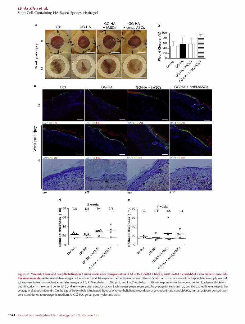
Figure 2. Wound closure and re-epithelialization 2 and 4weeks after transplantation of GG-HA, GG-HADhASCs, and GG-HADcondAhASCs into diabetic mice full-
thickness wounds. (a) Representative images of the wounds and (b) respective percentage of wound closure. Scale bar¼ 5mm. Control corresponds to an empty wound.
(c) Representative immunohistochemistry images of k5, k10 (scale bar ¼ 200 mm), and ki-67 (scale bar ¼ 50 mm) expression in the wound center. Epidermis thickness
quantification in thewound center (d) 2 and (e) 4weeks after transplantation. Eachmeasurement represents the average for each animal, and the dashed line represents the
average of diabeticmice skin.On the topof the symbols is indicated the total of re-epithelializedwounds per analyzed animals. condAhASCs, human adipose-derived stem
cells conditioned to neurogenic medium A; GG-HA, gellan gum-hyaluronic acid.
LP da Silva et al.Stem Cell-Containing HA-Based Spongy Hydrogel
Journal of Investigative Dermatology (2017), Volume 1371544
LP da Silva et al.Stem Cell-Containing HA-Based Spongy Hydrogel
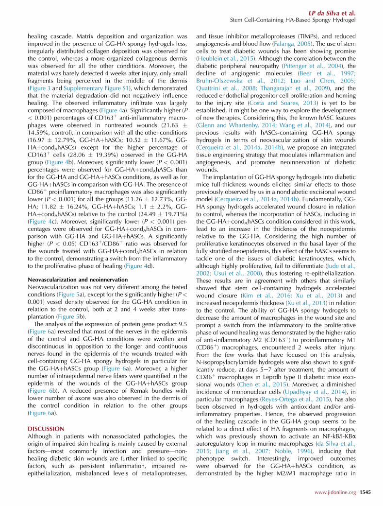
healing cascade. Matrix deposition and organization wasimproved in the presence of GG-HA spongy hydrogels less,irregularly distributed collagen deposition was observed forthe control, whereas a more organized collagenous dermiswas observed for all the other conditions. Moreover, thematerial was barely detected 4 weeks after injury, only smallfragments being perceived in the middle of the dermis(Figure 3 and Supplementary Figure S1), which demonstratedthat the material degradation did not negatively influencehealing. The observed inflammatory infiltrate was largelycomposed of macrophages (Figure 4a). Significantly higher (P< 0.001) percentages of CD163þ anti-inflammatory macro-phages were observed in nontreated wounds (21.63 �14.59%, control), in comparison with all the other conditions(16.97 � 12.79%, GG-HAþhASCs; 10.52 � 11.67%, GG-HAþcondAhASCs) except for the higher percentage ofCD163þ cells (28.06 � 19.39%) observed in the GG-HAgroup (Figure 4b). Moreover, significantly lower (P < 0.001)percentages were observed for GG-HAþcondAhASCs thanfor the GG-HA and GG-HAþhASCs conditions, as well as forGG-HAþhASCs in comparison with GG-HA. The presence ofCD86þ proinflammatory macrophages was also significantlylower (P < 0.001) for all the groups (11.26 � 12.73%, GG-HA; 11.82 � 16.24%, GG-HAþhASCs; 1.1 � 2.2%, GG-HAþcondAhASCs) relative to the control (24.49 � 19.71%)(Figure 4c). Moreover, significantly lower (P < 0.001) per-centages were observed for GG-HAþcondAhASCs in com-parison with GG-HA and GG-HAþhASCs. A significantlyhigher (P < 0.05) CD163þ/CD86þ ratio was observed forthe wounds treated with GG-HAþcondAhASCs in relationto the control, demonstrating a switch from the inflammatoryto the proliferative phase of healing (Figure 4d).
Neovascularization and neoinnervation
Neovascularization was not very different among the testedconditions (Figure 5a), except for the significantly higher (P <0.001) vessel density observed for the GG-HA condition inrelation to the control, both at 2 and 4 weeks after trans-plantation (Figure 5b).
The analysis of the expression of protein gene product 9.5(Figure 6a) revealed that most of the nerves in the epidermisof the control and GG-HA conditions were swollen anddiscontinuous in opposition to the longer and continuousnerves found in the epidermis of the wounds treated withcell-containing GG-HA spongy hydrogels in particular forthe GG-HAþhASCs group (Figure 6a). Moreover, a highernumber of intraepidermal nerve fibers were quantified in theepidermis of the wounds of the GG-HAþhASCs group(Figure 6b). A reduced presence of Remak bundles withlower number of axons was also observed in the dermis ofthe control condition in relation to the other groups(Figure 6a).
DISCUSSIONAlthough in patients with nonassociated pathologies, theorigin of impaired skin healing is mainly caused by externalfactors—most commonly infection and pressure—non-healing diabetic skin wounds are further linked to specificfactors, such as persistent inflammation, impaired re-epithelialization, misbalanced levels of metalloproteases,
and tissue inhibitor metalloproteases (TIMPs), and reducedangiogenesis and blood flow (Falanga, 2005). The use of stemcells to treat diabetic wounds has been showing promise(Heublein et al., 2015). Although the correlation between thediabetic peripheral neuropathy (Pittenger et al., 2004), thedecline of angiogenic molecules (Beer et al., 1997;Bruhn-Olszewska et al., 2012; Luo and Chen, 2005;Quattrini et al., 2008; Thangarajah et al., 2009), and thereduced endothelial progenitor cell proliferation and homingto the injury site (Costa and Soares, 2013) is yet to beestablished, it might be one way to explore the developmentof new therapies. Considering this, the known hASC features(Glenn and Whartenby, 2014; Wang et al., 2014), and ourprevious results with hASCs-containing GG-HA spongyhydrogels in terms of neovascularization of skin wounds(Cerqueira et al., 2014a, 2014b), we propose an integratedtissue engineering strategy that modulates inflammation andangiogenesis, and promotes neoinnervation of diabeticwounds.
The implantation of GG-HA spongy hydrogels into diabeticmice full-thickness wounds elicited similar effects to thosepreviously observed by us in a nondiabetic excisional woundmodel (Cerqueira et al., 2014a, 2014b). Fundamentally, GG-HA spongy hydrogels accelerated wound closure in relationto control, whereas the incorporation of hASCs, including inthe GG-HAþcondAhASCs condition considered in this work,lead to an increase in the thickness of the neoepidermisrelative to the GG-HA. Considering the high number ofproliferative keratinocytes observed in the basal layer of thefully stratified neoepidermis, this effect of the hASCs seems totackle one of the issues of diabetic keratinocytes, which,although highly proliferative, fail to differentiate (Jude et al.,2002; Usui et al., 2008), thus fostering re-epithelialization.These results are in agreement with others that similarlyshowed that stem cell-containing hydrogels acceleratedwound closure (Kim et al., 2016; Xu et al., 2013) andincreased neoepidermis thickness (Xu et al., 2013) in relationto the control. The ability of GG-HA spongy hydrogels todecrease the amount of macrophages in the wound site andprompt a switch from the inflammatory to the proliferativephase of wound healing was demonstrated by the higher ratioof anti-inflammatory M2 (CD163þ) to proinflammatory M1(CD86þ) macrophages, encountered 2 weeks after injury.From the few works that have focused on this analysis,N-isopropylacrylamide hydrogels were also shown to signif-icantly reduce, at days 5e7 after treatment, the amount ofCD86þ macrophages in Leprdb type II diabetic mice exci-sional wounds (Chen et al., 2015). Moreover, a diminishedincidence of mononuclear cells (Upadhyay et al., 2014), inparticular macrophages (Reyes-Ortega et al., 2015), has alsobeen observed in hydrogels with antioxidant and/or anti-inflammatory properties. Hence, the observed progressionof the healing cascade in the GG-HA group seems to berelated to a direct effect of HA fragments on macrophages,which was previously shown to activate an NF-kB/I-KBaautoregulatory loop in murine macrophages (da Silva et al.,2015; Jiang et al., 2007; Noble, 1996), inducing thatphenotype switch. Interestingly, improved outcomeswere observed for the GG-HAþhASCs condition, asdemonstrated by the higher M2/M1 macrophage ratio in
www.jidonline.org 1545
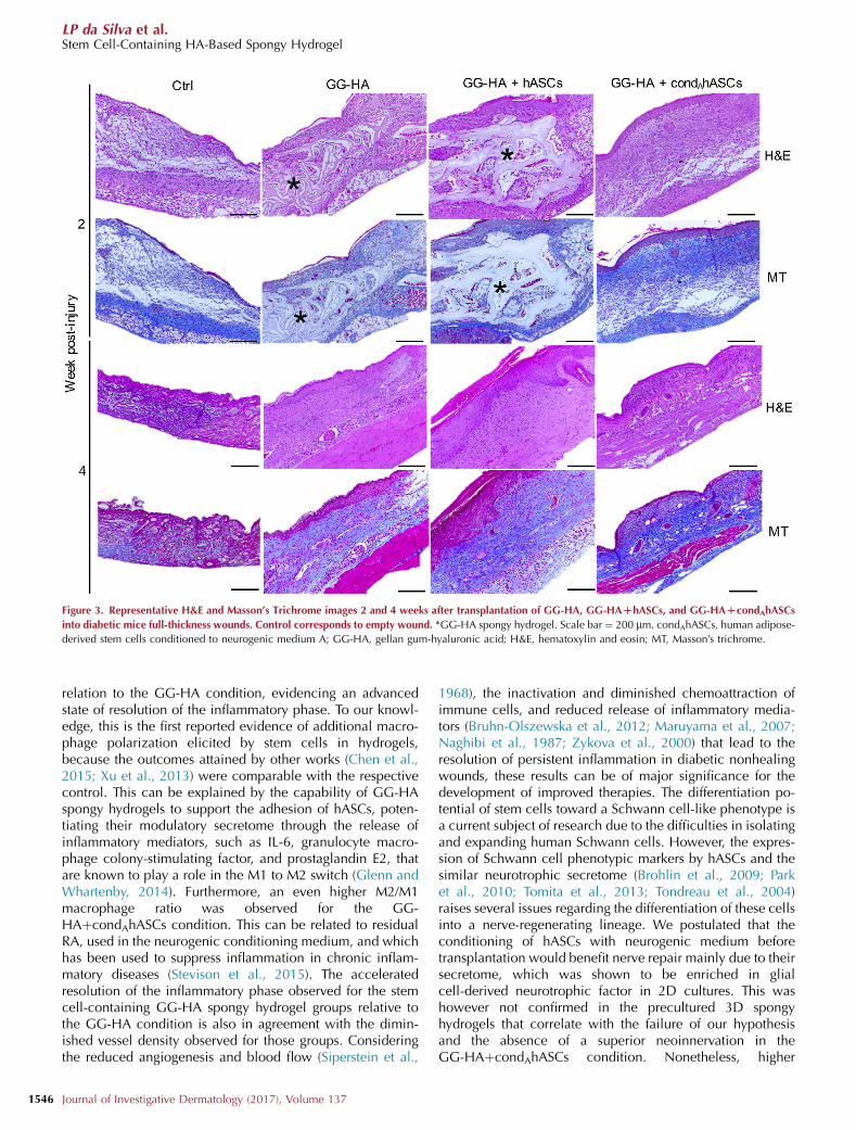
Figure 3. Representative H&E and Masson’s Trichrome images 2 and 4 weeks after transplantation of GG-HA, GG-HADhASCs, and GG-HADcondAhASCs
into diabetic mice full-thickness wounds. Control corresponds to empty wound. *GG-HA spongy hydrogel. Scale bar ¼ 200 mm. condAhASCs, human adipose-
derived stem cells conditioned to neurogenic medium A; GG-HA, gellan gum-hyaluronic acid; H&E, hematoxylin and eosin; MT, Masson’s trichrome.
LP da Silva et al.Stem Cell-Containing HA-Based Spongy Hydrogel
1546
relation to the GG-HA condition, evidencing an advancedstate of resolution of the inflammatory phase. To our knowl-edge, this is the first reported evidence of additional macro-phage polarization elicited by stem cells in hydrogels,because the outcomes attained by other works (Chen et al.,2015; Xu et al., 2013) were comparable with the respectivecontrol. This can be explained by the capability of GG-HAspongy hydrogels to support the adhesion of hASCs, poten-tiating their modulatory secretome through the release ofinflammatory mediators, such as IL-6, granulocyte macro-phage colony-stimulating factor, and prostaglandin E2, thatare known to play a role in the M1 to M2 switch (Glenn andWhartenby, 2014). Furthermore, an even higher M2/M1macrophage ratio was observed for the GG-HAþcondAhASCs condition. This can be related to residualRA, used in the neurogenic conditioning medium, and whichhas been used to suppress inflammation in chronic inflam-matory diseases (Stevison et al., 2015). The acceleratedresolution of the inflammatory phase observed for the stemcell-containing GG-HA spongy hydrogel groups relative tothe GG-HA condition is also in agreement with the dimin-ished vessel density observed for those groups. Consideringthe reduced angiogenesis and blood flow (Siperstein et al.,
Journal of Investigative Dermatology (2017), Volume 137
1968), the inactivation and diminished chemoattraction ofimmune cells, and reduced release of inflammatory media-tors (Bruhn-Olszewska et al., 2012; Maruyama et al., 2007;Naghibi et al., 1987; Zykova et al., 2000) that lead to theresolution of persistent inflammation in diabetic nonhealingwounds, these results can be of major significance for thedevelopment of improved therapies. The differentiation po-tential of stem cells toward a Schwann cell-like phenotype isa current subject of research due to the difficulties in isolatingand expanding human Schwann cells. However, the expres-sion of Schwann cell phenotypic markers by hASCs and thesimilar neurotrophic secretome (Brohlin et al., 2009; Parket al., 2010; Tomita et al., 2013; Tondreau et al., 2004)raises several issues regarding the differentiation of these cellsinto a nerve-regenerating lineage. We postulated that theconditioning of hASCs with neurogenic medium beforetransplantation would benefit nerve repair mainly due to theirsecretome, which was shown to be enriched in glialcell-derived neurotrophic factor in 2D cultures. This washowever not confirmed in the precultured 3D spongyhydrogels that correlate with the failure of our hypothesisand the absence of a superior neoinnervation in theGG-HAþcondAhASCs condition. Nonetheless, higher
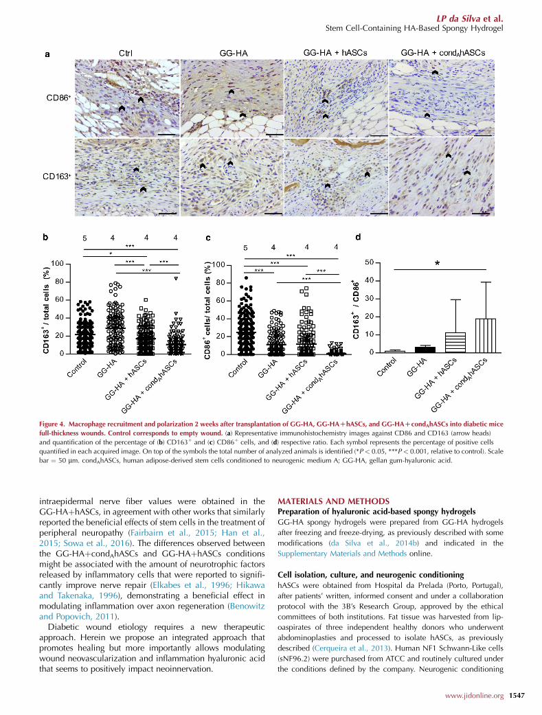
Figure 4. Macrophage recruitment and polarization 2 weeks after transplantation of GG-HA, GG-HADhASCs, and GG-HADcondAhASCs into diabetic mice
full-thickness wounds. Control corresponds to empty wound. (a) Representative immunohistochemistry images against CD86 and CD163 (arrow heads)
and quantification of the percentage of (b) CD163þ and (c) CD86þ cells, and (d) respective ratio. Each symbol represents the percentage of positive cells
quantified in each acquired image. On top of the symbols the total number of analyzed animals is identified (*P < 0.05, ***P < 0.001, relative to control). Scale
bar ¼ 50 mm. condAhASCs, human adipose-derived stem cells conditioned to neurogenic medium A; GG-HA, gellan gum-hyaluronic acid.
LP da Silva et al.Stem Cell-Containing HA-Based Spongy Hydrogel
intraepidermal nerve fiber values were obtained in theGG-HAþhASCs, in agreement with other works that similarlyreported the beneficial effects of stem cells in the treatment ofperipheral neuropathy (Fairbairn et al., 2015; Han et al.,2015; Sowa et al., 2016). The differences observed betweenthe GG-HAþcondAhASCs and GG-HAþhASCs conditionsmight be associated with the amount of neurotrophic factorsreleased by inflammatory cells that were reported to signifi-cantly improve nerve repair (Elkabes et al., 1996; Hikawaand Takenaka, 1996), demonstrating a beneficial effect inmodulating inflammation over axon regeneration (Benowitzand Popovich, 2011).
Diabetic wound etiology requires a new therapeuticapproach. Herein we propose an integrated approach thatpromotes healing but more importantly allows modulatingwound neovascularization and inflammation hyaluronic acidthat seems to positively impact neoinnervation.
MATERIALS AND METHODSPreparation of hyaluronic acid-based spongy hydrogels
GG-HA spongy hydrogels were prepared from GG-HA hydrogels
after freezing and freeze-drying, as previously described with some
modifications (da Silva et al., 2014b) and indicated in the
Supplementary Materials and Methods online.
Cell isolation, culture, and neurogenic conditioning
hASCs were obtained from Hospital da Prelada (Porto, Portugal),
after patients’ written, informed consent and under a collaboration
protocol with the 3B’s Research Group, approved by the ethical
committees of both institutions. Fat tissue was harvested from lip-
oaspirates of three independent healthy donors who underwent
abdominoplasties and processed to isolate hASCs, as previously
described (Cerqueira et al., 2013). Human NF1 Schwann-Like cells
(sNF96.2) were purchased from ATCC and routinely cultured under
the conditions defined by the company. Neurogenic conditioning
www.jidonline.org 1547
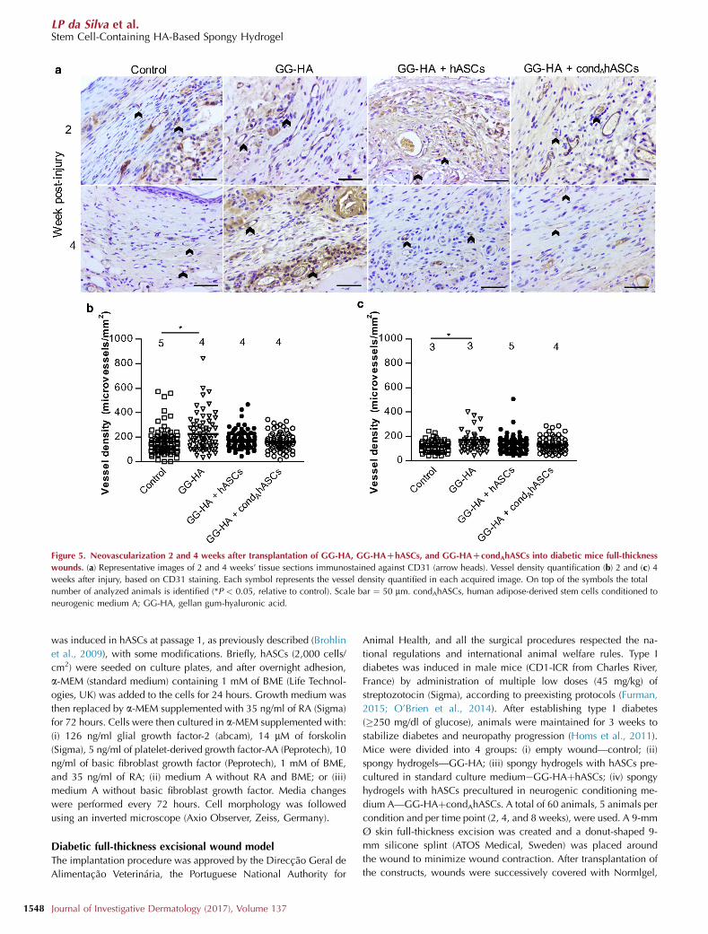
Figure 5. Neovascularization 2 and 4 weeks after transplantation of GG-HA, GG-HADhASCs, and GG-HADcondAhASCs into diabetic mice full-thickness
wounds. (a) Representative images of 2 and 4 weeks’ tissue sections immunostained against CD31 (arrow heads). Vessel density quantification (b) 2 and (c) 4
weeks after injury, based on CD31 staining. Each symbol represents the vessel density quantified in each acquired image. On top of the symbols the total
number of analyzed animals is identified (*P < 0.05, relative to control). Scale bar ¼ 50 mm. condAhASCs, human adipose-derived stem cells conditioned to
neurogenic medium A; GG-HA, gellan gum-hyaluronic acid.
LP da Silva et al.Stem Cell-Containing HA-Based Spongy Hydrogel
1548
was induced in hASCs at passage 1, as previously described (Brohlin
et al., 2009), with some modifications. Briefly, hASCs (2,000 cells/
cm2) were seeded on culture plates, and after overnight adhesion,
a-MEM (standard medium) containing 1 mM of BME (Life Technol-
ogies, UK) was added to the cells for 24 hours. Growth medium was
then replaced by a-MEM supplemented with 35 ng/ml of RA (Sigma)
for 72 hours. Cells were then cultured in a-MEM supplemented with:
(i) 126 ng/ml glial growth factor-2 (abcam), 14 mM of forskolin
(Sigma), 5 ng/ml of platelet-derived growth factor-AA (Peprotech), 10
ng/ml of basic fibroblast growth factor (Peprotech), 1 mM of BME,
and 35 ng/ml of RA; (ii) medium A without RA and BME; or (iii)
medium A without basic fibroblast growth factor. Media changes
were performed every 72 hours. Cell morphology was followed
using an inverted microscope (Axio Observer, Zeiss, Germany).
Diabetic full-thickness excisional wound model
The implantation procedure was approved by the Direccao Geral de
Alimentacao Veterinaria, the Portuguese National Authority for
Journal of Investigative Dermatology (2017), Volume 137
Animal Health, and all the surgical procedures respected the na-
tional regulations and international animal welfare rules. Type I
diabetes was induced in male mice (CD1-ICR from Charles River,
France) by administration of multiple low doses (45 mg/kg) of
streptozotocin (Sigma), according to preexisting protocols (Furman,
2015; O’Brien et al., 2014). After establishing type I diabetes
(�250 mg/dl of glucose), animals were maintained for 3 weeks to
stabilize diabetes and neuropathy progression (Homs et al., 2011).
Mice were divided into 4 groups: (i) empty wound—control; (ii)
spongy hydrogels—GG-HA; (iii) spongy hydrogels with hASCs pre-
cultured in standard culture mediumeGG-HAþhASCs; (iv) spongy
hydrogels with hASCs precultured in neurogenic conditioning me-
dium A—GG-HAþcondAhASCs. A total of 60 animals, 5 animals per
condition and per time point (2, 4, and 8 weeks), were used. A 9-mm
Ø skin full-thickness excision was created and a donut-shaped 9-
mm silicone splint (ATOS Medical, Sweden) was placed around
the wound to minimize wound contraction. After transplantation of
the constructs, wounds were successively covered with Normlgel,
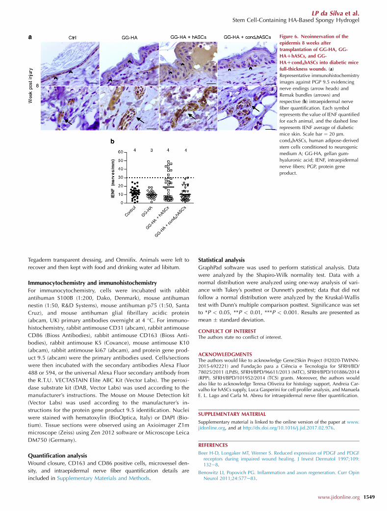
Figure 6. Neoinnervation of the
epidermis 8 weeks after
transplantation of GG-HA, GG-
HADhASCs, and GG-
HADcondAhASCs into diabetic mice
full-thickness wounds. (a)
Representative immunohistochemistry
images against PGP 9.5 evidencing
nerve endings (arrow heads) and
Remak bundles (arrows) and
respective (b) intraepidermal nerve
fiber quantification. Each symbol
represents the value of IENF quantified
for each animal, and the dashed line
represents IENF average of diabetic
mice skin. Scale bar ¼ 20 mm.
condAhASCs, human adipose-derived
stem cells conditioned to neurogenic
medium A; GG-HA, gellan gum-
hyaluronic acid; IENF, intraepidermal
nerve fibers; PGP, protein gene
product.
LP da Silva et al.Stem Cell-Containing HA-Based Spongy Hydrogel
Tegaderm transparent dressing, and Omnifix. Animals were left to
recover and then kept with food and drinking water ad libitum.
Immunocytochemistry and immunohistochemistry
For immunocytochemistry, cells were incubated with rabbit
antihuman S100B (1:200, Dako, Denmark), mouse antihuman
nestin (1:50, R&D Systems), mouse antihuman p75 (1:50, Santa
Cruz), and mouse antihuman glial fibrillary acidic protein
(abcam, UK) primary antibodies overnight at 4 �C. For immuno-
histochemistry, rabbit antimouse CD31 (abcam), rabbit antimouse
CD86 (Bioss Antibodies), rabbit antimouse CD163 (Bioss Anti-
bodies), rabbit antimouse K5 (Covance), mouse antimouse K10
(abcam), rabbit antimouse ki67 (abcam), and protein gene prod-
uct 9.5 (abcam) were the primary antibodies used. Cells/sections
were then incubated with the secondary antibodies Alexa Fluor
488 or 594, or the universal Alexa Fluor secondary antibody from
the R.T.U. VECTASTAIN Elite ABC Kit (Vector Labs). The peroxi-
dase substrate kit (DAB, Vector Labs) was used according to the
manufacturer’s instructions. The Mouse on Mouse Detection kit
(Vector Labs) was used according to the manufacturer’s in-
structions for the protein gene product 9.5 identification. Nuclei
were stained with hematoxylin (BioOptica, Italy) or DAPI (Bio-
tium). Tissue sections were observed using an Axioimager Z1m
microscope (Zeiss) using Zen 2012 software or Microscope Leica
DM750 (Germany).
Quantification analysis
Wound closure, CD163 and CD86 positive cells, microvessel den-
sity, and intraepidermal nerve fiber quantification details are
included in Supplementary Materials and Methods.
Statistical analysis
GraphPad software was used to perform statistical analysis. Data
were analyzed by the Shapiro-Wilk normality test. Data with a
normal distribution were analyzed using one-way analysis of vari-
ance with Tukey’s posttest or Dunnett’s posttest; data that did not
follow a normal distribution were analyzed by the Kruskal-Wallis
test with Dunn’s multiple comparison posttest. Significance was set
to *P < 0.05, **P < 0.01, ***P < 0.001. Results are presented as
mean � standard deviation.
CONFLICT OF INTERESTThe authors state no conflict of interest.
ACKNOWLEDGMENTSThe authors would like to acknowledge Gene2Skin Project (H2020-TWINN-2015-692221) and Fundacao para a Ciencia e Tecnologia for SFRH/BD/78025/2011 (LPdS), SFRH/BPD/96611/2013 (MTC), SFRH/BPD/101886/2014(RPP), SFRH/BPD/101952/2014 (TCS) grants. Moreover, the authors wouldalso like to acknowledge Teresa Oliveira for histology support, Andreia Car-valho for hASCs supply, Luca Gasperini for cell profiler analysis, and ManuelaE. L. Lago and Carla M. Abreu for intraepidermal nerve fiber quantification.
SUPPLEMENTARY MATERIAL
Supplementary material is linked to the online version of the paper at www.jidonline.org, and at http://dx.doi.org/10.1016/j.jid.2017.02.976.
REFERENCES
Beer H-D, Longaker MT, Werner S. Reduced expression of PDGF and PDGFreceptors during impaired wound healing. J Invest Dermatol 1997;109:132e8.
Benowitz LI, Popovich PG. Inflammation and axon regeneration. Curr OpinNeurol 2011;24:577e83.
www.jidonline.org 1549
LP da Silva et al.Stem Cell-Containing HA-Based Spongy Hydrogel
1550
Blais M, Grenier M, Berthod F. Improvement of nerve regeneration in tissue-engineered skin enriched with schwann cells. J Invest Dermatol 2009;129:2895e900.
Blakytny R, Jude EB. Altered molecular mechanisms of diabetic foot ulcers.Int J Low Extrem Wounds 2009;8:95e104.
Brohlin M, Mahay D, Novikov LN, Terenghi G, Wiberg M, Shawcross SG,et al. Characterisation of human mesenchymal stem cells followingdifferentiation into Schwann cell-like cells. Neurosci Res 2009;64:41e9.
Bruhn-Olszewska B, Korzon-Burakowska A, Gabig-Cimi�nska M, Olszewski P,Wegrzyn A, Jakobkiewicz-Banecka J. Molecular factors involved in thedevelopment of diabetic foot syndrome. Acta Biochim Pol 2012;59:507e13.
Caddick J, Kingham PJ, Gardiner NJ, Wiberg M, Terenghi G. Phenotypic andfunctional characteristics of mesenchymal stem cells differentiated along aSchwann cell lineage. Glia 2006;54:840e9.
Caissie R, Gingras M, Champigny M-F, Berthod F. In vivo enhancement ofsensory perception recovery in a tissue-engineered skin enriched withlaminin. Biomaterials 2006;27:2988e93.
Cerqueira MT, da Silva LP, Santos TC, Pirraco RP, Correlo VM, Marques AP,et al. Human skin cell fractions fail to self-organize within a gellan gum/hyaluronic acid matrix but positively influence early wound healing. TissueEng Part A 2014a;20:1369e78.
Cerqueira MT, da Silva LP, Santos TC, Pirraco RP, Correlo VM, Reis RL, et al.Gellan gum-hyaluronic acid spongy-like hydrogels and cells from adiposetissue synergize promoting neoskin vascularization. ACS Appl Mater In-terfaces 2014b;6:19668e79.
Cerqueira MT, Pirraco RP, Santos TC, Rodrigues DB, Frias AM, Martins AR,et al. Human adipose stem cells cell sheet constructs impact epidermalmorphogenesis in full-thickness excisional wounds. Biomacromolecules2013;14:3997e4008.
Chen S, Shi J, Zhang M, Chen Y, Wang X, Zhang L, et al. Mesenchymal stemcell-laden anti-inflammatory hydrogel enhances diabetic wound healing.Sci Rep 2015;5:18104.
Costa PZ, Soares R. Neovascularization in diabetes and its complications.Unraveling the angiogenic paradox. Life Sci 2013;92:1037e45.
da Silva LP, Cerqueira MC, Pirraco RP, Santos TC, Reis RL, Correlo VM, et al.Gellan gum-hyaluronate spongy-like hydrogels promote angiogenesis inhindlimb ischemia. 4th TERMIS World Congr. Boston, MA, Tissue Eng. PartA. 2015;21(Suppl. 1):S-1eS-413.
da Silva LP, Cerqueira MT, Sousa RA, Marques AP, Correlo VM, Reis RL.Gellan gum-based spongy-like hydrogels: methods and biomedical appli-cations thereof. US patent US20160325017 A1. 2014a.
da Silva LP, Cerqueira MT, Sousa RA, Reis RL, Correlo VM, Marques AP.Engineering cell-adhesive gellan gum spongy-like hydrogels for regenera-tive medicine purposes. Acta Biomater 2014b;10:4787e97.
da Silva LP, Pirraco RP, Santos TC, Novoa-Carballal R, Cerqueira MT, Reis RL,et al. Neovascularization induced by the hyaluronic acid-based spongy-like hydrogels degradation products. ACS Appl Mater Interfaces 2016;8:33464e74.
Edwards JL, Vincent AM, Cheng HT, Feldman EL, Edwards JL, Vincent AM,et al. Diabetic neuropathy: Mechanisms to management. Pharmacol Ther2008;120:1e34.
Elkabes S, DiCicco-Bloom EM, Black IB. Brain microglia/macrophages ex-press neurotrophins that selectively regulate microglial proliferation andfunction. J Neurosci 1996;16:2508e21.
Fairbairn NG, Meppelink AM, Ng-Glazier J, Randolph MA, Winograd JM.Augmenting peripheral nerve regeneration using stem cells: a review ofcurrent opinion. World J Stem Cells 2015;7:11e26.
Falanga V. Wound healing and its impairment in the diabetic foot. Lancet2005;366:1736e43.
Furman BL. Streptozotocin-induced diabetic models in mice and rats. CurrProtoc Pharmacol 2015;70:1e20.
GingrasM, Paradis I, Berthod F.Nerve regeneration in a collagen-chitosan tissue-engineered skin transplanted on nude mice. Biomaterials 2003;24:1653e61.
Glenn JD, Whartenby KA. Mesenchymal stem cells: emerging mechanisms ofimmunomodulation and therapy. World J Stem Cells 2014;6:526e39.
Han JW, Choi D, Lee MY, Huh YH, Yoon YS. Bone marrow-derived mesen-chymal stem cells improve diabetic neuropathy by direct modulation of
Journal of Investigative Dermatology (2017), Volume 137
both angiogenesis and myelination in peripheral nerves. Cell Transpl2015;25:1e38.
Heublein H, Bader A, Giri S. Preclinical and clinical evidence for stem cell ther-apies as treatment for diabetic wounds. Drug Discov Today 2015;20:703e17.
Hikawa N, Takenaka T. Myelin-stimulated macrophages release neurotrophicfactors for adult dorsal root ganglion neurons in culture. Cell Mol Neuro-biol 1996;16:517e28.
Homs J, Ariza L, Pages G, Verdu E, Casals L, Udina E, et al. Comparative studyof peripheral neuropathy and nerve regeneration in NOD and ICR diabeticmice. J Peripher Nerv Syst 2011;16:213e27.
Jiang D, Liang J, Noble PW. Hyaluronan in tissue injury and repair. Annu RevCell Dev Biol 2007;23:435e61.
Jude EB, Blakytny R, Bulmer J, Boulton AJM, Ferguson MWJ. Transforminggrowth factor-beta 1, 2, 3 and receptor type I and II in diabetic foot ulcers.Diabet Med 2002;19:440e7.
Kant V, Kumar D, Kumar D, Prasad R, Gopal A, Pathak NN, et al. Topicalapplication of substance P promotes wound healing in streptozotocin-induced diabetic rats. Cytokine 2015;73:144e55.
Kim EJ, Choi JS, Kim JS, Choi YC, Cho YW. Injectable and thermosensitivesoluble extracellular matrix and methylcellulose hydrogels for stem celldelivery in skin wounds. Biomacromolecules 2016;17:4e11.
Kingham PJ, Kalbermatten DF, Mahay D, Armstrong SJ, Wiberg M, Terenghi G.Adipose-derived stem cells differentiate into a Schwann cell phenotype andpromote neurite outgrowth in vitro. Exp Neurol 2007;207:267e74.
Laverdet B, Danigo A, Girard D, Magy L, Demiot C, Desmouliere A. Skininnervation: important roles during normal and pathological cutaneousrepair. Histol Histopathol 2015;30:875e92.
Leal EC, Carvalho EE, Tellechea A, Kafanas A, Tecilazich F, Kearney C, et al.Substance P promotes wound healing in diabetes by modulating inflam-mation and macrophage phenotype. Am J Pathol 2015;185:1638e48.
Luo JD, Chen AF. Nitric oxide: a newly discovered function on woundhealing. Acta Pharmacol Sin 2005;26:259e64.
Mahay D, Terenghi G, Shawcross SG. Schwann cell mediated trophic effects bydifferentiated mesenchymal stem cells. Exp Cell Res 2008a;314:2692e701.
Mahay D, Terenghi G, Shawcross SG. Growth factors in mesenchymal stemcells following glial-cell differentiation. Biotechnol Appl Biochem2008b;51:167.
Maruyama K, Asai J, Ii M, Thorne T, Losordo DW, D’Amore PA. Decreasedmacrophage number and activation lead to reduced lymphatic vessel for-mation and contribute to impaired diabetic wound healing. Am J Pathol2007;170:1178e91.
Moura LIF, Dias AMA, Suesca E, Casadiegos S, Leal EC, Fontanilla MR, et al.Neurotensin-loaded collagen dressings reduce inflammation and improvewound healing in diabetic mice. Biochim Biophys Acta 2014;1842:32e43.
Mulder G, Tenenhaus M, D’Souza GF. Reduction of diabetic foot ulcerhealing times through use of advanced treatment modalities. Int J LowExtrem Wounds 2014;13:335e46.
Naghibi M, Smith RP, Baltch AL, Gates SA, Wu DH, Hammer MC, et al. Theeffect of diabetes mellitus on chemotactic and bactericidal activity of hu-man polymorphonuclear leukocytes. Diabetes Res Clin Pract 1987;4:27e35.
Noble PW. Hyaluronan fragments activate an NF-kappa B/I-kappa B alphaautoregulatory loop in murine macrophages. J Exp Med 1996;183:2373e8.
O’Brien PD, Sakowski SA, Feldman EL. Mouse models of diabetic neuropathy.ILAR J 2014;54:259e72.
Park H-W, Lim M-J, Jung H, Lee S-P, Paik K-S, Chang M-S. Human mesen-chymal stem cell-derived Schwann cell-like cells exhibit neurotrophic ef-fects, via distinct growth factor production, in a model of spinal cord injury.Glia 2010;58:1118e32.
Pittenger GL, Ray M, Burcus NI, McNulty P, Basta B, Vinik AI. Intraepidermalnerve fibers are indicators of small-fiber neuropathy in both diabetic andnondiabetic patients. Diabetes Care 2004;27:1974e9.
Quattrini C, Jeziorska M, Boulton AJM, Malik RA. Reduced vascularendothelial growth factor expression and intra-epidermal nerve fiberloss in human diabetic neuropathy. Diabetes Care 2008;31:140e5.
Reyes-Ortega F, Cifuentes A, Rodrıguez G, Aguilar MR, Gonzalez-Gomez A,Solis R, et al. Bioactive bilayered dressing for compromised epidermal
LP da Silva et al.Stem Cell-Containing HA-Based Spongy Hydrogel
tissue regeneration with sequential activity of complementary agents. ActaBiomater 2015;23:103e15.
Siperstein MD, Unger RH, Madison LL. Studies of muscle capillary basementmembranes in normal subjects, diabetic, and prediabetic patients. J ClinInvest 1968;47:1973e99.
Sowa Y, Kishida T, Imura T, Numajiri T, Nishino K, Tabata Y, et al. Adipose-derived stem cells promote peripheral nerve regeneration in vivo withoutdifferentiation into Schwann-like lineage. Plast Reconstr Surg 2016;137:318ee30e.
Stevison F, Jing J, Tripathy S, Isoherranen N. Role of retinoic acid-metabolizing cytochrome P450s, CYP26, in inflammation and cancer.Adv Pharmacol 2015;74:373e412.
Thangarajah H, Yao D, Chang EI, Shi Y, Jazayeri L, Vial IN, et al. The mo-lecular basis for impaired hypoxia-induced VEGF expression in diabetictissues. Proc Natl Acad Sci USA 2009;106:13505e10.
Tomita K, Madura T, Mantovani C, Terenghi G. Differentiated adipose-derivedstem cells promote myelination and enhance functional recovery in a ratmodel of chronic denervation. J Neurosci Res 2012;90:1392e402.
Tomita K, Madura T, Sakai Y, Yano K, Terenghi G, Hosokawa K. Glial differ-entiation of human adipose-derived stem cells: implications for cell-basedtransplantation therapy. Neuroscience 2013;236:55e65.
Tondreau T, Lagneaux L, Dejeneffe M, Massy M, Mortier C, Delforge A, et al.Bone marrowederived mesenchymal stem cells already express specificneural proteins before any differentiation. Differentiation 2004;72:319e26.
Upadhyay A, Chattopadhyay P, Goyary D, Mitra Mazumder P, Veer V. Ixoracoccinea enhances cutaneous wound healing by upregulating the expres-sion of collagen and basic fibroblast growth factor. ISRN Pharmacol2014;2014:751824.
Usui ML, Mansbridge JN, Carter WG, Fujita M, Olerud JE. Keratinocyte migra-tion, proliferation, and differentiation in chronic ulcers from patients withdiabetes and normal wounds. J Histochem Cytochem 2008;56:687e96.
Wang Y, Chen X, Cao W, Shi Y. Plasticity of mesenchymal stem cells inimmunomodulation: pathological and therapeutic implications. NatImmunol 2014;15:1009e16.
Xu K, Cantu DA, Fu Y, Kim J, Zheng X, Hematti P, et al. Thiol-ene Michael-type formation of gelatin/poly(ethylene glycol) biomatrices for three-dimensional mesenchymal stromal/stem cell administration to cutaneouswounds. Acta Biomater 2013;9:8802e14.
Zykova SN, Jenssen TG, Berdal M, Olsen R, Myklebust R, Seljelid R. Alteredcytokine and nitric oxide secretion in vitro by macrophages from diabetictype II-like db/db mice. Diabetes 2000;49:1451e8.
www.jidonline.org 1551