staphylococcus aureus and staphylococcus epidermidis … · andmoredeeplyexploredthes. aureus and...
TRANSCRIPT
SC I ENCE TRANS LAT IONAL MED I C I N E | R E S EARCH ART I C L E
MICROB IOME
1Microbial Genomics Section, National Human Genome Research Institute, NationalInstitutes of Health (NIH), Bethesda, MD 20892, USA. 2Department of Bioinformatics,Boston University, Boston, MA 02215, USA. 3Mucosal Immunology Section, Laboratoryof Parasitic Diseases, National Institute of Allergy and Infectious Diseases (NIAID), NIH,Bethesda, MD 20892, USA. 4NIH Intramural Sequencing Center, National Human Ge-nome Research Institute, Bethesda, MD 20892, USA. 5NIAID Microbiome Program,Department of Intramural Research, NIAID, NIH, Bethesda, MD 20892, USA. 6Derma-tology Branch, Center for Cancer Research, National Cancer Institute, NIH, Bethesda,MD 20892, USA.*Corresponding author. Email: [email protected] (J.A.S.); [email protected] (H.H.K.)
Byrd et al., Sci. Transl. Med. 9, eaal4651 (2017) 5 July 2017
Copyright © 2017
The Authors, some
rights reserved;
exclusive licensee
American Association
for the Advancement
of Science. No claim
to original U.S.
Government Works
http://sD
ownloaded from
Staphylococcus aureus and Staphylococcus epidermidisstrain diversity underlying pediatric atopic dermatitisAllyson L. Byrd,1,2,3 Clay Deming,1 Sara K. B. Cassidy,1 Oliver J. Harrison,3 Weng-Ian Ng,1
Sean Conlan,1 NISC Comparative Sequencing Program,4 Yasmine Belkaid,3,5
Julia A. Segre,1* Heidi H. Kong6*
The heterogeneous course, severity, and treatment responses among patients with atopic dermatitis (AD; eczema)highlight the complexity of thismultifactorial disease. Prior studies haveused traditional typingmethods on cultivatedisolates or sequenced a bacterial marker gene to study the skin microbial communities of AD patients. Shotgunmeta-genomic sequence analysis provides much greater resolution, elucidating multiple levels of microbial communityassembly ranging from kingdom to species and strain-level diversification. We analyzedmicrobial temporal dynamicsfrom a cohort of pediatric AD patients sampled throughout the disease course. Species-level investigation of AD flaresshowed greater Staphylococcus aureus predominance in patients with more severe disease and Staphylococcusepidermidis predominance in patients with less severe disease. At the strain level, metagenomic sequencing analysesdemonstrated clonal S. aureus strains inmore severe patients and heterogeneous S. epidermidis strain communities inall patients. To investigate strain-level biological effects of S. aureus, we topically colonized mice with human strainsisolated from AD patients and controls. This cutaneous colonizationmodel demonstrated S. aureus strain–specific dif-ferences in eliciting skin inflammation and immune signatures characteristic of AD patients. Specifically, S. aureus iso-lates from AD patients with more severe flares induced epidermal thickening and expansion of cutaneous T helper 2(TH2) and TH17 cells. Integrating high-resolution sequencing, culturing, and animal models demonstrated how func-tional differences of staphylococcal strains may contribute to the complexity of AD disease.
tm.s
by guest on July 19, 2017ciencem
ag.org/
INTRODUCTIONAtopic dermatitis (AD; eczema) is a common inflammatory skin dis-order in industrialized countries, affecting 10 to 30% of children (1).Patients with AD suffer from chronic, relapsing, intensely itchy, andinflamed skin lesions and have an increased likelihood of developingasthma and/or hay fever (2). AD is a complex multifactorial diseasein which epidermal barrier impairment, type 2 immunity, and skin mi-crobes are each thought to potentially play a causative role (1). Morethan 30 susceptibility loci have been associated with AD, includingmu-tations in the gene encoding the skin barrier protein filaggrin (FLG) (3)and genes linked to the immune system (4).
In addition to host genetic susceptibility, the relationship betweenAD and skin bacteria is well recognized clinically. Patients with AD areoften treated with varying combinations of antimicrobial approaches(for example, antibiotics and dilute bleach baths) and anti-inflammatoryor immunosuppressive medications (5). The efficacy of these antimicro-bial treatments is associated with decreases in staphylococcal relativeabundances (6, 7). Staphylococcus aureus commonly colonizesAD skinand has been studied using colony-counting, sequencing typingmethods[for example, pulsed-field gel electrophoresis and SpA (S. aureus pro-tein A) typing] of selected cultivated isolates (8–11), and more recently,amplicon-based (marker gene) sequencing of the 16S ribosomal RNAgene (6, 7, 12, 13). However, sequence typing and amplicon sequencing
methods are unable to distinguish between genetically distinct strains,as determined by whole-genome sequencing (14, 15). By contrast,shotgun metagenomic sequencing of skin samples from healthy individ-uals provided deeper resolution and demonstrated the multiphyleticcomposition of commensal Staphylococcus (16).
With an increasing appreciation of functional differences be-tween strains within a single species, we performed shotgun meta-genomic sequencing of AD patient skin samples to capture the fullgenetic potential and strain-level differences of the skin micro-biome throughout the course of the disease. We confirmed an in-crease of staphylococcal species during disease flares in our cohortand more deeply explored the S. aureus and Staphylococcus epidermidisstrain diversity of each patient. To test the functional consequenceof strain-level differences between patients, we isolated staphylo-coccal strains from patients and healthy controls and investigatedthe cutaneous and immunologic effects when applied topically in amouse model.
RESULTSBacterial communities shift during AD disease progressionTo examine the relationship between the skin microbiota and AD,11 children with moderate to severe AD and 7 healthy children were re-cruited to the National Institutes of Health Clinical Center between June2012 and March 2015 (tables S1 and S2). Because AD has a chronic re-lapsing course, patients were sampled at stable/typical disease state(baseline), worsening of disease (flare), and 10 to 14 days after initiationof treatment using a combination of skin-directed therapies (post-flare).Because the use of topical medications on AD skin alters skin bacterialcommunities (6, 7), baseline samples were defined as those collected fromsubjects in their routine disease state who refrained from using skin-directed antimicrobial and anti-inflammatory treatments for 7 days, a
1 of 12

SC I ENCE TRANS LAT IONAL MED I C I N E | R E S EARCH ART I C L E
by guest on July 19, 2017http://stm
.sciencemag.org/
Dow
nloaded from
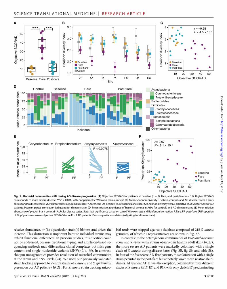
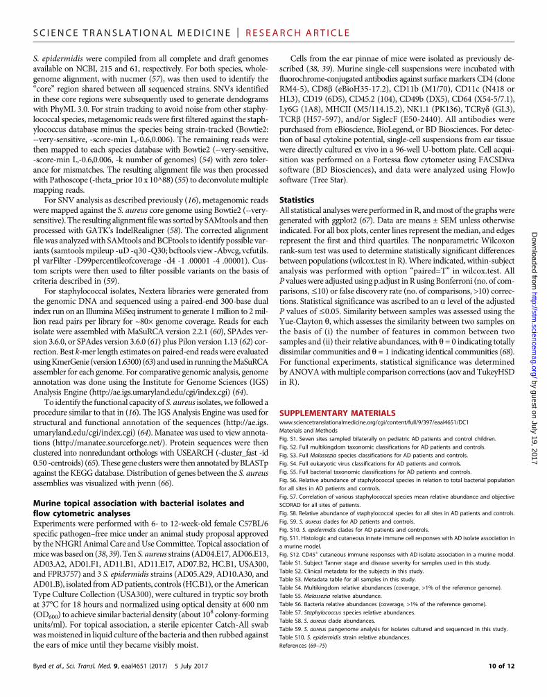
duration of time determined on the basis of prior findings (6). Flareswere defined as time points when patients experienced worsening inthe clinical severity of their typical AD, had not used skin-directed anti-microbial and anti-inflammatory treatments for 7 days, and did nothave clinical skin infection (for example, yellow crusts or pustules).At each time point, disease severity was determined with objectiveSCORAD (SCORingAtopicDermatitis), a validated clinical severity as-sessment tool (17–19). Subjects were sampled bilaterally at sites of dis-ease predilection: the inner elbow [antecubital crease (Ac)] and behindthe knees [popliteal crease (Pc)], along with five additional sites to in-vestigate defined areas with different skin physiologies (fig. S1). Becauseof the clinical severity of their AD, 6 of the 11 patients experienced ex-acerbations of their skin disease with the 7-day skin preparation regi-men and could not provide baseline time point samples, reflecting thespectrum of the natural course of AD. Because the skin microbial dys-biosis during AD flares was of greatest interest, most of the analysesfocused on comparisons between flare and post-flare time points. Intotal, we performed shotgun metagenomic sequencing of 422 samples,generating 191 giga–base pairs (Gbp) of microbial sequence data from27 AD patient visits and 7 healthy control visits (table S3). During pa-tient flares, ADdisease severitywas significantly elevated as indicated byhighermeanobjective SCORAD(38±2.9) as compared tobaseline (9.4±1.6; P < 4.5 × 10⁻⁴) and post-flare (11 ± 1.6; P < 2.8 × 10⁻⁶) (Fig. 1A).
To compare the microbial community composition across timepoints, we mapped microbial reads to a multikingdom referencedatabase. As seen in healthy adults (16, 20, 21), Bacteria was the mostpredominant kingdom across time points and body sites (fig. S2 andtable S4). Malassezia species, particularly Malassezia restricta andMalassezia globosa, predominated the fungal communities (fig. S3and table S5), and eukaryotic DNA viral communities were mostlypolyomaviruses or papillomaviruses depending on the individual(fig. S4). No significant differences in the fungal or viral components overtime were identified; therefore, we focused on bacterial communitiesthat demonstrated the greatest shifts in this cohort (fig. S5 and tableS6). We first determined the Shannon diversity index, an ecologicalmeasure of richness (total number of bacterial species) and evenness(relative proportion of the bacterial species), to evaluate the overall com-munity structure/composition across body sites and time points. Dur-ing flares, Ac and Pc, which are the sites of AD predilection, exhibited amarked reduction in Shannon diversity compared to baseline, post-flare, and healthy controls, a trend observed to a lesser extent acrossother sites (Fig. 1B). Because changes in bacterial diversity were mostpronounced at sites of disease predilection and Ac and Pc have similarmicrobial communities (21), we averaged these sites per subject andused the composite “AcPc” for subsequent analyses. Similar to our pre-vious analysis of microbial diversity in an AD patient cohort (6), thepartial correlation between objective SCORAD and Shannon diversity,adjusting for disease state, was significantly inversely correlated (r =−0.58, P = 4.5 × 10⁻⁴) (Fig. 1C), indicating that reduced skin bacterialdiversity corresponds to worse disease severity, primarily at sites of dis-ease predilection (fig. S5A).
To determine which taxa were contributing to the loss of diversity,we compared the relative abundances of the most prominent taxa (Fig.1D and fig. S5B). Of the four most prominent genera in the AcPc, onlyStaphylococcuswas significantly increased in flares (45 ± 10.2%) as com-pared to post-flares (9.2 ± 2.4%; P < 0.0078) and healthy controls (6.6 ±4.1%; P < 0.033) (Fig. 1E). This increase in Staphylococcus relative abun-dances was positively correlated with objective SCORAD (r = 0.67, P <8.1× 10⁻⁶) (Fig. 1F), indicating that severeADwas associatedwith high-
Byrd et al., Sci. Transl. Med. 9, eaal4651 (2017) 5 July 2017
er staphylococcal relative abundances at sites of disease predilection. Inaddition, therewas a positive correlation for the forehead, retroauricularcrease, andvolar forearm(fig. S5C), sites that canbeaffected inmore severedisease. However, differences inCorynebacterium, Propionibacterium,and Streptococcus relative abundances between flares and post-flareswere not statistically significant (Fig. 1E).
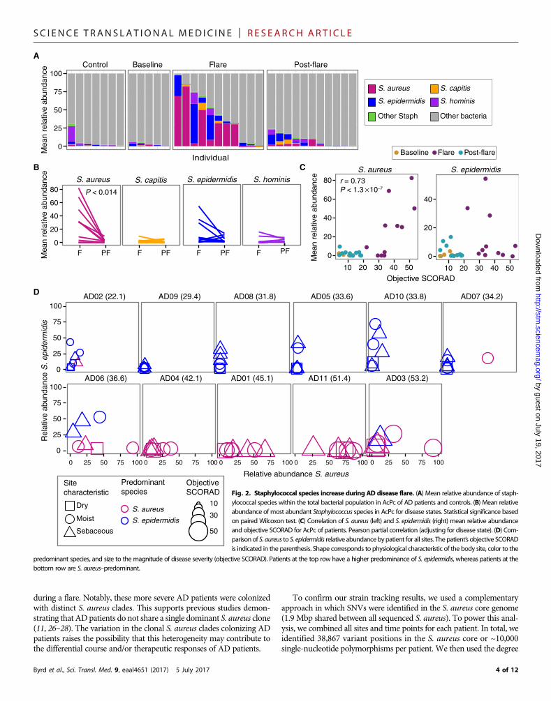
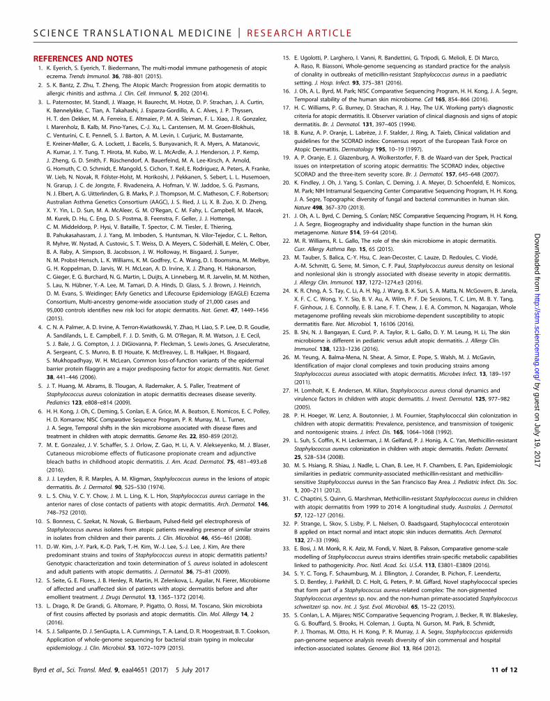
AD flare severity is linked with specificstaphylococcal speciesTo further examine the positive correlation between Staphylococcus andAD disease course observed in this study and in prior studies (22), weidentified the relative abundances of staphylococcal species includingS. aureus, S. epidermidis, Staphylococcus hominis, and Staphylococcuscapitis (Fig. 2A and fig. S6). Only relative abundances of S. aureusweresignificantly increased from flares (28 ± 8.8%) to post-flares (2.3 ±0.8%; P < 0.014) (Fig. 2B). Although S. epidermidis relative abun-dances were also higher during flares (13 ± 5.4%) as compared topost-flares (3.7 ± 1.4%), results did not reach statistical significance.For all patients, relative abundances of S. aureus were positivelycorrelated with objective SCORAD (r = 0.73; P < 1.0 × 10⁻⁷), whereasS. epidermidiswas not correlated (Fig. 2C and fig. S7). This associationbetween S. aureus and AD severity (23) has been observed in prior stu-dies. Neither S. hominis nor S. capitis demonstrated significant shifts inrelative abundances between time points (Fig. 2B) or was correlatedwith disease severity (fig. S7).
To further explore the relationship between disease severity andstaphylococcal species, we sorted the patients by their objective SCORADandplotted the relative abundances of S. aureus and S. epidermidis at flare(Fig. 2D). We observed a trend whereby patients with more severe ADflares (objective SCORAD, 45 ± 3.0) had higher relative abundances ofS. aureus (Fig. 2D, bottom row; fig. S8; and table S7). In contrast,patients with less severe AD flares, as well as lower objective SCORAD(31 ± 1.9;P< 0.004, in comparison to themore severe flares), had higherrelative abundances of S. epidermidis (Fig. 2D, top row) across sampledsites. Specifically,more severeAD flare patients had relative abundancesof 34 ± 8.7% S. aureuswith 7.4 ± 4.2% S. epidermidis, and less severeADflare patients had relative abundances of 3.8 ± 1.7% S. aureus with 13 ±3.9% S. epidermidis averaged across all sites during flare. The range ofS. aureus relative abundances based on sequencing was variable: 3 of11 patients had no S. aureus on their skin, and 3 of 11 patients hadrelative abundances of S. aureus on their skin exceeding 50%, similarto prior studies of S. aureus relative abundances on AD skin (6, 24, 25).
To compare these metagenomic results with more traditional studies,we cultured bacteria from skin and nares swabs collected concurrentlywith genomic samples. Cultures of S. aureus from skin clinical samplescorrelatedwith themicroorganismdetection by sequencing.Notably, twoless severe AD flare patients were culture-positive for S. aureus only intheir nares, a common site of carriage. The S. aureus culture-positive ratesin this cohort were consistent with those in other studies (6–8, 11–13).The genomic analyses were internally consistent with cultivationresults, and both supported the strong association betweenADdiseaseseverity and S. aureus.
S. aureus strains in AD demonstrate monoclonalityAlthough the differential association of S. aureus and S. epidermidiswithAD severity defined an intriguing feature of disease heterogeneity, theunderlying strain communities of these species during the diseasecourse remained unknown. Two alternative scenarios could underliemicrobial shifts in a disease flare: (i) All strains equally increase in
2 of 12
SC I ENCE TRANS LAT IONAL MED I C I N E | R E S EARCH ART I C L E
by guest on July 19, 2017http://stm
.sciencemag.org/
Dow
nloaded from
relative abundance, or (ii) a particular strain(s) blooms and drives theincrease. This distinction is important because individual strains mayexhibit functional differences. In previous studies, this question couldnot be addressed, because traditional typing and amplicon-based se-quencing methods may differentiate clonal complexes but miss genecontent and single-nucleotide variants (SNVs) (14, 15). In contrast,shotgun metagenomics provides resolution of microbial communitiesat the strain and SNV levels (24). We used our previously validatedstrain tracking approach to identify strains of S. aureus and S. epidermidispresent on our AD patients (16, 21). For S. aureus strain tracking, micro-
Byrd et al., Sci. Transl. Med. 9, eaal4651 (2017) 5 July 2017
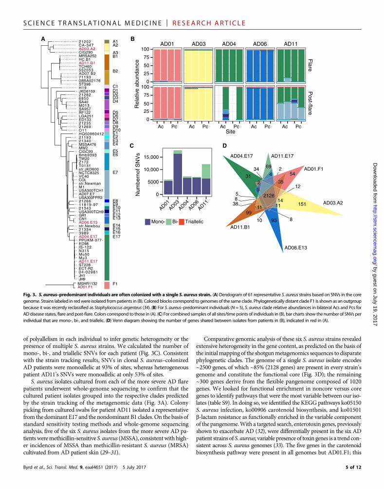
bial reads were mapped against a database composed of 215 S. aureusgenomes, of which 61 representatives are shown in Fig. 3A.
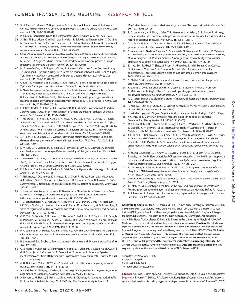
In contrast to the heterogenous communities of Propionibacteriumacnes and S. epidermidis strains observed in healthy adult skin (16, 21),the more severe AD patients were markedly colonized with a singleclade of S. aureus during disease flares (Fig. 3B, fig. S9, and table S8).In four of the five severe AD flare patients, this colonization with a singlestrain persisted in the post-flare but at notably lowermean relative abun-dances. AD patient AD11 was the exception, colonized by three differentclades of S. aureus (E17, E7, and B1), with only clade E17 predominating
Control Baseline Flare Post-flare
0
25
50
75
100
Individual
Mea
n re
lativ
e ab
unda
nce
ActinobacteriaCorynebacteriaceaePropionibacteriaceae
BacteroidetesFirmicutes
StaphylococcaceaStreptococcaceae
ProteobacteriaBetaproteobacteriaGammaproteobacteria
Other bacteria
B
1
2
3
4
10 20 30 40 50
Objective SCORAD
Sha
nnon
div
ersi
ty in
dex
C
D
E
1.5
2.0
2.5
3.0
3.5
Vf Ac Ic Pc Fh Oc RaSite
Sha
nnon
div
ersi
ty in
dex
F
Control
r = –0.58P < 4.5 × 10⁻⁴
0
25
50
75
100
10 20 30 40 50
Objective SCORAD
Pro
port
ion
Sta
phyl
ococ
cus
BaselineFlarePost-flare
BaselineFlarePost-flare
Baseline
Post-flareFlare
A
10
20
30
40
50
Baseline Flare Post-flare
Obj
ectiv
e S
CO
RA
D
******
Mea
n re
lativ
e ab
unda
nce Corynebacterium Propionibacterium
0
25
50
75
100
F PF F PF
Staphylococcus Streptococcus
F PF F PF
P < 0.0078
r = 0.67P < 8.1 × 10⁻⁶
Fig. 1. Bacterial communities shift during AD disease progression. (A) Objective SCORAD for patients at baseline (n = 5), flare, and post-flare (n = 11). Higher SCORADcorresponds to more severe disease. ***P < 0.001, with nonparametric Wilcoxon rank-sum test. (B) Mean Shannon diversity ± SEM in controls and AD disease states. Colorscorrespond to disease state. Vf, volar forearm; Ic, inguinal crease; Fh, forehead; Oc, occiput; Ra, retroauricular crease. (C) Shannon diversity versus objective SCORAD for AcPc of ADpatients. Pearson partial correlation (adjusting for disease state). (D) Mean relative abundance of bacterial genera in AcPc for controls and AD disease states. (E) Mean relativeabundance of predominant genera in AcPc for disease states. Statistical significance based on pairedWilcoxon test and Bonferroni correction. F, flare; PF, post-flare. (F) Proportionof Staphylococcus versus objective SCORAD for AcPc of AD patients. Pearson partial correlation (adjusting for disease state).
3 of 12
AD07 (34.2)D08 (31.8) AD10 (33.8)
AD03 (53.2)
0 25 50 75 100
D01 (45.1)
5 50 75 100
AD11 (51.4)
0 25 50 75 100
Relative abundance S. aureus
AD05 (33.6)
C S. aureus
0
20
40
60
80 r = 0.73P < 1.3 10–7
10 20 30 40 50
Objective SCORAD
0
20
40
10 20 30 40 50
S. epidermidis
Post-flare
S. hominis
F PF
Mea
n re
lativ
e ab
unda
nce
Other bacteria
S. aureus S. capitis
S. epidermidis S. hominis
Other Staph
Baseline Flare Post-flare
SC I ENCE TRANS LAT IONAL MED I C I N E | R E S EARCH ART I C L E
by guest on July 19, 2017http://stm
.sciencemag.org/
Dow
nloaded from
during a flare. Notably, these more severe AD patients were colonizedwith distinct S. aureus clades. This supports previous studies demon-strating that ADpatients do not share a single dominant S. aureus clone(11, 26–28). The variation in the clonal S. aureus clades colonizing ADpatients raises the possibility that this heterogeneity may contribute tothe differential course and/or therapeutic responses of AD patients.
Byrd et al., Sci. Transl. Med. 9, eaal4651 (2017) 5 July 2017
To confirm our strain tracking results, we used a complementaryapproach in which SNVs were identified in the S. aureus core genome(1.9 Mbp shared between all sequenced S. aureus). To power this anal-ysis, we combined all sites and time points for each patient. In total, weidentified 38,867 variant positions in the S. aureus core or ~10,000single-nucleotide polymorphisms per patient. We then used the degree
A
S. aureus S. capitis
0
20
40
60
80
F PF F PF
B
P < 0.014
D AD02 (22.1) AD09 (29.4) A
0
25
50
75
100
0
25
50
75
100AD06 (36.6)
0 25 50 75 100
AD04 (42.1)
0 25 50 75 100
A
0 2
Rel
ativ
e ab
unda
nce
S. e
pide
rmid
is
Objective SCORAD
Site characteristic
Dry
Moist
Sebaceous
10
30
50
S. aureus
S. epidermidis
Predominantspecies
0
25
50
75
100
Individual
Mea
n re
lativ
e ab
unda
nce Control Baseline Flare
S. epidermidis
PFFMea
n re
lativ
e ab
unda
nce
Fyaoapis
4 of 12
-
-
ig. 2. Staphylococcal species increase during AD disease flare. (A) Mean relative abundance of staphlococcal species within the total bacterial population in AcPc of AD patients and controls. (B) Mean relativebundance of most abundant Staphylococcus species in AcPc for disease states. Statistical significance basedn paired Wilcoxon test. (C) Correlation of S. aureus (left) and S. epidermidis (right) mean relative abundancend objective SCORAD for AcPc of patients. Pearson partial correlation (adjusting for disease state). (D) Comarison of S. aureus to S. epidermidis relative abundancebypatient for all sites. The patient’s objective SCORADindicated in the parenthesis. Shape corresponds to physiological characteristic of the body site, color to the
predominant species, and size to the magnitude of disease severity (objective SCORAD). Patients at the top row have a higher predominance of S. epidermidis, whereas patients at thebottom row are S. aureus–predominant.
SC I ENCE TRANS LAT IONAL MED I C I N E | R E S EARCH ART I C L E
by guest on July 19, 2017http://stm
.sciencemag.org/
Dow
nloaded from
of polyallelism in each individual to infer genetic heterogeneity or thepresence of multiple S. aureus strains. We calculated the number ofmono-, bi-, and triallelic SNVs for each patient (Fig. 3C). Consistentwith the strain tracking results, SNVs in clonal S. aureus–colonizedAD patients were monoallelic at 93% of sites, whereas heterogeneouspatient AD11’s SNVs were monoallelic at only 53% of sites.
S. aureus isolates cultured from each of the more severe AD flarepatients underwent whole-genome sequencing to confirm that thecultured patient isolates grouped into the respective clades predictedby the strain tracking of the metagenomic data (Fig. 3A). Colonypicking from cultured swabs for patient AD11 isolated a representativefrom the dominant E17 and the nondominant B1 clades. On the basis ofstandard sensitivity testing methods and whole-genome sequencinganalysis, five of the six S. aureus isolates from the more severe AD pa-tientsweremethicillin-sensitive S. aureus (MSSA), consistentwith high-er incidences of MSSA than methicillin-resistant S. aureus (MRSA)cultivated from AD patient skin (29–31).
Byrd et al., Sci. Transl. Med. 9, eaal4651 (2017) 5 July 2017
Comparative genomic analysis of these six S. aureus strains revealedextensive heterogeneity in the gene content, as predicted on the basis ofthe initialmapping of the shotgunmetagenomics sequences to disparatephylogenetic clades. The genome of a single S. aureus isolate encodes~2500 genes, of which ~85% (2128 genes) are present in every strain’sgenome and constitute the functional core (Fig. 3D); the remaining~300 genes derive from the flexible pangenome composed of 1020genes. We looked for functional enrichment in noncore versus coregenes to identify pathways that were the most variable between our iso-lates (table S9). In doing so, we identified the KEGG pathways ko05150S. aureus infection, ko00906 carotenoid biosynthesis, and ko01501b-lactam resistance as functionally enriched in the variable componentof the pangenome.With a targeted search, enterotoxin genes, previouslyshown to exacerbate AD (32), were differentially present in the six ADpatient strains of S. aureus; variable presence of toxin genes is a trend con-sistent across S. aureus genomes (33). The five genes in the carotenoidbiosynthesis pathway were present in all genomes but AD01.F1; this
0
5000
10,000
15,000
AD01AD03
AD04AD06
AD11
Num
bern
of S
NV
s
A B
C D
0
25
50
75
100
0
25
50
75
100
SiteR
elat
ive
abun
danc
e
AD03
Ac Pc
AD04
Ac Pc
AD01
Ac Pc
AD06
Ac Pc
AD11
Ac Pc
Post-flare
Flare
Mono- Bi- Triallelic
31
69
54
151
93
34
2
55
2
14
4
12
8
1199
10
38
35
11
82128
AD04.E17 AD11.E17
AD01.F1
AD03.A2
AD06.E13
AD11.B1
MSHR1132AD01.F1
AD11.E17
21340
ED133
NCTC8325
HC.B1
VC40
SA40
08BA02176
RF122
MW2
CA-347
T0131
AD11.B1
MSSA476
21334
Mu50
ED98
USA300FPR3
N315
SA957
AD04.E17
str.Newman
O11
CIGC93
MRSA252
CN1
JH1
552053AD07.B2
21343
21266
M013
21202
3989
IS-122
USA300TCH9
71193
21235
JH9
TCH60
Bmb9393TW20
Mu3
ST228
11819-97
ST398
6850
H19
ECT-R2
str.JKD600
COL
GR1
CIG290
LGA251
Z172
PPUKM-377-
AD03.A2
str.Newbou
21262
USA300TCH1
AD06.E13
04-02981
JKD6159
21269
HO5096041221193
M1
A3
A2A1
B1
B2
C1D1D2D3D4
D5D6D7D8D9D10E1
E10
E14E15
E12E13
E11
E8E9
E16E17
E2E3E4
E5E6
E7
AD07.E7
F1
Fig. 3. S. aureus–predominant individuals are often colonized with a single S. aureus strain. (A) Dendogram of 61 representative S. aureus strains based on SNVs in the coregenome. Strains labeled in redwere isolated frompatients in (B). Colored blocks correspond to genomes of the same clade. Phylogenetically distant clade F1 is shownas an outgroupbecause it was recently reclassified as Staphylococcus argenteus (34). (B) For S. aureus–predominant individuals (N = 5), S. aureus clade relative abundances in bilateral Acs and Pcs forADdisease states, flare and post-flare. Colors correspond to those in (A). (C) For combined samples of all sites/time points of individuals in (B), bar charts show the number of SNVs perindividual that are mono-, bi-, and triallelic. (D) Venn diagram showing the number of genes shared between isolates from patients in (B), indicated in red in (A).
5 of 12
SC I ENCE TRANS LAT IONAL MED I C I N E | R E S EARCH ART I C L E
httD
ownloaded from
isolate is the most closely related to strainMSHR1132 that was recentlyreclassified as S. argenteus and can be visually distinguished by its whitepigment versus yellow pigment (34). Finally, variability of genes in theb-lactam resistance family, including the mec cassette, was consistentwith our previous result that only isolate AD11.E17 was an MRSA.Overall, this strain-level gene variation generates additional questionsregarding the potential role of specific strains on disease pathogenesisand host factors on clonal strain selection.
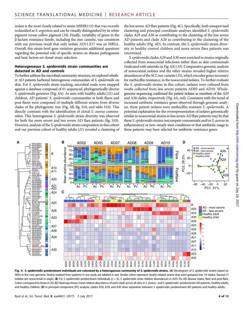
Heterogeneous S. epidermidis strain communities aredetected in AD and controlsTo further address themicrobial community structure,we exploredwheth-er AD patients harbored heterogenous communities of S. epidermidis onskin. For S. epidermidis strain tracking, microbial reads were mappedagainst a database composed of 61 sequenced, phylogenetically diverseS. epidermidis genomes (Fig. 4A). As seen with healthy adults (21) andchildren, AD patients’ S. epidermidis communities at both flares andpost-flares were composed of multiple different strains from diverseclades of the phylogenetic tree (Fig. 4B, fig. S10, and table S10). Thisdirectly contrasts with the identification of clonal S. aureus commu-nities. This heterogenous S. epidermidis strain diversity was observedfor both the more severe and less severe AD flare patients (fig. S10).However, analysis of the S. epidermidis strain composition in this cohortand our previous cohort of healthy adults (21) revealed a clustering of
Byrd et al., Sci. Transl. Med. 9, eaal4651 (2017) 5 July 2017
the less severe AD flare patients (Fig. 4C). Specifically, both unsupervisedclustering and principal coordinate analyses identified S. epidermidisclades A29 and A30 as contributing to the clustering of the less severeAD patients and clade A20 as contributing to the clustering of thehealthy adults (Fig. 4D). In contrast, the S. epidermidis strain diver-sity in healthy control children and more severe flare patients wasintermixed.
S. epidermidis cladesA29 andA30were enriched in strains originallycollected from nosocomial infections rather than as skin commensals(indicatedwith asterisks in Fig. 4A) (35). Comparative genomic analysisof nosocomial isolates and the other strains revealed higher relativeabundances of the SCCmec cassette (35), which encodes genes necessaryformethicillin resistance, in the nosocomial isolates. To further evaluatethe S. epidermidis strains in this cohort, isolates were cultured fromswabs collected from less severe patients AD05 and AD10. Whole-genome sequencing confirmed the patient isolates as members of the A29and A30 clades, respectively (Fig. 4A, red). Consistent with the trend ofincreased antibiotic resistance genes observed through genomic analy-sis, these patient isolates were methicillin-resistant S. epidermidis. Apotential explanation for the overrepresentation of isolates genomicallysimilar to nosocomial strains in less severeAD flare patientsmay be thatthese S. epidermidis strains outcompete commensals and/or S. aureus ininflammatory or non–steady-state conditions or that antibiotic usage inthese patients may have selected for antibiotic resistance genes.
by guest on July 19, 2017p://stm
.sciencemag.org/
BA20A24A30A29A22A26A28A21A27A11A10A23A25
−3
−2
−1
0
1
2
3
AD - more severe
Healthy adultHealthy child
AD - less severe
0
25
50
75
100
0
25
50
75
100
Flare
Postflare
Mea
n re
lativ
e ab
unda
nce
Ac Pc Ac Pc Ac Pc Ac Pc Ac Pc Ac Pc
A B
C
NIH05001*
VCU057
NIHLM015
NIHLM039
AD01.B
NIHLM088
NIH04003*
VCU123
VCU045
VCU126
VCU129
NIH05003*
NIHLM001
NIHLM003
NIHLM020
VCU120
NIHLM037
VCU144
VCU127
NIH051475*
VCU117
14.1.R1.SE
NIHLM067
M23864_W2g
AD05.A29
BVS058A4
ATCC12228
NIHLM023
VCU065
NIH051668*
NIHLM087
VCU037
AU12-03
NIH08001*
NIH05005*
NIHLM049
NIH06004*
NIHLM040
SK135
VCU109
NIHLM031
VCU071
BCM-HMP006
NIHLM021
VCU105
NIHLM095
NIHLM070
VCU125
NIH04008*
AD10.A30
VCU041
W23144
RP62A
NIHLM057NIHLM053
NIHLM008
VCU118
NIHLM061
12142587
VCU128
FRI909
B
A10A11
A20
A21A22
A23
A24
A25
A26
A27A28
A29
A30
VCU081VCU028NIHLM018
DAD02 AD05 AD07 AD08 AD09 AD10
AD
08
AD
05
AD
09
HC
07
HC
03
AD
01
AD
04
AD
10
AD
02
AD
07
AD
03
HV
02
HC
01
HC
02
HV
12
HC
06
HV
03
HV
08
HV
07
HV
10
HV
13
HV
05
HV
09
AD
11
HC
05
HV
01
HV
06
HV
11
HV
04
HV
14
HC
04
HV
15
AD
06
−60 −40 −20 0 20
−20
0
20
40
PC1, 31%P
C2,
26.
2%
A20
A29
A30
B
AD - more severeAD - less severeHealthy adultHealthy child
Fig. 4. S. epidermidis–predominant individuals are colonized by a heterogenous community of S. epidermidis strains. (A) Dendogram of S. epidermidis strains based onSNVs in the core genome. Strains isolated from patients in our study are labeled in red. Similar colors represent closely related strains that were grouped into 14 clades. Starred (*)isolates are nosocomial in origin. (B) For S. epidermidis–predominant individuals (n = 6), S. epidermidis strain relative abundances in AcPc for AD disease states, flare and post-flare.Colors correspond to those in (A). (C) Heatmap showsmean relative abundance of each clade across all sites in S. aureus– and S. epidermidis–predominant ADpatients, healthy adults,and healthy children. (D) In principal component (PC) analysis, clades A20, A29, and A30 drive separation between S. epidermidis–predominant AD patients and healthy adults.
6 of 12
SC I ENCE TRANS LAT IONAL MED I C I N E | R E S EARCH ART I C L E
by guest on July 19, 2017http://stm
.sciencemag.org/
Dow
nloaded from
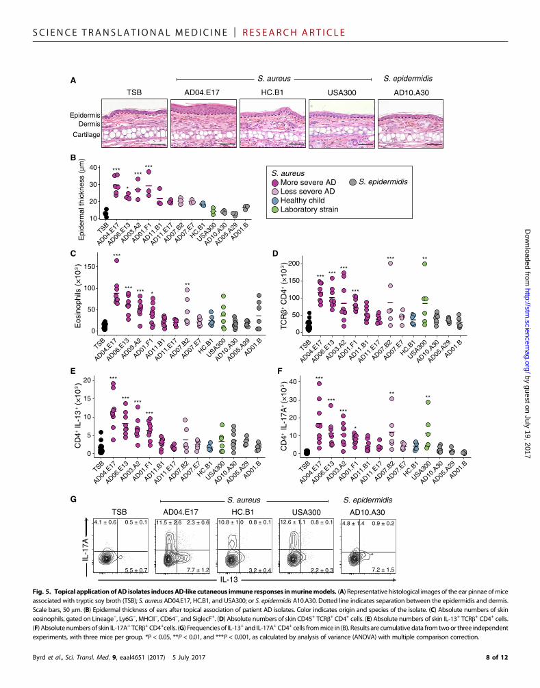
Strains elicit differential cutaneous immune responses in amurine modelWhereas S. aureus has been tightly linked with AD, it is still debatedwhether S. aureus is a cause or effect, that is, whether S. aureus can elicitand/or worsen AD skin disease or is a bystander that flourishes withincreased access to extracellular matrix or other products of inflamma-tion in eczematous skin (36, 37). By observing that individual strains ofS. aureus predominated during AD flares in our more severe flare pa-tients, we sought to investigate whether these clonal strains elicited abiological response distinct from other strains of staphylococci. By har-nessing the combined power of shotgun metagenomic sequencing ofclinical samples and whole-genome sequencing of bacteria cultivatedfrom concurrently collected skin swabs, we next analyzed (i) whetherstrains associated with AD flares would be sufficient to elicit skin in-flammation in the absence of any known genetic predisposition or priorbarrier disruption and (ii) whether therewere strain-specific differences.To test this, we topically applied staphylococcal strains cultivated fromAD patients and healthy controls onto intact skin of specific pathogen–free C57BL/6 wild-type mice, with a method previously developed totest the immune response to skin commensals. We individually tested10 phylogenetically distinct S. aureus isolates: 6 S. aureus isolatescultivated directly from the flared skin of patients with more severeflares, 2 S. aureus isolates from the skin of the less severe patientAD07’s flare time point, 1 S. aureus isolate from a healthy control,and 1 common pathogenic S. aureus USA300 FPR3757 isolate (high-lighted in red in fig. S9A). In addition, we tested three S. epidermidisisolates from AD patients: a representative each from clades A29 andA30, which predominated in the skin of less severe AD patients, and arepresentative from the ubiquitous B clade (highlighted in red in fig.S10A). In contrast to the noninflammatory responses observed after as-sociationwith either skin commensals (38, 39) orADpatient S. epidermidisisolates, topical application of the S. aureus isolates, particularly thoseassociated with more severe AD flare patients, was sufficient to induceepidermal thickening and inflammatory responses (Fig. 5, A and B,and fig. S11A) as well as immune cell infiltrate composed of neutro-phils and eosinophils (Fig. 5C and fig. S11, B and C). The USA300isolate, commonly used as a representative S. aureus in functionalexperiments, induced only a modest immune response as comparedtomany of the isolates cultivated from severe AD patients, underscor-ing the importance of using matched clinical isolates.
In addition to innate immune cells, infiltration of T cell receptor(TCR)ab+ and gdlow cells was also observed inmice colonized onlywithspecific S. aureus strains (fig. S12A).Most of the TCRb+ cells wereCD4+
with variable effector potential, depending on the associated isolate (Fig.5D). Notably, four S. aureus isolates frommore severe AD flare patientsinduced production of the cytokine interleukin-13 (IL-13) (Fig. 5E),which is commonly associated with allergic inflammation. CutaneousT helper 17 (TH17) cells were also identified when mice were colonizedwith these four IL-13–inducing strains, in addition to AD07.B2 andUSA300 (Fig. 5, F and G). Recent reports have identified the presenceof TH17 cells in AD lesions (40, 41), particularly in Asian patient popu-lations (42).
Similar to CD4+T cells, the gdT cells ofmice associatedwith specificstrains of S. aureus isolates also had the potential to make higher levelsof IL-17A (fig. S12B). Notably, four of the S. aureus isolates [two fromthe more severe flare patient AD11 (AD11.B1 and AD11.E17), onefrom the less severe flare patient AD07 (AD07.E7), and one from ahealthy child (HC.B1)] induced minimal immune responses in allcategories. Overall, association of S. aureus strains isolated from more
Byrd et al., Sci. Transl. Med. 9, eaal4651 (2017) 5 July 2017
severe AD flare patients to wild-type mice without prior barrier disrup-tion induced immune responses in the skin that were significantlygreater than those inducedwith S. epidermidis or S. aureus isolates fromless severe AD flare patients or controls. Thus, these findings suggestthat specific strains of S. aureusmay be sufficient to elicit and/or exac-erbate skin inflammation as part of AD disease pathogenesis.
DISCUSSIONAD is a complex disease with many contributing factors, including skinbarrier integrity, innate and adaptive immunity, and the microbiome.The heterogeneity of the course, severity, and clinical response in ADpatients underscores the diversity of phenotypic presentations, aswell asthe probable differences in disease pathogenesis, within this one diag-nosis. In addition to the various genetic susceptibility loci for AD,deeper investigation into the skin microbiome could provide a betterunderstanding of the microbial heterogeneity of AD and its potentialcontributions to disease.
Although there have been many efforts to identify bacteria in ADskin, studies have generally relied on methods that do not distinguishmicrobes beyond the species level or can misclassify genomically distinctclones (14, 15). Here, we combined shotgun metagenomic sequencing ofclinical samples with whole-genome sequencing of patient-derived iso-lates to investigate the microbial communities of AD skin down to thestrain- and SNV-level resolution. Because topical anti-inflammatoryand antimicrobial treatments alter the skinmicrobiota (6, 7), the baselineand flare time points in this cohort were strictly defined by skin prepar-atory regimens to capture the natural history of the skin disease and toavoid potential confounders. As compared to healthy controls, the ADpatients exhibitedmarked skin bacterial dysbiosis during flares. This dys-biosis was related to the increased relative abundance of staphylococci,consistent with prior cohorts. On the basis of the disease severity (definedby objective SCORAD) during flares, we observed a strong correlationbetween severe AD flares and S. aureus relative abundances. These find-ings demonstrated that despite the relatively small numbers of subjects inthis study, our cohort of patients is representative of other published pa-tient cohorts as defined by validated diagnostic criteria.
Shotgun metagenomic sequencing enables strain-level examinationof microbes within the broad microbial community of bacteria, fungi,and viruses. Strain tracking identified marked outgrowth of clonalS. aureus strains in the skin of flaring AD patients with more severedisease; these same strains persisted post-flare at lower relative abun-dances. Othermethods have examinedwhether S. aureus expansion inthe skin of AD flares was related to either proportional increases in theentire community of S. aureus strains or the increase of a single or afew dominant clones; however, these studies were limited by the in-ability to examine these possibilities in the context of the whole skinmicrobial community. Although the fungal and viral communities werenot significantly different in this study, expansion of reference data-bases/genomes and studies into the microbial “dark matter” in metage-nomic data may provide further insights into AD microbiota. Ourfindings demonstrate that AD skin flares in patients with more severedisease are tightly linked with clonal S. aureus isolates.
In addition to characterizing strain communities during the courseof AD, we found that less severe AD patients were colonized with moremethicillin-resistant strains, whereas the more severe AD patients wereprimarily colonized with methicillin-sensitive strains. Although methi-cillin resistance is not as common in AD as would be predicted on thebasis of the high rates of S. aureus colonization in this disease, the
7 of 12
SC I ENCE TRANS LAT IONAL MED I C I N E | R E S EARCH ART I C L E
by guest on July 19, 2017http://stm
.sciencemag.org/
Dow
nloaded from
IL-1
7AA
EpidermisDermis
Cartilage
TSB AD04.E17 USA300 AD10.A30
C D
E F
IL-13
G
HC.B1
***
***
***
*
** **
0
10
20
30
40
CD
4IL
-17A
++
***
*** ***
***
0
5
10
15
20
CD
4IL
-13
++
***
*** *** ***
0
50
100
150
Eos
inop
hils
E
pide
rmal
thic
knes
s (µ
m)
*** ******
***
*** **
0
50
100
150
200
+ C
D4
TC
Rβ
+ (
×10
3 )
S. aureus S. epidermidis
TSB AD04.E17 USA300 AD10.A30HC.B1
S. aureus S. epidermidis
B
More severe ADLess severe ADHealthy childLaboratory strain
S. epidermidisS. aureus***
*
******
10
20
30
40
TSB
AD04.E
17
AD06.E
13
AD03.A
2
AD01.F
1
AD11.B
1
AD11.E
17
AD07.B
2
AD07.E
7
HC.B1
USA300
AD10.A
30
AD05.A
29
AD01.B
TSB
AD04.E
17
AD06.E
13
AD03.A
2
AD01.F
1
AD11.B
1
AD11.E
17
AD07.B
2
AD07.E
7
HC.B1
USA300
AD10.A
30
AD05.A
29
AD01.B
TSB
AD04.E
17
AD06.E
13
AD03.A
2
AD01.F
1
AD11.B
1
AD11.E
17
AD07.B
2
AD07.E
7
HC.B1
USA300
AD10.A
30
AD05.A
29
AD01.B
TSB
AD04.E
17
AD06.E
13
AD03.A
2
AD01.F
1
AD11.B
1
AD11.E
17
AD07.B
2
AD07.E
7
HC.B1
USA300
AD10.A
30
AD05.A
29
AD01.B
TSB
AD04.E
17
AD06.E
13
AD03.A
2
AD01.F
1
AD11.B
1
AD11.E
17
AD07.B
2
AD07.E
7
HC.B1
USA300
AD10.A
30
AD05.A
29
AD01.B
(×
103 )
(×
103 )
(×
103 )
7.2 ± 1.5
0.9 ± 0.24.8 ± 1.411.5 ± 2.6 2.3 ± 0.6
7.7 ± 1.2 3.2 ± 0.4
0.8 ± 0.110.8 ± 1.0
2.2 ± 0.3
0.8 ± 0.112.6 ± 1.14.1 ± 0.6 0.5 ± 0.1
5.5 ± 0.7
Fig. 5. Topical application of AD isolates induces AD-like cutaneous immune responses inmurinemodels. (A) Representative histological images of the ear pinnae ofmiceassociated with tryptic soy broth (TSB); S. aureus AD04.E17, HC.B1, and USA300; or S. epidermidis A10.A30. Dotted line indicates separation between the epidermidis and dermis.Scale bars, 50 mm. (B) Epidermal thickness of ears after topical association of patient AD isolates. Color indicates origin and species of the isolate. (C) Absolute numbers of skineosinophils, gated on Lineage−, Ly6G−, MHCII−, CD64−, and SiglecF+. (D) Absolute numbers of skin CD45+ TCRb+ CD4+ cells. (E) Absolute numbers of skin IL-13+ TCRb+ CD4+ cells.(F) Absolutenumbers of skin IL-17A+ TCRb+CD4+cells. (G) Frequencies of IL-13+ and IL-17A+ CD4+ cells frommice in (B). Results are cumulativedata from twoor three independentexperiments, with three mice per group. *P < 0.05, **P < 0.01, and ***P < 0.001, as calculated by analysis of variance (ANOVA) with multiple comparison correction.
Byrd et al., Sci. Transl. Med. 9, eaal4651 (2017) 5 July 2017 8 of 12
SC I ENCE TRANS LAT IONAL MED I C I N E | R E S EARCH ART I C L E
by guest on July 19, 2017http://stm
.sciencemag.org/
Dow
nloaded from
finding ofMSSA andmethicillin-resistant S. epidermidis predominancemay contribute to differential responses to therapies in AD patients (43).The contrasts between S. aureus and S. epidermidis observed in this studylikely also relate to the differences in microbial genetics and populationdynamics at both the species and strain levels. Additional investigations ofthese microbiome phenotypic differences may improve the understand-ing of ADpathogenesis and lead tomore targeted therapeutics, includingthe potential use of commensals to protect against S. aureus (44). Birthcohort studies may address whether these patients acquired bacterialstrains from family members and/or environmental sources as part ofmicrobial inheritance (45). Testing of S. aureus strains in gnotobioticmice, similar to Bacteroides gut commensal studies, may functionally ad-dress whether colonization by clonal S. aureus occurs through limited ex-posure or colonization resistance (46).
Using strains isolated from the skin of AD flares and a healthy con-trol as well as a known laboratory strain, we examined the potentialbiological differences between staphylococcal strains. In a murinemodel without prior skin barrier disruption and with intact immunity,S. aureus strains from flare time points inmore severe ADpatients weresufficient to inducemanifestations of skin inflammation, such as epider-mal thickening and cutaneous infiltration of TH2 and TH17 cells. Themagnitude of different immunologic effects varied depending on theisolated strain but was not strictly related to the disease severity ofthe source patient. Notably, murine colonization with either isolateAD11.B1 or AD11.E17 induced minimal immune responses, althoughpatientAD11had an objective SCORADof 51.4.However, AD11 is alsoheterozygous for anullmutation in theFLGgene (S757X), suggesting thatAD11’s strains of S. aureus may be immunogenic in the setting of animpaired skin barrier, which, as shown by previous studies, allowsS. aureus to breach the epidermis into the dermis where it can triggerexpression of proinflammatory cytokines (47). Caveats of these findingsin the murine model are the relatively small number of isolates from thiscohort that were fully sequenced and studied in the murine model andthe observation of varied host responses when testing isolates from thesame clade (AD04 and AD11), highlighting the need to examine a largernumber of isolates including strains from similar and different cladesand from healthy individuals and AD patients. An important additionallimitation is the recognition that this murine model and others do notrecapitulate the multiple complexities of human AD.
In mouse models, S. aureus enterotoxins have been shown to act assuperantigens that can initiate TH17 responses (48), whereas S. aureusd-toxin can induce degranulation of mast cells (49). These genes wereboth present in the noninflammatory S. aureus isolates, indicating thatstrain variability exists not only in gene content but also in gene expres-sion. Because healthy control–associated S. aureus strains were limitedin our cohort because of the small percentage of healthy individuals co-lonized with S. aureus, future studies with additional S. aureus isolatesfrom healthy individuals are necessary to tease apart the mechanismsunderlying functional differences between S. aureus strains. In the con-text of prior studies demonstrating cutaneous immunologic responsesto skin commensals (38, 39) and exacerbation of eczematous skin inADmouse models by S. aureus (38, 39, 49, 50), our findings demonstratethat staphylococcal strains may play an important role in AD diseaseprogression in a strain-specific manner.
Here, we used shotgun metagenomic sequencing to examine strain-level microbial compositions of AD skin, coupled with whole-genomesequencing of patient isolates.With increasing recognition of highly in-dividualized skin microbiomes (16), the presence of patient-specificstrains underscores the individuality of the disease course and therapeu-
Byrd et al., Sci. Transl. Med. 9, eaal4651 (2017) 5 July 2017
tic response and may represent an opportunity for precision medicine.Our functional studies with cutaneous colonization of AD patient–associated strains of S. aureus and S. epidermidis demonstrated strain-specific differences in the ability to elicit histologic and immunologicalterations. AD typically has an age of onset in the first year of life whenthe human immune system is developing and being tuned by the en-dogenous microbial community. Recent studies have shown that earlyexposures canmodulate host immunity to subsequent exposure and in-duce tolerance (51, 52). Thus, in light of the known links between severeAD and subsequent development of asthma and hay fever (“the atopicmarch”), targeted modulation of an AD patient’s particular staphylo-coccal strains has the potential to ameliorate the broader developmentof atopic disorders.
MATERIALS AND METHODSStudy designAD patients and similarly aged healthy controls were recruited to par-ticipate in a natural history study approved by the institutional reviewboard of the National Human Genome Research Institute (NHGRI)(www.clinicaltrials.gov/ct2/show/NCT00605878). Written informedconsentwas obtained fromparents or guardians of all participating chil-dren. Patients were diagnosed with AD on the basis of the UKWorkingParty definition (53). Eligibility criteria included ages of 2 to 18 years,moderate to severe disease (objective SCORAD, ≥15), presence of ≥1affected Ac or Pc at enrollment, and >3 weeks off of systemic antibioticsand corticosteroids (17–19). After skin preparation regimen, standar-dized skin sampling was performed from prespecified skin sites bilater-ally and at defined time points (baseline, flare, and post-flare). Skinsamples for metagenomic sequencing and negative controls were ob-tained as previously described (16, 21), with additional swabs of theAc, retroauricular crease, and the nares collected concurrently for sub-sequent culture analyses.
Microbiome sequencing and analysisProcedures for library generation with Nextera DNA Library Prep Kitand sequencing 2 × 125–bp reads with a target of 15 million to 50 mil-lion clusters onan IlluminaHiSeq instrumentwereperformedasdescribedpreviously (21). In total, for 18 individuals (11 patients and 7 controls)sampled at seven body sites at different stages of disease forADpatients,we obtained 422 samples and 2.26 trillion reads (or 191 Gbp) of non-human, quality-filtered reads.
Microbial reads were assigned taxonomic classifications as previous-ly described (21). Included in the microbial reference genome databaseare 2342 bacterial, 389 fungal, 1375 viral, and 67 archaeal genomes. Inaddition, a staphylococcus database was compiled from 315 completeand draft genomes from theNational Center for Biological Information(NCBI) (www.ncbi.nlm.nih.gov) as of October 2014. Nonhuman readswere separately mapped to both genome collections using Bowtie2’s“--very-sensitive” parameter -k 10 to retrieve the top 10 hits (54). Theresulting alignment files were processed with Pathoscope v1.0 (55)to assign multiply mapped reads to their most likely genome of or-igin. Read hit counts were then normalized by genome and scaled tosum to 1. Coverages of each output genome were calculated usinggenomeCoverageBed in the bedtools suite (56). To reduce the effectsof spurious classifications from low-abundance organisms, only specieswith ≥1% coverage of the genome were considered (21).
Strain tracking of S. aureus and S. epidermidiswas performed as pre-viously described (21). Briefly, reference databases for S. aureus and
9 of 12
SC I ENCE TRANS LAT IONAL MED I C I N E | R E S EARCH ART I C L E
by guest on July 19, 2017http://stm
.sciencemag.org/
Dow
nloaded from
S. epidermidis were compiled from all complete and draft genomesavailable on NCBI, 215 and 61, respectively. For both species, whole-genome alignment, with nucmer (57), was then used to identify the“core” region shared between all sequenced strains. SNVs identifiedin these core regions were subsequently used to generate dendogramswith PhyML 3.0. For strain tracking to avoid noise from other staphy-lococcal species,metagenomic reads were first filtered against the staph-ylococcus database minus the species being strain-tracked (Bowtie2:--very-sensitive, -score-min L,-0.6,0.006). The remaining reads werethen mapped to each species database with Bowtie2 (--very-sensitive,-score-min L,-0.6,0.006, -k number of genomes) (54) with zero toler-ance for mismatches. The resulting alignment file was then processedwith Pathoscope (-theta_prior 10 x 10^88) (55) to deconvolutemultiplemapping reads.
For SNV analysis as described previously (16), metagenomic readswere mapped against the S. aureus core genome using Bowtie2 (--very-sensitive). The resulting alignment filewas sorted by SAMtools and thenprocessed with GATK’s IndelRealigner (58). The corrected alignmentfilewas analyzedwith SAMtools andBCFtools to identify possible var-iants (samtoolsmpileup -uD -q30 -Q30; bcftools view -Abvcg, vcfutils.pl varFilter -D99percentileofcoverage -d4 -1 .00001 -4 .00001). Cus-tom scripts were then used to filter possible variants on the basis ofcriteria described in (59).
For staphylococcal isolates, Nextera libraries were generated fromthe genomic DNA and sequenced using a paired-end 300-base dualindex run on an IlluminaMiSeq instrument to generate 1million to 2mil-lion read pairs per library for ~80× genome coverage. Reads for eachisolate were assembled with MaSuRCA version 2.2.1 (60), SPAdes ver-sion 3.6.0, or SPAdes version 3.6.0 (61) plus Pilon version 1.13 (62) cor-rection. Best k-mer length estimates on paired-end readswere evaluatedusingKmerGenie (version1.6300) (63) andused in running theMaSuRCAassembler for each genome. For comparative genomic analysis, genomeannotation was done using the Institute for Genome Sciences (IGS)Analysis Engine (http://ae.igs.umaryland.edu/cgi/index.cgi) (64).
To identify the functional capacity of S. aureus isolates, we followed aprocedure similar to that in (16). The IGS Analysis Engine was used forstructural and functional annotation of the sequences (http://ae.igs.umaryland.edu/cgi/index.cgi) (64). Manatee was used to view annota-tions (http://manatee.sourceforge.net/). Protein sequences were thenclustered into nonredundant orthologs with USEARCH (-cluster_fast -id0.50 -centroids) (65). These gene clusterswere then annotatedbyBLASTpagainst the KEGGdatabase. Distribution of genes between the S. aureusassemblies was visualized with jvenn (66).
Murine topical association with bacterial isolates andflow cytometric analysesExperiments were performed with 6- to 12-week-old female C57BL/6specific pathogen–free mice under an animal study proposal approvedby theNHGRIAnimal Care andUseCommittee. Topical association ofmicewas based on (38, 39). Ten S. aureus strains (AD04.E17,AD06.E13,AD03.A2, AD01.F1, AD11.B1, AD11.E17, AD07.B2, HC.B1, USA300,and FPR3757) and 3 S. epidermidis strains (AD05.A29, AD10.A30, andAD01.B), isolated fromADpatients, controls (HC.B1), or theAmericanType Culture Collection (USA300), were cultured in tryptic soy brothat 37°C for 18 hours and normalized using optical density at 600 nm(OD600) to achieve similar bacterial density (about 108 colony-formingunits/ml). For topical association, a sterile epicenter Catch-All swabwasmoistened in liquid culture of the bacteria and then rubbed againstthe ears of mice until they became visibly moist.
Byrd et al., Sci. Transl. Med. 9, eaal4651 (2017) 5 July 2017
Cells from the ear pinnae of mice were isolated as previously de-scribed (38, 39). Murine single-cell suspensions were incubated withfluorochrome-conjugated antibodies against surfacemarkers CD4 (cloneRM4-5), CD8b (eBioH35-17.2), CD11b (M1/70), CD11c (N418 orHL3), CD19 (6D5), CD45.2 (104), CD49b (DX5), CD64 (X54-5/7.1),Ly6G (1A8), MHCII (M5/114.15.2), NK1.1 (PK136), TCRgd (GL3),TCRb (H57-597), and/or SiglecF (E50-2440). All antibodies werepurchased from eBioscience, BioLegend, or BD Biosciences. For detec-tion of basal cytokine potential, single-cell suspensions from ear tissuewere directly cultured ex vivo in a 96-well U-bottom plate. Cell acqui-sition was performed on a Fortessa flow cytometer using FACSDivasoftware (BD Biosciences), and data were analyzed using FlowJosoftware (Tree Star).
StatisticsAll statistical analyses were performed inR, andmost of the graphsweregenerated with ggplot2 (67). Data are means ± SEM unless otherwiseindicated. For all box plots, center lines represent themedian, and edgesrepresent the first and third quartiles. The nonparametric Wilcoxonrank-sum test was used to determine statistically significant differencesbetween populations (wilcox.test in R).Where indicated, within-subjectanalysis was performed with option “paired=T” in wilcox.test. AllP valueswere adjusted using p.adjust in Rusing Bonferroni (no. of com-parisons,≤10) or false discovery rate (no. of comparisons, >10) correc-tions. Statistical significance was ascribed to an a level of the adjustedP values of ≤0.05. Similarity between samples was assessed using theYue-Clayton q, which assesses the similarity between two samples onthe basis of (i) the number of features in common between twosamples and (ii) their relative abundances, with q = 0 indicating totallydissimilar communities and q = 1 indicating identical communities (68).For functional experiments, statistical significance was determinedbyANOVAwithmultiple comparison corrections (aov andTukeyHSDin R).
SUPPLEMENTARY MATERIALSwww.sciencetranslationalmedicine.org/cgi/content/full/9/397/eaal4651/DC1Materials and MethodsFig. S1. Seven sites sampled bilaterally on pediatric AD patients and control children.Fig. S2. Full multikingdom taxonomic classifications for AD patients and controls.Fig. S3. Full Malassezia species classifications for AD patients and controls.Fig. S4. Full eukaryotic virus classifications for AD patients and controls.Fig. S5. Full bacterial taxonomic classifications for AD patients and controls.Fig. S6. Relative abundance of staphylococcal species in relation to total bacterial populationfor all sites in AD patients and controls.Fig. S7. Correlation of various staphylococcal species mean relative abundance and objectiveSCORAD for all sites of patients.Fig. S8. Relative abundance of staphylococcal species for all sites in AD patients and controls.Fig. S9. S. aureus clades for AD patients and controls.Fig. S10. S. epidermidis clades for AD patients and controls.Fig. S11. Histologic and cutaneous innate immune cell responses with AD isolate association ina murine model.Fig. S12. CD45+ cutaneous immune responses with AD isolate association in a murine model.Table S1. Subject Tanner stage and disease severity for samples used in this study.Table S2. Clinical metadata for the subjects in this study.Table S3. Metadata table for all samples in this study.Table S4. Multikingdom relative abundances (coverage, >1% of the reference genome).Table S5. Malassezia relative abundance.Table S6. Bacteria relative abundances (coverage, >1% of the reference genome).Table S7. Staphylococcus species relative abundances.Table S8. S. aureus clade abundances.Table S9. S. aureus pangenome analysis for isolates cultured and sequenced in this study.Table S10. S. epidermidis strain relative abundances.References (69–75)
10 of 12
SC I ENCE TRANS LAT IONAL MED I C I N E | R E S EARCH ART I C L E
by guest on July 19, 2017http://stm
.sciencemag.org/
Dow
nloaded from
REFERENCES AND NOTES1. K. Eyerich, S. Eyerich, T. Biedermann, The multi-modal immune pathogenesis of atopic
eczema. Trends Immunol. 36, 788–801 (2015).
2. S. K. Bantz, Z. Zhu, T. Zheng, The Atopic March: Progression from atopic dermatitis toallergic rhinitis and asthma. J. Clin. Cell. Immunol. 5, 202 (2014).
3. L. Paternoster, M. Standl, J. Waage, H. Baurecht, M. Hotze, D. P. Strachan, J. A. Curtin,K. Bønnelykke, C. Tian, A. Takahashi, J. Esparza-Gordillo, A. C. Alves, J. P. Thyssen,H. T. den Dekker, M. A. Ferreira, E. Altmaier, P. M. A. Sleiman, F. L. Xiao, J. R. Gonzalez,I. Marenholz, B. Kalb, M. Pino-Yanes, C.-J. Xu, L. Carstensen, M. M. Groen-Blokhuis,C. Venturini, C. E. Pennell, S. J. Barton, A. M. Levin, I. Curjuric, M. Bustamante,E. Kreiner-Møller, G. A. Lockett, J. Bacelis, S. Bunyavanich, R. A. Myers, A. Matanovic,A. Kumar, J. Y. Tung, T. Hirota, M. Kubo, W. L. McArdle, A. J. Henderson, J. P. Kemp,J. Zheng, G. D. Smith, F. Rüschendorf, A. Bauerfeind, M. A. Lee-Kirsch, A. Arnold,G. Homuth, C. O. Schmidt, E. Mangold, S. Cichon, T. Keil, E. Rodríguez, A. Peters, A. Franke,W. Lieb, N. Novak, R. Fölster-Holst, M. Horikoshi, J. Pekkanen, S. Sebert, L. L. Husemoen,N. Grarup, J. C. de Jongste, F. Rivadeneira, A. Hofman, V. W. Jaddoe, S. G. Pasmans,N. J. Elbert, A. G. Uitterlinden, G. B. Marks, P. J. Thompson, M. C. Matheson, C. F. Robertson;Australian Asthma Genetics Consortium (AAGC), J. S. Ried, J. Li, X. B. Zuo, X. D. Zheng,X. Y. Yin, L. D. Sun, M. A. McAleer, G. M. O’Regan, C. M. Fahy, L. Campbell, M. Macek,M. Kurek, D. Hu, C. Eng, D. S. Postma, B. Feenstra, F. Geller, J. J. Hottenga,C. M. Middeldorp, P. Hysi, V. Bataille, T. Spector, C. M. Tiesler, E. Thiering,B. Pahukasahasram, J. J. Yang, M. Imboden, S. Huntsman, N. Vilor-Tejedor, C. L. Relton,R. Myhre, W. Nystad, A. Custovic, S. T. Weiss, D. A. Meyers, C. Söderhäll, E. Melén, C. Ober,B. A. Raby, A. Simpson, B. Jacobsson, J. W. Holloway, H. Bisgaard, J. Sunyer,N. M. Probst-Hensch, L. K. Williams, K. M. Godfrey, C. A. Wang, D. I. Boomsma, M. Melbye,G. H. Koppelman, D. Jarvis, W. H. McLean, A. D. Irvine, X. J. Zhang, H. Hakonarson,C. Gieger, E. G. Burchard, N. G. Martin, L. Duijts, A. Linneberg, M. R. Jarvelin, M. M. Nöthen,S. Lau, N. Hübner, Y.-A. Lee, M. Tamari, D. A. Hinds, D. Glass, S. J. Brown, J. Heinrich,D. M. Evans, S. Weidinger; EArly Genetics and Lifecourse Epidemiology (EAGLE) EczemaConsortium, Multi-ancestry genome-wide association study of 21,000 cases and95,000 controls identifies new risk loci for atopic dermatitis. Nat. Genet. 47, 1449–1456(2015).
4. C. N. A. Palmer, A. D. Irvine, A. Terron-Kwiatkowski, Y. Zhao, H. Liao, S. P. Lee, D. R. Goudie,A. Sandilands, L. E. Campbell, F. J. D. Smith, G. M. O’Regan, R. M. Watson, J. E. Cecil,S. J. Bale, J. G. Compton, J. J. DiGiovanna, P. Fleckman, S. Lewis-Jones, G. Arseculeratne,A. Sergeant, C. S. Munro, B. El Houate, K. McElreavey, L. B. Halkjaer, H. Bisgaard,S. Mukhopadhyay, W. H. McLean, Common loss-of-function variants of the epidermalbarrier protein filaggrin are a major predisposing factor for atopic dermatitis. Nat. Genet.38, 441–446 (2006).
5. J. T. Huang, M. Abrams, B. Tlougan, A. Rademaker, A. S. Paller, Treatment ofStaphylococcus aureus colonization in atopic dermatitis decreases disease severity.Pediatrics 123, e808–e814 (2009).
6. H. H. Kong, J. Oh, C. Deming, S. Conlan, E. A. Grice, M. A. Beatson, E. Nomicos, E. C. Polley,H. D. Komarow; NISC Comparative Sequence Program, P. R. Murray, M. L. Turner,J. A. Segre, Temporal shifts in the skin microbiome associated with disease flares andtreatment in children with atopic dermatitis. Genome Res. 22, 850–859 (2012).
7. M. E. Gonzalez, J. V. Schaffer, S. J. Orlow, Z. Gao, H. Li, A. V. Alekseyenko, M. J. Blaser,Cutaneous microbiome effects of fluticasone propionate cream and adjunctivebleach baths in childhood atopic dermatitis. J. Am. Acad. Dermatol. 75, 481–493.e8(2016).
8. J. J. Leyden, R. R. Marples, A. M. Kligman, Staphylococcus aureus in the lesions of atopicdermatitis. Br. J. Dermatol. 90, 525–530 (1974).
9. L. S. Chiu, V. C. Y. Chow, J. M. L. Ling, K. L. Hon, Staphylococcus aureus carriage in theanterior nares of close contacts of patients with atopic dermatitis. Arch. Dermatol. 146,748–752 (2010).
10. S. Bonness, C. Szekat, N. Novak, G. Bierbaum, Pulsed-field gel electrophoresis ofStaphylococcus aureus isolates from atopic patients revealing presence of similar strainsin isolates from children and their parents. J. Clin. Microbiol. 46, 456–461 (2008).
11. D.-W. Kim, J.-Y. Park, K.-D. Park, T.-H. Kim, W.-J. Lee, S.-J. Lee, J. Kim, Are therepredominant strains and toxins of Staphylococcus aureus in atopic dermatitis patients?Genotypic characterization and toxin determination of S. aureus isolated in adolescentand adult patients with atopic dermatitis. J. Dermatol. 36, 75–81 (2009).
12. S. Seite, G. E. Flores, J. B. Henley, R. Martin, H. Zelenkova, L. Aguilar, N. Fierer, Microbiomeof affected and unaffected skin of patients with atopic dermatitis before and afteremollient treatment. J. Drugs Dermatol. 13, 1365–1372 (2014).
13. L. Drago, R. De Grandi, G. Altomare, P. Pigatto, O. Rossi, M. Toscano, Skin microbiotaof first cousins affected by psoriasis and atopic dermatitis. Clin. Mol. Allergy 14, 2(2016).
14. S. J. Salipante, D. J. SenGupta, L. A. Cummings, T. A. Land, D. R. Hoogestraat, B. T. Cookson,Application of whole-genome sequencing for bacterial strain typing in molecularepidemiology. J. Clin. Microbiol. 53, 1072–1079 (2015).
Byrd et al., Sci. Transl. Med. 9, eaal4651 (2017) 5 July 2017
15. E. Ugolotti, P. Larghero, I. Vanni, R. Bandettini, G. Tripodi, G. Melioli, E. Di Marco,A. Raso, R. Biassoni, Whole-genome sequencing as standard practice for the analysisof clonality in outbreaks of meticillin-resistant Staphylococcus aureus in a paediatricsetting. J. Hosp. Infect. 93, 375–381 (2016).
16. J. Oh, A. L. Byrd, M. Park; NISC Comparative Sequencing Program, H. H. Kong, J. A. Segre,Temporal stability of the human skin microbiome. Cell 165, 854–866 (2016).
17. H. C. Williams, P. G. Burney, D. Strachan, R. J. Hay, The U.K. Working party’s diagnosticcriteria for atopic dermatitis. II. Observer variation of clinical diagnosis and signs of atopicdermatitis. Br. J. Dermatol. 131, 397–405 (1994).
18. B. Kunz, A. P. Oranje, L. Labrèze, J. F. Stalder, J. Ring, A. Taïeb, Clinical validation andguidelines for the SCORAD index: Consensus report of the European Task Force onAtopic Dermatitis. Dermatology 195, 10–19 (1997).
19. A. P. Oranje, E. J. Glazenburg, A. Wolkerstorfer, F. B. de Waard-van der Spek, Practicalissues on interpretation of scoring atopic dermatitis: The SCORAD index, objectiveSCORAD and the three-item severity score. Br. J. Dermatol. 157, 645–648 (2007).
20. K. Findley, J. Oh, J. Yang, S. Conlan, C. Deming, J. A. Meyer, D. Schoenfeld, E. Nomicos,M. Park; NIH Intramural Sequencing Center Comparative Sequencing Program, H. H. Kong,J. A. Segre, Topographic diversity of fungal and bacterial communities in human skin.Nature 498, 367–370 (2013).
21. J. Oh, A. L. Byrd, C. Deming, S. Conlan; NISC Comparative Sequencing Program, H. H. Kong,J. A. Segre, Biogeography and individuality shape function in the human skinmetagenome. Nature 514, 59–64 (2014).
22. M. R. Williams, R. L. Gallo, The role of the skin microbiome in atopic dermatitis.Curr. Allergy Asthma Rep. 15, 65 (2015).
23. M. Tauber, S. Balica, C.-Y. Hsu, C. Jean-Decoster, C. Lauze, D. Redoules, C. Viodé,A.-M. Schmitt, G. Serre, M. Simon, C. F. Paul, Staphylococcus aureus density on lesionaland nonlesional skin is strongly associated with disease severity in atopic dermatitis.J. Allergy Clin. Immunol. 137, 1272–1274.e3 (2016).
24. K. R. Chng, A. S. Tay, C. Li, A. H. Ng, J. Wang, B. K. Suri, S. A. Matta, N. McGovern, B. Janela,X. F. C. C. Wong, Y. Y. Sio, B. V. Au, A. Wilm, P. F. De Sessions, T. C. Lim, M. B. Y. Tang,F. Ginhoux, J. E. Connolly, E. B. Lane, F. T. Chew, J. E. A. Common, N. Nagarajan, Wholemetagenome profiling reveals skin microbiome-dependent susceptibility to atopicdermatitis flare. Nat. Microbiol. 1, 16106 (2016).
25. B. Shi, N. J. Bangayan, E. Curd, P. A. Taylor, R. L. Gallo, D. Y. M. Leung, H. Li, The skinmicrobiome is different in pediatric versus adult atopic dermatitis. J. Allergy Clin.Immunol. 138, 1233–1236 (2016).
26. M. Yeung, A. Balma-Mena, N. Shear, A. Simor, E. Pope, S. Walsh, M. J. McGavin,Identification of major clonal complexes and toxin producing strains amongStaphylococcus aureus associated with atopic dermatitis. Microbes Infect. 13, 189–197(2011).
27. H. Lomholt, K. E. Andersen, M. Kilian, Staphylococcus aureus clonal dynamics andvirulence factors in children with atopic dermatitis. J. Invest. Dermatol. 125, 977–982(2005).
28. P. H. Hoeger, W. Lenz, A. Boutonnier, J. M. Fournier, Staphylococcal skin colonization inchildren with atopic dermatitis: Prevalence, persistence, and transmission of toxigenicand nontoxigenic strains. J. Infect. Dis. 165, 1064–1068 (1992).
29. L. Suh, S. Coffin, K. H. Leckerman, J. M. Gelfand, P. J. Honig, A. C. Yan, Methicillin-resistantStaphylococcus aureus colonization in children with atopic dermatitis. Pediatr. Dermatol.25, 528–534 (2008).
30. M. S. Hsiang, R. Shiau, J. Nadle, L. Chan, B. Lee, H. F. Chambers, E. Pan, Epidemiologicsimilarities in pediatric community-associated methicillin-resistant and methicillin-sensitive Staphylococcus aureus in the San Francisco Bay Area. J. Pediatric Infect. Dis. Soc.1, 200–211 (2012).
31. C. Chaptini, S. Quinn, G. Marshman, Methicillin-resistant Staphylococcus aureus in childrenwith atopic dermatitis from 1999 to 2014: A longitudinal study. Australas. J. Dermatol.57, 122–127 (2016).
32. P. Strange, L. Skov, S. Lisby, P. L. Nielsen, O. Baadsgaard, Staphylococcal enterotoxinB applied on intact normal and intact atopic skin induces dermatitis. Arch. Dermatol.132, 27–33 (1996).
33. E. Bosi, J. M. Monk, R. K. Aziz, M. Fondi, V. Nizet, B. Palsson, Comparative genome-scalemodelling of Staphylococcus aureus strains identifies strain-specific metabolic capabilitieslinked to pathogenicity. Proc. Natl. Acad. Sci. U.S.A. 113, E3801–E3809 (2016).
34. S. Y. C. Tong, F. Schaumburg, M. J. Ellington, J. Corander, B. Pichon, F. Leendertz,S. D. Bentley, J. Parkhill, D. C. Holt, G. Peters, P. M. Giffard, Novel staphylococcal speciesthat form part of a Staphylococcus aureus-related complex: The non-pigmentedStaphylococcus argenteus sp. nov. and the non-human primate-associated Staphylococcusschweitzeri sp. nov. Int. J. Syst. Evol. Microbiol. 65, 15–22 (2015).
35. S. Conlan, L. A. Mijares; NISC Comparative Sequencing Program, J. Becker, R. W. Blakesley,G. G. Bouffard, S. Brooks, H. Coleman, J. Gupta, N. Gurson, M. Park, B. Schmidt,P. J. Thomas, M. Otto, H. H. Kong, P. R. Murray, J. A. Segre, Staphylococcus epidermidispan-genome sequence analysis reveals diversity of skin commensal and hospitalinfection-associated isolates. Genome Biol. 13, R64 (2012).
11 of 12
SC I ENCE TRANS LAT IONAL MED I C I N E | R E S EARCH ART I C L E
by guest on July 19, 2017http://stm
.sciencemag.org/
Dow
nloaded from
36. S.-H. Cho, I. Strickland, M. Boguniewicz, D. Y. M. Leung, Fibronectin and fibrinogencontribute to the enhanced binding of Staphylococcus aureus to atopic skin. J. Allergy Clin.Immunol. 108, 269–274 (2001).
37. P. Kuusela, Fibronectin binds to Staphylococcus aureus. Nature 276, 718–720 (1978).38. S. Naik, N. Bouladoux, C. Wilhelm, M. J. Molloy, R. Salcedo, W. Kastenmuller, C. Deming,
M. Quinones, L. Koo, S. Conlan, S. Spencer, J. A. Hall, A. Dzutsev, H. Kong, D. J. Campbell,G. Trinchieri, J. A. Segre, Y. Belkaid, Compartmentalized control of skin immunity byresident commensals. Science 337, 1115–1119 (2012).
39. S. Naik, N. Bouladoux, J. L. Linehan, S.-J. Han, O. J. Harrison, C.Wilhelm, S. Conlan, S. Himmelfarb,A. L. Byrd, C. Deming, M. Quinones, J. M. Brenchley, H. H. Kong, R. Tussiwand, K. M. Murphy,M. Merad, J. A. Segre, Y. Belkaid, Commensal–dendritic-cell interaction specifies a uniqueprotective skin immune signature. Nature 520, 104–108 (2015).
40. M. Suárez-Fariñas, N. Dhingra, J. Gittler, A. Shemer, I. Cardinale, C. de Guzman Strong,J. G. Krueger, E. Guttman-Yassky, Intrinsic atopic dermatitis shows similar TH2 and higherTH17 immune activation compared with extrinsic atopic dermatitis. J. Allergy Clin.Immunol. 132, 361–370 (2013).
41. C. Koga, K. Kabashima, N. Shiraishi, M. Kobayashi, Y. Tokura, Possible pathogenic role ofTH17 cells for atopic dermatitis. J. Invest. Dermatol. 128, 2625–2630 (2008).
42. S. Noda, M. Suárez-Fariñas, B. Ungar, S. J. Kim, C. de Guzman Strong, H. Xu, X. Peng,Y. D. Estrada, S. Nakajima, T. Honda, J. U. Shin, H. Lee, J. G. Krueger, K.-H. Lee,K. Kabashima, E. Guttman-Yassky, The Asian atopic dermatitis phenotype combinesfeatures of atopic dermatitis and psoriasis with increased TH17 polarization. J. Allergy Clin.Immunol. 136, 1254–1264 (2015).
43. F. J. Bath-Hextall, A. J. Birnie, J. C. Ravenscroft, H. C. Williams, Interventions to reduceStaphylococcus aureus in the management of atopic eczema: An updated Cochranereview. Br. J. Dermatol. 163, 12–26 (2010).
44. T. Nakatsuji, T. H. Chen, S. Narala, K. A. Chun, A. M. Two, T. Yun, F. Shafiq, P. F. Kotol,A. Bouslimani, A. V. Melnik, H. Latif, J.-N. Kim, A. Lockhart, K. Artis, G. David, P. Taylor,J. Streib, P. C. Dorrestein, A. Grier, S. R. Gill, K. Zengler, T. R. Hata, D. Y. M. Leung, R. L. Gallo,Antimicrobials from human skin commensal bacteria protect against Staphylococcusaureus and are deficient in atopic dermatitis. Sci. Transl. Med. 9, eaah4680 (2017).
45. J. J. Faith, J.-F. Colombel, J. I. Gordon, Identifying strains that contribute to complexdiseases through the study of microbial inheritance. Proc. Natl. Acad. Sci. U.S.A. 112,633–640 (2015).
46. S. M. Lee, G. P. Donaldson, Z. Mikulski, S. Boyajian, K. Ley, S. K. Mazmanian, Bacterialcolonization factors control specificity and stability of the gut microbiota. Nature 501,426–429 (2013).
47. T. Nakatsuji, T. H. Chen, A. M. Two, K. A. Chun, S. Narala, R. S. Geha, T. R. Hata, R. L. Gallo,Staphylococcus aureus exploits epidermal barrier defects in atopic dermatitis to triggercytokine expression. J. Invest. Dermatol. 136, 2192–2200 (2016).
48. E. S. Macias, F. A. Pereira, W. Rietkerk, B. Safai, Superantigens in dermatology. J. Am. Acad.Dermatol. 64, 455–472 (2011).
49. Y. Nakamura, J. Oscherwitz, K. B. Cease, S. M. Chan, R. Muñoz-Planillo, M. Hasegawa,A. E. Villaruz, G. Y. C. Cheung, M. J. McGavin, J. B. Travers, M. Otto, N. Inohara, G. Nuñez,Staphylococcus d-toxin induces allergic skin disease by activating mast cells. Nature 503,397–401 (2013).
50. T. Kobayashi, M. Glatz, K. Horiuchi, H. Kawasaki, H. Akiyama, D. H. Kaplan, H. H. Kong,M. Amagai, K. Nagao, Dysbiosis and Staphylococcus aureus colonization drivesinflammation in atopic dermatitis. Immunity 42, 756–766 (2015).
51. T. C. Scharschmidt, K. S. Vasquez, H.-A. Truong, S. V. Gearty, M. L. Pauli, A. Nosbaum,I. K. Gratz, M. Otto, J. J. Moon, J. Liese, A. K. Abbas, M. A. Fischbach, M. D. Rosenblum, Awave of regulatory t cells into neonatal skin mediates tolerance to commensal microbes.Immunity 43, 1011–1021 (2015).
52. G. D. Toit, G. Roberts, P. H. Sayre, H. T. Bahnson, S. Radulovic, A. F. Santos, H. A. Brough,D. Phippard, M. Basting, M. Feeney, V. Turcanu, M. L. Sever, M. Gomez Lorenzo, M. Plaut,G. Lack; LEAP Study Team, Randomized trial of peanut consumption in infants at risk forpeanut allergy. N. Engl. J. Med. 372, 803–813 (2015).
53. H. C. Williams, P. G. Burney, A. C. Pembroke, R. J. Hay, The U.K. Working Party’s diagnosticcriteria for atopic dermatitis. III. Independent hospital validation. Br. J. Dermatol. 131,406–416 (1994).
54. B. Langmead, S. L. Salzberg, Fast gapped-read alignment with Bowtie 2. Nat. Methods 9,357–359 (2012).
55. O. E. Francis, M. Bendall, S. Manimaran, C. Hong, N. L. Clement, E. Castro-Nallar, Q. Snell,G. B. Schaalje, M. J. Clement, K. A. Crandall, W. E. Johnson, Pathoscope: Speciesidentification and strain attribution with unassembled sequencing data. Genome Res. 23,1721–1729 (2013).
56. A. R. Quinlan, I. M. Hall, BEDTools: A flexible suite of utilities for comparing genomicfeatures. Bioinformatics 26, 841–842 (2010).
57. A. L. Delcher, A. Phillippy, J. Carlton, S. L. Salzberg, Fast algorithms for large-scale genomealignment and comparison. Nucleic Acids Res. 30, 2478–2483 (2002).
58. A. McKenna, M. Hanna, E. Banks, A. Sivachenko, K. Cibulskis, A. Kernytsky, K. Garimella,D. Altshuler, S. Gabriel, M. Daly, M. A. DePristo, The Genome Analysis Toolkit: A
Byrd et al., Sci. Transl. Med. 9, eaal4651 (2017) 5 July 2017
MapReduce framework for analyzing next-generation DNA sequencing data. Genome Res.20, 1297–1303 (2010).
59. T. D. Lieberman, K. B. Flett, I. Yelin, T. R. Martin, A. J. McAdam, G. P. Priebe, R. Kishony,Genetic variation of a bacterial pathogen within individuals with cystic fibrosis provides arecord of selective pressures. Nat. Genet. 46, 82–87 (2014).
60. A. V. Zimin, G. Marcais, D. Puiu, M. Roberts, S. L. Salzberg, J. A. Yorke, The MaSuRCAgenome assembler. Bioinformatics 29, 2669–2677 (2013).
61. A. Bankevich, S. Nurk, D. Antipov, A. A. Gurevich, M. Dvorkin, A. S. Kulikov, V. M. Lesin,S. I. Nikolenko, S. Pham, A. D. Prjibelski, A. V. Pyshkin, A. V. Sirotkin, N. Vyahhi, G. Tesler,M. A. Alekseyev, P. A. Pevzner, SPAdes: A new genome assembly algorithm and itsapplications to single-cell sequencing. J. Comput. Biol. 19, 455–477 (2012).
62. B. J. Walker, T. Abeel, T. Shea, M. Priest, A. Abouelliel, S. Sakthikumar, C. A. Cuomo,Q. D. Zeng, J. Wortman, S. K. Young, A. M. Earl, Pilon: An integrated tool forcomprehensive microbial variant detection and genome assembly improvement.PLOS ONE 9, e112963 (2014).
63. R. Chikhi, P. Medvedev, Informed and automated k-mer size selection for genomeassembly. Bioinformatics 30, 31–37 (2014).
64. K. Galens, J. Orvis, S. Daugherty, H. H. Creasy, S. Angiuoli, O. White, J. Wortman,A. Mahurkar, M. G. Giglio, The IGS standard operating procedure for automatedprokaryotic annotation. Stand Genomic Sci. 4, 244–251 (2011).
65. R. C. Edgar, Search and clustering orders of magnitude faster than BLAST. Bioinformatics26, 2460–2461 (2010).
66. P. Bardou, J. Mariette, F. Escudie, C. Djemiel, C. Klopp, jvenn: An interactive Venn diagramviewer. BMC Bioinformatics 15, 293 (2014).
67. H. Wickham, ggplot2: Elegant Graphics for Data Analysis (Use R!, Springer, 2009), 213 pp.68. J. C. Yue, M. K. Clayton, A similarity measure based on species proportions.
Commun Stat. Theory Method 34, 2123–2131 (2005).69. M. I. Asher, U. Keil, H. R. Anderson, R. Beasley, J. Crane, F. Martinez, E. A. Mitchell, N. Pearce,
B. Sibbald, A. W. Stewart, et al., International Study of Asthma and Allergies inChildhood (ISAAC): Rationale and methods. Eur. Respir. J. 8, 483–491 (1995).
70. J. K. Teer, L. L. Bonnycastle, P. S. Chines, N. F. Hansen, N. Aoyama, A. J. Swift, A. J. Swift,H. O. Abaan; NISC Comparative Sequencing Program, E. H. Margulies, E. D. Green,F. S. Collins, J. C. Mullikin, L. G. Biesecker, Systematic comparison of three genomicenrichment methods for massively parallel DNA sequencing. Genome Res. 20, 1420–1431(2010).
71. K. Zhang, J. Sparling, B. L. Chow, S. Elsayed, Z. Hussain, D. L. Church, D. B. Gregson,T. Louie, J. M. Conly, New quadriplex PCR assay for detection of methicillin and mupirocinresistance and simultaneous discrimination of Staphylococcus aureus from coagulase-negative staphylococci. J. Clin. Microbiol. 42, 4947–4955 (2004).
72. F. Martineau, F. J. Picard, P. H. Roy, M. Ouellette, M. G. Bergeron, Species-specific andubiquitous DNA-based assays for rapid identification of Staphylococcus epidermidis.J. Clin. Microbiol. 34, 2888–2893 (1996).
73. Clinical and Laboratory Standards Institute (CLSI), M100-S23—Performance Standards forAntimicrobial Susceptibility Testing (CLSI, 2013).
74. T. Lefébure, M. J. Stanhope, Evolution of the core and pan-genome of Streptococcus:Positive selection, recombination, and genome composition. Genome Biol. 8, R71 (2007).
75. R. Schmieder, R. Edwards, Quality control and preprocessing of metagenomic datasets.Bioinformatics 27, 863–864 (2011).
Acknowledgments: We thank P. Thomas, M. Park, E. A. Kennedy, S. Phang, A. Pradhan, V. S. Pillai,I. Bozhenko [Harris Corporation employee working under contract with the National CancerInstitute (NCI)], and K. Beacht for the underlying efforts, and Segre lab,M. C. Udey, and K. Nagao forthe helpful discussions. This study used the high-performance computational capabilitiesof the NIH Biowulf Linux cluster. IGS Analysis Engine at the University of Maryland School ofMedicine provided structural and functional annotation of genomes. Funding: This study wassupported by NHGRI, NCI, and National Institute of Allergy and Infectious Diseases IntramuralResearch Programs. Sequencingwas funded by a grant from theNIH (4UH3AR057504-02).Authorcontributions: A.L.B., Y.B., J.A.S., and H.H.K. designed the study and drafted the manuscript.Sequencing was carried out by NISC. A.L.B. analyzed microbial sequence data. C.D., S.K.B.C.,O.J.H., S.C., and W.-I.N. performed the experiments and analyses. Competing interests: Theauthors declare that they have no competing interests. Data and materials availability: Thesequencing data for this study are linked to the NCBI BioProject 46333.
Submitted 23 November 2016Accepted 22 April 2017Published 5 July 201710.1126/scitranslmed.aal4651
Citation: A. L. Byrd, C. Deming, S. K. B. Cassidy, O. J. Harrison, W.-I. Ng, S. Conlan, NISC ComparativeSequencing Program, Y. Belkaid, J. A. Segre, H. H. Kong, Staphylococcus aureus and Staphylococcusepidermidis strain diversity underlying pediatric atopic dermatitis. Sci. Transl. Med. 9, eaal4651 (2017).
12 of 12
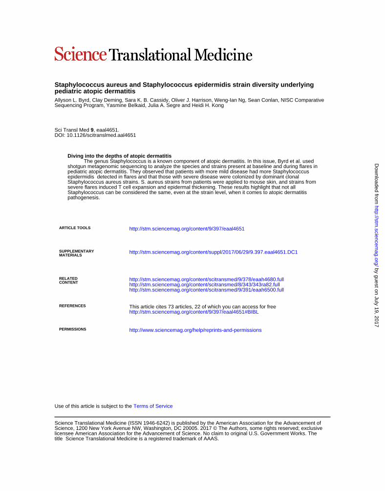
pediatric atopic dermatitis strain diversity underlyingStaphylococcus epidermidis and Staphylococcus aureus
Sequencing Program, Yasmine Belkaid, Julia A. Segre and Heidi H. KongAllyson L. Byrd, Clay Deming, Sara K. B. Cassidy, Oliver J. Harrison, Weng-Ian Ng, Sean Conlan, NISC Comparative
DOI: 10.1126/scitranslmed.aal4651, eaal4651.9Sci Transl Med
pathogenesis. can be considered the same, even at the strain level, when it comes to atopic dermatitisStaphylococcus
severe flares induced T cell expansion and epidermal thickening. These results highlight that not all strains from patients were applied to mouse skin, and strains fromS. aureus strains. Staphylococcus aureus
detected in flares and that those with severe disease were colonized by dominant clonal epidermidisStaphylococcuspediatric atopic dermatitis. They observed that patients with more mild disease had more
shotgun metagenomic sequencing to analyze the species and strains present at baseline and during flares in . usedet al is a known component of atopic dermatitis. In this issue, Byrd StaphylococcusThe genus
Diving into the depths of atopic dermatitis
ARTICLE TOOLS http://stm.sciencemag.org/content/9/397/eaal4651
MATERIALSSUPPLEMENTARY http://stm.sciencemag.org/content/suppl/2017/06/29/9.397.eaal4651.DC1
CONTENTRELATED
http://stm.sciencemag.org/content/scitransmed/9/391/eaah6500.fullhttp://stm.sciencemag.org/content/scitransmed/8/343/343ra82.fullhttp://stm.sciencemag.org/content/scitransmed/9/378/eaah4680.full
REFERENCES
http://stm.sciencemag.org/content/9/397/eaal4651#BIBLThis article cites 73 articles, 22 of which you can access for free
PERMISSIONS http://www.sciencemag.org/help/reprints-and-permissions
Terms of ServiceUse of this article is subject to the
is a registered trademark of AAAS.Science Translational Medicinetitle licensee American Association for the Advancement of Science. No claim to original U.S. Government Works. TheScience, 1200 New York Avenue NW, Washington, DC 20005. 2017 © The Authors, some rights reserved; exclusive
(ISSN 1946-6242) is published by the American Association for the Advancement ofScience Translational Medicine
by guest on July 19, 2017http://stm
.sciencemag.org/
Dow
nloaded from