spectrophotometric estimation of nucleic acid of plant leaves · spectrophotometric estimation of...
TRANSCRIPT

Spectrophotometric Estimation of Nucleic Acidof Plant Leaves 1
R. H. Nieman & L. L. PoulsenU. S. Salinity Laboratory, Riverside, California
In the course of studies on leaf growth, we havefrequently been faced with the problem of estimatingthe nucleic acid content of very young leaves wherethe amounts, particularly of DNA, were so small asto make phosphorus or pentose analysis unreliable.The sensitivity of a spectrophotometric procedurenmakes it the method of choice for such small samples,if sufficiently pure nucleic acid can be isolated.Isolation procedures successfully employed with ani-mal tissue or even plant root tips were not directlyapplicable to leaves, chiefly because of the largeamounts of interfering substances present and be-cause of difficulties in achieving complete extractionof nucleic acid. Of several procedures that we havetried. the nmost satisfactory one was based on theSchmidt and Thannhauser procedure (12). Theprelinminary extraction of the leaf was patterned afterthe procedures of Ogur and Rosen (10) and Mark-ham (8). The extraction of nucleic acid by alkalifollowed the suggestions of Davi(dson and Smellie(2). Spectrophotometric estimates of nucleic acidwere compared with phosphate analysis and with thediphenylamine procedure for DNA.
MaterialsPlant Material: Leaves were obtained from bush
bean (Phaseolus vtlgaris L.), cotton (Gossypifumhirstitum L.), grape (Vitis zinifera L.), radish (Ra-phanus sativus L.), spinach (Spinacea oleracea L.),strawberry (Fragaria ananasse Baily), and sunflower(Helianthus annus L.). Bean plants were grown onnutrient solution cultures (9) in a control room withartificial light (1500 ft-c from fluorescent plus tung-sten lamps). The plants were on a 12-hour light-and-dark cycle (30 C in the light, 21 C in the dark).Both cotton and grape were grown out of doors:cotton in soil, grape in nutrient-irrigated sand beds(4). Radish seedlings were grown in flats of ver-miculite moistened with de-ionized water and kept ina control room (1,000 ft-c constant light, 25 C).Cotyledonary leaves were harvested from 5-day oldseedlings. Spinach was obtained at the local market.Strawberries were grown on nutrient-irrigated gravelcultures in the greenhouse (6). Sunflowers wvere
Received June 19, 1962.
31
grown in soil cultures in the greenhouse.Reagents: Yeast RNA and sperm DNA were
obtained froni Nutritional Biochemicals Corp. Di-phenvlamine was recrystallized from 70 % ethanol.Other chemiicals were of reagent grade where pos-sible.
General MethodsPhosphate was determined by the Fiske-Subba-
Row procedure (5). The determination of DNAwith the diphenylamine reagent was by the procedureof Dische (3). All spectrophotometric measure-ments were made with the Beckman DU spectro-photonmeter and with quartz cuvettes having a lightpath of 1 cIml.
Chloroplasts were isolated (in the cold) fromspinach leaves and radish cotyledonary leaves bygrindinlg in a mortar and pestle with 1 ml of Jagen-dorf's sucrose medium (7) per gram of fresh leaf.The homogenate was squeezed through cheese clothand filtered through S and S No. 588 filter paper.The filtrate was centrifuged at 0 C. The fractionsedimenting at 120 X g (2 min) was discarded.That sedimenting at 755 X g (15 min) equals theunwaslhed chloroplasts. The plastids were washedby resuspending in the grinding medium and centri-fuging at 755 X g. Chloroplasts. suspended inJagendorf's meedium, were counted in a hemocyto-meter.
ProcedurePreliminary Extraction of Leaves: The pro-
cedure outlined below is for samples of 1 gram freshweight. The volumes of reagents used are reducedproportionately for smaller samples. The fewnmod-ifications employed in handling larger saniples areindicated in a later section.
The frozen sample was homogenized in a chilled,Potter-Elvehjenm homogenizer with 10 ml of cold(-10 C) 95 % ethanol. The homogenate was rinsedinto a centrifuge tube with another 10 ml of coldethanol and centrifuged. Unless otherwise noted,centrifuging was done at room temperature. Thesupernatant was discarded. The sediment was ex-tracted successively, by suspending and centrifuging,with 20 nml of each of the reagents indicated below.The supernataint was discarded in each case.
www.plantphysiol.orgon January 21, 2020 - Published by Downloaded from Copyright © 1963 American Society of Plant Biologists. All rights reserved.
PLANT PHYSIOLOGY
1. 95 % ethanol at room temiperature, once. 2.50% ethanol adjusted to a pH of 4.5 with glacialacetic acid, room temperature, twice. 3. Chilled(4 C) 0.2 N perchloric acid (PCA), twice. Thesuspension was held at 4 C for 15 minutes beforecentrifuging. 4. 95 % ethanol at room temperature,once. 5. Boiling absolute ethanol-ether (3: 1) for3 minutes, twice. 6. Ether at room temi)erature,once.
Nucleic Acid Extraction: The leaf powdler wassuspended with 5 ml of 0.3 N NaOH an(d held at 30 Cfor 18 hours. The se(liment was removed by centri-fuging andl washed once with 5 ml of 0.3 N NaOH.The extract and wash were combined and ma(le to10 ml with 0.3 N NaOH. An 8-ml aliquot. reserving2 ml for protein determination, was acidified to pH1 with 15 % PCA, held at 4 C for 40 minutes, andthen centrifuged. The supernatant equals the RNAfraction. The DNA-protein sediment was resuspend-ed with 2 ml of water followed by the addition of 2ml of 1 N PCA. The suspension was held at 4 Cfor 20 minutes, then centrifuged, and the supernatantadded to the RNA fraction. This resuspensionprocess was repeated until the optical density (OD)of the supernatant indicated the absence of RNAnucleotides. Usually, their concentration was negli-gible in the supernatant from the second resuspension,and this was discardedl. The final RNA extract was
made to the nearest \\lole volume with water.The DNA-protein se(dimiient was suspended with
3 ml of 0.5N PCA, heated at 70 C for 15 minutes,an(l centrifuged at 2 C. The superniatant equals theDNA fraction. The protein sedimiienlt w\\as washedwith 2 ml of 0.5 N PCA at 2 C and the wash addedto the DNA fraction. The final DNA extract was
made to 5 ml with 0.5 N PCA.Mlodificationsg for Large Samples: Leaves with
a fresh weight of more than 1 gram were first im-mersed in 20 volumes of boiling 95 ethanol for2 minutes. They were allowed to drain at room
temperature and then ground in a cold, glass mortar.The hot ethanol extract, which contained some sus-
pended solids, was chilled in an ice bath and addedto the mortar as the grinding progressedl. The ho-mogenate was centrifuged and the supernatant dis-carcled. Thereafter, the extraction procedure was
the same as for the small samples but with propor-
tionately larger volumes of reagents. The fractiona-tion of the nucleic acids was carried out with a 10-ml aliquot of the alkaline extract.
Nucleic Acid Estimation: The OD of the nucleicacid extracts was routinely measured at 250. 260,265, 280, and 310 m,u. The DNA extract was meas-ured directly with 0.5 N PCA as a blank. An aliquotof the RNA extract was first diluted (1 + 5) witlwater (final pH in the range of 1-2) and the ODmeasured with water as a blank. (The OD of a re-
agent blank for the diluted RNA extract was negli-gible.) The OD at 260 m,u, divided by the appropri-ate factor, gave the concentration of nucleic acidI in
the cuvette in the units, Ag nucleic acid phosphortusper ml.
Both the yeast RNA and sperm DNA employedas standards contained a small amount of free phos-phate. This phosphorus was subtracted from thetotal to give the nucleic acidl phosphorus. Samplesof the standards were dissolved in 0.3 N NaOH andtreate(l like the unknowns. The OD at 260 m,u ofa solution of hydrolyzed nucleic acid containing 1 ,gof phosphorus per ml was: RNA (in the presenceof 0.04 or 0.2 N PCA) - 0.346, DNA (in the pres-ence of 0.5 N PCA) - 0.290. These values agreewith others in the literature for hvdrolyzed nucleicaci(d in an acid medium (10, 11).
Discussion & EvaluationOf the compounds present in leaves that could
interfere w\ith a spectrophotometric estimation ofnucleic acid, the phenolics are, perhaps, the mostimportant. Their concentration is high in mostleaves, and they absorb strongly in the ultraviolet.The preliminary extraction (lescribed effectively re-nmoved these compounds from leaves of bean, grape,ra(lislh, spinach, and sunflower. Leaves of cotton andstrawberry, on the other hand, required as many aseight extractions with 95 (4 ethanol. The extrac-tion wvas repeated until the supernatant remainedcolorless on addition of a few drops of 1 N NaOH.Some comiipound (s) resembling chlorogenic acid werenot extracted by ethanol until after the extractionwith 0.2 N PCA. Leaves that resist ethanol extrac-tion may yield more readily to other solvents such asthe methanol-formic acidl system recommended bySmillie and Krotkov (14).
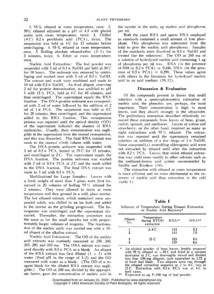
The extraction witlh ethanol at roomii temperatureis more efficient and no miiore detrimental to the re-coverv of nucleic acid than extr-action in the cold(table I).
Table IInfluenice of Temperature During Etlhaniol Extraction
on Nucleic Acid Recovery*
Aliquot TemperatureN. during ETOH RNA** DNA\**extraction
1 2-4C 114 8.22 115 7.9
3 25 C 123 8.64 119 8.6
* An alcohol powder of bean leaves, freshly preparedwith 95 % ethanol at -10 C and dried in a vacuumdesiccator at 2C, -%\as thoroughly mixed and dividedinto four 150-mg aliquots, each equivalent to 1.72 gof fresh leaf blade. Two aliquots were run throughthe ethanol extraction sequence at 2 to 4C, twvo at25 C. Extraction with 0.2 N PCA was at 4 C inboth cases.
** Expressed as ,g P/100 mg of leaf pow-der.
32
www.plantphysiol.orgon January 21, 2020 - Published by Downloaded from Copyright © 1963 American Society of Plant Biologists. All rights reserved.
NIEMAN & POULSEN-ESTIMATION OF NUCLEIC ACID OF LEAVES
An unidentified brown pigment developed duringthe extraction with alkali if the leaf residue containedtraces of ethanol. This pigment contaminated boththe RNA and DNA extracts. It was avoided bythe final preliminary extraction with pure ether.With this procedure, the alkaline extract was nearlycolorless.
The hydrolysis of DNA with 0.5 N PCA is a
critical step if DNA is to be estimated spectrophoto-metrically. Prolonged heating or the use of tempera-
tures much above 70 C caused an appreciable con-
tamination of the DNA extract of bean leaves byprotein degradation products. This was indicatedby a shift in the absorption maximum from 265 mytoward a longer wavelength. Heating at 70 C for15 minutes resulted in essentially complete extractionof purine and pyrimidine moities (see later section)without appreciable contamination by protein.
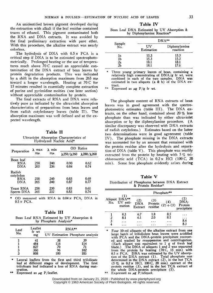
The final extracts of RNA and DNA were rela-tively pure as indicated by the ultraviolet absorptioncharacteristics of preparations from bean leaves andfrom radish cotyledonary leaves (table II). Theabsorption maximum was well defined and at the ex-
pected wavelength.
Table II
Ultraviolet Absorption Characteristics ofHydrolyzed Nucleic Acid*
Preparation A max X min OD Ratiosm,u iA X250/X260 X280/X260
Bean leafRNA 258 240 0.90 0.62DNA 265 236 0.84 0.74
Radishcotyledon
RNA 258 240 0.83 0.69DNA 265 240 0.87 0.77
Yeast RNA 258 230 0.83 0.61Sperm DNA 265 232 0.83 0.74
* (n mpCtsr AX74.1 DA in nAN PCA TX A insLs ~ ~ ~ ~ ~ ~FLaur(0.5 N PCA.
Table IIIBean Leaf RNA Estimated by UV Absorption &
by Phosphate Analysis*
Leaf Leaflet RNA**No. fr wt
mg UV Estimation Phosphate analysis
1 41 38 391 484 116 1141 827 70 713 360 96 1023 808 180 185
* Lateral leaflets from the first and third trifolioateleaf at different stages of development. The firsttrifolioate leaf indicated a loss of RNA during mat-uration.
** Expressed as ug P/leaflet.
Table IVBean Leaf DNA Estimated by UV Absorption &
by Diphenylamine Reaction*
DNA**SampleNo. UV Diphenylamine
Estimation reaction
la 15.3 15.3lb 15.3 15.32a 18.1 18.12b 18.1 17.6
* Three young primary leaves of bean, containing arelatively high concentration of DNA/g fr wt, werecombined in each of the two samples. DNA wasestimated in two aliquots (a & b) of the DNA ex-tract.
** Expressed as ,ug P/g fr wt.
The phosphate content of RNA extracts of beanleaves was in good agreement with the spectro-photometric estimates (table III). The DNA ex-tracts, on the other hand, contained about 20 % lessphosphate than was indicated by either ultravioletabsorption or by the diphenylamine procedure. (Asimilar discrepancy was observed with DNA extractsof radish cotyledons.) Estimates based on the lattertwo determinations were in good agreement (tableIV). The phosphate missing in the DNA extractswas accounted for by an amount that remained withthe protein residue after the hydrolysis and separa-tion of DNA (table V). This phosphate was readilyextracted from the protein by heating with 5 % tri-chloroacetic acid (TCA) in 0.2 N HCI (100 C, 20min). Some free phosphate evidently arises during
Table VDistribution of Phosphorus between DNA Extract
& Protein Residue*
(1) Phosphate**
Aliquot DNA** (2) (3) (4) (5)No. UV esti- DNA rotin NA-
mation EDxtNract rei (2) + (3) ProteinExtractresidue precipitate1 8.3 6.7 1.8 8.52 8.1 6.1 2.0 8.13 ... ... ... ... 8.44 ... ... ... ... 8.3
* Four 10-ml aliquots of the alkaline extract from onelarge batch of trifoliolate bean leaves were acidifiedwith PCA and the DNA-protein precipitate recover-ed and washed by resuspension and centrifugation.(Each aliquot was equivalent to 1 g of fresh leafblade.) The DNA of aliquots 1 and 2 was separatedfrom the protein by heating (70 C, 15 min) with0.5 N PCA. DNA was estimated by the UV absorp-tion of the DNA extract (1). Total phosphate wasdetermined in the DNA extract (2), in the hot TCA(5 % in 0.2N HCI, 100 C, 20 min) extract of theprotein residue (3), and in the hot TCA extract ofthe whole DNA-protein precipitate (5).
** Expressed as ,ug P/aliquot.
33
niNl lIH U.U" N rtz, Ntl IIl
www.plantphysiol.orgon January 21, 2020 - Published by Downloaded from Copyright © 1963 American Society of Plant Biologists. All rights reserved.

PLANT PHYSIOLOGY
the lhydrolysis with 0.5 N PCA, presumably from in-terpurine linkages (1, 13), and is adsorbed by theprotein. (No free phosphate was detected in theDNA extract.)
The extraction of purine and pyrimidine moitiesby PCA wtas essentially complete as indicated bythe agreement between the spectrophotometric esti-mation of DNA in the extract and the phosphateextracted by hot TCA from the DNA-protein pre-cipitate (table V).
The sum of the RNA and DNA. estimated spec-trophotometrically, should equal the total phosphatecontent of the whole alkali extract, if the estimatesare reliable and if the preliminary extraction waseffective in removing non-nucleic acid phosphate.The (lata of table VI show that this was the case withextracts from bean leaves.
Table VIComparison of Total Estimated
Total Phosphate in AlkalinBean Leaves
Sample Nucleic acid*No. RNA DNA
4D-1 126 5.454D-2 121 5.546B-1 139 6.346B-2 136 6.25
suspension were used for counting chloroplasts. TheRNA and DNA extracts had the expected ultraviolet
Table VIIIRNA & DNA of Washed & Unwashed
Spinach Chloroplast Preparations
No. of Nucleic acid/chloroplastwashes RNA* DNA*
038
21186.9
1.61.51.2
* Expressed as ,ug P X 109.
aDso1rptin CnaraCIer1Is1CS. LS11smaesIo 1XNZA. anaDNA per spinach chloroplast, before and after wash-ing, are given in table VIII. The pattern of RNAand DNA disappearance during washing is more sug-
Nucleic Acid with gestive of degradation than removal of an impurity.e Extract of As a matter of fact, RNA disappears rapidly if a
chloroplast preparation is allowed to stand at roomtemperature. DNA is more resistant. Whatever
Total the origin of the chloroplast-associated DNA, it cer-Total phosphate tainly resists removal by repeated washing.132127145142
128124145141
* Expresse(l as Ag P /g fr wvt.
The reproducibility of the procedure is (lemon-strated by the small variation in estimates of RNAand DNA in three replicate samples of radish coty-ledonary- leaves (table VII). The standard errorwas 3.3 °c of the mean for the fresh weight but only1.2 and 1.4 %, of the mean for RNA and DNA, re-spectively.
The procedure is also useful in the analysis ofleaf homogenates and fractions thereof. As one ex-ample, chloroplast preparations of spinach leaves andradish cotyledonary leaves indicate small but fairlyrconstant amounts of both RNA and DNA. Foranalysis, aliquots of a chloroplast suspension wereblown into four volumes of boiling 95 % ethanol.chilled. centrifuged, and then carried througlh theusual extraction procedure. Other aliquots of the
Table VIIRNA and DNA of Radish Cotyledonary Leaves
Sample fr wt RNA* DNA*mg Weight/20 cotyledons**
365 ± 12 95.3 ± 1.1 5.84 + 0.08* Expressed as ,ug P.
** Mean ± standard error for three groups of 20 coty-ledons from 5-day old seedlings.
SummaryA procedure is described for extracting and esti-
mating, spectrophotometrically. the RNA and DNAcontent of leaves and leaf fractions. Estimates ofRNA checked with phosphate analysis. Estimatesof DNA checked with values obtained by the di-phenylamine procedure and with phosphate analysis,when the behavior of phosphate during the hydrolysisof DNA with perchloric acid was ascertained. Somevalues are given for the RNA and DNA content ofbean leaves, radlish cotyledonary leaves, and spinachchloroplasts.
Literature Cited1. COHN, W. E. & E. VOLKIN. 1957. Acid degrada-
tion products of deoxyribonucleic acid. Biochim.Biophys. Acta 24: 359-364.
2. DAVIDSON, J. N. & R. M. S. SMELLIE. 1952. Phos-phorus compounds in the cell. II. Separation byionophoresis on paper of the constituent nucleotidesof ribonucleic acid. Biochem. J. 52: 594-599.
3. DiSCHE, Z. 1955. Color reactions of nucleic acidcomponents. In: The Nucleic Acids, E. Char-gaff & J. N. Davidson, eds. Academic Press,New York. Vol. I, pp. 285-305.
4. EHLIG, C. F. 1960. Effects of salinity on 4 varie-ties of table grapes grown in sand culture. Proc.Am. Soc. Hortic. Sci. 76: 323-331.
5. FiSKE, C. H. & Y. SUBBAROW. 1925. The colori-metric determination of phosphorus. J. Biol.Chem. 66: 375-400.
6. GAUCH, H. G. & C. H. WADLEIGH. 1943. A newtype of intermittently-irrigated sand culture equip-ment. Plant Physiol. 18: 543-547.
34
11 r-%" A-In t-n C+; - C t; c 4-; ti-i n +,a L- --; P T*J A , "A
www.plantphysiol.orgon January 21, 2020 - Published by Downloaded from Copyright © 1963 American Society of Plant Biologists. All rights reserved.
NIEMAN & POULSEN-ESTIMATION OF NUCLEIC ACID OF LEAVES
7. JAGENDORF, A. T. & MARIE SMITH. 1962. Un-coupling phosphorylation in spinach chloroplastsby absence of cations. Plant Physiol. 37: 135-141.
8. MARKHAM, R. 1955. Nucleic acids, their compo-nents, & related compounds. In: Modern Methodsof Plant Analysis, K. Peach & M. V. Tracey, eds.Springer-Verlag, Berlin. Vol. IV, pp. 246-304.
9. NIEMAN, R. H. & L. BERNSTEIN. 1959. Interactiveeffects of gibberellic acid & salinity on the growthof beans. Am. J. Botany 46: 667-670.
10. OGUR, M. & G. ROSEN. 1950. The nucleic acidsof plant tissues. I. The extraction & estimationof dosoxypentose nucleic acid & pentose nucleicacid. Arch. Biochem. 25: 262-276.
11. SCHMIDT, G. 1957. Preparation of ribonucleic acidfrom yeast & animal tissues. In: Methods in
Enzymology, S. P. Colowick & N. 0. Kaplan, eds.Academic Press, New York. Vol. III, pp.687-691.
12. SCHMIDT, G. & S. J. THANNHAUSER. 1945. Amethod for the determination of desoxyribonucleicacid, ribonucleic acid, & phosphoproteins in animaltissues. J. Biol. Chem. 161: 83-89.
13. SHAPIRO, H. S. & E. CHARGAFF. 1960. Studies onthe nucleotide arrangement in deoxyribonucleicadds. IV. Patterns of nucleotide sequence in thedeoxyribonucleic acid of rye germ & its fractions.Biochim. Biophys. Acta 39: 68-82.
14. SMILLIE, R. M. & G. KROTKOV. 1960. The estima-tion of nucleic acids in some algae & higher plants.Can. J. Botany 38: 31-49.
Design of a Rotary Shaking MachineLeonard Machbis
Department of Botany, University of California, Berkeley, California
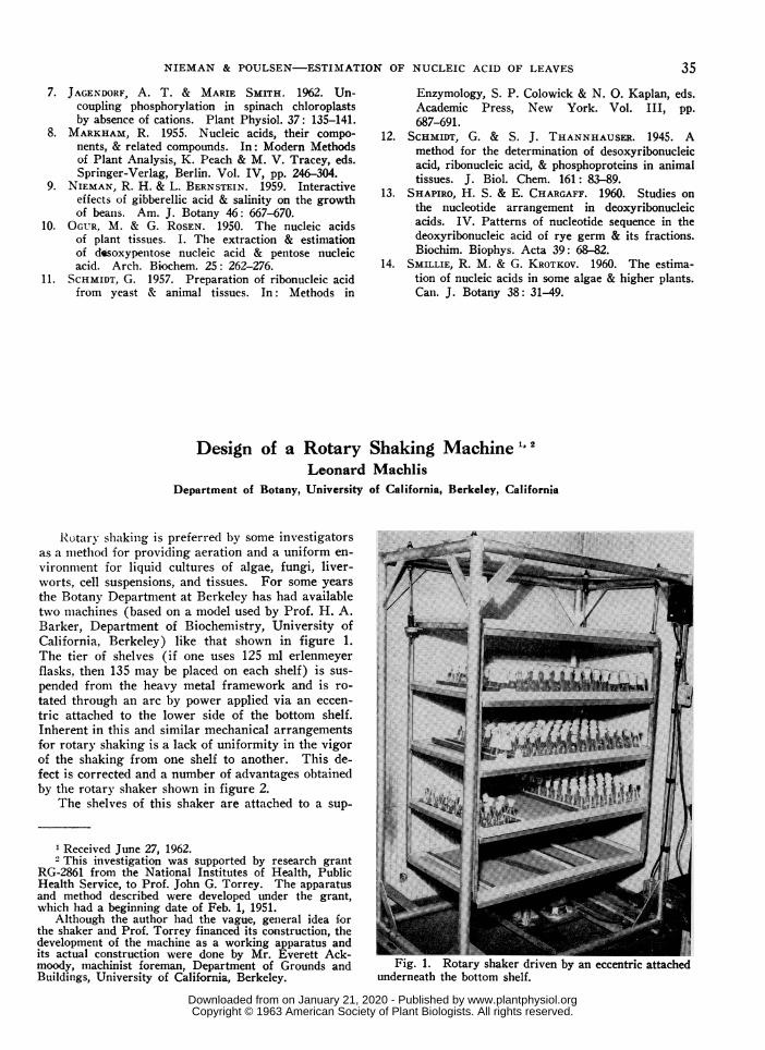
Rotarv shaking is preferred by some investigatorsas a method for providing aeration and a uniform en-vironment for liquid cultures of algae, fungi, liver-worts, cell suspensions, and tissues. For some yearsthe Botany Department at Berkeley has had availabletwo machines (based on a model used by Prof. H. A.Barker, Department of Biochemistry, University ofCalifornia, Berkeley) like that shown in figure 1.The tier of shelves (if one uses 125 ml erlenmeyerflasks, then 135 may be placed on each shelf) is sus-pended from the heavy metal framework and is ro-tated through an arc by power applied via an eccen-tric attached to the lower side of the bottom shelf.Inherent in this and similar mechanical arrangementsfor rotary shaking is a lack of uniformity in the vigorof the shaking from one shelf to another. This de-fect is corrected and a number of advantages obtainedby the rotary shaker shown in figure 2.
The shelves of this shaker are attached to a sup-
1 Received June 27, 1962.2 This investigation was supported by research grant
RG-2861 from the National Institutes of Health, PublicHealth Service, to Prof. John G. Torrey. The apparatusand method described were developed under the grant,which had a beginning date of Feb. 1, 1951.
Although the author had the vague, general idea forthe shaker and Prof. Torrey financed its construction, thedevelopment of the machine as a working apparatus andits actual construction were done by Mr. Everett Ack-moody, machinist foreman, Department of Grounds and Fig. 1. Rotary shaker driven IBuildings, University of California, Berkeley. underneath the bottom shelf.
by an ecceni
35
www.plantphysiol.orgon January 21, 2020 - Published by Downloaded from Copyright © 1963 American Society of Plant Biologists. All rights reserved.