spectral characterization of dna sarah aldosari. dna ‘deoxy ribonucleic acid’
TRANSCRIPT

SPECTRAL CHARACTERIZATION OF DNA
Sarah AlDosari

DNA ‘DEOXY RIBONUCLEIC ACID’

DEOXY RIBONUCLEIC ACID (DNA)
• DNA is made of 2 polynucleotide chains which run in opposite direction.”antiparallel ”
• DNA has a double helical structure.
• Each polynucleotide chain of DNA consists of monomer units.
• A monomer unit consists of 3 main components that are:
1. A sugar, 2. a phosphate, 3. a nitrogenous base.

NUCLEOTIDE
Monomer

DNA STRUCTURE
1. Deoxyribose sugar:
• Is a monosaccharide 5-Carbon Sugar, Its name indicates that it is a deoxy sugar, meaning that
[ it is derived from the sugar ribose by loss of an oxygen atom ].
2. Phosphate Group:
• The sugars are joined together by phosphate groups that form phosphodiester bonds between the third and fifth carbon atoms of adjacent sugar rings.

DNA STRUCTURE3. Nitrogenous bases:
• is a nitrogen-containing organic molecule having the chemical properties of a base
• They are classified as the derivatives of two parent compounds,
1. Purine. • [ Adenine, Guanine ]
2. Pyrimidine. • [ Cytosine, Thymine ]
DNA STRUCTURE
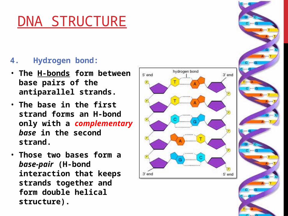
4. Hydrogen bond:
• The H-bonds form between base pairs of the antiparallel strands.
• The base in the first strand forms an H-bond only with a complementary base in the second strand.
• Those two bases form a base-pair (H-bond interaction that keeps strands together and form double helical structure).

DNA STRUCTURE
• The hydrophobic bases are inside the double helix of DNA, give the hydrophobic effect to stabilizes the double helix.
• while sugars and phosphates are located outside of the double helical structure.

OPTICAL DENSITY OF DNA
• Nucleic acid would be expected to have maximum absorbance at 260.
• It absorbs at this wavelength because of the nitrogenous bases (A, G, C and T) of DNA.
• In a spectrophotometer, a sample is exposed to ultraviolet light at 260 nm, and a photo-detector measures the light that passes through the sample.
Wave length (nm)
Abs
orb
ance
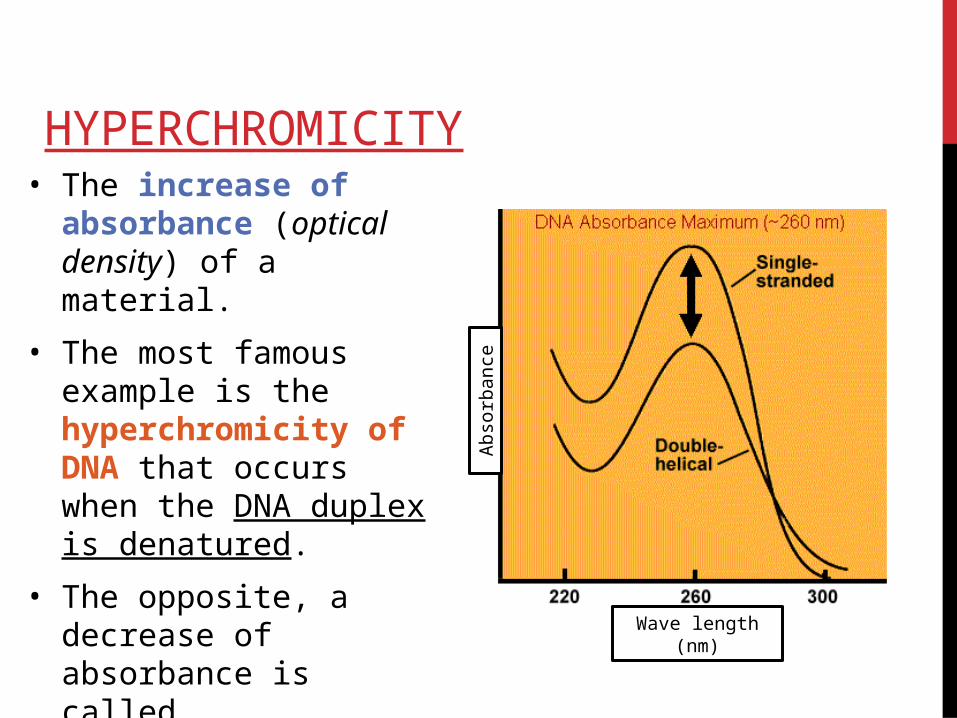
HYPERCHROMICITY• The increase of
absorbance (optical density) of a material.
• The most famous example is the hyperchromicity of DNA that occurs when the DNA duplex is denatured.
• The opposite, a decrease of absorbance is called hypochromicity.
Wave length (nm)
Abs
orb
ance

DENATURATION OF DNA
• Many different substances or environmental conditions can denature DNA, such as:• strong acids,
organic solvent• heating
EXPERIMENT OF DAY
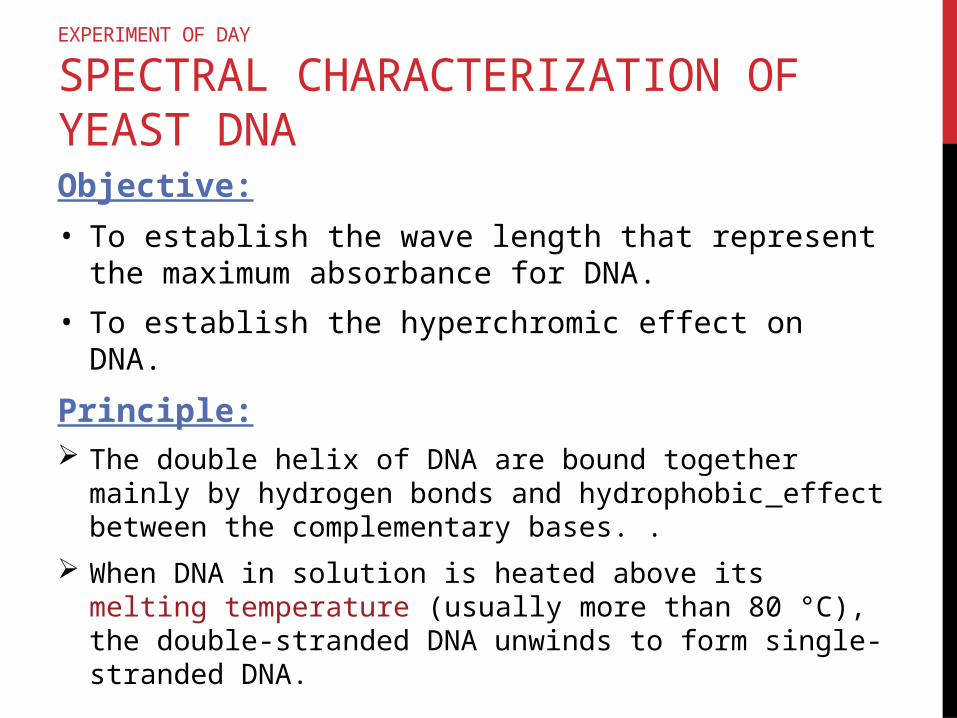
SPECTRAL CHARACTERIZATION OF YEAST DNAObjective:
• To establish the wave length that represent the maximum absorbance for DNA.
• To establish the hyperchromic effect on DNA.
Principle: The double helix of DNA are bound together mainly by
hydrogen bonds and hydrophobic effect between the complementary bases. .
When DNA in solution is heated above its melting temperature (usually more than 80 °C), the double-stranded DNA unwinds to form single-stranded DNA.

Principle: In single stranded DNA the bases become unstacked and
can thus absorb more light.
In their native state, the bases of DNA absorb light in the 260-nm wavelength region.
When the bases become unstacked, the wavelength of maximum absorbance does not change, but the amount absorbed increases by 30-40%.
a double strand DNA dissociating to single strands produces a sharp cooperative transition.
EXPERIMENT OF DAY
SPECTRAL CHARACTERIZATION OF YEAST DNA

Materials:• DNA concentrated sample( extracted from yeast).
• 1X saline solution ( NaCl with Tri Sodium Citrate).
• Quartz Cuvtte.
• Spectrophotometer.
EXPERIMENT OF DAY
SPECTRAL CHARACTERIZATION OF YEAST DNA

Method:• Set and lable 6 test tube : D1, D2, D3,D4,D5,D6
1. In D1 pipette 0.5 ml of isolated DNA (extracted from Yeast) and add to it 4.5ml of 1X saline-citrate. Mix it very will.
• Measure the absorbance of D1 at 260nm if it is > 3 :
2. In D2 pipette 0.5 ml of D1 ,add to it 4.5ml of 1X saline-citrate. Mix it very will.
• Measure the absorbance of D2 (if the absorbance is greater than 1,dilute the solution until you obtain A260 of 1 or slightly less).
EXPERIMENT OF DAY
SPECTRAL CHARACTERIZATION OF YEAST DNA

Method:• When the absorbance of solution (A260≈1.0) is obtained
read the absorbance of the solution at the following wave lengths:
(240,245,250,255,260,265,270,275,280)
• using 1X saline as a blank.
EXPERIMENT OF DAY
SPECTRAL CHARACTERIZATION OF YEAST DNA

Method:• Now take the dilution tube which give an
absorbance=1 ,cover the tube and put it in boiling water bath for 15 min
• Immediately measure the absorbance at the following wave lengths:
(240,245,250,255,260,265,270,275,280)
• using 1X saline as a blank.
EXPERIMENT OF DAY
SPECTRAL CHARACTERIZATION OF YEAST DNA

Results: Plot The absorption spectra of
the native DNA solution and the denatured DNA against wave lengths.
Record Your result and write your comment in the discussion.
EXPERIMENT OF DAY
SPECTRAL CHARACTERIZATION OF YEAST DNA
Absorbance of heated
DNA
Absorbance of isolated
DNA
Wave length (nm)
240
245
250
255
260
265
270
275
280