smoothened adopts multiple active and inactive ...chuang/pdf/plosone44e5182.pdf · smoothened...
TRANSCRIPT
Smoothened Adopts Multiple Active and InactiveConformations Capable of Trafficking to the PrimaryCiliumChristopher W. Wilson, Miao-Hsueh Chen, Pao-Tien Chuang*
Cardiovascular Research Institute, University of California San Francisco, San Francisco, California, United States of America
Abstract
Activation of Hedgehog (Hh) signaling requires the transmembrane protein Smoothened (Smo), a member of the G-proteincoupled receptor superfamily. In mammals, Smo translocates to the primary cilium upon binding of Hh ligands to theirreceptor, Patched (Ptch1), but it is unclear if ciliary trafficking of Smo is sufficient for pathway activation. Here, wedemonstrate that cyclopamine and jervine, two structurally related inhibitors of Smo, force ciliary translocation of Smo.Treatment with SANT-1, an unrelated Smo antagonist, abrogates cyclopamine- and jervine-mediated Smo translocation.Further, activation of protein kinase A, either directly or through activation of Gas, causes Smo to translocate to a proximalregion of the primary cilium. We propose that Smo adopts multiple inactive and active conformations, which influence itslocalization and trafficking on the primary cilium.
Citation: Wilson CW, Chen M-H, Chuang P-T (2009) Smoothened Adopts Multiple Active and Inactive Conformations Capable of Trafficking to the PrimaryCilium. PLoS ONE 4(4): e5182. doi:10.1371/journal.pone.0005182
Editor: Rafael Linden, Universidade Federal do Rio de Janeiro (UFRJ), Instituto de Biofı́sica da UFRJ, Brazil
Received December 25, 2008; Accepted March 15, 2009; Published April 13, 2009
Copyright: � 2009 Wilson et al. This is an open-access article distributed under the terms of the Creative Commons Attribution License, which permitsunrestricted use, distribution, and reproduction in any medium, provided the original author and source are credited.
Funding: National Institutes of Health (NIH) and American Lung Association (ALA). The funders had no role in study design, data collection and analysis, decisionto publish, or preparation of the manuscript.
Competing Interests: The authors have declared that no competing interests exist.
* E-mail: [email protected]
Introduction
The Hedgehog (Hh) signal transduction cascade is critical for
many aspects of embryonic development, and aberrant regulation
of the pathway results in a wide variety of congenital defects and
cancers [1]. Activation of the pathway is initiated by binding of Hh
ligand to the core receptor Patched (Ptch1 in mammals), a twelve-
pass membrane protein with distant similarity to the resistance-
nodulation division (RND) of bacterial transporters [2]. The
interaction of Hh with Ptch1 relieves inhibition of Smoothened
(Smo), a seven-pass transmembrane protein with structural
similarity to G-protein coupled receptors (GPCRs), via unknown
mechanisms. Once released from Ptch1-mediated inhibition, Smo
communicates the status of pathway activation to the Ci/Gli
transcription factors, which commence transcription of Hh target
genes. This is achieved through the production of Gli activators,
derived from full-length Gli proteins, and a concomitant reduction
in levels of Gli repressors resulting from limited proteolysis of full-
length Gli proteins [3]. The mechanistic details of Smo activation
are unclear and may differ between invertebrates and vertebrates
[4,5]. In addition, the means by which Smo relays the status of
pathway activation to the Gli proteins do not appear to be
evolutionarily conserved [4], particularly the cellular microenvi-
ronment in which Smo is activated and the downstream
components it interacts with. Nevertheless, two general features
of Smo activation that are shared between species are a change in
its subcellular distribution after relief of Ptch1 inhibition [6,7], and
conformational changes in the extracellular and cytosolic domains
[8]. A conserved series of arginine (Arg) residues in the C-tail of
both fly and mammalian Smo plays a critical role in modulation of
conformation. How these events lead to Smo activation remains a
central unresolved issue in understanding the molecular mecha-
nisms of Hh signaling.
In mammals, the primary cilium is essential for proper
interpretation of the Hh signal. Cilia contain a long microtubular
axoneme, extending from the basal body and surrounded by an
external membrane that is continuous with the plasma membrane.
Assembly and maintenance of the primary cilium are mediated by
the process called intraflagellar transport (IFT), which involves
bidirectional movement of IFT particles powered by anterograde
kinesin (Kif3a, b and c) and retrograde dynein motors [9,10].
Mutations that abolish the biogenesis or function of the primary
cilium lead to defective Hh signaling [11]. Further, the production
of both Gli activators and repressors is affected in the absence of
the cilium, leading to a loss of Gli repressive activity without a
corresponding gain of transcriptional activation [10,12,13]. Smo
localization to the primary cilium is associated with Hh pathway
activation, and other components of the pathway, including Gli
proteins and Ptch1, are also found in this organelle [14,15].
Mutations in Smo that confer constitutive Hh pathway activation
(SmoA1) promote ciliary localization of Smo in the absence of Hh
stimulation; conversely mutations that abolish ciliary localization
(CLDSmo) appear to render the protein incapable of activating
the pathway in the presence of the primary cilium [7]. Ptch1
localizes to the cilium in the absence of Hh ligand, and traffics off
the cilium after Hh binding, allowing movement of Smo to the
axoneme [15]. It has been proposed that the cilium acts as a
scaffold or provides a specialized microenvironment for relaying
the Hh signal [10,16]. This led to a model in which Smo adopts an
active conformation upon localizing to the primary cilium, which
PLoS ONE | www.plosone.org 1 April 2009 | Volume 4 | Issue 4 | e5182

is capable of coupling to yet-to-be identified downstream
components, thus resulting in stimulation of Gli activators,
reduction in Gli repressors, and induction of target gene
expression. Here, we show that a distinct class of Smo antagonists
which suppress Smo-mediated pathway activation also unexpect-
edly stimulate translocation of Smo to the primary cilium. In
addition, modulation of protein kinase A (PKA) activity by
chemical means causes a partial accumulation of Smo on a
proximal segment of the primary cilium. We propose that multiple
conformational changes of Smo are required for ciliary translo-
cation and subsequent pathway activation.
Results and Discussion
Smo localizes to the cilium upon both activation andrepression of the Hh pathway
We generated antibodies against the C-terminal domain of
mouse Smo [17] to examine the ciliary localization of endogenous
Smo in response to known Hh pathway agonists and antagonists.
When exposed to conditioned media (CM) collected from cells
expressing the N-terminal signaling fragment of Sonic hedgehog
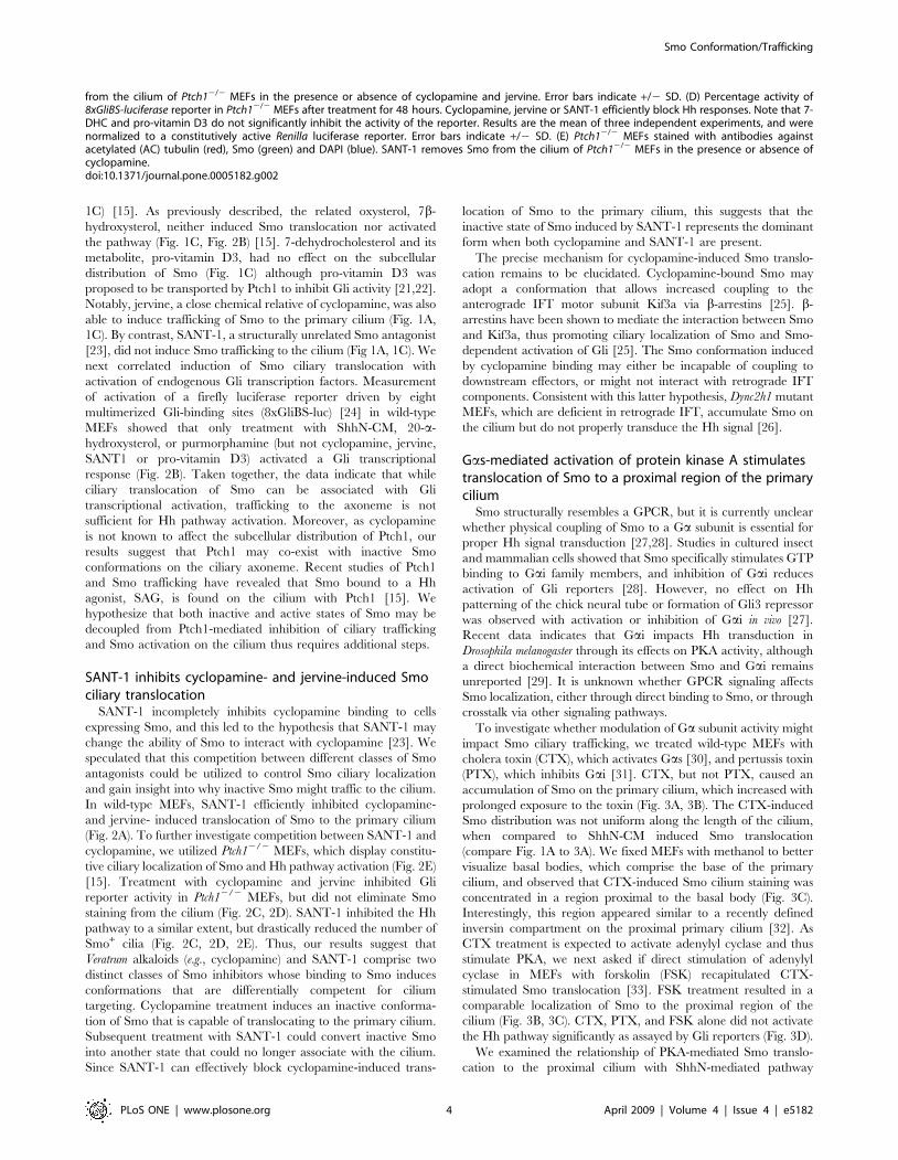
(ShhN), wild-type mouse embryonic fibroblasts (MEFs) accumu-
lated Smo in primary cilia, and nearly 100% of cilia were positive
for Smo (Smo+) after 6 hours of treatment (Fig. 1A, 1B). In
agreement with previously published results [7], a reduced number
of cilia were Smo+ after brief (1 hour) treatment with cyclopamine,
a teratogen derived from the Veratrum genus of plants and a well-
defined Smo antagonist known to bind to the heptahelical bundle
of Smo (Fig. 1A, 1B) [18]. Surprisingly, prolonged treatment of
MEFs with cyclopamine resulted in a significant number of Smo+
cilia, with roughly 70% displaying strong Smo signal along the
entire length of the cilium after 24 hours of exposure to
cyclopamine (Fig. 1A, 1B). We speculate that the increased time
of cyclopamine treatment required to generate a high number of
Smo+ cilia underlies the difference between our observation and
prior reports. This finding also provided a unique opportunity to
examine the relationship between ciliary localization of Smo and
Hh pathway activation.
This unexpected result prompted us to screen a number of
previously reported modulators of Smo activity for their ability to
induce or prevent Smo translocation to the primary cilium. The
Smo agonists 20-a-hydroxysterol [19] and purmorphamine [20]
caused a significant translocation of Smo to the cilium,
comparable to that seen after treatment with ShhN-CM (Fig. 1A,
Figure 1. Hh pathway agonists and antagonists stimulate Smotranslocation to the primary cilium. (A) Wild-type mouseembryonic fibroblasts (MEFs) stained with antibodies against endoge-nous Smo (green), acetylated (AC) tubulin (red), and DAPI (blue). MEFstreated with ShhN-conditioned media (ShhN-CM), purmorphamine,cyclopamine, and jervine display Smo staining along the entire lengthof the cilium. Note that fixation with paraformaldehyde producesartifactual nuclear staining with the Smo antibody; this nuclear stainingis not visible after fixation in methanol (See Fig. 3C). (B) Quantificationof the percentage of Smo-positive (Smo+) cilia in MEFs after treatmentwith ShhN CM and cyclopamine for the indicated times. Cyclopamineinduces Smo translocation to a significant number of cilia after 6 hoursof treatment, although this is less than that induced by ShhN CM. Allquantification of cilia staining in this and subsequent figures representsat least two separate experiments with a minimum of 100 cilia scoredper time point and condition. Error bars indicate +/2 standarddeviation (SD). (C) Quantification of Smo+ cilia in MEFs after treatmentwith indicated compounds for 24 hours. 20-a-hydroxysterol (20a-OHC),purmorphamine, cyclopamine, and jervine induce Smo translocation tothe cilium. Error bars indicate +/2 SD.doi:10.1371/journal.pone.0005182.g001
Smo Conformation/Trafficking
PLoS ONE | www.plosone.org 2 April 2009 | Volume 4 | Issue 4 | e5182

Figure 2. SANT-1 inhibits cyclopamine-and jervine- induced Smo translocation to the primary cilium. (A) Quantification of Smo+ cilia inwild-type MEFs after treatment with indicated compounds for 24 hours. SANT-1 inhibits cyclopamine- and jervine-induced ciliary translocation ofSmo. Error bars indicate +/2 SD. (B) Fold activation of 8xGliBS-luciferase Hh reporter in wild-type MEFs after agonist and antagonist treatment for48 hours. Treatment with ShhN-CM, 20-a-hydroxysterol (20a-OHC), or purmorphamine [but not 7b-hydroxysterol (7b-OHC), pro-vitamin D3, 7-dehydrocholesterol (7-DHC), cyclopamine, jervine or SANT-1] activated a Gli transcriptional response. Results are representative of three experimentsin two wild-type MEF lines, and were normalized to a constitutively active Renilla luciferase reporter. Error bars indicate +/2 SD. (C) Quantification ofSmo+ cilia in Ptch12/2 MEFs after treatment for 24 hours. Cyclopamine and jervine do not disrupt constitutive Smo localization. SANT-1 removes Smo
Smo Conformation/Trafficking
PLoS ONE | www.plosone.org 3 April 2009 | Volume 4 | Issue 4 | e5182
1C) [15]. As previously described, the related oxysterol, 7b-
hydroxysterol, neither induced Smo translocation nor activated
the pathway (Fig. 1C, Fig. 2B) [15]. 7-dehydrocholesterol and its
metabolite, pro-vitamin D3, had no effect on the subcellular
distribution of Smo (Fig. 1C) although pro-vitamin D3 was
proposed to be transported by Ptch1 to inhibit Gli activity [21,22].
Notably, jervine, a close chemical relative of cyclopamine, was also
able to induce trafficking of Smo to the primary cilium (Fig. 1A,
1C). By contrast, SANT-1, a structurally unrelated Smo antagonist
[23], did not induce Smo trafficking to the cilium (Fig 1A, 1C). We
next correlated induction of Smo ciliary translocation with
activation of endogenous Gli transcription factors. Measurement
of activation of a firefly luciferase reporter driven by eight
multimerized Gli-binding sites (8xGliBS-luc) [24] in wild-type
MEFs showed that only treatment with ShhN-CM, 20-a-
hydroxysterol, or purmorphamine (but not cyclopamine, jervine,
SANT1 or pro-vitamin D3) activated a Gli transcriptional
response (Fig. 2B). Taken together, the data indicate that while
ciliary translocation of Smo can be associated with Gli
transcriptional activation, trafficking to the axoneme is not
sufficient for Hh pathway activation. Moreover, as cyclopamine
is not known to affect the subcellular distribution of Ptch1, our
results suggest that Ptch1 may co-exist with inactive Smo
conformations on the ciliary axoneme. Recent studies of Ptch1
and Smo trafficking have revealed that Smo bound to a Hh
agonist, SAG, is found on the cilium with Ptch1 [15]. We
hypothesize that both inactive and active states of Smo may be
decoupled from Ptch1-mediated inhibition of ciliary trafficking
and Smo activation on the cilium thus requires additional steps.
SANT-1 inhibits cyclopamine- and jervine-induced Smociliary translocation
SANT-1 incompletely inhibits cyclopamine binding to cells
expressing Smo, and this led to the hypothesis that SANT-1 may
change the ability of Smo to interact with cyclopamine [23]. We
speculated that this competition between different classes of Smo
antagonists could be utilized to control Smo ciliary localization
and gain insight into why inactive Smo might traffic to the cilium.
In wild-type MEFs, SANT-1 efficiently inhibited cyclopamine-
and jervine- induced translocation of Smo to the primary cilium
(Fig. 2A). To further investigate competition between SANT-1 and
cyclopamine, we utilized Ptch12/2 MEFs, which display constitu-
tive ciliary localization of Smo and Hh pathway activation (Fig. 2E)
[15]. Treatment with cyclopamine and jervine inhibited Gli
reporter activity in Ptch12/2 MEFs, but did not eliminate Smo
staining from the cilium (Fig. 2C, 2D). SANT-1 inhibited the Hh
pathway to a similar extent, but drastically reduced the number of
Smo+ cilia (Fig. 2C, 2D, 2E). Thus, our results suggest that
Veratrum alkaloids (e.g., cyclopamine) and SANT-1 comprise two
distinct classes of Smo inhibitors whose binding to Smo induces
conformations that are differentially competent for cilium
targeting. Cyclopamine treatment induces an inactive conforma-
tion of Smo that is capable of translocating to the primary cilium.
Subsequent treatment with SANT-1 could convert inactive Smo
into another state that could no longer associate with the cilium.
Since SANT-1 can effectively block cyclopamine-induced trans-
location of Smo to the primary cilium, this suggests that the
inactive state of Smo induced by SANT-1 represents the dominant
form when both cyclopamine and SANT-1 are present.
The precise mechanism for cyclopamine-induced Smo translo-
cation remains to be elucidated. Cyclopamine-bound Smo may
adopt a conformation that allows increased coupling to the
anterograde IFT motor subunit Kif3a via b-arrestins [25]. b-
arrestins have been shown to mediate the interaction between Smo
and Kif3a, thus promoting ciliary localization of Smo and Smo-
dependent activation of Gli [25]. The Smo conformation induced
by cyclopamine binding may either be incapable of coupling to
downstream effectors, or might not interact with retrograde IFT
components. Consistent with this latter hypothesis, Dync2h1 mutant
MEFs, which are deficient in retrograde IFT, accumulate Smo on
the cilium but do not properly transduce the Hh signal [26].
Gas-mediated activation of protein kinase A stimulatestranslocation of Smo to a proximal region of the primarycilium
Smo structurally resembles a GPCR, but it is currently unclear
whether physical coupling of Smo to a Ga subunit is essential for
proper Hh signal transduction [27,28]. Studies in cultured insect
and mammalian cells showed that Smo specifically stimulates GTP
binding to Gai family members, and inhibition of Gai reduces
activation of Gli reporters [28]. However, no effect on Hh
patterning of the chick neural tube or formation of Gli3 repressor
was observed with activation or inhibition of Gai in vivo [27].
Recent data indicates that Gai impacts Hh transduction in
Drosophila melanogaster through its effects on PKA activity, although
a direct biochemical interaction between Smo and Gai remains
unreported [29]. It is unknown whether GPCR signaling affects
Smo localization, either through direct binding to Smo, or through
crosstalk via other signaling pathways.
To investigate whether modulation of Ga subunit activity might
impact Smo ciliary trafficking, we treated wild-type MEFs with
cholera toxin (CTX), which activates Gas [30], and pertussis toxin
(PTX), which inhibits Gai [31]. CTX, but not PTX, caused an
accumulation of Smo on the primary cilium, which increased with
prolonged exposure to the toxin (Fig. 3A, 3B). The CTX-induced
Smo distribution was not uniform along the length of the cilium,
when compared to ShhN-CM induced Smo translocation
(compare Fig. 1A to 3A). We fixed MEFs with methanol to better
visualize basal bodies, which comprise the base of the primary
cilium, and observed that CTX-induced Smo cilium staining was
concentrated in a region proximal to the basal body (Fig. 3C).
Interestingly, this region appeared similar to a recently defined
inversin compartment on the proximal primary cilium [32]. As
CTX treatment is expected to activate adenylyl cyclase and thus
stimulate PKA, we next asked if direct stimulation of adenylyl
cyclase in MEFs with forskolin (FSK) recapitulated CTX-
stimulated Smo translocation [33]. FSK treatment resulted in a
comparable localization of Smo to the proximal region of the
cilium (Fig. 3B, 3C). CTX, PTX, and FSK alone did not activate
the Hh pathway significantly as assayed by Gli reporters (Fig. 3D).
We examined the relationship of PKA-mediated Smo translo-
cation to the proximal cilium with ShhN-mediated pathway
from the cilium of Ptch12/2 MEFs in the presence or absence of cyclopamine and jervine. Error bars indicate +/2 SD. (D) Percentage activity of8xGliBS-luciferase reporter in Ptch12/2 MEFs after treatment for 48 hours. Cyclopamine, jervine or SANT-1 efficiently block Hh responses. Note that 7-DHC and pro-vitamin D3 do not significantly inhibit the activity of the reporter. Results are the mean of three independent experiments, and werenormalized to a constitutively active Renilla luciferase reporter. Error bars indicate +/2 SD. (E) Ptch12/2 MEFs stained with antibodies againstacetylated (AC) tubulin (red), Smo (green) and DAPI (blue). SANT-1 removes Smo from the cilium of Ptch12/2 MEFs in the presence or absence ofcyclopamine.doi:10.1371/journal.pone.0005182.g002
Smo Conformation/Trafficking
PLoS ONE | www.plosone.org 4 April 2009 | Volume 4 | Issue 4 | e5182
Smo Conformation/Trafficking
PLoS ONE | www.plosone.org 5 April 2009 | Volume 4 | Issue 4 | e5182
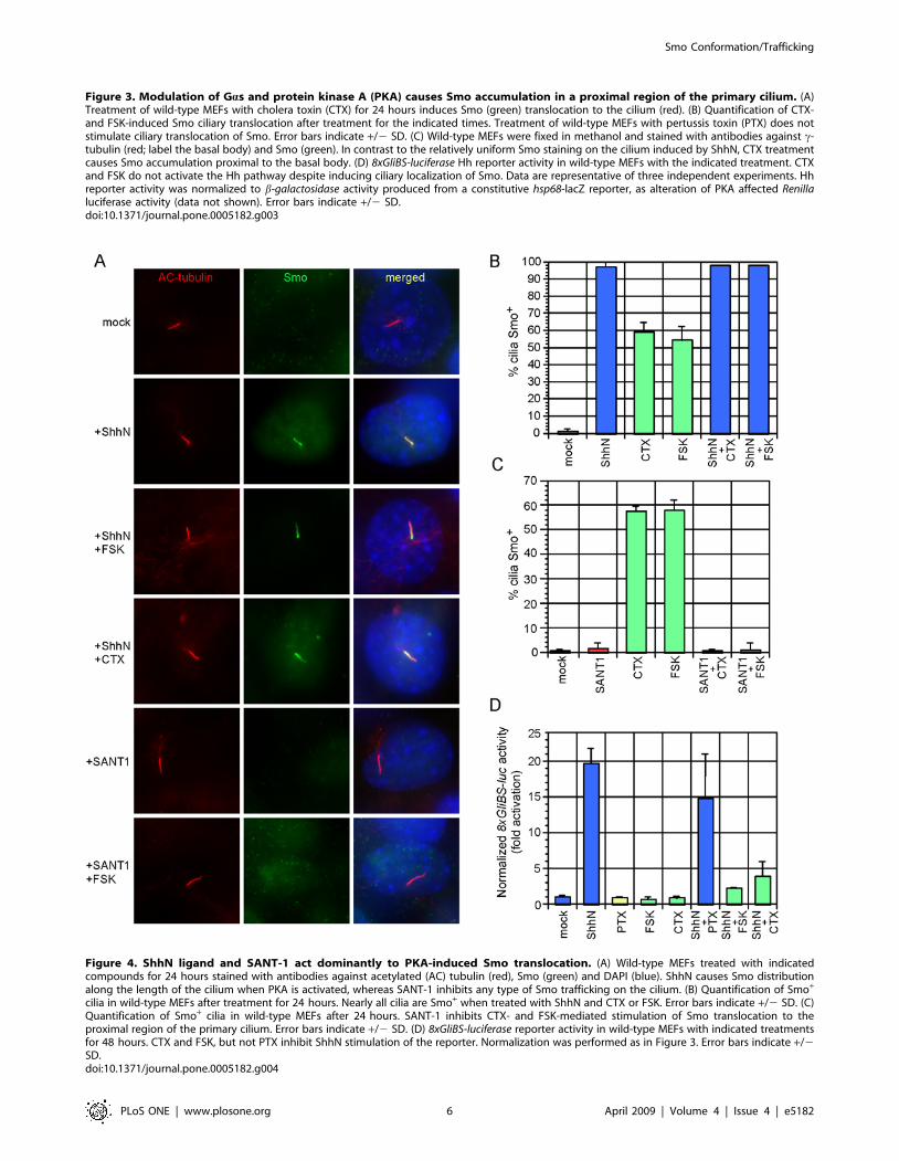
Figure 4. ShhN ligand and SANT-1 act dominantly to PKA-induced Smo translocation. (A) Wild-type MEFs treated with indicatedcompounds for 24 hours stained with antibodies against acetylated (AC) tubulin (red), Smo (green) and DAPI (blue). ShhN causes Smo distributionalong the length of the cilium when PKA is activated, whereas SANT-1 inhibits any type of Smo trafficking on the cilium. (B) Quantification of Smo+
cilia in wild-type MEFs after treatment for 24 hours. Nearly all cilia are Smo+ when treated with ShhN and CTX or FSK. Error bars indicate +/2 SD. (C)Quantification of Smo+ cilia in wild-type MEFs after 24 hours. SANT-1 inhibits CTX- and FSK-mediated stimulation of Smo translocation to theproximal region of the primary cilium. Error bars indicate +/2 SD. (D) 8xGliBS-luciferase reporter activity in wild-type MEFs with indicated treatmentsfor 48 hours. CTX and FSK, but not PTX inhibit ShhN stimulation of the reporter. Normalization was performed as in Figure 3. Error bars indicate +/2SD.doi:10.1371/journal.pone.0005182.g004
Figure 3. Modulation of Gas and protein kinase A (PKA) causes Smo accumulation in a proximal region of the primary cilium. (A)Treatment of wild-type MEFs with cholera toxin (CTX) for 24 hours induces Smo (green) translocation to the cilium (red). (B) Quantification of CTX-and FSK-induced Smo ciliary translocation after treatment for the indicated times. Treatment of wild-type MEFs with pertussis toxin (PTX) does notstimulate ciliary translocation of Smo. Error bars indicate +/2 SD. (C) Wild-type MEFs were fixed in methanol and stained with antibodies against c-tubulin (red; label the basal body) and Smo (green). In contrast to the relatively uniform Smo staining on the cilium induced by ShhN, CTX treatmentcauses Smo accumulation proximal to the basal body. (D) 8xGliBS-luciferase Hh reporter activity in wild-type MEFs with the indicated treatment. CTXand FSK do not activate the Hh pathway despite inducing ciliary localization of Smo. Data are representative of three independent experiments. Hhreporter activity was normalized to b-galactosidase activity produced from a constitutive hsp68-lacZ reporter, as alteration of PKA affected Renillaluciferase activity (data not shown). Error bars indicate +/2 SD.doi:10.1371/journal.pone.0005182.g003
Smo Conformation/Trafficking
PLoS ONE | www.plosone.org 6 April 2009 | Volume 4 | Issue 4 | e5182

activation and SANT-1 inhibition. Exposure of MEFs to both
ShhN and CTX or FSK resulted in restoration of Smo staining
along the entire length of the primary cilium (Fig. 4A, 4B). In
contrast, SANT-1 inhibited PKA stimulation of Smo trafficking to
the proximal cilium (Fig. 4A, 4C). Two conclusions may be drawn
from these results. First, the Smo conformation adopted when
bound to SANT-1 is refractory to both cyclopamine- and PKA-
stimulated cilium trafficking, suggesting that SANT-1 may act
upstream to sequester Smo from or inhibit its interaction with b-
arrestins or IFT particles [25]. Second, addition of ShhN overrides
accumulation of Smo in the proximal region of the primary cilium.
Restricted Smo localization to this region could be a prerequisite
for pathway activation, or a means to inhibit Smo trafficking or
coupling to downstream components. Consistent with previous
reports [34–37], we observed that activation of the Hh reporter
was blocked by CTX- and FSK-mediated PKA stimulation, but
PTX treatment produced only a modest reduction (Fig 4D). We
speculate that if PKA positively regulates Smo while promoting
Gli repressor production, any potential positive effect on Hh
signaling due to partial Smo translocation induced by CTX or
FSK may be offset by negative regulation of Gli factors by PKA
[38]. Additional studies using endogenous levels of PKA-refractory
forms of the Gli proteins will be necessary to rigorously resolve this
issue.
D. melanogaster Smo utilizes a series of PKA sites in its C-terminal
tail to neutralize the inhibitory effects of several clusters of Arg
residues [8]. These Arg clusters, but not the PKA sites, are
conserved in vertebrates and are critical for maintenance of Smo
in an inhibited state that is incapable of trafficking to the cell
surface [4,5]. Further investigation is required to clarify if
repression of the autoinhibitory Arg clusters results in Smo ciliary
translocation, and if PKA acts directly or indirectly on Smo for
targeting to a proximal region of the cilium. Nonetheless, our data
raise the possibility that signaling through Gas-coupled receptors
and activation of PKA could influence Smo trafficking both to and
on the primary cilium. This could provide a means for crosstalk
between signaling pathways.
In this study, we show that Hh pathway agonists, a specific class
of antagonists, and PKA activity induce accumulation of Smo in
different regions of the primary cilium. Smo localization to the
cilium is therefore necessary, but not sufficient, for activation of
the Hh pathway. Taken together, the data suggest that Smo can
adopt multiple conformations that allow ciliary trafficking, but
only a subset of these are competent to activate the Hh cascade. In
addition, the precise location of Smo on the primary cilium may
be important for activation of the pathway. Further analysis of
Smo conformations and identification of factors that communicate
these conformations to Gli proteins are necessary for a
comprehensive understanding of the role of the primary cilium
in Smo function.
Materials and Methods
Generation and treatment of cell linesMouse embryonic fibroblasts (MEFs) were derived from wild-
type embryos at embryonic days (E) 9.5 or E10.5, and from
Ptch12/2 embryos at E9.5. These cells were subsequently
immortalized with recombinant retroviruses encoding the simian
virus (SV) 40 large T antigen [39]. MEFs were maintained in
DMEM (Cellgro) supplemented with 10% fetal bovine serum
(Cellgro), L-glutamine (Gibco), penicillin/streptomycin (Gibco),
and 200 mg/ml G418 (Gibco). ShhN-conditioned media (CM) was
produced as previously described [40]. Briefly, HEK293T cells
were transfected with 10 mg of pcDNA3::ShhN using Lipofecta-
mine 2000 (Invitrogen). Conditioned media was harvested 48 and
96 hours post-transfection, pooled, filtered through a 0.22 mm
PES syringe filter (Millipore), and buffered with 5 mM HEPES,
pH 7.5. Compounds used for treatment of MEFs and their
concentrations were as follows: 20-a-hydroxysterol (10 mM,
Sigma), 7-b-hydroxysterol (10 mM, Sigma), 7-dehydrocholesterol
(10 mM, Sigma), cholecalciferol (pro-vitamin D3, 10 mM, Sigma),
cyclopamine (10 mM, Toronto Research Chemicals), jervine
(10 mM, Toronto Research Chemicals), SANT-1 (10 mM, Cal-
biochem), cholera toxin (CTX, 100 ng/ml, Sigma), pertussis toxin
(PTX, 100 ng/ml, Calbiochem), forskolin (FSK, 10 mM, Sigma).
ImmunostainingConfluent MEFs were grown on gelatin-coated glass coverslips
(Fisher) and treated with conditioned media or compounds in
DMEM supplemented with 0.5% newborn calf serum and
penicillin/streptomycin for the indicated times. Cells were washed
with PBS, fixed in 4% paraformaldehyde in PBS (Sigma) for
15 min at room temperature, and permeabilized in 0.2% Triton
X-100/PBS for 10 min. Alternatively, to better visualize basal
bodies, MEFs were fixed in ice-cold methanol for 10 min at
220uC. Blocking was performed in 10% sheep serum/0.02%Tri-
ton X-100/PBS for 1 hour at room temperature. Cells were
stained with the following primary antibodies in 2% sheep serum/
0.02% Triton X-100/PBS at 4uC, overnight: rabbit anti-Smo
(1:500, [17]), mouse anti-acetylated tubulin (1:2000, Sigma), and
mouse anti-c-tubulin (1:1000, Sigma). Cells were washed three
times in 0.02% Triton X-100/PBS and probed with the following
secondary antibodies: mouse Alexa 594 (1:2000) and rabbit Alexa
488 (1:2000, Invitrogen). Cells were washed as before, stained with
DAPI (1:10,000, Sigma), and mounted in VectaShield (Vector
Laboratories, Burlingame, CA).
ImagingImages were taken with a Nikon Eclipse E1000 epifluorescence
microscope using a Plan Apochromat 1006/1.40 oil objective
(Nikon) and a SPOT 2.3 RT Slider cooled CCD camera. Images
were acquired using SPOT Advanced software (Diagnostic
Instruments), converted to 24 bit (RGB) images, and the RGB
histograms were adjusted to reduce background fluorescence.
Activity assaysMEFs were seeded in 24-well plates at a concentration of 56104
cells/ml the day prior to transfection. Fugene 6 (Roche) was used
for transfection of reporter constructs according to manufacturer’s
instructions. We transfected a mix of pcDNA3 (Invitrogen) :
8xGliBS-firefly luciferase [24] : pRL-TK (Promega) or hsp68-lacZ
with a ratio of 4:5:1. 48 hours post-transfection, media was
changed to low serum, and ShhN-CM or compounds were added
for 36–48 hours. Cells were harvested and reporter activity
measured using a Dual Luciferase kit (Promega) and a Lumines-
cent b-galactosidase detection kit II (Clontech), on an LmaxII 384
luminometer (Molecular Devices).
Acknowledgments
We thank Brian Black for the hsp68-lacZ plasmid and David Casso, Arif
Hussein, Jean Regard, and members of the Chuang laboratory for helpful
discussions.
Author Contributions
Conceived and designed the experiments: CW PTC. Performed the
experiments: CW. Analyzed the data: CW PTC. Contributed reagents/
materials/analysis tools: CW MHC. Wrote the paper: CW PTC.
Smo Conformation/Trafficking
PLoS ONE | www.plosone.org 7 April 2009 | Volume 4 | Issue 4 | e5182

References
1. McMahon AP, Ingham PW, Tabin CJ (2003) Developmental roles and clinical
significance of hedgehog signaling. Curr Top Dev Biol 53: 1–114.2. Taipale J, Cooper MK, Maiti T, Beachy PA (2002) Patched acts catalytically to
suppress the activity of Smoothened. Nature 418: 892–897.3. Jiang J, Hui CC (2008) Hedgehog signaling in development and cancer. Dev
Cell 15: 801–812.
4. Huangfu D, Anderson KV (2006) Signaling from Smo to Ci/Gli: conservationand divergence of Hedgehog pathways from Drosophila to vertebrates.
Development 133: 3–14.5. Varjosalo M, Li SP, Taipale J (2006) Divergence of hedgehog signal
transduction mechanism between Drosophila and mammals. Dev Cell 10:
177–186.6. Zhu AJ, Zheng L, Suyama K, Scott MP (2003) Altered localization of
Drosophila Smoothened protein activates Hedgehog signal transduction. GenesDev 17: 1240–1252.
7. Corbit KC, Aanstad P, Singla V, Norman AR, Stainier DY, et al. (2005)Vertebrate Smoothened functions at the primary cilium. Nature 437:
1018–1021.
8. Zhao Y, Tong C, Jiang J (2007) Hedgehog regulates smoothened activity byinducing a conformational switch. Nature 450: 252–258.
9. Rosenbaum JL, Witman GB (2002) Intraflagellar transport. Nat Rev Mol CellBiol 3: 813–825.
10. Eggenschwiler JT, Anderson KV (2007) Cilia and developmental signaling.
Annu Rev Cell Dev Biol 23: 345–373.11. Huangfu D, Liu A, Rakeman AS, Murcia NS, Niswander L, et al. (2003)
Hedgehog signalling in the mouse requires intraflagellar transport proteins.Nature 426: 83–87.
12. Huangfu D, Anderson KV (2005) Cilia and Hedgehog responsiveness in themouse. Proc Natl Acad Sci U S A 102: 11325–11330.
13. Liu A, Wang B, Niswander LA (2005) Mouse intraflagellar transport proteins
regulate both the activator and repressor functions of Gli transcription factors.Development 132: 3103–3111.
14. Haycraft CJ, Banizs B, Aydin-Son Y, Zhang Q, Michaud EJ, et al. (2005) Gli2and Gli3 localize to cilia and require the intraflagellar transport protein polaris
for processing and function. PLoS Genet 1: e53.
15. Rohatgi R, Milenkovic L, Scott MP (2007) Patched1 regulates hedgehogsignaling at the primary cilium. Science 317: 372–376.
16. Davenport JR, Yoder BK (2005) An incredible decade for the primary cilium: alook at a once-forgotten organelle. Am J Physiol Renal Physiol 289:
F1159–1169.17. Gerber AN, Wilson CW, Li YJ, Chuang PT (2007) The hedgehog regulated
oncogenes Gli1 and Gli2 block myoblast differentiation by inhibiting MyoD-
mediated transcriptional activation. Oncogene 26: 1122–1136.18. Chen JK, Taipale J, Cooper MK, Beachy PA (2002) Inhibition of Hedgehog
signaling by direct binding of cyclopamine to Smoothened. Genes Dev 16:2743–2748.
19. Corcoran RB, Scott MP (2006) Oxysterols stimulate Sonic hedgehog signal
transduction and proliferation of medulloblastoma cells. Proc Natl Acad Sci U S A103: 8408–8413.
20. Sinha S, Chen JK (2006) Purmorphamine activates the Hedgehog pathway bytargeting Smoothened. Nat Chem Biol 2: 29–30.
21. Koide T, Hayata T, Cho KW (2006) Negative regulation of Hedgehog signalingby the cholesterogenic enzyme 7-dehydrocholesterol reductase. Development
133: 2395–2405.
22. Bijlsma MF, Spek CA, Zivkovic D, van de Water S, Rezaee F, et al. (2006)Repression of smoothened by patched-dependent (pro-)vitamin D3 secretion.
PLoS Biol 4: e232.
23. Chen JK, Taipale J, Young KE, Maiti T, Beachy PA (2002) Small molecule
modulation of Smoothened activity. Proc Natl Acad Sci U S A 99:
14071–14076.
24. Sasaki H, Hui C, Nakafuku M, Kondoh H (1997) A binding site for Gli proteins
is essential for HNF-3beta floor plate enhancer activity in transgenics and can
respond to Shh in vitro. Development 124: 1313–1322.
25. Kovacs JJ, Whalen EJ, Liu R, Xiao K, Kim J, et al. (2008) Beta-arrestin-
mediated localization of smoothened to the primary cilium. Science 320:
1777–1781.
26. Ocbina PJ, Anderson KV (2008) Intraflagellar transport, cilia, and mammalian
Hedgehog signaling: analysis in mouse embryonic fibroblasts. Dev Dyn 237:
2030–2038.
27. Low WC, Wang C, Pan Y, Huang XY, Chen JK, et al. (2008) The decoupling of
Smoothened from Galphai proteins has little effect on Gli3 protein processing
and Hedgehog-regulated chick neural tube patterning. Dev Biol 321: 188–196.
28. Riobo NA, Saucy B, Dilizio C, Manning DR (2006) Activation of heterotrimeric
G proteins by Smoothened. Proc Natl Acad Sci U S A 103: 12607–12612.
29. Ogden SK, Fei DL, Schilling NS, Ahmed YF, Hwa J, et al. (2008) G protein
Galphai functions immediately downstream of Smoothened in Hedgehog
signalling. Nature 456: 967–970.
30. Gill DM, Meren R (1978) ADP-ribosylation of membrane proteins catalyzed by
cholera toxin: basis of the activation of adenylate cyclase. Proc Natl Acad
Sci U S A 75: 3050–3054.
31. Codina J, Hildebrandt J, Iyengar R, Birnbaumer L, Sekura RD, et al. (1983)
Pertussis toxin substrate, the putative Ni component of adenylyl cyclases, is an
alpha beta heterodimer regulated by guanine nucleotide and magnesium. Proc
Natl Acad Sci U S A 80: 4276–4280.
32. Shiba D, Yamaoka Y, Hagiwara H, Takamatsu T, Hamada H, et al. (2009)
Localization of Inv in a distinctive intraciliary compartment requires the C-
terminal ninein-homolog-containing region. J Cell Sci 122: 44–54.
33. Seamon KB, Daly JW (1981) Forskolin: a unique diterpene activator of cyclic
AMP-generating systems. J Cyclic Nucleotide Res 7: 201–224.
34. Hammerschmidt M, Bitgood MJ, McMahon AP (1996) Protein kinase A is a
common negative regulator of Hedgehog signaling in the vertebrate embryo.
Genes Dev 10: 647–658.
35. Fan CM, Porter JA, Chiang C, Chang DT, Beachy PA, et al. (1995) Long-range
sclerotome induction by sonic hedgehog: direct role of the amino-terminal
cleavage product and modulation by the cyclic AMP signaling pathway. Cell 81:
457–465.
36. Epstein DJ, Marti E, Scott MP, McMahon AP (1996) Antagonizing cAMP-
dependent protein kinase A in the dorsal CNS activates a conserved Sonic
hedgehog signaling pathway. Development 122: 2885–2894.
37. Pan Y, Wang C, Wang B (2009) Phosphorylation of Gli2 by protein kinase A is
required for Gli2 processing and degradation and the Sonic Hedgehog-regulated
mouse development. Dev Biol 326: 177–189.
38. Wang B, Fallon JF, Beachy PA (2000) Hedgehog-regulated processing of Gli3
produces an anterior/posterior repressor gradient in the developing vertebrate
limb. Cell 100: 423–434.
39. Brown M, McCormack M, Zinn KG, Farrell MP, Bikel I, et al. (1986) A
recombinant murine retrovirus for simian virus 40 large T cDNA transforms
mouse fibroblasts to anchorage-independent growth. J Virol 60: 290–293.
40. Chen MH, Li YJ, Kawakami T, Xu SM, Chuang PT (2004) Palmitoylation is
required for the production of a soluble multimeric Hedgehog protein complex
and long-range signaling in vertebrates. Genes Dev 18: 641–659.
Smo Conformation/Trafficking
PLoS ONE | www.plosone.org 8 April 2009 | Volume 4 | Issue 4 | e5182