small intestinal submucosa: a tissue-derived extracellular matrix that promotes tissue-specific...
TRANSCRIPT
TISSUE ENGINEERINGVolume 4, Number 2, 1998Mary Ann Liebert, Inc.
Small Intestinal Submucosa: A Tissue-DerivedExtracellular Matrix That Promotes Tissue-Specific
Growth and Differentiation of Cells in Vitro
SHERRY L. VOYTIK-HARBIN, M.S.E.E., Ph.D.,1 ANDREW O. BRIGHTMAN, Ph.D.,1
BEVERLY Z. WAISNER, B.S., 2 J. PAUL ROBINSON, Ph.D.,2 andCARLTON H. LAMAR, D.V.M., Ph.D.2
ABSTRACT
The importance of understanding cell-extracellular matrix (ECM) interaction is now evi-dent as scientists, engineers, and physicians search for novel scaffolds that support and main-tain tissue-specific cellular growth and function both in vivo and in vitro. Small intestinalsubmucosa (SIS) represents an ECM that has been derived from porcine intestine while pre-serving its natural composition and architecture. More recently, an extract of this physio-logic ECM, which forms a three-dimensional gel in vitro, has been developed. When com-pared to routinely used culture substrata (e.g., plastic, Vitrogen, and Matrigel), intact SISand SIS-derived gel possess unique compositional and architectural features. Simple squa-mous epithelial (pulmonary artery), fibroblastic (Swiss 3T3), glandular epithelial (adeno-carcinoma), and smooth muscle-like (urinary bladder) cells were seeded upon intact SIS andSIS-derived gel and their morphologic response evaluated. For each of the four cell typesstudied, intact SIS and SIS-derived gel were equivalent or superior in their ability to sup-port and maintain expression of tissue-specific phenotype when compared to the routinelyused substrata, plastic, Vitrogen, and Matrigel. Therefore, SIS may provide a novel biolog-ically derived scaffold for the growth and study of a variety of cell types in vitro. Such in-formation regarding the influence of substrate structure and function on cell behavior willbe useful in the development of successful tissue engineering strategies.
INTRODUCTION
INTERACTION OF CELLS WITH THEIR EXTRACELLULAR MATRIX (ECM) plays a crucial role in the organization,homeostasis, and function of all tissues and organs. It is the continuous cross-talk between cells and the
surrounding matrix environment that orchestrates critical processes such as the acquisition and maintenanceof differentiated phenotypes during embryogenesis, the development of form (morphogenesis), angiogene-
1 Hillenbrand Biomedical Engineering Center and 2Department of Basic Medical Science, School of Veterinary Med-icine, Purdue University, West Lafayette, Indiana 47907.
This work was presented in part at the Inaugral Meeting of the Tissue Engineering Society, held in Orlando, Florida,December 13-15, 1996.
157
VOYTIK-HARBIN ET AL.
sis, wound healing, and even tumor metastasis. The cell and its ECM are said to exist in a state of "dy-namic reciprocity".12 Both biochemical and biophysical signals from the ECM modulate fundamental cel-lular activities, including adhesion, migration, proliferation, differential gene expression, and programmedcell death.3-4 In turn, the cell can modify its ECM environment by modulating synthesis and degradationof specific matrix components. The realization of the significance of cell-ECM communication has led toa renewed interest in characterizing ECM constituents and the basic mechanisms of cell-ECM interaction.
To provide an in vitro culture environment that would more closely mimic cell-ECM interaction in vivo,purified ECM components such as collagen, fibronectin, laminin, and glycosaminoglycans (e.g., hyaluronicacid, heparin sulfate) have been used to derivatize artificial substrata for augmentation of cell adhesion,growth, and morphology. Three-dimensional culture matrices also have been fashioned from purified ECMcomponents, specifically fibrin clots5 and collagen gels.6 Investigations with these matrices have demon-strated the importance of three-dimensional architecture in the establishment of a tissue-like histology.
More complex scaffolds representing combinations of ECM components in a natural or processed formhave also been studied. These include reconstituted basement membrane extract from Engelbreth HolmSwarm tumor (Matrigel), and allogeneic and xenogeneic tissues (including lens capsule,7 liver,8 amnion,9
and chorioallantoic membrane10). Although these substrata allow the study of cell growth and differentia-tion in a more physiologically relevant system, their use has been limited by availability and amenabilityto disinfection, sterilization, and manufacturing processes.
Previous in vivo studies have shown that SIS modulates the cellular activities of the remodeling and re-pair responses when implanted in a number of different tissue microenvironments, including lower urinarytract,11-12 body wall,13 tendon,14 ligament,15 bone (Voytik-Harbin, unpublished data), and blood vessels.16
Compositional analyses of the material have shown that, like other normal connective tissues, the materialconsists predominantly of fibrillar collagens (types I, III, V; unpublished data), with lesser quantities of pro-teoglycans, glycosaminoglycans (e.g., hyaluronic acid),17 glycoproteins (e.g., fibronectin),18 and growth fac-tors (e.g., FGF-2 and TGF/3-related protein).19 It appears likely that SIS, because of its complex composi-tion and architecture, could be useful for in vitro cell culture, where maintenance of differentiated phenotypeis important.
In this communication, we report on the ability of SIS to support the survival and growth of several dif-ferent cell types, including fibroblasts (Swiss mouse 3T3), simple squamous epithelium (rat pulmonaryartery endothelium), glandular epithelium (canine prostate adenocarcinoma), and smooth muscle-like cells(human urinary bladder). The morphologic response of these cell types to intact SIS, SIS-derived gel, andother widely used substrata (plastic, Matrigel, and Vitrogen) was evaluated and compared. In addition, thestructural and compositional features of the substrata were compared.
MATERIALS AND METHODS
Cell Culture
Swiss mouse 3T3 fibroblasts were obtained from American Type Culture Collection (ATCC) (Rockville,MD). Primary human urinary bladder stromal cells (HUBS) were derived from bladders of patients under-going ureteral reimplantation for vesicoureteral reflux and generously provided by Dr. E. Cheng, North-western University (Chicago, IL). Primary canine prostate carcinoma cells were established from a primarytumor of a dog with prostate adenocarcinoma and were kindly provided by Dr. D. Waters, Purdue Univer-sity (West Lafayette, IN). Rat pulmonary endothelial cells were isolated from rat pulmonary arteries, puri-fied by flow cytometric fluorescence-activated cell sorting.20 The cell types, source information, and mediumconditions involved in these experiments are summarized in Table 1.
Substrata
Vitrogen and Matrigel were obtained from Collagen Corporation (Fremont, CA) and Collaborative Bio-medical (Bedford, MA), respectively. All tissue culture plastics were obtained from Corning Inc. (Coming, NY).
158
SMALL INTESTINAL SUBMUCOSA
TABLE 1. SUMMARY OF CELL TYPES AND NUTRIENTS
Name Origin/Source Medium
3T3
RPEC
HUBS
Clemons
Swiss mouse embryo fibroblasts;American Type Culture Collection,CRL 1658
Rat pulmonary artery endothelial cellsJ. P. Robinson, Purdue University
Human urinary bladder stromal cells;E. Cheng, Northwestern University
Canine prostate adenocarcinoma;D. Waters, Purdue University
DMEM (Dulbecco's modified Eagle's medium) with1.5 g/L NaHCO3, 10% NNCS (neonatal calf serum),100 U/ml penicillin, 100 /Ag/ml streptomycin,2 mM L-glutamine
RPMI 1640, 5% NCS (newborn calf serum), 5% FBS(fetal bovine serum), 100 U/ml penicillin, 100 /u.g/mlstreptomycin, 2 mM L-glutamine
Modified medium 199 supplemented with 10% NCS(newborn calf serum), 2.5 /^g/ml fungizone and50 jug/ml gentamicin (Baskin et al., 1993); mediummodifications included the addition of sodiumbicarbonate (2.2 g/L), bactopeptone (0.5 g/L), glucose(3.0 g/L), L-glutamine (0.29 g/L), HEPES (3.57 g/L),100X BME vitamins (10 ml/L, Flow Labs), and100X BME amino acids (10 ml/L, Gibco, GrandIsland, New York)
RPMI 1640, 10% FBS, 100 U/ml penicillin, 100 fig/mlstreptomycin, 2 mM L-glutamine
Preparation of intact SIS and SIS-derived gel. Intestinal submucosa was prepared from the small intestinesof market weight pigs obtained from a local meat processing plant. In brief, intestine was rinsed free ofcontents and everted, and the superficial layers of the mucosa were removed by mechanical delamination.The tissue was reverted to its original orientation and the external muscle layer removed. The prepared in-testinal submucosa tube was split open longitudinally and rinsed extensively in water to lyse any cells as-sociated with the matrix and to eliminate cell degradation products. Immediately after rinsing, intestinalsubmucosa was disinfected with 0.1 % peracetic acid for cell culture or frozen in liquid nitrogen and storedat — 80°C for preparation of SIS-derived gel.
For preparation of SIS-derived gel, frozen tissue first was pulverized under liquid nitrogen with an in-dustrial blender and stored at -80°C prior to use. SIS powder (5% w/v) was suspended in 0.5 M aceticacid containing 0.1% pepsin and vigorously stirred for 72 h at 4°C. The mixture then was centrifuged at12,000 rpm for 20 min at 4°C to remove undigested tissue. The supernatant was dialyzed extensively against0.01 M acetic acid at 4°C in spectrapor tubing (MWCO 3500, Spectrum Medical Industries). To obtain asterile preparation, the solution was dialyzed against 0.01 M acetic acid containing chloroform (approxi-mately 0.5% v/v), followed by several changes of sterile 0.01 M acetic acid. To polymerize the SIS extract,1.2 ml 10X PBS (1.37 M NaCl, 26.8 mM KC1, 0.1 M Na2HPO4, and 17.6 mM KH2PO4, and 5 mg/L phe-nol red, pH 7.4) and 1.2 ml 0.1 NaOH were added to 8 ml of SIS extract. This solution was brought topH 7 with 0.1 M HC1, aliquotted into 24-well plates, and gelled at 37°C for 30-60 min.
Scanning Electron Microscopy
Substrata were fixed in 3% glutaraldehyde in Millonig's buffer and post fixed in 1% osmium tetroxide.Fixed specimens were dehydrated in a graded series of acetones, critical point dried, affixed to scanningstubs, and sputter coated with gold/pallidium. Specimens were viewed with a JEOL JSM-840 scanning elec-tron microscope. Gelled substrata also were quick frozen by plunging into a liquid nitrogen slush withoutprior fixation or dehydration. The sample was transferred into a CT1000 coldstage attachment (Oxford In-struments North America, Inc., Concord, MA), and the surface was fractured and coated with gold prior toviewing at temperatures of - 150°C with the SEM.
159
VOYTIK-HARBIN ET AL.
Cell Growth on Substrata
Twenty-four-well tissue culture plates were prepared with Matrigel (500 ^I/well), Vitrogen (500 /il/well),SIS, SIS-derived gel (500 ju-l/well), or no substrate. SIS material was affixed in polypropylene frames, withthe mucosal surface facing upward to create a well area of 0.5 cm2. All substrata were equilibrated withsterile PBS, pH 7.4, prior to the application of cells.
Cells were harvested in complete medium (Table 1) and seeded upon substrata at 60,000 cells/cm2. Forcertain experiments, cells were labeled with the fluorescent cell membrane dye PKH26 (Sigma, St. Louis,MO) prior to seeding upon the substrata. Culture plates were incubated at 37°C in a humidified atmosphereof 5% CO2 in air and fed two to three times weekly. On days 1, 4, 7, and 14, the cells and associated sub-strate (intact SIS only) were fixed and processed for light or fluorescence microscopy.
Fluorescence microscopy. Cells labeled with fluorescent markers were fixed in 4% paraformaldehyde andobserved using a fluorescence microscope (Labophot, Nikon).
Light microscopy. Cell growth on plastic, Matrigel, Vitrogen, and SIS-derived gel was observed daily us-ing a standard inverted microscope. Digital images were collected on days 1, 4, 7, 11, and 14 using an in-verted microscope, video camera (Sanyo, Japan), and Digital Video Producer software (Asymetrix) on a755CD laptop computer (IBM).
Histology Cells and associated substrata were fixed in neutral buffered formalin, embedded in paraffin,sectioned to 6 (xm, and stained with H&E. Morphological evaluation was conducted using light microscopy.
RESULTS
Morphological Appearance and Characterization
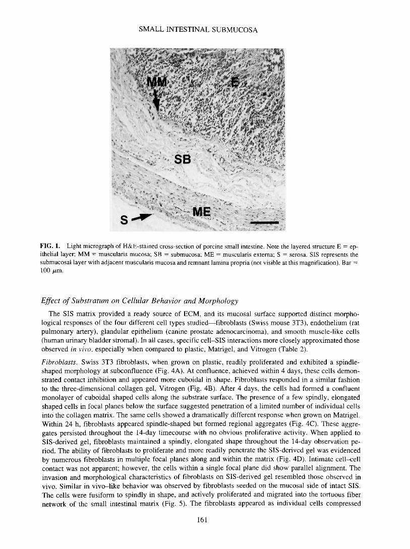
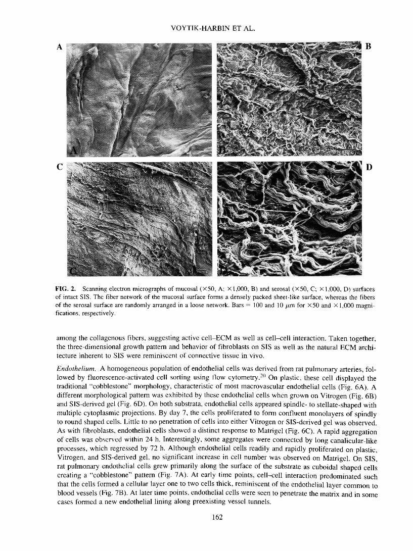
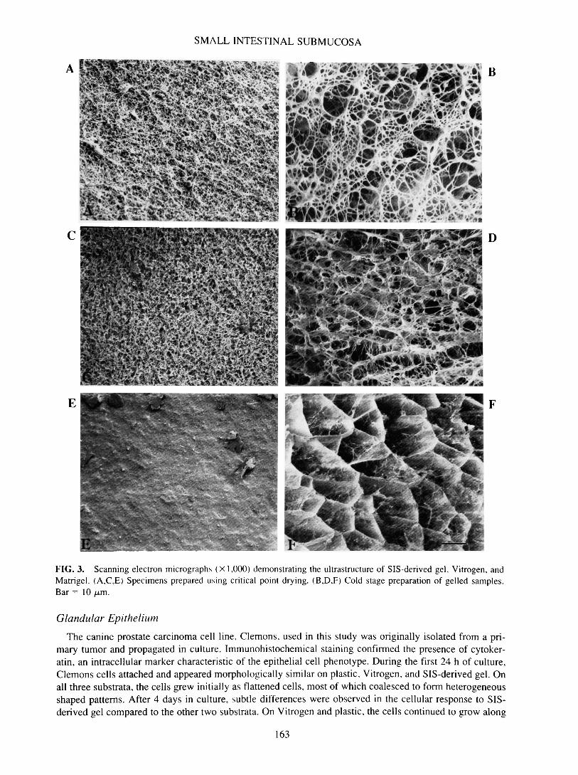
It has been well documented that physical, geometrical, and topological features of substrata affect cellbehavior both in vitro and in vivo.21 Therefore, architectural features of SIS, SIS-derived gel, Vitrogen, andMatrigel were determined and subsequently compared. Small intestine represents a multilayered organ con-sisting of mucosa, lamina propria, muscularis mucosa, submucosa, muscularis externa, and serosa (Fig. 1).Preparation of SIS involved mechanical removal of the outermost epithelial and muscle layers. Treatmentof the remaining submucosa, muscularis mucosa, and remnant lamina propria layers under hypotonic con-ditions provided an acellular ECM designated SIS. Initial structural analysis was performed using routinecritical point drying of specimens followed by scanning electron microscopy (SEM). Low-magnificationSEM demonstrated the disparity in the topography of the mucosal and serosal surfaces of SIS (Fig. 2). Therelatively smooth mucosal surface, which once supported the epithelial lining of the intestine, showed mul-tiple folds and involutions (Fig. 2A), whereas the fibrillar nature of the serosal side was evidenced by itsmore ragged appearance (Fig. 2C). Ultrastructurally, the mucosal surface was characterized by more denselypacked fibers, which form discontinuous layers varying in orientation (Fig. 2B). Alternatively, the serosalside exhibited a fine network of loosely organized fibers, most of which are < 1 /am in diameter (Fig. 2D).Although most fibers appeared to be organized randomly, some formed assemblies to create larger fibers.Analysis of SIS-derived gel (Fig. 3A) and Vitrogen (Fig. 3C) prepared using critical point drying techniquesdemonstrated a tightly woven network of small-diameter fibrils with extensive lateral association. Matrigel,on the other hand, featured a more densely compact, sheet-like surface (Fig. 3E). Although some appreci-ation of substrate architecture was obtained from critical point dried specimens, excessive shrinkage wasnoted. To minimize the possibility of structural artifacts induced by such preparatory techniques, quick-freeze, cold-stage SEM was employed. This technique obviated the need for both chemical fixation and de-hydration. Results obtained using this method more accurately represent the detailed macromolecular struc-ture of samples with high water content. With cold-stage SEM, the marked differences in the ultrastructureof the three substrata were obvious. SIS-derived gel consisted of a network of loosely organized fibrils thatvaried in size (Fig. 3B). The fine fibers composing Vitrogen appeared more randomly oriented and formedregions of dense aggregates with extensive cross-branching (Fig. 3D). Matrigel featured a honeycomb lat-tice with very fine, cobweb-like fibers decorating the individual honeycomb cells (Fig. 3F).
160
SMALL INTESTINAL SUBMUCOSA
. * * • » .
' . V - ME
FIG. 1. Light micrograph of H&E-stained cross-section of porcine small intestine. Note the layered structure E = ep-ithelial layer; MM = muscularis mucosa; SB = submucosa; ME = muscularis externa; S = serosa. SIS represents thesubmucosal layer with adjacent muscularis mucosa and remnant lamina propria (not visible at this magnification). Bar =100 iu.m.
Effect of Substratum on Cellular Behavior and Morphology
The SIS matrix provided a ready source of ECM, and its mucosal surface supported distinct morpho-logical responses of the four different cell types studied—fibroblasts (Swiss mouse 3T3), endothelium (ratpulmonary artery), glandular epithelium (canine prostate adenocarcinoma), and smooth muscle-like cells(human urinary bladder stromal). In all cases, specific cell-SIS interactions more closely approximated thoseobserved in vivo, especially when compared to plastic, Matrigel, and Vitrogen (Table 2).
Fibroblasts. Swiss 3T3 fibroblasts, when grown on plastic, readily proliferated and exhibited a spindle-shaped morphology at subconfluence (Fig. 4A). At confluence, achieved within 4 days, these cells demon-strated contact inhibition and appeared more cuboidal in shape. Fibroblasts responded in a similar fashionto the three-dimensional collagen gel, Vitrogen (Fig. 4B). After 4 days, the cells had formed a confluentmonolayer of cuboidal shaped cells along the substrate surface. The presence of a few spindly, elongatedshaped cells in focal planes below the surface suggested penetration of a limited number of individual cellsinto the collagen matrix. The same cells showed a dramatically different response when grown on Matrigel.Within 24 h, fibroblasts appeared spindle-shaped but formed regional aggregates (Fig. 4C). These aggre-gates persisted throughout the 14-day timecourse with no obvious proliferative activity. When applied toSIS-derived gel, fibroblasts maintained a spindly, elongated shape throughout the 14-day observation pe-riod. The ability of fibroblasts to proliferate and more readily penetrate the SIS-derived gel was evidencedby numerous fibroblasts in multiple focal planes along and within the matrix (Fig. 4D). Intimate cell-cellcontact was not apparent; however, the cells within a single focal plane did show parallel alignment. Theinvasion and morphological characteristics of fibroblasts on SIS-derived gel resembled those observed invivo. Similar in vivo-like behavior was observed by fibroblasts seeded on the mucosal side of intact SIS.The cells were fusiform to spindly in shape, and actively proliferated and migrated into the tortuous fibernetwork of the small intestinal matrix (Fig. 5). The fibroblasts appeared as individual cells compressed
161
VOYTIK-HARBIN ET AL.
FIG. 2. Scanning electron micrographs of mucosal (X50, A; X 1,000, B) and serosal (X50, C; X 1,000, D) surfacesof intact SIS. The fiber network of the mucosal surface forms a densely packed sheet-like surface, whereas the fibersof the serosal surface are randomly arranged in a loose network. Bars = 100 and 10 /urn for X50 and X 1,000 magni-fications, respectively.
among the collagenous fibers, suggesting active cell-ECM as well as cell-cell interaction. Taken together,the three-dimensional growth pattern and behavior of fibroblasts on SIS as well as the natural ECM archi-tecture inherent to SIS were reminiscent of connective tissue in vivo.
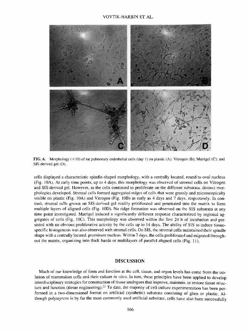
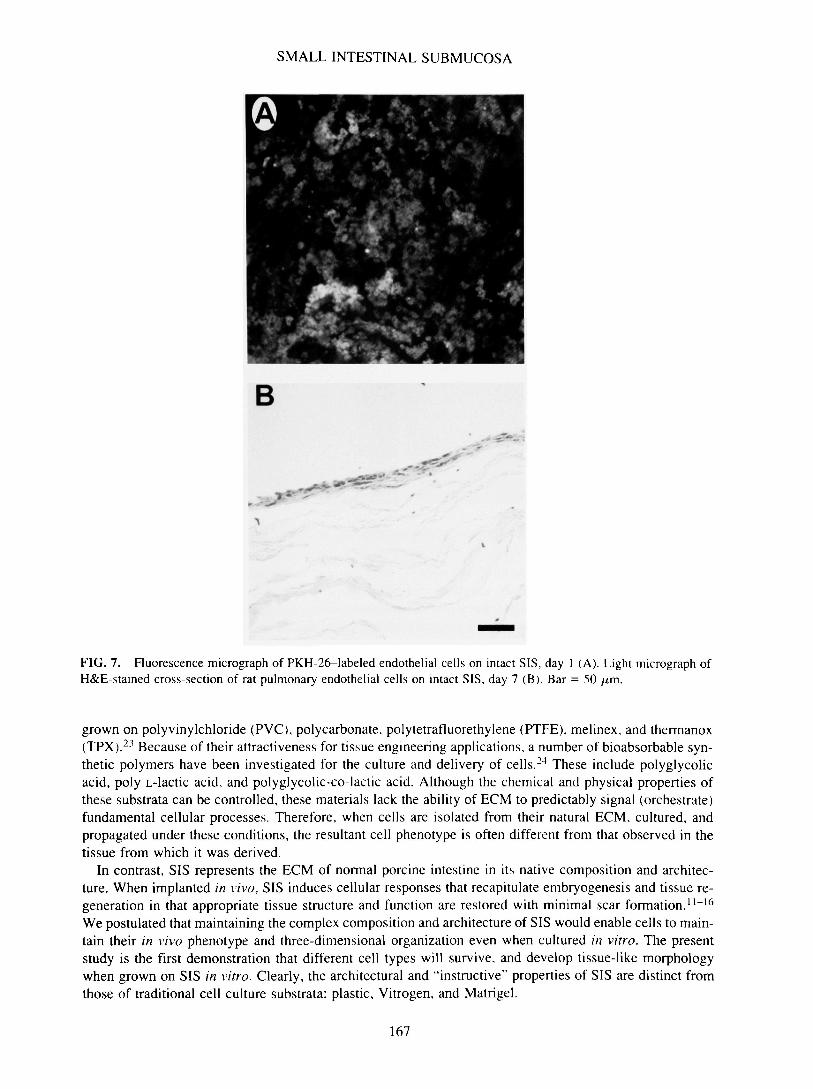
Endothelium. A homogeneous population of endothelial cells was derived from rat pulmonary arteries, fol-lowed by fluorescence-activated cell sorting using flow cytometry.20 On plastic, these cell displayed thetraditional "cobblestone" morphology, characteristic of most macrovascular endothelial cells (Fig. 6A). Adifferent morphological pattern was exhibited by these endothelial cells when grown on Vitrogen (Fig. 6B)and SIS-derived gel (Fig. 6D). On both substrata, endothelial cells appeared spindle- to stellate-shaped withmultiple cytoplasmic projections. By day 7, the cells proliferated to form confluent monolayers of spindlyto round shaped cells. Little to no penetration of cells into either Vitrogen or SIS-derived gel was observed.As with fibroblasts, endothelial cells showed a distinct response to Matrigel (Fig. 6C). A rapid aggregationof cells was observed within 24 h. Interestingly, some aggregates were connected by long canalicular-likeprocesses, which regressed by 72 h. Although endothelial cells readily and rapidly proliferated on plastic,Vitrogen, and SIS-derived gel, no significant increase in cell number was observed on Matrigel. On SIS,rat pulmonary endothelial cells grew primarily along the surface of the substrate as cuboidal shaped cellscreating a "cobblestone" pattern (Fig. 7A). At early time points, cell-cell interaction predominated suchthat the cells formed a cellular layer one to two cells thick, reminiscent of the endothelial layer common toblood vessels (Fig. 7B). At later time points, endothelial cells were seen to penetrate the matrix and in somecases formed a new endothelial lining along preexisting vessel tunnels.
162
SMALL INTESTINAL SUBMUCOSA
E
FIG. 3. Scanning electron micrographs (X 1,000) demonstrating the infrastructure of SIS-derived gel, Vitrogen, andMatrigel. (A,C,E) Specimens prepared using critical point drying. (B,D,F) Cold stage preparation of gelled samples.Bar = 10 jLtm.
Glandular Epithelium
The canine prostate carcinoma cell line, Clemons, used in this study was originally isolated from a pri-mary tumor and propagated in culture. Immunohistochemical staining confirmed the presence of cytoker-atin, an intracellular marker characteristic of the epithelial cell phenotype. During the first 24 h of culture,Clemons cells attached and appeared morphologically similar on plastic, Vitrogen, and SIS-derived gel. Onall three substrata, the cells grew initially as flattened cells, most of which coalesced to form heterogeneousshaped patterns. After 4 days in culture, subtle differences were observed in the cellular response to SIS-derived gel compared to the other two substrata. On Vitrogen and plastic, the cells continued to grow along
163
VOYTIK-HARBIN ET AL.
TABLE 2. EFFECT OF SUBSTRATA ON BEHAVIOR AND MORPHOLOGY OF DIFFERENT CELL TYPES
Cell type
FibroblastsShape
Prolif
Morph
EndotheliumShapeProlifMorph
Glandularepithelium
ShapeProlifMorph
Smoothmuscle-likecells
Shape
ProlifMorph
Plastic
Spindle; cuboidalupon confluence
Yes; contactinhibition
Two-dimensionalmonolayer
CobblestoneYesTwo-dimensional
monolayer
Round (variable)YesTwo-dimensional
monolayer
Spindle, nucleusprominent
YesMonolayer with
aggregatedridges
Vitrogen
Spindle; cuboidalupon confluence
Yes; contactinhibition
Two-dimensionalmonolayer;limited matrixpenetration
Spindle to stellateYesThree-dimensional
multilayers (1-3cells thick);limited matrixpenetration
Round (variable)YesThree-dimensional
multilayers;limited matrixpenetration
Spindle, nucleusprominent
YesMonolayer with
aggregatedridges; limitedmatrixpenetration
Substrate
Matrigel
Spindle to round
Limited to none
Rapid aggregateformation
VariableLimited to noneRapid aggregate
formation;canalicular-likeprocesses
Round (variable)Limited to noneRapid aggregate
formation;canalicular-likeprocesses
Round
Little to noneRapid aggregate
formation
SIS-derived gel
Spindle
Yes
Three-dimensional;spindle-shapedcells alongsurface of andwithin matrix
Spindle to stellateYesThree-dimensional
multilayers (1-3cells thick);limited matrixpenetration
Round (variable)YesThree-dimensional
multilayers;limited matrixpenetration
Spindle; nucleusprominent
YesThree-dimensional
multilayeredarrays; matrixpenetration
Intact SIS
Spindle
Yes
Three-dimensional;spindle-shapedcells alongsurface of andwithin matrix
CobblestoneYesThree-dimensional
multilayers (1-3cells thick);matrixpenetration
Round (variable)YesThree-dimensional
multilayers; aciniformation;limited matrixpenetration
Spindle; nucleusprominent
YesThree-dimensional
multilayeredarrays; matrixpenetration
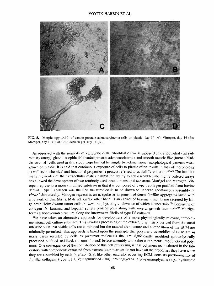
the substrate surface as a single layer of flattened cells. In contrast, SIS-derived gel induced regional pil-ing of cells into multilayered aggregates. Cells reached confluence between days 7 and 11 on plastic, Vit-rogen, and SIS-derived gel. However, even at confluence, cells on plastic maintained a two-dimensionalgrowth pattern (Fig. 8A). While some evidence of multilayer cell aggregation was noted on Vitrogen (Fig.8B), the most extensive three-dimensional pattern was developed by cells on SIS-derived gel (Fig. 8D).When grown on Matrigel, Clemons cells attached and formed aggregates within 24 h (Fig. 8C). In somecases, the aggregates were connected by long canalicular-like processes. At later time points, the intercon-necting structures receded, leaving dense aggregates scattered along the surface. On Matrigel, no signifi-cant increase in cell number was noted at any time point up to 14 days. Unlike Matrigel, intact SIS inducedattachment, proliferation, and polarization of Clemons cells in vitro. Initially, cells grew as aggregates one
164

SMALL INTESTINAL SUBMUCOSA
* A• • • 1 H
s
m -
B
* . DFIG. 4. Morphology (X 10) of mouse fibroblasts on plastic, day 1 (A); Vitrogen day 4 (B); Matrigel, day 1 (C); andSIS-derived gel. day 4 <D).
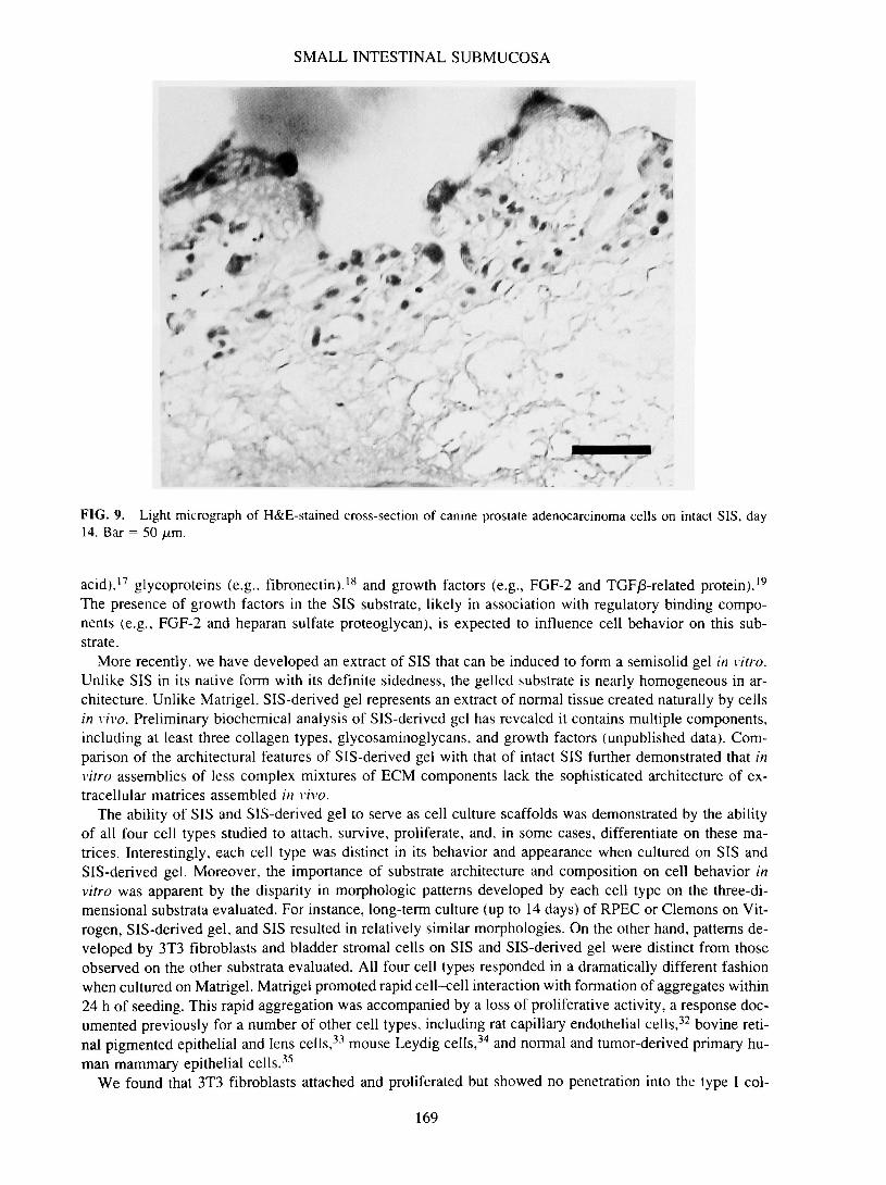
to three cells thick along the surface of the SIS. By day 7, confluent layer of cells covered the surface ofSIS, with some organized areas resembling early follicle formation. By day 14, numerous structures com-posed of epithelial cells organized around a central lumen were evident, reminiscent of acini (Fig. 9). Al-though these cells grew primarily along the surface of SIS, in some sections, isolated foci of cells wereidentified within the matrix (data not shown).
Smooth Muscle-Like Cells
Primary cultures of stromal cells were derived from human urinary bladders and subsequently propagatedin vitro. Immunohistochemical staining confirmed the presence of vimentin, smooth muscle a-actin, andsmooth muscle myosin, characteristic of the smooth muscle phenotype. When grown on plastic, stromal
FIG. 5. Light micrograph of H&E-stained cross-section of mouse fibroblasts on intact SIS; day 14. Bar = 50
165
VOYTIK-HARBIN ET AL.
FIG. 6. Morphology (X 10) of rat pulmonary endothelial cells (day 1) on plastic (A); Vitrogen (B); Matrigel (C); andSIS-derived gel (D).
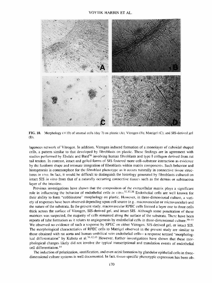
cells displayed a characteristic spindle-shaped morphology, with a centrally located, round to oval nucleus(Fig. 10A). At early time points, up to 4 days, this morphology was observed of stromal cells on Vitrogenand SIS-derived gel. However, as the cells continued to proliferate on the different substrata, distinct mor-phologies developed. Stromal cells formed aggregated ridges of cells that were grossly and microscopicallyvisible on plastic (Fig. 10A) and Vitrogen (Fig. 10B) as early as 4 days and 7 days, respectively. In con-trast, stromal cells grown on SIS-derived gel readily proliferated and penetrated into the matrix to formmultiple layers of aligned cells (Fig. 10D). No ridge formation was observed on the SIS substrata at anytime point investigated. Matrigel induced a significantly different response characterized by regional ag-gregates of cells (Fig. IOC). This morphology was observed within the first 24 h of incubation and per-sisted with no obvious proliferative activity by the cells up to 14 days. The ability of SIS to induce tissue-specific histogenesis was also observed with stromal cells. On SIS, the stromal cells maintained their spindleshape with a centrally located, prominent nucleus. Within 7 days, the cells proliferated and migrated through-out the matrix, organizing into thick bands or multilayers of parallel aligned cells (Fig. 11).
DISCUSSION
Much of our knowledge of form and function at the cell, tissue, and organ levels has come from the iso-lation of mammalian cells and their culture in vitro. In turn, these principles have been applied to developinterdisciplinary strategies for construction of tissue analogues that improve, maintain, or restore tissue struc-ture and function (tissue engineering).22 To date, the majority of cell culture experimentation has been per-formed in a two-dimensional format on artificial (synthetic) substrata consisting of glass or plastic. Al-though polystyrene is by far the most commonly used artificial substrate, cells have also been successfully
166
SMALL INTESTINAL SUBMUCOSA
B
-• - - • .
FIG. 7. Fluorescence micrograph of PKH-26-labeled endothelial cells on intact SIS, day 1 (A). Light micrograph ofH&E-stained cross-section of rat pulmonary endothelial cells on intact SIS, day 7 (B). Bar = 50 /i,m.
grown on polyvinylchloride (PVC), polycarbonate, polytetrafluorethylene (PTFE), melinex, and thermanox(TPX).23 Because of their attractiveness for tissue engineering applications, a number of bioabsorbable syn-thetic polymers have been investigated for the culture and delivery of cells.24 These include polyglycolicacid, poly L-lactic acid, and polyglycolic-co-lactic acid. Although the chemical and physical properties ofthese substrata can be controlled, these materials lack the ability of ECM to predictably signal (orchestrate)fundamental cellular processes. Therefore, when cells are isolated from their natural ECM, cultured, andpropagated under these conditions, the resultant cell phenotype is often different from that observed in thetissue from which it was derived.
In contrast, SIS represents the ECM of normal porcine intestine in its native composition and architec-ture. When implanted in vivo, SIS induces cellular responses that recapitulate embryogenesis and tissue re-generation in that appropriate tissue structure and function are restored with minimal scar formation.11"16
We postulated that maintaining the complex composition and architecture of SIS would enable cells to main-tain their in vivo phenotype and three-dimensional organization even when cultured in vitro. The presentstudy is the first demonstration that different cell types will survive, and develop tissue-like morphologywhen grown on SIS in vitro. Clearly, the architectural and "instructive" properties of SIS are distinct fromthose of traditional cell culture substrata: plastic, Vitrogen, and Matrigel.
167
VOYTIK-HARBIN ET AL.
DFIG. 8. Morphology (X10) of canine prostate adenocarcinoma cells on plastic, day 14 (A); Vitrogen, day 14 (B);Matrigel, day 1 (C); and SIS-derived gel, day 14 (D).
As observed with the majority of vertebrate cells, fibroblastic (Swiss mouse 3T3), endothelial (rat pul-monary artery), glandular epithelial (canine prostate adenocarcinoma), and smooth muscle-like (human blad-der stromal) cells used in this study were limited to simple two-dimensional morphological patterns whengrown on plastic. It is said that continuous exposure of cells to plastic often results in loss of morphologyas well as biochemical and functional properties, a process referred to as dedifferentiation.23-26 The fact thatmany molecules of the extracellular matrix exhibit the ability to self-assemble into highly ordered arrayshas allowed the development of two routinely used three-dimensional substrata. Matrigel and Vitrogen. Vit-rogen represents a more simplified substrate in that it is composed of Type I collagen purified from bovinedermis. Type I collagen was the first macromolecule to be shown to undergo spontaneous assembly invitro.21 Structurally. Vitrogen represents an irregular arrangement of dense fibrillar aggregates laced witha network of thin fibrils. Matrigel, on the other hand, is an extract of basement membrane secreted by En-gelbreth Holm Swarm tumor cells in vitro, the physiologic relevance of which is uncertain.28 Consisting ofcollagen IV, laminin, and heparan sulfate proteoglycan along with several growth factors,2930 Matrigelforms a honeycomb structure along the interwoven fibrils of type IV collagen.
We have taken an alternative approach for development of a more physiologically relevant, three-di-mensional cell culture substrate. This involves processing of the extracellular matrix derived from the smallintestine such that viable cells are eliminated but the natural architecture and composition of the ECM areminimally perturbed. This approach is based upon the principle that polymeric assemblies of ECM are inmany cases secreted by cells as precursor molecules that are significantly modified (proteolyticallyprocessed, sulfated, oxidized, and cross-linked) before assembly with other components into functional poly-mers. One consequence of the contribution of this cell processing is that polymers reconstituted in the lab-oratory with components extracted from extracellular matrices do not have all the properties they have whenthey are assembled by cells in v/vo.31 SIS, like other naturally occurring ECM, consists predominantly offibrillar collagens (type I, III, V; unpublished data), proteoglycans, glycosaminoglycans (e.g., hyaluronic
168
SMALL INTESTINAL SUBMUCOSA
FIG. 9. Light micrograph of H&E-stained cross-section of canine prostate adenocarcinoma cells on intact SIS, day14. Bar = 50 /xm.
acid).17 glycoproteins (e.g., fibronectin),18 and growth factors (e.g., FGF-2 and TGF/3-related protein).19
The presence of growth factors in the SIS substrate, likely in association with regulatory binding compo-nents (e.g., FGF-2 and heparan sulfate proteoglycan), is expected to influence cell behavior on this sub-strate.
More recently, we have developed an extract of SIS that can be induced to form a semisolid gel in vitro.Unlike SIS in its native form with its definite sidedness, the gelled substrate is nearly homogeneous in ar-chitecture. Unlike Matrigel, SIS-derived gel represents an extract of normal tissue created naturally by cellsin vivo. Preliminary biochemical analysis of SIS-derived gel has revealed it contains multiple components,including at least three collagen types, glycosaminoglycans, and growth factors (unpublished data). Com-parison of the architectural features of SIS-derived gel with that of intact SIS further demonstrated that invitro assemblies of less complex mixtures of ECM components lack the sophisticated architecture of ex-tracellular matrices assembled in vivo.
The ability of SIS and SIS-derived gel to serve as cell culture scaffolds was demonstrated by the abilityof all four cell types studied to attach, survive, proliferate, and, in some cases, differentiate on these ma-trices. Interestingly, each cell type was distinct in its behavior and appearance when cultured on SIS andSIS-derived gel. Moreover, the importance of substrate architecture and composition on cell behavior invitro was apparent by the disparity in morphologic patterns developed by each cell type on the three-di-mensional substrata evaluated. For instance, long-term culture (up to 14 days) of RPEC or Clemons on Vit-rogen, SIS-derived gel, and SIS resulted in relatively similar morphologies. On the other hand, patterns de-veloped by 3T3 fibroblasts and bladder stromal cells on SIS and SIS-derived gel were distinct from thoseobserved on the other substrata evaluated. All four cell types responded in a dramatically different fashionwhen cultured on Matrigel. Matrigel promoted rapid cell-cell interaction with formation of aggregates within24 h of seeding. This rapid aggregation was accompanied by a loss of proliferative activity, a response doc-umented previously for a number of other cell types, including rat capillary endothelial cells,32 bovine reti-nal pigmented epithelial and lens cells,33 mouse Leydig cells,34 and normal and tumor-derived primary hu-man mammary epithelial cells.35
We found that 3T3 fibroblasts attached and proliferated but showed no penetration into the type I col-
169
VOYTIK-HARBIN ET AL.
« • • ' • • • •
FIG. 10.(D).
Morphology (X 10) of stromal cells (day 7) on plastic (A); Vitrogen (B); Matrigel (C); and SIS-derived gel
lagenous network of Vitrogen. In addition, Vitrogen induced formation of a monolayer of cuboidal shapedcells, a pattern similar to that developed by fibroblasts on plastic. These findings are in agreement withstudies performed by Elsdale and Bard36 involving human fibroblasts and type I collagen derived from rattail tendon. In contrast, intact and gelled forms of SIS fostered more cell-substrate interaction as evidenceby the fusiform shape and intimate integration of fibroblasts within matrix components. Such behavior andhistogenesis is commonplace for the fibroblast phenotype as it occurs naturally in connective tissue struc-tures in vivo. In fact, it would be difficult to distinguish the histology generated by fibroblasts cultured onintact SIS in vitro from that of a naturally occurring connective tissues such as the dermis or submucosalayer of the intestine.
Previous investigations have shown that the composition of the extracellular matrix plays a significantrole in influencing the behavior of endothelial cells in vitro}2-31 *3i Endothelial cells are well known fortheir ability to form "cobblestone" morphology on plastic. However, in three-dimensional culture, a vari-ety of responses have been observed depending upon cell source (e.g., macrovascular or microvascular) andthe nature of the substrata. In the present study, macrovascular RPEC cells formed a layer one to three cellsthick across the surface of Vitrogen, SIS-derived gel, and intact SIS. Although some penetration of thesematrices was suspected, the majority of cells remained along the surface of the substrata. There have beenreports of tube formation as it relates to angiogenesis by endothelial cells in three-dimensional culture.39"41
We observed no evidence of such a response by RPEC on either Vitrogen, SIS-derived gel, or intact SIS.The morphological characteristics of RPEC cells to Matrigel observed in the present study are similar tothose obtained with rat aorta and human umbilical vein endothelial cells—a response termed "morpholog-ical differentiation" by Kubota et al.3x4243 However, further investigations have shown that these mor-phological changes likely did not involve the typical transcriptional and translation events of endothelialcell differentiation.44
The induction of polarization, stratification, and even acini formation by glandular epithelial cells in three-dimensional culture systems is well documented. In fact, tissue-specific phenotypic expression has been ob-
170

SMALL INTESTINAL SUBMUCOSA
F I G . 11 . Fluorescence micrograph of PKH-26- lahe led stromal cells on intact SIS, day 7 (A). Light micrograph of
H&E-stained cross-section of stromal cells on intact SIS, day 14 (B). Bar = 50 /xm.
served with both normal and tumorigenic epithelial cells, including human mammary epithelial cells,45-46
rectal adenocarcinoma cells,47 and thyroid epithelial cells.4X Herein, we demonstrated the ability of canineprostate adenocarcinoma cells to develop three-dimensional morphological patterns when cultured on Vit-rogen, Matrigel, SlS-derived gel, and intact SIS but not on plastic. Interestingly, the rapid aggregation onMatrigel stabilized within 24-4X h, with no obvious proliferation. In contrast, both proliferation and dif-ferentiation were noted on Vitrogen, SIS-derived gel and intact SIS. The acini formed resembled those thatwould be expressed by this cell type in vivo.
The culture of smooth muscle cells in vitro has routinely been difficult due to loss of expression of smoothmuscle-specific protein markers (e.g., a-actin, myosin, caldesmon) along with contractile function.4g Thisis the first demonstration of the influence of different ECM substrata on the growth and behavior of humanbladder stromal cells. The formation of multilayered arrays without contact inhibition of growth by fetalbovine and human bladder smooth muscle cells on plastic has been reported previously by Baskin.50 Al-though we observed a similar growth pattern with short culture periods on plastic, Vitrogen, SIS-derivedgel, and intact SIS, long-term persistence of this pattern (>14 days) occurred only on intact SIS and SIS-derived gel. Stromal cells not only penetrated the matrix but also formed bundles or arrays of parallel alignedcells characteristic of the stromal layer of urogcnital tissues from which they were derived.
The ability of SIS to induce tissue-specific morphogenesis of cells was demonstrated initially in vivo andnow in vitro. In summary, for the four cell types investigated, intact SIS and SIS-derived gel were equiv-alent or superior in their ability to support and maintain expression of tissue-specific phenotype and be-
171
VOYTIK-HARBIN ET AL.
havior when compared to the routinely used three-dimensional substrata Vitrogen and Matrigel. Future stud-ies will provide a more detailed and quantitative evaluation of how SIS influences adhesion, proliferation,differentiation, and migration of specific cell types. We believe SIS shows promise as a cell culture toolfor basic studies of ECM-cell interaction, tissue physiology, metabolism, and morphogenesis as well as forengineering tissue constructs for medical applications.
ACKNOWLEDGMENTS
We are grateful to Deborah Van Horn and Phyllis Lockard of the Electron Microscopy Laboratory of theSchool of Veterinary Medicine at Purdue University for their technical assistance with electron microscopyand related photography. All SEM was performed at the Electron Microscope Center in Purdue's Schoolof Agriculture under the expert consultation of Debra Sherman. This work was supported by grants fromPurdue University Research Foundation (TRASK) and Cook Biotech Inc., West Lafayette, IN.
REFERENCES
1. Bissell, M.J., Hall, H.G., and Parry, G. How does the extracellular matrix direct gene expression? J. Theor. Biol.99, 31, 1982.
2. Bissell, M.J., and Aggeler, J. Dynamic reciprocity: How do extracellular matrix and hormones direct gene expres-sion? In: Cabot, M.C., and McKeehan, W.L., eds. Mechanisms of Signal Transduction by Hormones and GrowthFactors. New York: Alan R. Liss, 1987, pp. 251-262.
3. Adams, J.C., and Watt, F.M. Regulation of development and differentiation by the extracellular matrix. Develop-ment 117, 1183, 1993.
4. Lelievre, S., Weaver, V.M., and Bissell, MJ. Extracellular matrix signaling from the cellular membrane skeletonto the nuclear skeleton: A model of gene regulation. Recent Prog. Horm. Res. 51, 417, 1996.
5. Leighton, J. Radial histophysiologic gradient culture chamber: Rationale and preparation. In Vitro Cell. Dev. Biol.27A, 786, 1991.
6. Douglas, W.H., McAteer, J.A., Dell'Orco, R.T., and Phelps, D. Visualization of cellular aggregates cultured on athree-dimensional collagen sponge matrix. In Vitro Cell. Dev. Biol. 16, 306, 1980.
7. Dehm, P., and Kefalides, N.A. The collagenous component of lens basement membrane. The isolation and char-acterization of an alpha chain size collagenous peptide and its relationship to newly synthesized lens components.J. Biol. Chem. 253, 6680, 1978.
8. Rojkind, M., Gatmaitan, Z., Mackensen, S., Giambrone, M.A., Ponce, P., and Reid, L.M. Connective tissue bio-matrix: Its isolation and utilization for long-term cultures of normal rat hepatocytes. I. Cell Biol. 87, 255, 1980.
9. Liotta, L.A., Lee, C.W., and Morakis, D.J. New method for preparing large surfaces of intact human basementmembrane for tumor invasion studies. Cancer Lett. 11, 141, 1980.
10. Armstrong, P.B., and Quigley, J.B. Transepithelial invasion and intramesenchymal infiltration of the chick embryochorioallantois by tumor cell lines. Cancer Res. 42, 1826, 1982.
11. Knapp, P.M., Lingeman, J.E., Siegel, Y.I., Badylak, S.F., and Demeter, R.J. Biocompatibility of small-intestinalsubmucosa in urinary tract as augmentation cystoplasty graft and injectable suspension. J. Endourol. 8, 125, 1994.
12. Kropp, B.P., Rippy, M.K., Badylak, S.F., Adams, M.C., Keating, M.A., Rink, R.C., and Thor, K.B. Regenerativeurinary bladder augmentation using small intestinal submucosa: Urodynamic and histopathological assessment inlong-term canine bladder augmentation. J. Urol. 155, 2098, 1996.
13. Prevel, CD., Eppley, B.L., Summerlin, D.J., Jackson, J.R., McCarty, M., and Badylak, S.F. Small intestinal sub-mucosa (SIS): Utilization for repair of rodent abdominal wall defects. Ann. Plast. Surg. 35, 374, 1995.
14. Badylak, S.F., Tullius, R., Kokini, K., Shelbourne, K.D., Klootwyk, T., Voytik, S.L., Kraine, M.R., and Simmons,C. The use of xenogeneic small intestinal submucosa as a biomaterial for Achille's tendon repair in a dog model.J. Biomed. Mater. Res. 29, 977, 1995.
15. Aiken, S.W., Badylak, S.F., Toombs, J.P., Shelbourne, K.D., Hiles, M.C., Lantz, G.C., and Van Sickle, D. Smallintestinal submucosa as an intra-articular ligamentous graft material: A pilot study in dogs. V.C.O.T. 7, 124, 1994.
16. Lantz, G.C., Badylak, S.F., Hiles, M.C., Coffey, A.C., Geddes, L.A., Kokini, K., Sandusky, G.E., and Morff, R.J.Small intestinal submucosa as a vascular graft: A review. J. Invest. Surg. 6, 297. 1993.
172
SMALL INTESTINAL SUBMUCOSA
17. Hodde, J.P., Badylak, S.F., Brightman, A.O., and Voytik-Harbin, S.L. Glycosaminoglycan content of small in-testinal submucosa: A bioscaffold for tissue replacement. Tissue Eng. 2, 209, 1996.
18. McPherson, T., and Badylak, S.F. Characterization of fibronectin derived from porcine small intestinal submucosa.Tissue Eng. 4, 75, 1998.
19. Voytik-Harbin, S.L., Brightman, A.O., Waisner, B., Lamar, C.H., and Badylak, S.F. Identification of extractablegrowth factors from small intestinal submucosa. J. Cell. Biochem. 4, 478, 1997.
20. Carter, W.O., Narayanan, P.K., and Robinson, J.P. Intracellular hydrogen peroxide and superoxide anion detectionin endothelial cells. J. Leuk. Biol. 55, 253. 1994.
21. Singhvi, R., Stephanopoulos, G., and Wang, D. Review: Effects of substratum morphology on cell physiology.Biotechnol. Bioeng. 43, 764, 1994.
22. Martins-Green, M. The dynamics of cell-ECM interactions with implications for tissue engineering. In: Lanza, R.P.,Langer, R., and Chick, W.L., eds. Principles of Tissue Engineering. Austin: R.G. Landes Company, 1997, pp.23-46.
23. Freshney, R. Culture of Animal Cells. A Manual of Basic Technique. New York: Wiley-Liss, Inc., 1994.24. Langer, R.. Vacanti, J., Vacanti, C, Atala, A., Freed, L., and Vunjak-Novakovic, G. Tissue engineering: Biomed-
ical applications. Tissue Eng. 1, 151, 1995.25. Horster, M. Tissue culture in nephrology: Potential and limits for the study of renal disease. Klin. Wochenschr. 58,
965. 1980.26. Lefebvre, V., Peeters-Joris, C, and Vaes, G. Production of collagens, collagenase and collagenase inhibitor during
the dedifferentiation of articular chondrocytes by serial subcultures. Biochem. Biophys. Acta 1051, 266, 1990.27. Gross, J.. Highberger, J., and Schmitt, F. Collagen structures considered as states of aggregation of a kinetic unit.
The tropocollagen particle. Proc. Natl. Acad. Sci. U.S.A. 40, 679, 1954.28. Kleinman, H.K., McGarvey, M.L., Hassell, J.R., and Martin, G.R. Formation of a supramolecular complex is in-
volved in the reconstitution of basement membrane components. Biochemistry 22, 4969, 1983.29. Kleinman, H.K., McGarvey, M.L., Liotta, L.A., Robey, P.G., Tryggvason, K., and Martin, G.R. Isolation and char-
acterization of type IV procollagen, laminin, and heparan sulfate proteoglycan from the EHS sarcoma. Biochem-istry 21,6188, 1982.
30. Kleinman, H.K.. McGarvey, M.L., Hassell, J.R., Star, V.L., Cannon, F.B., Laurie, G.W., and Martin, G.R. Base-ment membrane complexes with biological activity. Biochemistry 25, 312, 1986.
31. Olsen, B. Matrix molecules and their ligands. In: Lanza, R.P., Langer, R., and Chick, W.L., eds. Principles of Tis-sue Engineering. Austin: R.G. Landes Company, 1997, pp. 47-65.
32. Madri, J.A., and Williams, S.K. Capillary endothelial cell cultures: Phenotypic modulation by matrix components.J. Cell Biol. 97, 153, 1983.
33. Kennedy, A., Frank, R.N., Sotolongo, L.B., Das, A., and Zhang, N.L. Proliferative response and macromolecularsynthesis by ocular cells cultured on extracellular matrix materials. Curr. Eye Res. 9, 307, 1990.
34. Vernon, R.B., Lane, T.F., Angello, J.C., and Sage, H. Adhesions, shape, proliferation, and gene expression of mouseLeydig cells are influenced by extracellular matrix in vitro. Biol. Reprod. 44, 157, 1991.
35. Bergstraesser, L.M., and Weitzman, S.A. Culture of normal and malignant primary human mammary epithelialcells in a physiological manner simulates in vivo growth patterns and allows discrimination of cell type. CancerRes. 53, 2644, 1993.
36. Elsdale, T., and Bard, J. Collagen substrata for studies on cell behavior. J. Cell Biol. 54, 626, 1972.37. McGuire, P.G., and Orkin, R.W. Isolation of rat aortic endothelial cells by primary explant techniques and their
phenotypic modulation by defined substrata. Lab. Invest. 57, 94, 1987.38. Kubota, Y., Kleinman, H.K., Martin, G.R., and Lawley, T.J. Role of laminin and basement membrane in the mor-
phological differentiation of human endothelial cells into capillary-like structures. J. Cell Biol. 107, 1589, 1988.39. Folkman, J., and Haudenschild, C. Angiogenesis in vitro. Nature 288, 551, 1980.40. Maciag, T., Kadish, J., Wilkins, L., Stemerman, M.B., and Weinstein, R. Organizational behavior of human um-
bilical vein endothelial cells. J. Cell Biol. 94, 511, 1982.41. Montesano, R., Orci, L., and Vassalli, P. In vitro rapid organization of endothelial cells into capillary-like networks
is promoted by collagen matrices. J. Cell Biol. 97, 1648, 1983.42. Vernon, R., Angello, J., Iruela-Arispe, M., Lane, T., and Sage, E. Reorganization of basement membrane matrices
by cellular traction promotes the formation of cellular networks in vitro. Lab. Invest. 66, 536, 1992.43. Grant, D.S., Lelkes, P.I., Fukuda, K., and Kleinman, H.K. Intracellular mechanisms involved in basement mem-
brane induced blood vessel differentiation in vitro. In Vitro Cell. Dev. Biol. 27A, 327, 1991.44. Zimrin, A.B., Villeponteau, B., and Maciag, T. Models of in vitro angiogenesis: Endothelial cell differentiation on
fibrin but not matrigel is transcriptionally dependent. Biochem. Biophys. Res. Commun. 213, 630, 1995.
173
VOYTIK-HARBIN ET AL.
45. Barcellos-Hoff, M.H., Aggeler, J., Ram, T.G., and Bissell, M.J. Functional differentiation and alveolar morpho-genesis of primary mammary cultures on reconstituted basement membrane. Development 105, 223, 1989.
46. Yang, J., Richards, J., Guzman, R., Imagawa, W., and Nandi, S. Sustained growth in primary culture of normalmammary epithelial cells embedded in collagen gels. Proc. Natl. Acad. Sci. U.S.A. 77, 2088, 1980.
47. Kirkland, S.C. Polarity and differentiation of human rectal adenocarcinoma cells in suspension and collagen gelcultures. J. Cell. Sci. 91, 615, 1988.
48. Chambard, M., Gabrion, J., and Mauchamp, J. Influence of collagen gel on the orientation of epithelial cell polar-ity: Follicle formation from isolated thyroid cells and from preformed monolayers. J. Cell Biol. 91, 157, 1981.
49. Birukov, K.G., Frid, M.G., Rogers, J.D., Shirinsky, V.P., Koteliansky, V.E., Campbell, J.H., and Campbell, G.R.Synthesis and expression of smooth muscle phenotype markers in primary culture of rabbit aortic smooth musclecells: Influence of seeding density and media and relation to cell contractility. Exp. Cell Res. 204, 46, 1993.
50. Baskin, L.S., Howard, P.S., Duckett, J.W., Snyder, H.M., and Macarak, E.J. Bladder smooth muscle cells in cul-ture. I. Identification and characterization. J. Urol. 149, 190, 1993.
Address reprint requests to:Sherry L. Voytik-Harbin, M.S.E.E., Ph.D.
Purdue UniversityHillenbrand Biomedical Engineering Center
Hansen Building, Room B050West Lafayette, Indiana 47907
174