sleep and medical disorders - saithan.netsaithan.net/sleep disorder/sleep and medical...
TRANSCRIPT
Med Clin N Am 88 (2004) 679–703
Sleep and medical disorders
Richard B. Berry, MD, D-ABSMa,*,Susan M. Harding, MD, D-ABSMb
aSleep Disorders Centers Shands at AGH, Malcom Randall Veterans Affairs
Medical Center, University of Florida, Box 100225 HSC, Gainesville, FL 32610, USAbDivision of Pulmonary, Allergy, and Critical Care Medicine, University of Alabama at
Birmingham Sleep–Wake Disorders Center, University of Alabama at Birmingham, 1900
University Boulevard, THT-215, Birmingham, AL 35294, USA
Sleep disturbance is a common problem in many medical disorders.Impairment of sleep may worsen symptoms in these disorders or evenworsen the prognosis. Sleep quality may be reduced in many ways. The totalsleep time may be reduced or brief awakenings from sleep (arousals) may befrequent and prevent sleep from being restorative. Frequent arousals canalso reduce the amount of stage 3 and 4 or rapid eye movement (REM)sleep. Primary sleep disorders, such as sleep apnea, may adversely affectpatients with medical disorders [1–4]. Because obstructive sleep apnea(OSA) is very common (2% to 4% or more) in medical patients, the internisttaking care of patients with medical problems should be well aware of theimpact of sleep apnea on these disorders. One study of mortality in sleepapnea patients found an increased risk for death in men 30–35 years of age[3]. This highlights the importance of early intervention. Because nearlyevery medical disorder has an interaction with sleep, space limitationsmandate that only selected topics can be addressed. This article reviews theeffects of sleep and sleep disorders on selected medical disorders includinghypertension, congestive heart failure, coronary artery disease, arrhythmias,asthma and chronic obstructive pulmonary disease (COPD), gastroesoph-ageal reflux (GER), renal disease, infectious diseases, selected endocrinedisorders, and the fibromyalgia syndrome.
Cardiovascular disease
Normal sleep is usually a time of rest for the cardiovascular system witha reduction in heart rate, arterial blood pressure, and cardiac output [1].
* Corresponding author.
E-mail address: [email protected] (R.B. Berry).
0025-7125/04/$ - see front matter � 2004 Elsevier Inc. All rights reserved.
doi:10.1016/j.mcna.2004.01.006
680 R.B. Berry, S.M. Harding / Med Clin N Am 88 (2004) 679–703
Sympathetic tone decreases and parasympathetic tone increases. Thepresence of OSA or central sleep apnea (CSA) impairs this period of restand some recent evidence suggests that clinical outcomes of patients withcardiovascular disease may be adversely affected by the sleep-disorderedbreathing (SDB).
Arterial hypertension
Hypertensive patients without sleep apnea have a nocturnal fall in bloodpressure. Twenty percent to 40% of patients with OSA, however, fail to havethe normal nocturnal fall in systemic blood pressure (nondippers) [5]. Bloodpressure tends to rise slightly during apnea and then rise abruptly at apneatermination secondary to arousal from sleep and sympathetic activation.There is continued controversy about whether OSA can cause daytime(diurnal) hypertension. Animal models of simulated OSA suggest that it can[6] but the evidence in humans is less clear-cut. Several studies have foundthat OSA is very common in adult populations with hypertension ([30%)[7]. This association does not prove causality because patients withhypertension and OSA share common potentially causative factors, suchas obesity. Carlson et al [8] found that age, obesity, and sleep apnea wereindependent and additive risk factors for the presence of hypertension. TheWisconsin cohort study has shown that the presence of even mild OSAincreases the risk for the presence of hypertension after adjusting forconfounding factors, such as obesity, age, and smoking [9]. The Sleep HeartHealth Study also found a modest increased risk of having hypertensionwhen even mild levels of OSA were present [2]. Even if sleep apnea does notcause hypertension, it may well worsen the physiologic impact of the disorderor impair treatment efficacy. For example, Verdecchia et al [10] found thathypertensive patients who failed to have a 10% nocturnal fall in bloodpressure had greater left ventricular hypertrophy.
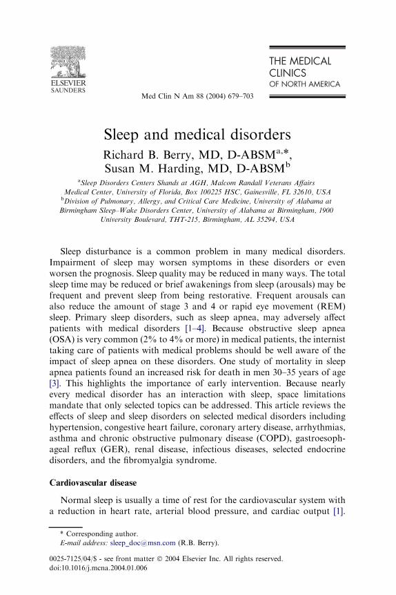
If sleep apnea is effectively treated, does hypertension improve? Thisquestion has been approached by a number of studies that have determinedthe effect of nasal continuous positive airway pressure (CPAP) on nocturnaland daytime blood pressure in patients with OSA. Becker et al [11] foundthat effective treatment of sleep apnea with nasal CPAP for 9 weeks or morelowered both nocturnal and daytime blood pressure by about 10 mm Hgusing a placebo-controlled study (Fig. 1). Other investigations have shownsmaller [12,13] or no effects on daytime blood pressure [14,15]. Theseconflicting results may reflect inadequate CPAP treatment (poor adher-ence); too short a treatment interval; or less severe sleep apnea populations.In general, most hypertensive patients with sleep apnea still continue torequire antihypertensive medications when treated with CPAP. Twenty-four–hour control of blood pressure, however, may improve on CPAPtreatment.
681R.B. Berry, S.M. Harding / Med Clin N Am 88 (2004) 679–703
Congestive heart failure
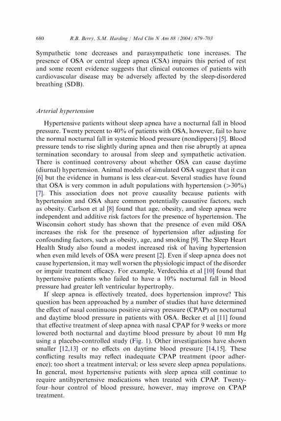
Clinicians may incorrectly dismiss complaints of frequent nocturnalawakenings, daytime sleepiness, or insomnia as simply reflecting the sleepdisturbance associated with congestive heart failure (orthopnea, paroxysmaldyspnea). Recent evidence suggests, however, that occult SDB is common inpatients with congestive heart failure [16,17]. OSA, CSA of the Cheyne-Stokes respiration type (CSA-CSR), and a mixture of OSA and CSA-CSRmay be present in patients with heart failure. CSA-CSR refers to acrescendo-decrescendo pattern of respiration with central apnea at the nadirin effort (Fig. 2). Arousal usually occurs at the peak of the ventilatory phase.There is commonly a delay in the nadir in the arterial oxygen saturation(SaO2) following the event reflecting an increased circulation time. Althougha few patients may exhibit the CSR pattern of breathing during wakefulness,it is usually exhibited only during sleep. Sin et al [18] retrospectivelyevaluated a group of patients with significant left ventricular failure referredto the sleep laboratory and found that risk factors for CSA-CSR includedthe male gender, awake hypocapnia, age greater than 60 years, and the
Fig. 1. (A) The 24-hour blood pressure at baseline (black dots) is reduced after CPAP treatment
(gray dots). The greatest reductions are during the night and in the morning. (B) There is no
difference between baseline and subtherapeutic CPAP. (From Becker HF, Jerrentrup A, Ploch
T, Grote L, Penzel T, Sullivan CE, et al. Effect of nasal continuous positive airway pressure
treatment on blood pressure in patients with obstructive sleep apnea. Circulation 2003;107:68–
73; with permission.)
682 R.B. Berry, S.M. Harding / Med Clin N Am 88 (2004) 679–703
presence of atrial fibrillation. The risk factors for OSA included an increasedbody mass index for men and increased age for women.
In patients with congestive heart failure and OSA, the negative intra-thoracic pressure, hypoxemia, and increased sympathetic tone associatedwith the apneas are believed to impact ventricular function negatively [1].Treatment of OSA with nasal CPAP in patients with cardiomyopathy wasfound to improve the ejection fraction and symptoms [19,20]. This seemsto occur because of a reduction in sympathetic tone and a decrease inventricular afterload [1].
The primary cause of CSA-CSR in patients with congestive heart failurewas once believed to be the long circulation time (delayed feedback tochemoreceptors) causing an overshoot in ventilation. During sleep if thePCO2 falls below a critical level (apneic threshold), central apnea occurs [21].Studies of groups of patients with congestive heart failure, however, havefound equivalent circulation time (and ejection fraction) in congestive heartfailure patients with and without CSA-CSR [22]. Patients with CSR tend tohave daytime hypocapnia, higher ventilatory responses to CO2, and sleepingPCO2 values nearer the apneic threshold [21]. These characteristics increasethe likelihood of instability in ventilatory control and the presence of CSR.The etiology of the hypocapnia seems related at least in part to greaterpulmonary congestion and stimulation of ventilation by J receptors in thelung [23].
Patients with congestive heart failure and CSA-CSR seem to have a worseprognosis than patients with equivalent ejection fractions but without CSR[24–26]. Lanfranchi et al [25] evaluated a cohort of patients with reduced leftventricular ejection fraction by portable monitoring. Patients with atrialfibrillation or an obstructive apnea-hypopnea index greater than 5 per hourwere excluded. Echocardiography to assess chamber size and Holter EKG
Fig. 2. A tracing of central apnea and Cheyne-Stokes respiration in a patient with congestive
heart failure is shown. Note the crescendo-decrescendo pattern of respiration between apneas.
The long delay in the nadir of the arterial oxygen saturation (53.5 seconds) is shown. This is
caused by a prolonged circulation time.
683R.B. Berry, S.M. Harding / Med Clin N Am 88 (2004) 679–703
monitoring to detect heart rate variability were also performed. After a meanfollow-up of 28 months, characteristics of survivors and nonsurvivors weredetermined. As a group, nonsurvivors did have worse symptoms and a lowerejection fraction. Multivariate analysis showed, however, that an apnea-hypopnea index greater than 30 per hour (amount of CSR) and a large leftatrial size were the only independent predictors of cardiac death.
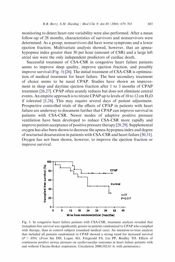
Successful treatment of CSA-CSR in congestive heart failure patientsseems to improve sleep quality, improve ejection fraction, and possiblyimprove survival (Fig. 3) [26]. The initial treatment of CSA-CSR is optimiza-tion of medical treatment for heart failure. The best secondary treatmentof choice seems to be nasal CPAP. Studies have shown an improve-ment in sleep and daytime ejection fraction after 1 to 3 months of CPAPtreatment [26,27]. CPAP often acutely reduces but does not eliminate centralevents. An empiric approach is to titrate CPAPup to levels of 10 to 12 cmH2Oif tolerated [1,26]. This may require several days of patient adjustment.Prospective controlled trials of the effects of CPAP in patients with heartfailure are underway to document further that CPAP can improve survival inpatients with CSA-CSR. Newer modes of adaptive positive pressureventilation have been developed to reduce CSA-CSR more rapidly andimprove patient acceptance of positive pressure therapy [28,29]. Supplementaloxygen has also been shown to decrease the apnea-hypopnea index and degreeof nocturnal desaturation in patients with CSA-CSR and heart failure [30,31].Oxygen has not been shown, however, to improve the ejection fraction orimprove survival.
Fig. 3. In congestive heart failure patients with CSA-CSR, treatment analysis revealed that
transplant-free survival was significantly greater in patients randomized to CPAP who complied
with therapy, than in control subjects (standard medical care). An intention-to-treat analysis
that included all patients randomized to CPAP showed a strong trend for increased survival
(P= .059). (From Sin DD, Logan AG, Fitzgerald FS, Liu PP, Bradley TD. Effects of
continuous positive airway pressure on cardiovascular outcomes in heart failure patients with
and without Cheyne-Stokes respiration. Circulation 2000;102:61–6; with permission.)
684 R.B. Berry, S.M. Harding / Med Clin N Am 88 (2004) 679–703
Arrhythmias and pacing
New information has also emerged concerning the effect of sleep dis-orders on arrhythmias. Patients with OSA typically have a slowing of theheart rate with the onset of apnea and an increase in heart rate followingapnea termination. Although this has been called the brady-tachy pattern,many OSA patients have heart rates that actually remain between 50 and100 beats per minute. The incidence of more significant rhythm disturbancesin patients with OSA has varied between studies. A recent prospective studyof 45 recently diagnosed OSA patients used Holter monitoring for 18 hoursafter diagnosis and again after 2 to 3 days of CPAP treatment [32]. Only 8 ofthe 45 had significant rhythm disturbances including ventricular tachycar-dia, atrial fibrillation, supraventricular tachycardia, and second- or third-degree heart block. In seven of these eight patients CPAP resulted in theabolition of the arrhythmia. Javaheri and Corbett [33] performed Holtermonitoring, polysomnography, and arterial blood gas testing in 59 patientswith stable congestive heart failure and an ejection fraction of 45% or less.Patients with hypocapnia were more likely to have CSA and the presence ofventricular tachycardia was 20 times as great in the hypocapnic patients.
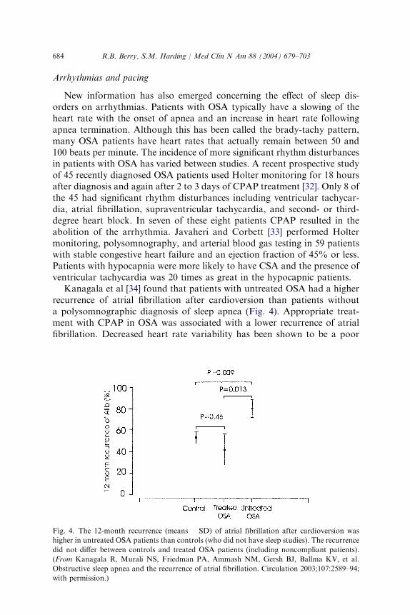
Kanagala et al [34] found that patients with untreated OSA had a higherrecurrence of atrial fibrillation after cardioversion than patients withouta polysomnographic diagnosis of sleep apnea (Fig. 4). Appropriate treat-ment with CPAP in OSA was associated with a lower recurrence of atrialfibrillation. Decreased heart rate variability has been shown to be a poor
Fig. 4. The 12-month recurrence (means � SD) of atrial fibrillation after cardioversion was
higher in untreated OSA patients than controls (who did not have sleep studies). The recurrence
did not differ between controls and treated OSA patients (including noncompliant patients).
(From Kanagala R, Murali NS, Friedman PA, Ammash NM, Gersh BJ, Ballma KV, et al.
Obstructive sleep apnea and the recurrence of atrial fibrillation. Circulation 2003;107:2589–94;
with permission.)
685R.B. Berry, S.M. Harding / Med Clin N Am 88 (2004) 679–703
prognostic sign in patients with cardiovascular disease. In many patientsthis may represent an increase in sympathetic predominance. Khoo et al [35]found that CPAP treatment of OSA improved vagal heart rate control andthat the degree of improvement varied directly with the amount of adherencewith CPAP use.
Because patients with OSA may have nocturnal bradycardia andparoxysmal tachyarrhythmias one group investigated the effects of atrialpacing. An unexpected finding was that atrial overdrive pacing actuallyreduced the number of CSAs and OSAs [36]. The mechanism for this actionis unknown.
Coronary artery disease
There is a circadian peak in the onset of acute myocardial infarction atmidmorning. One retrospective study of 3309 patients found that 26% hadthe onset of acute myocardial infarction during sleep [37]. The patientstended to have lower ejection fraction and older age. Sleep studies werenot performed on the patients. The Sleep Heart Health Study, a largeprospective cohort of patients, found evidence of a modest increase in risk ofhaving self-reported coronary artery disease at even low levels of sleep apnea[2]. Peker et al [38] found an increase in mortality in patients with coronaryartery disease who had untreated OSA.
There have also been a growing number of studies showing changesin blood components or indicators of inflammation in OSA that may beassociated with an increased risk of atherosclerosis or thrombosis. In OSAthere is an increase in the early morning hematocrit [39] and fibrinogenlevels [40] that decreased after CPAP treatment. The levels of vascularendothelial growth factor [41], amount of neutrophil [42,43], and plateletactivation [44] are also reduced with CPAP treatment of patients with OSA.Inflammation is now believed to play a role in atherosclerosis or plaquerupture. The level of C-reactive protein (a marker of inflammation) isreduced with CPAP treatment [45,46]. One study found a reduction in leptin(a hormone secreted by adipose tissue) and a reduction in visceral fat onCPAP treatment [47]. Increased visceral fat is associated with an increasedrisk of cardiac disease.
Sleep and respiratory disease
Asthma
Nocturnal worsening of symptoms and sleep disturbance are significantproblems for patients with asthma. In one study, up to 40% of asthmaticsexperienced symptoms every night [48]. The normal circadian variation inairway function, with the highest airflow in the late afternoon (4:00 PM) andthe lowest in the early morning (4:00 AM), is exaggerated in patients with
686 R.B. Berry, S.M. Harding / Med Clin N Am 88 (2004) 679–703
obstructive airway diseases [49]. In patients with nocturnal asthma theforced expiratory volume in 1 second or peak flow can fall as much as 20%to 40% in the morning hours (morning dippers). The etiology of this var-iation is multifactorial and includes circadian changes in the amounts ofcirculating steroids and epinephrine, cholinergic tone, and possibly in-flammatory mediators in the lungs [49,50]. Sleep also seems to have anadverse effect on asthma, independent of other factors. The easiest way todiagnose severe nocturnal worsening of asthma is to have the patient recordpeak flow measurements at bedtime and on awakening.
Treatment of patients with nocturnal asthma should begin with inhaledcorticosteroids [51]. This medication has been shown to reduce the circadianfluctuation in airway tone. Patients with continued nocturnal symptomsdespite an adequate dose of inhaled corticosteroids can then be treated witha long-acting bronchodilator. Theophylline has been proved effective despitethe stimulatory effects of the medication [49,52,53]. In dosing theophylline,the goal should be to obtain the highest levels during the time of greatestairflow obstruction (at night and early morning). Long-acting inhaled betaagonists (salmeterol and formoterol) are also useful for control of symptomsin nocturnal asthma and potentially might cause less sleep disruption thantheophylline. Selby et al [52], however, found only a slight advantage forsalmeterol compared with theophylline in sleep quality (fewer arousals). Thefalls in morning flow rates were similar but awakenings were less frequent onsalmeterol. Weigand et al [53] found salmeterol to be more effective thantheophylline at preventing the morning drop in flow rates. The drugs did notdiffer in polysomnographic findings but patients perceived better sleep withsalmeterol than theophylline. If OSA is also present, nocturnal asthma mayimprove with effective treatment [54].
Sleep and chronic obstructive pulmonary disease
Patients with COPD often have multiple sleep complaints, such asinsomnia (difficulty initiating or maintaining sleep) and frequent awakeningswith shortness of breath or cough. The sleep of patients with COPD is poor,with low total sleep times, and reduced amounts of slow wave and REMsleep. Airflow obstruction typically worsens in the early morning hourssimilar to asthmatics [55]. Those with moderate to severe COPD may alsoexhibit significant falls in the oxygen saturation during sleep. COPD patientswith an awake PO2 of 50 to 60 mmHg have desaturation during sleep as evennormal persons have a fall in PO2 of 8 to 10 mm Hg during non-REM(NREM) sleep. The most severe desaturations occur during REM sleep,however, when there is skeletal muscle hypotonia and periods of hypo-ventilation characterized by irregular breathing, reduced respiratoryeffort, and small tidal volumes (Fig. 5) [56,57]. Of note, REM-associatednonapneic hypoventilation may result in severe hypoxemia even if thedaytime PO2 is greater than or equal to 60 mm Hg. NREM and REM sleep
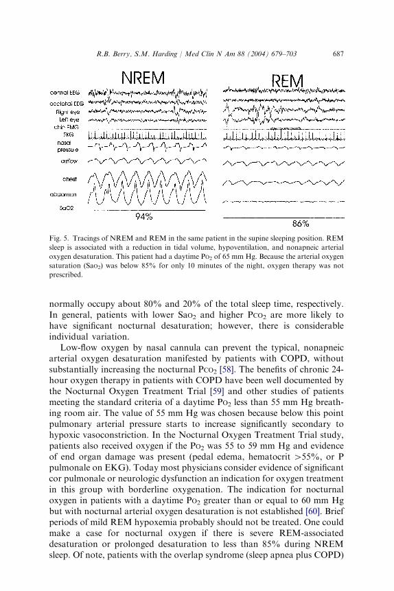
687R.B. Berry, S.M. Harding / Med Clin N Am 88 (2004) 679–703
normally occupy about 80% and 20% of the total sleep time, respectively.In general, patients with lower SaO2 and higher PCO2 are more likely tohave significant nocturnal desaturation; however, there is considerableindividual variation.
Low-flow oxygen by nasal cannula can prevent the typical, nonapneicarterial oxygen desaturation manifested by patients with COPD, withoutsubstantially increasing the nocturnal PCO2 [58]. The benefits of chronic 24-hour oxygen therapy in patients with COPD have been well documented bythe Nocturnal Oxygen Treatment Trial [59] and other studies of patientsmeeting the standard criteria of a daytime PO2 less than 55 mm Hg breath-ing room air. The value of 55 mm Hg was chosen because below this pointpulmonary arterial pressure starts to increase significantly secondary tohypoxic vasoconstriction. In the Nocturnal Oxygen Treatment Trial study,patients also received oxygen if the PO2 was 55 to 59 mm Hg and evidenceof end organ damage was present (pedal edema, hematocrit [55%, or Ppulmonale on EKG). Today most physicians consider evidence of significantcor pulmonale or neurologic dysfunction an indication for oxygen treatmentin this group with borderline oxygenation. The indication for nocturnaloxygen in patients with a daytime PO2 greater than or equal to 60 mm Hgbut with nocturnal arterial oxygen desaturation is not established [60]. Briefperiods of mild REM hypoxemia probably should not be treated. One couldmake a case for nocturnal oxygen if there is severe REM-associateddesaturation or prolonged desaturation to less than 85% during NREMsleep. Of note, patients with the overlap syndrome (sleep apnea plus COPD)
Fig. 5. Tracings of NREM and REM in the same patient in the supine sleeping position. REM
sleep is associated with a reduction in tidal volume, hypoventilation, and nonapneic arterial
oxygen desaturation. This patient had a daytime PO2 of 65 mm Hg. Because the arterial oxygen
saturation (SaO2) was below 85% for only 10 minutes of the night, oxygen therapy was not
prescribed.
688 R.B. Berry, S.M. Harding / Med Clin N Am 88 (2004) 679–703
are best treated with positive airway pressure (CPAP or bilevel positiveairway pressure) with the addition of supplemental oxygen if needed. Givingsuch patients oxygen alone may increase apnea duration, incompletelyreverse desaturation, and may result in large increases in nocturnal PCO2
[58].Treatment of nocturnal symptoms in patients with COPD includes
theophylline and long-acting beta agonists. Sustained-action theophyllineimproves morning pulmonary function compared with short-acting betaagonists without negatively impacting sleep [61]. Theophylline and long-acting beta agonists have not been compared in COPD subjects. In asthma,however, some studies showed a possible slight advantage for long-actingbeta agonists [52]. Ipratropium bromide at bedtime has also been shownto be useful in COPD [62] and the only problem with this medication isthe relatively short duration of action. Many COPD patients complain ofinsomnia despite bronchodilator treatment. Studies have found thatbenzodiazepine receptor agonists are generally safe [63]. COPD patientswith hypoventilation or those with coexistent sleep apnea, however, shouldnot be prescribed hypnotics.
Sleep and gastroesophageal reflux
Important physiologic changes in esophageal function occur during sleepand following arousal from sleep [64,65]. The upper esophageal sphincterpressure decreases from 40 to 20 mm Hg with sleep onset and furtherdecreases to 8 mm Hg during stable sleep. This increases the possibility thatesophageal contents can reach the upper airway or be aspirated into the lung[65]. The lower esophageal sphincter (LES) is the primary antireflux barrier.The LES normally relaxes with swallowing. When the LES relaxes withouta swallow, a transient LES relaxation is said to occur. Although one mightassume that gastroesophageal reflux (GER) occurs because of inadequateLES pressure, most patients with GER have normal LES pressure. TransientLES relaxations are the primary GER mechanism, accounting for 63% to100% of GER episodes [66,67]. Other reflux episodes are secondary to stressreflux (increases in gastric pressure) or free reflux (sustained reduction in LESpressure). In normal subjects, nearly all episodes of GER are caused bytransient LES relaxations. Transient LES relaxations are confined to waketime and following brief arousals from sleep [68]. Sleep also affects esophagealacid clearance mechanisms. Production of saliva (neutralizes acid) andswallowing rapidly clear acid from the esophagus during wakefulness. Salivaproduction ceases during sleep, however, impeding the ability to neutralizerefluxate [69]. Furthermore, swallowing frequency is almost nonexistentduring stable sleep, with swallowing occurring only during brief arousals [70].For these reasons, esophageal acid clearance is markedly prolonged duringsleep, and requires arousal from sleep [70]. If reflux does occur during sleep,acid migrates further upward in the esophagus [71].
689R.B. Berry, S.M. Harding / Med Clin N Am 88 (2004) 679–703
Sleep-related gastroesophageal reflux
Gastroesophageal reflux during sleep is common with up to 10% of thepopulation reporting symptoms of nocturnal reflux in surveys studies [72].In a recent Gallup poll of heartburn patients, 79% reported nighttimeheartburn, of which 75% noted that heartburn negatively affected theirsleep. Despite medical therapy for GER, only 49% had adequate control oftheir nocturnal symptoms [73]. Nocturnal GER is potentially more injuriousthan diurnal GER because acid clearance mechanisms are impaired duringsleep. Freidin et al [68] compared normal subjects and patients with refluxesophagitis with nocturnal monitoring of pH, esophageal manometry, andsleep stage. The LES pressures were similar in normal subjects and patients.Both groups had similar LES pressure during both wakefulness and sleep.The patients had many more reflux episodes, however, with most nocturnalreflux episodes occurring during wake periods and some occurring followingbrief arousals from sleep. Transient LES relaxations accounted for most ofthese episodes.
Because symptomatic reflux is a risk factor for the development ofBarrett’s esophagus (a precursor for carcinoma) [74], nocturnal GER couldpose a significant role in the development of Barrett’s esophagus. One studyof GER patients found that a history of nocturnal reflux increased the riskof having Barrett’s esophagus [75]. Another study, however, did notreplicate this finding [76].
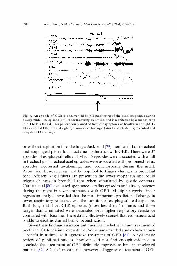
Symptoms of nocturnal GER include multiple awakenings, substernalburning or chest discomfort, indigestion, and heartburn. Other symptomsinclude a sour or bitter taste in the mouth, regurgitation, water brash,coughing, and choking. Diagnostic methods for detecting nocturnal GERinclude esophageal pH testing that has a sensitivity and specificity ofapproximately 90% [77]. It can be integrated with polysomnography (sleepmonitoring) for temporal correlation of sleep-related events, such asarousals. Esophageal pH testing is performed by placing a pH probe inthe distal esophagus (5 cm above the LES). Many laboratories include dualpH probes, where a proximal pH probe is also placed at the upperesophageal sphincter or in the pharynx. A GER episode is defined by thepresence of material that has a pH of less than 4. Fig. 6 shows a GER eventfollowing an arousal from sleep. GER episodes should be suspected onroutine polysomnography if there is an arousal followed by a prolongedperiod of increased chin electromyogram (manifestation of swallowing).
Nocturnal asthma and gastroesophageal reflux
Recently there has been considerable interest in the relationship betweennocturnal GER and asthma. Gislason et al [78] noted that 5% of randomlyselected subjects had nocturnal GER more than once a week, and thatasthma was more frequent in those with nocturnal GER. There is someexperimental evidence to suggest that GER can worsen airway function with
690 R.B. Berry, S.M. Harding / Med Clin N Am 88 (2004) 679–703
or without aspiration into the lungs. Jack et al [79] monitored both trachealand esophageal pH in four nocturnal asthmatics with GER. There were 37episodes of esophageal reflux of which 5 episodes were associated with a fallin tracheal pH. Tracheal acid episodes were associated with prolonged refluxepisodes, nocturnal awakenings, and bronchospasm during the night.Aspiration, however, may not be required to trigger changes in bronchialtone. Afferent vagal fibers are present in the lower esophagus and couldtrigger changes in bronchial tone when stimulated by gastric contents.Cuttitta et al [80] evaluated spontaneous reflux episodes and airway patencyduring the night in seven asthmatics with GER. Multiple stepwise linearregression analysis revealed that the most important predictor of change inlower respiratory resistance was the duration of esophageal acid exposure.Both long and short GER episodes (those less than 5 minutes and thoselonger than 5 minutes) were associated with higher respiratory resistancecompared with baseline. These data collectively suggest that esophageal acidis able to elicit nocturnal bronchoconstriction.
Given these findings an important question is whether or not treatment ofnocturnal GER can improve asthma. Some uncontrolled studies have showna benefit in asthma with aggressive treatment of GER [81]. A systematicreview of published studies, however, did not find enough evidence toconclude that treatment of GER definitely improves asthma in unselectedpatients [82]. A 2- to 3-month trial, however, of aggressive treatment of GER
Fig. 6. An episode of GER is documented by pH monitoring of the distal esophagus during
a sleep study. The episode (arrow) occurs during an arousal and is manifested by a sudden drop
in pH to less than 4. This patient complained of frequent symptoms of heartburn at night. L-
EOG and R-EOG, left and right eye movement tracings; C4-A1 and O2-A1, right central and
occipital EEG tracings.
691R.B. Berry, S.M. Harding / Med Clin N Am 88 (2004) 679–703
in individual symptomatic patients with uncontrolled nocturnal asthma andGER should be considered.
Sleep-related laryngospasm
Gastroesophageal reflux also has a role in sleep-related laryngospasm.Patients abruptly awaken with an intense feeling of suffocation oftenaccompanied with stridor and choking sensations [83]. Other featuresinclude intense anxiety, rapid heart rate, sensation of impending death, andresidual hoarseness. Differential diagnosis for sleep-related laryngospasmincludes OSA, epilepsy, sleep choking syndrome, sleep terrors, vocal corddysfunction, and other upper airway pathologies. Thurnheer et al [83] notedthat 9 of 10 patients with sleep-related laryngospasm had GER documentedby esophageal pH testing [83]. Six patients responded to antireflux therapy,showing that GER may be associated with sleep-related laryngospasm.
Gastroesophageal reflux and obstructive sleep apnea
Given the negative intrathoracic pressure during obstructive apnea andthe frequent arousals from sleep, one would suspect that nocturnal GERis common in patients with OSA. Green et al [84] prospectively examined331 OSA patients. Significant nighttime GER was found in 62% ofsubjects before OSA treatment. Patients compliant with CPAP had a 48%improvement in their nocturnal GER symptoms. There was no change innighttime reflux symptoms if patients did not use CPAP. Furthermore, therewas a strong correlation between higher CPAP pressures and improvementin nocturnal GER symptom scores. This study shows that nocturnal reflux iscommon in OSA patients and that nasal CPAP decreases the frequency ofnocturnal GER symptoms. Of note, the fact that nocturnal GER is commonin OSA patients and that CPAP reduces GER does not necessarily provethat OSA causes GER. In some studies episodes of GER were not correlatedwith apneic events [85]. CPAP by increasing the pressure gradient betweenthe thorax and the stomach may also reduce GER independent of the effectsof CPAP on OSA.
Therapy of sleep-related gastroesophageal reflux
Adequate treatment of GER requires a comprehensive approachincluding lifestyle modifications and medications [86]. Patients should noteat for at least 2 hours before bedtime and avoid foods that promote GER,including high fat–containing foods, caffeine, chocolate, mint, alcohol,tomato products, citrus, and sodas. Medications that promote reflux shouldbe avoided, including calcium channel blockers, theophylline, prostaglan-dins, and bisphosphonates. Smoking significantly decreases LES pressure,so all patients should be encouraged to stop smoking. Patients should looseweight if they are obese and sleep in loose-fitting clothing. Positional
692 R.B. Berry, S.M. Harding / Med Clin N Am 88 (2004) 679–703
therapy can also be used. Sleeping with the head of the bed elevated 6 incheswith a full-length wedge or placing blocks under the head of the bed may beuseful. The right lateral decubitus position worsens GER, whereas the leftlateral decubitus posture seems to be the best sleep position for sleep-relatedGER [87].
Medications to treat sleep-related GER include antacids for acutesymptom control, H2 receptor antagonists, proton pump inhibitors, andprokinetic agents. H2 receptor antagonists provide heartburn relief in 60%of patients and can be given before sleep onset. Proton pump inhibitorsprovide superior gastric acid suppression. One study found that that 40 mgof omeprazole with dinner, or omeprazole, 20 mg, before breakfast and withdinner, resulted in better gastric acid suppression than giving 40 mg beforebreakfast only [88]. Recent data suggest there may be nocturnal acidbreakthrough despite proton pump inhibitor therapy [89]. Whethernocturnal gastric acid breakthrough is clinically important in GER is notknown. Metoclopramide is the only prokinetic agent available in the UnitedStates and has a high prevalence (20% to 50%) of central nervous systemside effects. Prokinetic agents can be used concomitantly with gastric acidsuppressive agents. Antireflux surgery, primarily fundoplication (both openand laparoscopic methods), is successful in 80% to 90% of patients. Long-term results, however, show that many surgically treated patients useantireflux medications regularly [90]. Nasal CPAP therapy has also beenshown to reduce sleep-related GER symptoms [91].
Sleep disturbances in patients with renal disease
Sleep disturbances are also very common in renal disease patients [92].Most investigations examine patients with end-stage renal disease who areon chronic hemodialysis or continuous ambulatory peritoneal dialysis. Sleepcomplaints occur in up to 80% of dialysis patients [92]. Holley et al [93]reported that the most common sleep complaints were nighttime awaken-ings in 67%, early morning awakenings in 80%, restless legs in 72%, jerkinglegs in 83%, and daytime sleepiness in 28% of patients. Dialysis patientsalso have alterations in their objectively recorded sleep architecture. Poly-somnographic features include reduced total sleep times, sleep efficiencies aslow as 66%, and large amounts of wake time [92].
There is also a higher prevalence of OSA in renal disease patientscompared with the general population. Kimmel et al [94], performingpolysomnography in 30 patients with chronic renal failure, found that 73%of patients had sleep apnea. Continuous ambulatory peritoneal dialysispatients had increased sleep fragmentation and lower oxygen saturationsfrom apneas on nights when fluid was present in their abdomens.
Patients with renal disease also have a high prevalence of restless legssyndrome and periodic limb movement of sleep disorder [95]. The restlesslegs syndrome is characterized by unpleasant sensations in the legs or an
693R.B. Berry, S.M. Harding / Med Clin N Am 88 (2004) 679–703
irresistible urge to move the limbs during wakefulness that is temporarilyimproved by movement and is present only or worse in the evening. Uremiais considered a secondary cause of restless legs syndrome. Periodic limbmovement of sleep disorder is defined as complaints of disturbed sleep,insomnia, or daytime sleepiness secondary to periodic movements of the legsduring sleep. Up to 80% of patients with restless legs syndrome also haveperiodic limb movement of sleep disorder. The restless legs syndrome isextremely distressing to many renal patients, and occurs in approximately80% of dialysis patients. Renal transplantation can often result in cure ofthis problem. Treatments of restless legs syndrome include treatment of irondeficiency (if present); dopaminergic agents; narcotics; selected antiepilepticmedications; and benzodiazepines [96].
Patients with end-stage renal disease often complain of daytime sleepinessand objective testing has confirmed this [97]. Daytime sleepiness is oftenmultifactorial. Possible causes include sleep disorders such as insufficientsleep caused by dialysis schedules, OSA, restless legs syndrome or periodiclimb movement of sleep disorder, and direct effects of renal failure. Otherpotential causes of daytime sleepiness include uremic encephalopathy,parathyroid hormone excess (which could have neurotoxic effects), andalterations in neurotransmitter levels. Dialysis may also release cytokinesthat have somnogenic properties, including interleukin-1 and tumor necrosisfactor-a. Rapid changes in the acid-base balance and serum osmolality mayalso affect alertness.
Sleep in infectious disease patients
Most of us notice an increased propensity to sleep when we have aninfection. Infectious products induce release of somnogenic cytokinesincluding interleukin-1 [98]. These immune responses are partially re-sponsible for sleep alterations. This section briefly discusses sleep alterationsin HIV patients and other infections. This section does not discuss centralnervous system infections including African sleeping sickness (Trypanosomabrucei), meningeal encephalitis, and prion diseases.
HIV infection
Patients with HIV infection report many sleep-related symptomsincluding daytime sleepiness, difficulty in initiating and maintaining sleep,and multiple nocturnal arousals (Table 1) [99]. HIV infection alters cytokinesand other immune regulators that may impact sleep. Also, secondaryinfectious processes or medications used to treat HIV infection may disruptsleep. For instance, some patients report insomnia with the use of zidovudine(AZT). Vivid dreams have been reported in patients taking the nonnucleo-side reverse transcriptase inhibitors nevirapine and efavirenz [100].
694 R.B. Berry, S.M. Harding / Med Clin N Am 88 (2004) 679–703
Many investigators have examined sleep architecture in HIV patients.Norman et al [99] found an increase in delta sleep that was more prevalentduring the later part of the sleep time. The effects of HIV infection on sleepvary with the stage of the disease [101]. During the early stages of HIVinfection (CD4 counts[400/mm3), there is mild difficulty in initiating andmaintaining sleep associated with mild and intermittent periods of daytimefatigue. Polysomnographic findings in these patients include an increase intotal slow wave sleep percentage with more slow wave sleep occurringduring the later sleep cycles. Alpha intrusion may also be noted along withmild alterations in the REM-NREM sleep cycles. In patients with moderateHIV infection (CD4 count greater than 200/mm3 but less than 400/mm3),there is more difficulty in maintaining sleep and patients note increasingfatigue. Polysomnographic findings include a decrease in sleep efficiencywith lesser amounts of delta sleep and more difficultly discriminating theNREM-REM sleep cycles. In the terminal stages of HIV infection (CD4counts less than 200/mm3), patients have more difficulty with daytimefatigue and severe difficulty in maintaining sleep. Polysomnographicfindings include further decreases and sometimes even the absence of deltasleep, poor sleep efficiency, and difficulty in recognizing NREM-REM sleepcycles with many spontaneous arousals. It has been postulated that thedecrease in delta sleep as the disease progresses may be related tofluctuations in cytokines and neurologic involvement from the infection[101,102].
Therapy of sleep disturbances in HIV-positive individuals includes goodsleep hygiene practices; avoidance of alcohol, caffeine, and other sleepdisruptive substances; and screening for treatable sleep disorders. Forinstance, lipodystrophy may predispose patients to OSA development.Patients with HIV also have a high prevalence of depression, so this shouldbe screened for and treated if present. Some practitioners use intermittentsedatives and hypnotics for insomnia, which may be helpful in selectedpatients. As more aggressive therapeutic modalities are available to treatHIV infection, clinical outcomes may be improved, and further attention tosleep disturbances may improve the patient’s overall quality of life.
Table 1
Symptoms and signs of sleep symptoms in patients with HIV infection
Stage of disease Symptoms Sleep study findings
Early CD4[400/mm3 Mild insomnia Increased SWS
Mild daytime fatigue Alpha intrusion may occur
400/mm3[ CD4[200 Significant insomnia Decreased SWS
Moderate daytime fatigue
CD4\200/mm3 Severe insomnia Absent SWS
Severe fatigue Fragmented sleep architecture
Abbreviation: SWS, slow wave sleep (stage 3, 4 NREM sleep).
695R.B. Berry, S.M. Harding / Med Clin N Am 88 (2004) 679–703
Other infectious disease and immunization effects
Patients with infectious mononucleosis experience malaise and fatigueduring active infection. Some patients develop chronic sleepiness and fatigueand have prolonged sleep periods and nap throughout the day. Otherchronic infections that may potentially cause fatigue include cytomegalo-virus, hepatitis B and C, Lyme disease [103], and brucellosis. There has alsobeen some interesting evidence showing that sleep deprivation can impairimmune function. For example, preceding sleep deprivation can impair theresponse to vaccination [104].
Sleep in endocrine disorders
Hypothyroidism has been associated with sleep apnea [105]. There are nolarge cohort studies evaluating the prevalence of sleep apnea in hypothyroidsubjects. Obesity may be a significant confounding factor. Pelttari et al [106]examined 26 patients with hypothyroidism and 188 euthyroid controlsubjects finding that 50% of hypothyroid patients and 29% of controlsubjects had significant respiratory events [106]. Whereas some physiciansorder thyroid function tests on all OSA patients, this may not be not costeffective [107]. Postmenopausal women with OSA (who are at higher risk forhypothyroidism) or OSA patients without predisposing OSA risk factors,might warrant thyroid studies. There are case reports showing resolution ofOSA after attaining normal thyroid function; however, it takes an extendedperiod of time. Hypothyroid sleep apnea should be treated as usual (nasalCPAP), while the euthyroid state is being restored, and until a repeat sleepstudy off treatment shows the absence of OSA. Hypothyroidism hasdiffering effects on sleep in patients without OSA, including complaints ofexcessive daytime sleepiness and a reduction in delta sleep percentage.Hyperthyroidism has been associated with insomnia. There are conflictingdata concerning hyperthyroidism’s effect on sleep architecture.
Growth hormone excess resulting in acromegaly is also associated withsleep apnea. Grunstein et al [108] noted that 60% of unselected acromegalypatients have sleep apnea. Potential pathophysiologic mechanisms of thisassociation include macroglossia and increased muscle mass of the upperairway. Because CSA is also noted in acromegalic patients, alterations incentral ventilatory control may also play a role [108]. Acromegalic patientswithout OSA may also have excessive daytime sleepiness with an increase inREM sleep [109]. There are limited data examining sleep characteristics withgrowth hormone deficiency. One study showed a reduction in delta sleep,although more research is needed to make any conclusions.
Adrenocorticosteroid excess, as seen in Cushing’s disease, is associatedwith sleep apnea in approximately 30% of patients [110]. Other inves-tigations have shown shortened REM latencies and poor sleep efficiencies,although more data are needed to draw further conclusions.
696 R.B. Berry, S.M. Harding / Med Clin N Am 88 (2004) 679–703
Studies have suggested that patients with OSA have impaired glucosetolerance, but unfortunately obesity is a major confounding factor. Somestudies have suggested that OSA impairs glucose tolerance independent ofthe associated obesity [111,112]. A large cohort study, however, did notdocument that SDB was an independent risk factor for diabetes [113].Diabetic patients did seem to have more central apnea or periodic breathing.OSA patients with smaller degrees of obesity had a more clear-cutimpairment of glucose control secondary to SDB. The impaired glucosecontrol in OSA patients that is independent of obesity is thought secondaryto increased sympathetic activity. Harsch et al [114] found that CPAPtreatment rapidly improves insulin sensitivity in OSA patients. Theimprovement was greater in patients with lower body mass. Significantimprovement in glucose control with long-term CPAP has yet to bedemonstrated.
Mild to moderate chronic sleep deprivation is a chronic behavior in manyindustrialized societies. Spiegel et al [115] found sleep restriction to 4 hoursof sleep at night impaired glucose tolerance, increased the evening cortisol,and increased sympathetic activation in normal subjects. The authorshypothesized that sleep debt may increase the severity of age-related chronicdisorders.
Fibromyalgia syndrome
Fibromyalgia syndrome is defined by the American College ofRheumatology as the presence of widespread musculoskeletal pain for atleast 3 months, which is bilateral above and below the waist, including axialpain and the presence of 11 of 18 tender points [116]. Fibromyalgiasyndrome should be considered a syndrome rather than a disease process.Fibromyalgia affects about 3% of the population aged 30 to 50 years and70% to 90% of patients are women [117]. Depression is common in thedisorder. The pathophysiology of fibromyalgia syndrome is very complex[118–121]. The main mechanism is thought to be central sensitization ofnociceptive neurons in the dorsal horn of the spinal cord with activationof N-methyl-D-aspartate receptors [121]. This central sensitization resultsin generalized heightened pain sensitivity caused by pathologic nociceptiveprocessing within the central nervous system. There is a threefold increase insubstance P and a decrease in serotonin levels in the cerebrospinal fluid [120].
Sleep complaints are common and include nonrestorative sleep, frag-mented sleep, and insomnia. Poor sleep seems to worsen pain symptomsin 67% of the patients [118]. Sleep studies in fibromyalgia syndrome patientshave shown decreased total sleep time, decreased slow wave and REM sleep,and increased arousals [117]. An interesting EEG pattern (alpha sleep oralpha-NREM anomaly) was first described in fibromyalgia syndromepatients by Moldofsky [122,123]. This is characterized by prominent alphaactivity (8–13 Hz) persisting into NREM sleep (alpha intrusion). Alpha
697R.B. Berry, S.M. Harding / Med Clin N Am 88 (2004) 679–703
activity is normally present during relaxed wake and following briefawakenings (arousals) but is normally virtually absent during stages 2 to 4of NREM sleep. Alpha intrusion into slow wave (delta) sleep is called alpha-delta sleep. Since that time it has been recognized that the alpha-NREMsleep anomaly is not specific for fibromyalgia and is not present in allpatients with this syndrome. Other groups in which the alpha-NREM sleepanomaly can be found include patients with chronic pain syndromes,depression, and diverse causes of nonrestorative sleep. Indeed, alpha sleephas been seen in up to 15% of normal subjects [124]. A variant of alpha sleep(phasic alpha activity) in which alpha intrusion is seen mainly during slowwave sleep rather than being present diffusely in NREM sleep seemed to bepresent in fibromyalgia syndrome patients with prominent sleep distur-bance, subjective feeling of superficial sleep, and more pain and stiffness[125].
Treatments for fibromyalgia include antidepressants, hypnotics, musclerelaxers, cognitive therapy, exercise, biofeedback, and hypnosis [123].Patients should incorporate good sleep hygiene habits. Furthermore,screening for primary sleep disorders is also indicated. Of note, fibromyalgiapatients have a higher prevalence of restless legs syndrome than controls[126]. Some fibromyalgia syndrome patients have clinical improvement withlow doses of antidepressants, whereas others require the usual doses neededfor an antidepressant effect. Antidepressants that have been used includetrazodone, fluoxetine, amitriptyline, and venlafaxine, sometimes in combi-nation [127]. Recently sodium oxybate (c-hydroxybutyrate) was found to beeffective in improving subjective and objective sleep quality (assessed bypolysomnography) and daytime symptoms using a placebo-controlled design[128]. This medication is currently approved by the Food and DrugAdministration only for treatment of cataplexy in narcolepsy. The 5HT3receptor antagonist tropisetron was also found to be effective [129]. The roleof these new treatments in the routine treatment of fibromyalgia remains tobe determined.
References
[1] Leung RST, Bradley TD. Sleep apnea and cardiovascular disease. State of the art. Am J
Respir Crit Care Med 2001;164:2147–65.
[2] Shahar E, Whitney CW, Redline S, Lee ET, Newman AB, Javier Nieto F, et al. Sleep
disordered breathing and cardiovascular disease: cross-sectional results of the Sleep Heart
Health Study. Am J Respir Crit Care Med 2001;163:19–25.
[3] Lavie P, Herer P, Peled R, Berger I, Yoffe N, Zomer J, et al. Mortality in sleep apnea
patients: a multivariate analysis of risk factors. Sleep 1997;20:377–80.
[4] Peker Y, Hedner J, Norum J, Kraiczi H, Carlson J. Increased incidence of cardiovascular
disease in middle-aged men with obstructive sleep apnea. Am J Respir Crit Care Med
2002;166:159–65.
[5] Suzuki M, Guilleminault G, Otsuka K, Shimomi T. Blood pressure ‘‘dipping’’ and ‘‘non-
dipping’’ in obstructive sleep apnea syndrome patients. Sleep 1996;19:382–7.
698 R.B. Berry, S.M. Harding / Med Clin N Am 88 (2004) 679–703
[6] Brooks D, Horner RL, Kozar LF, Render-Teixeira CL, Phillipson EA. Obstructive sleep
apnea as a cause of systemic hypertension: evidence from a canine model. J Clin Invest
1997;99:106–9.
[7] Kales A, Bixler EO, Cadieux RJ, Schneck DW, Shaw LC III, Locke TW, et al. Sleep
apnea in a hypertensive population. Lancet 1984;3:1005–8.
[8] Carlson JT, Hedner JA, Ejnell H, Peterson LE. High prevalence of hypertension in sleep
apnea patients independent of obesity. Am J Respir Crit Care Med 1994;150:72–7.
[9] Peppard PE, Young T, Palta M, Skatrud J. Prospective study of the association between
sleep- disordered breathing and hypertension. N Engl J Med 2000;342:1378–84.
[10] Verdecchia P, Schillaci G, Guerrieri M, Gatteschi C, Benemio G, Boldrini F, et al. Cir-
cadian blood pressure changes and left ventricular hypertrophy in essential hypertension.
Circulation 1990;81:528–36.
[11] Becker HF, Jerrentrup A, Ploch T, Grote L, Penzel T, Sullivan CE, et al. Effect of nasal
continuous positive airway pressure treatment on blood pressure in patients with obstruc-
tive sleep apnea. Circulation 2003;107:68–73.
[12] Pepperell JCT, Ramdassingh-Dow S, Crosthwaite N, Mullins R, Jenkinson C, Stradling
JR, et al. Ambulatory blood pressure after therapeutic and subtherapeutic nasal contin-
uous positive airway pressure for obstructive sleep apnea: a randomized parallel trial.
Lancet 2002;359:204–10.
[13] Faccendia J, Mackay TW, Bood NA, Douglas NJ. Randomized placebo-controlled trial
of continuous positive airway pressure on blood pressure in the sleep apnea-hypopnea
syndrome. Am J Respir Crit Care Med 2001;163:344–8.
[14] Engleman HM, Gough K, Martin SE, Kingshott RN, Padfiled PL, Douglas NJ.
Ambulatory blood pressure on and off continuous positive airway pressure therapy for
the sleep apnea-hypopnea syndrome: effects in ‘‘non-dippers. Sleep 1996;19:378–81.
[15] Dimsdale JE, Loredo JS, Profant J. Effect of continuous positive pressure on blood
pressure placebo trial. Hypertension 2000;35:144–7.
[16] Javaheri S, Parker TJ, Wexler L, Michaels SE, Stanberry E, Nishyama H, et al. Occult
sleep- disordered breathing in stable congestion heart failure. Ann Intern Med 1995;122:
487–92.
[17] Lanfranchi PA, Somers VK, Braghiroli A, Corra U, Eleuteri E, Giannuzzi P. Central
sleep apnea in left ventricular dysfunction: prevalence and implications for arrhythmic
risk. Circulation 2003;107:727–32.
[18] Sin D, Fitzgerald F, Parker J. Risk factors for central and obstructive sleep apnea in 450
men and women with congestive heart failure. Am J Respir Crit Care Med 1999;160:
1101–6.
[19] Malone S, Liu PP, Holloway R, Rutherford R, Xie A, Bradley TD. Obstructive sleep
apnea in patients with dilated cardiomyopathy: effects of continuous positive airway
pressure. Lancet 1991;33:1480–4.
[20] Kaneko Y, Floras JS, Usui K, Plante J, Tkacova R, Kubo T, et al. Cardiovascular effects
of continuous positive airway pressure in patients with heart failure and obstructive sleep
apnea. N Engl J Med 2003;348:1233–41.
[21] Xie A, Skatrud JB, Puleo S, Rahko PS, Dempsey JA. Apnea-hypopnea threshold for CO2
in patients with congestive heart failure. Am J Respir Crit Care Med 2002;165:1245–50.
[22] Naughton M, Benard D, Tam A, Rutherford R, Bradley TD. Role of hyperventilation in
the pathogenesis of central sleep apnea in patients with congestive heart failure. Am Rev
Respir Dis 1993;148:330–8.
[23] Solin P, Bergin P, Richardson M, Kaye DM, Walters EH, Naughton MT. Influence of
pulmonary capillary wedge pressure on central apnea in heart failure. Circulation 1999;
99:1574–9.
[24] Hanly PJ, Zuberi-Khokhar N. Increased mortality associated with Cheyne-Stokes
respiration in patients with congestive heart failure. Am J Respir Crit Care Med 1996;153:
272–6.
699R.B. Berry, S.M. Harding / Med Clin N Am 88 (2004) 679–703
[25] Lanfrachi PA, Braghiroli A, Bosimini E, Mazzuero G, Colombo R, et al. Prognostic
values of Cheyne-Stokes respiration in chronic heart failure. Circulation 1999;99:1435–40.
[26] Sin DD, Logan AG, Fitzgerald FS, Liu PP, Bradley TD. Effects of continuous positive
airway pressure on cardiovascular outcomes in heart failure patients with and without
Cheyne-Stokes respiration. Circulation 2000;102:61–6.
[27] Naughton MW, Liu P, Benard DC, Goldstein RS, Bradley TD. Treatment of congestive
heart failure and Cheyne-Stokes respiration during sleep by continuous positive airway
pressure. Am J Respir Crit Care Med 1995;151:92–7.
[28] Teschler H, Dohring J, Wang YM, Berthon-Jones M. Adaptive pressure support servo-
ventilation: a novel treatment for Cheyne-Stokes respiration in heart failure. Am J Respir
Crit Care Med 2001;164:614–9.
[29] Pepperell JCT, Maskell NA, Jones DR, Lanford-Wiley BA, Crosthwaite N, Stradling JR,
et al. A randomized controlled trial of adaptive ventilation for Cheyne-Stokes breathing
in heart failure. Am J Respir Crit Care Med 2003;168:1109–14.
[30] Javaheri S. Pembrey’s dream: the time has come for a long term trial of nocturnal
supplemental oxygen to treat central sleep apnea in congestive heart failure. Chest 2003;
123:322–5.
[31] Krachman SL, D’Alonzo GE, Berger TJ, Eisen HJ. Comparison of oxygen therapy with
nasal continuous positive airway pressure on Cheyne-Stokes respiration during sleep in
congestive heart failure. Chest 1999;116:1550–7.
[32] Harbison J, O’Reilly P, McNicholas WT. Cardiac rhythm disturbances in obstructive
sleep apnea syndrome: effects of nasal continuous positive airway pressure therapy. Chest
2000;118:591–5.
[33] Javaheri S, Corbett WS. Association of low PCO2 with central sleep apnea and ventricular
arrhythmias in ambulatory patients with stable heart failure. Ann Intern Med 1998;28:
204–7.
[34] Kanagala R, Murali NS, Friedman PA, Ammash NM, Gersh BJ, Ballma KV, et al.
Obstructive sleep apnea and the recurrence of atrial fibrillation. Circulation 2003;107:
2589–94.
[35] Khoo MC, Belozeroff V, Berry RB, Sassoon CSH. Cardiac autonomic control in
obstructive sleep apnea: effects of long term CPAP therapy. Am J Respir Crit Care Med
2001;164:807–12.
[36] Garrigue S, Bordier P, Jais P, Shah DC, Hocini M, Raherison C, et al. Benefit of atrial
pacing in sleep apnea syndrome. N Engl J Med 2002;346:404–12.
[37] Peters RW, Zoble RG, Brooks MM. Onset of myocardial infarction during sleep. Clin
Cardiol 2002;25:237–41.
[38] Peker Y, Hender J, Kraiczi H, Loth S. Respiratory disturbance index: an independent
predictor of mortality in coronary artery disease. Am J Respir Crit Care Med 2000;162:
81–6.
[39] Kreiger J, Sforza E, Barthelmebs M, et al. Overnight decreases in hematocrit after nasal
CPAP with patients with OSA. Chest 1990;97:729–30.
[40] Chin K, Ohi M, Kita H, et al. Effects of NCPAP therapy on fibrinogen levels in
obstructive sleep apnea syndrome. Am J Respir Crit Care Med 1996;153:1972–6.
[41] Lavie L, Kraiczi H, Hefetz A, Ghandour H, Perelman A, Hedner J, et al. Plasma vascular
endothelial growth factor in sleep apnea syndrome: effects of nasal continuous positive air
pressure treatment. Am J Respir Crit Care Med 2002;165:1624–8.
[42] Dyugovskaya L, Lavie P, Lavie L. Increased adhesion molecules expression and produc-
tion of reactive oxygen species in leukocytes of sleep apnea patients. Am J Respir Crit
Care Med 2002;165:934–9.
[43] Schulz R, Mahmoudi S, Hattar K, Sibelius U, Olschewski H, Mayer K, et al. Enhanced
release of superoxide from polymorphonuclear neutrophils in obstructive sleep apnea:
impact of continuous positive airway pressure therapy. Am J Respir Crit Care Med 2000;
162:566–70.
700 R.B. Berry, S.M. Harding / Med Clin N Am 88 (2004) 679–703
[44] Bokinsky G, Miller M, Ault K, Husband P, Mitchell J. Spontaneous platelet activation
and aggregation during obstructive sleep apnea and its response to therapy with nasal
continuous positive airway pressure: a preliminary investigation. Chest 1995;108:625–30.
[45] Shamsuzzaman AS, Winnicki M, Lanfranchi P, Wolk R, Kara T, Accurso V, et al.
Elevated C-reactive protein in patients with obstructive sleep apnea. Circulation 2002;105:
2462–4.
[46] Yokoe T, Minoguchi K, Matsuo H, Oda N, Minoguchi H, Yoshino G, et al. Elevated
levels of C-reactive protein and interleukin-6 in patients with obstructive sleep apnea
syndrome are decreased by nasal continuous positive airway pressure. Circulation 2003;
107:1129–34.
[47] Shimizu K, Chin K, Nakamura T, Masuzaki H, Ogawa Y, Hosokawa R, et al. Plasma
leptin levels and cardiac sympathetic function in patients with obstructive sleep apnoea-
hypopnoea syndrome. Thorax 2002;57:429–34.
[48] Turner Warwick. Epidemiology of nocturnal asthma. Am J Med 1988;85:6–8.
[49] Martin RJ. Nocturnal asthma: circadian rhythms and therapeutic interventions. Am Rev
Respir Dis 1993;147:525–8.
[50] Beam WR, Weiner DE, Martin RJ. Timing of prednisone and alterations of airways in-
flammation in nocturnal asthma. Am Rev Respir Dis 1992;146:1524–30.
[51] Weersink EJM, Douma RR, Postma DS, Koeter GH. Fluticasone propionate, salmeterol
xinafoate, and their combination in the treatment of nocturnal asthma. Am J Respir Crit
Care Med 1997;155:1241–6.
[52] Selby C, Engleman HM, Fitzpatrick MF, Sime PM, Mackay TW, Douglas NJ. Inhaled
salmeterol or oral theophylline in nocturnal asthma? Am J Respir Crit Care Med 1997;
155:104–8.
[53] Wiegand L, Mende CN, Zaidel G, Zwillich CW, Petrocella VJ, Yancey S, et al.
Salmeterol vs Theophylline: sleep and efficacy outcomes in patients with nocturnal
asthma. Chest 1999;115:1525–32.
[54] Chan CS, Woolcock AJ, Sullivan CE. Nocturnal asthma: role of snoring and obstructive
sleep apnea. Am Rev Respir Dis 1988;137:1502–4.
[55] Douglas NJ, Flenley DC. Breathing during sleep in patients with obstructive lung disease.
Am Rev Respir Dis 1990;141:1055–69.
[56] Fletcher EC, Gray BA, Levin DC. Nonapneic mechanisms of arterial oxygen
desaturation during rapid-eye-movement sleep. J Appl Physiol 1983;54:632–9.
[57] Hudgel DW, Martin RJ, Capehart M, et al. Contribution of hypoventilation to sleep
oxygen desaturation in chronic obstructive pulmonary disease. J Appl Physiol 1983;55:
669–77.
[58] Goldstein RS, Ramcharan V, Bowes G, et al. Effect of supplemental nocturnal oxygen on
gas exchange in patients with severe obstructive lung disease. N Engl J Med 1984;310:
425–9.
[59] Nocturnal Oxygen Therapy Trial Group. Continuous or nocturnal oxygen therapy in
hypoxemic chronic obstructive lung disease. Ann Intern Med 1980;93:391–8.
[60] Chaouat A, Weitzenblum E, Kessler R, et al. Outcome of COPD patients with mild
daytime hypoxemia with or without sleep-related oxygen desaturation. Eur Respir J 2001;
17:848–55.
[61] Berry RB, Desa MM, Branum JP, Light RW. Effect of theophylline on sleep and sleep-
disordered breathing in patients with chronic obstructive pulmonary disease. Am Rev
Respir Dis 1991;143:245–50.
[62] Martin RJ, Bartelson BL, Smith P, Hudgel DW, Lewis D, Pohl G, et al. Effect of
ipratropium bromide treatment on oxygen saturation and sleep quality in COPD. Chest
1999;115:1338–45.
[63] Girault C, Muir JF, Mihaltan F, et al. Effects of repeated administration of zolpidem on
sleep, diurnal and nocturnal respiratory function, vigilance, and physical performance in
patients with COPD. Chest 1996;110:1203–11.
701R.B. Berry, S.M. Harding / Med Clin N Am 88 (2004) 679–703
[64] Orr WC, Johnson LF, Robinson MG. The effect of sleep on swallowing, esophageal
peristalsis, and acid clearance. Gastroenterology 1984;86:814–9.
[65] Kahrilas PJ, Dodds WJ, Dent J, Haeberle B, Hogan WJ, Arndorfer RC. Effect of sleep,
spontaneous gastroesophageal reflux, and a meal on upper esophageal sphincter pressure
in normal human volunteers. Gastroenterology 1987;92:466–71.
[66] Mittal RK, Holloway RH, Panagini R, Blackshaw A, Dent J. Transient lower esophageal
sphincter relaxation. Gastroenterology 1995;109:601–10.
[67] Mittal R, Balaban DH. The esophageal junction. N Engl J Med 1997;336:924–32.
[68] Freidin N, Fisher MJ, Taylor W, Boyd D, Surratt P, McCallum RW, et al. Sleep and
nocturnal acid reflux in normal subjects and patients with reflux oesophagitis. Gut 1991;
32:1275–9.
[69] Schneyer LH, Pigman W, Hanahan L, et al. Rate of flow of human parotid, sublingual,
and submaxillary secretion during sleep. J Dent Res 1956;35:109–14.
[70] Orr WC, Johnson LF. Responses to different levels of esophageal acidification during
waking and sleep. Dig Dis Sci 1998;43:241–5.
[71] Orr WC, Elsenbruch S, Harnish MJ, et al. Proximal migration of esophageal acid
perfusions during waking and sleep. Am J Gastroenterol 2000;95:37–42.
[72] Farup C, Kleinman L, Sloan S, Ganoczy D, Chee D, Lee C, et al. The impact of
nocturnal symptoms associated with gastroesophageal reflux disease on health-related
quality of life. Arch Intern Med 2001;161:45–70.
[73] Shaker R, Castell DO, Schoenfeld PS, Spechler SJ. Nighttime heartburn is an under-
appreciated clinical problem that impacts sleep and daytime function: the results of a
Gallup Survey conducted on behalf of the American Gastroenterological Association.
Am J Gastroenterol 2003;98:1487–93.
[74] Lagergren J, Bergstrom R, Lindgren A, Nuyren O. Symptomatic gastroesophageal reflux
as a risk factor or esophageal adenocarcinoma. N Engl J Med 1999;340:825–31.
[75] Gerson LB, Edson R, Lavori PW, Triadafilopoulos G. Use of a simple symptom
questionnaire to predict Barrett’s esophagus in patients with symptoms of gastroesoph-
ageal reflux. Am J Gastroenterol 2001;96:2005–12.
[76] Eloubeidi MA, Provenzale D. Clinical and demographic predictors of Barrett’s esophagus
among patients with gastroesophageal reflux disease: a multivariable analysis in veterans.
J Clin Gastroenterol 2001;33:306–9.
[77] Kahrilas PJ, Quigley EMM. Clinical esophageal pH recording: a technical review for
practice guideline development. Gastroenterology 1996;110:1982–96.
[78] Gislason T, Janson C, Vermeire P, Plaschke P, Bjornsson E, Gislason D, et al.
Respiratory symptoms and nocturnal gastroesophageal reflux: a population-based study
of young adults in three European countries. Chest 2002;121:158–63.
[79] Jack CIA, Calverley PMA, Donnelly RJ, Tran J, Russell G, Hind CRK, et al.
Simultaneous tracheal and oesophageal pH measurement in asthmatic patients with
gastro-oesophageal reflux. Thorax 1995;50:201–4.
[80] Cuttitta G, Cibella F, Visconti A. Spontaneous gastroesophageal reflux and airway
patency during the night in adult asthmatics. Am J Respir Crit CareMed 2000;151:177–81.
[81] Harding SM, Richter JE, Guzzo MR, Schan CA, et al. Asthma and gastroesophageal
reflux: acid suppressive therapy improves asthma outcome. Am J Med 1996;100:395–405.
[82] Gibson PG, Henry RL, Coughlan JL. Gastro-oesophageal reflux treatment for asthma in
adults and children. Cochran Database Syst Rev 2003;2:CD001496.
[83] Thurnheer R, Henz S, Knoblauch A. Sleep-related laryngospasm. Eur Respir J 1997;10:
2084–6.
[84] Green BT, Broughton WA, O’Connor JB. Marked improvement in nocturnal
gastroesophageal reflux in a large cohort of patients with obstructive sleep apnea. Arch
Intern Med 2003;163:41–5.
[85] Graf KI, Karaus M, Heinemann S, et al. Gastroesophageal reflux in patients with sleep
apnea syndrome. Z Gastroenterol 1995;33:689–93.
702 R.B. Berry, S.M. Harding / Med Clin N Am 88 (2004) 679–703
[86] DeVault KR, Castell DO. Updated guidelines for the diagnosis and treatment of
gastroesophageal reflux disease. The Practice Parameters Committee of the American
College of Gastroenterology. Am J Gastroenterol 1999;94:1434–42.
[87] Khoury RM, Camacho-Lobato L, Katz PO, Mohiuddin MA, Castell DO. Influence of
spontaneous sleep positions on nighttime recumbent reflux in patients with gastroesoph-
ageal reflux disease. Am J Gastroenterol 1999;94:2069–73.
[88] Kuo B, Castell DO. Optimal dosing of omeprazole 40 mg daily: effects on gastric and
esophageal pH and serum gastrin in healthy controls. Am J Gastroenterol 1996;91:
1532–8.
[89] Peghini PL, Katz PO, Bracy NA, et al. Nocturnal recovery of gastric acid secretion with
twice-daily dosing of proton pump inhibitors. Am J Gastroenterol 1998;93:763–7.
[90] Spechler SJ, Lee E, Ahnen D, et al. Long-term outcome of medical and surgical therapies
for gastroesophageal reflux disease: follow-up of a randomized controlled trial. JAMA
2001;285:2331–8.
[91] Kerr P, Shoenut JP, Steens RD, Millar T, Miclfliker AB, Kryger MH. Nasal continuous
positive airway pressure: a new treatment for nocturnal gastroesophageal reflux? J Clin
Gastroenterol 1993;17:276–80.
[92] Parker KP. Sleep disturbances in dialysis patients. Sleep Med Rev 2003;7:131–43.
[93] Holley JL, Nespor S, Rault R. Characterizing sleep disorders in chronic hemodialysis
patients. ASAIO Trans 1991;37:M456–7.
[94] Kimmel PL, Miller G, Mendelson WB. Sleep apnea syndrome in chronic renal disease.
Am J Med 1989;86:308–14.
[95] Winkelman JW, Chertow GM, Lazarus JM. Restless legs syndrome in end-stage renal
disease. Am J Kidney Dis 1996;28:372–8.
[96] Earley CJ. Clinical practice: restless legs syndrome. N Engl J Med 2003;348:2103–9.
[97] Stepanski E, Faber M, Zorick F, et al. Sleep disorders in patients on continuous ambu-
latory peritoneal dialysis. J Am Soc Nephrol 1995;6:192–7.
[98] Krueger JM, Majde JA, Obale F. Sleep in host defense. Brain Behav Immun 2003;
17(Suppl 1):S41–7.
[99] Norman SE, Chediak AD, Freeman C, et al. Sleep disturbances in HIV infected men.
AIDS 1996;4:775–81.
[100] Morlese JF, Qazi NA, Gazzard BG, et al. Nevirapine-induced neuropsychiatric compli-
cations, a class effect of non-nucleoside reverse transcriptase inhibitors? AIDS 2002;16:
1840–1.
[101] Darko DF, McCutchan J, Kripke D, Gillin JC, Golshan S. Fatigue, sleep disturbances,
disability and indices of progression in HIV infection. Am J Psychiatry 1992;149:514–20.
[102] Norman SE, Chediak AD, Freeman C, Kiel M, Mendez A, Duncan R, et al. Sleep
disturbances in men with asymptomatic human immunodeficiency virus (HIV) infection.
Sleep 1992;15:150–5.
[103] Greenberg HE, Ney G, Scharf SM, Ravdin L, Hilton E. Sleep quality in Lyme disease.
Sleep 1995;18:912–6.
[104] Spiegel K, Sheridan JF, Van Cauter E. Effect of sleep deprivation on response to im-
munization. JAMA 2002;288:1471–2.
[105] Grunstein RR, Sullivan CE. Sleep apnea and hypothyroidism: mechanisms and manage-
ment. Am J Med 1988;85:775–9.
[106] Pelttari L, Rauhala E, Polo O, et al. Upper airway obstruction in hypothyroidism.
J Intern Med 1994;236:177–81.
[107] Winkelman JW, Goldman H, Piscatelli N, et al. Are thyroid function tests necessary in
patients with suspected sleep apnea? Sleep 1996;19:790–3.
[108] Grunstein RR, Ho KY, Sullivan CE. Sleep apnea in acromegaly. Ann Intern Med 1991;
115:527–32.
[109] Astrom C, Christensen L, Gjerris F, et al. Sleep in acromegaly before and after treatment
with adrenalectomy. Neuroendocrinology 1991;53:328–31.
703R.B. Berry, S.M. Harding / Med Clin N Am 88 (2004) 679–703
[110] Shipley JE, Schteingart DE, Tandon R, et al. Sleep architecture and sleep apnea in
patients with Cushing’s disease. Sleep 1992;15:514–8.
[111] Punjabi NM, Sorkin JD, Katzel LI, Goldberg AP, Schwartz AR, Smith PL. Sleep-
disordered breathing and insulin resistance in middle aged and overweight men. Am J
Respir Crit Care Med 2002;165:677–82.
[112] Meslier N, Gagnadoux F, Giraud P, Person C, Ouksel H, Urban T, et al. Impaired
glucose-insulin metabolism in males with obstructive sleep apnea syndrome. Eur Respir J
2003;22:156–60.
[113] Sanders MH, Givelber R. Sleep disordered breathing may not be an independent risk
factor for diabetes, but diabetes may contribute to the occurrence of periodic breathing in
sleep. Sleep Med 2003;4:349–50.
[114] Harsch IA, Pour Schahin S, Radespiel-Troger M, Weintz O, Jahreiss H, Fuchs FS, et al.
CPAP treatment rapidly improves insulin sensitivity in patients with OSAS. Am J Respir
Crit Care Med 2004;169:159–62.
[115] Spiegel K, Leproult R, Van Cauter E. Impact of sleep debt on metabolic and endocrine
function. Lancet 1999;354:1435–9.
[116] Wolfe F, Smythe HA, Yunus MB, Bennett RM, Bombadier C, Goldenberg DL, et al. The
American College of Rheumatology 1990 Criteria for the classification of fibromyalgia:
report of the multi-center criteria committee. Arthritis Rheum 1990;33:160–72.
[117] Harding SM. Sleep in fibromyalgia patients: subjective and objective findings. Am J Med
Sci 1998;315:367–76.
[118] Okifugi A, Turk DC. Stress and psychophysiological dysregulation in patients with
fibromyalgia syndrome. Appl Psychophysiol Biofeedback 2002;27:129–41.
[119] Neeck G. Pathogenic mechanisms of fibromyalgia. Ageing Res Rev 2002;1:243–55.
[120] Pillemer SR, Bradley LA, Crofford LJ, Moldofsky H, Chrousos GP. The neuroscience
and endocrinology of fibromyalgia. Arthritis Rheum 1997;40:1928–39.
[121] Neeck G, Crofford LJ. Neuroendocrine perturbations in fibromyalgia and chronic fatigue
syndrome. Rheum Dis Clin North Am 2000;26:989–1002.
[122] Branco J, Atalaia A, Paiva T. Sleep cycles and alpha-delta sleep in fibromyalgia syndrome.
J Rheumatol 1994;21:1113–7.
[123] Moldofsky H. Management of sleep disorders in fibromyalgia. Rheum Dis Clin North
Am 2002;28:353–68.
[124] Mahowald ML, Mahowald MW. Nighttime sleep and daytime functioning (sleepiness
and fatigue) in less well-defined chronic rheumatic diseases with particular reference to the
‘‘alpha-delta NREM sleep anomaly.’’ Sleep Med 2000;1:195–207.
[125] Roizenblatt S, Moldofsky H, Benedito-Silva AA, Tufik S. Alpha sleep characteristics in
fibromyalgia. Arthritis Rheum 2001;44:222–30.
[126] Yunus MB, Aldag J. Restless legs syndrome and leg cramps in fibromyalgia syndrome:
a controlled study. BMJ 1996;312:1339.
[127] Goldenberg D, Mayskiy M, Mossey C, Ruthazer R, Schmid C. A randomized, double-
blind crossover trial of fluoxetine and amitriptyline in the treatment of fibromyalgia.
Arthritis Rheum 1996;39:1852–9.
[128] Scharf MB, Baumann M, Berkowitz DV. The effects of sodium oxybate on clinical
symptoms and sleep patterns in patients with fibromyalgia. J Rheumatol 2003;30:1070–4.
[129] Farber L, Stratz TH, Bruckle W, Spath M, Pongratz D, Lautenschlager J, et al. Short-
term treatment of primary fibromyalgia with the 5–HT3-receptor antagonist tropisetron:
results of a randomized, double-blind, placebo-controlled multicenter trial in 418 patients.
Int J Clin Pharmacol Res 2001;21:1–13.