skin and wound care training module · pdf filerisk assessment of skin breakdown and ......
TRANSCRIPT

RENFREW VICTORIA HOSPITAL
SKIN AND WOUND CARE PROGRAM TRAINING
RISK ASSESSMENT OF SKIN BREAKDOWN AND TREATMENT OF WOUNDS AND PRESSURE ULCERS
SELF-LEARNING MODULE For Registered Nurses and Registered Practical Nurses
DIRECTIONS: 1. READ MATERIAL IN MODULE 2. ATTEND A SKIN AND WOUND CARE SESSION 3. PERFORM A RISK ASSESSMENT (BRADEN) & CARE PLAN AND REVIEW WITH
INSTRUCTOR 4. RECEIVE CERTIFICATE OF COMPLETION
June 2000
Prepared by: Rose-Marie Dolinar, R.N., MScN
Reviewed by: Donna Keon, R.N. Lee-Ann Somerville, R.P.N. Sandra Tramer, R.N., Nurse Manager Approved by: Nancy Kelly, R.N., B.Sc.N., M.P.A., Assistant Executive
Director, Renfrew Victoria Hospital

2
TABLE OF CONTENTS
1. Objective of the Skin and Wound Care Program Training.............................................................................. 3 2. RVH Skin and Wound Care Program Administrative Policy .......................................................................... 4 3. Predisposing Factors to Skin Breakdown.......................................................................................................... 5 4. Braden Scale for Predicting Pressure Sore Risk................................................................................................ 5 5. Cardinal Rules of Prevening Skin Breakdown................................................................................................... 8 6. Classification of Pressure Ulcers and Wounds................................................................................................ 8 7. The Wound Healing Scale ® (Krasner, 1996).................................................................................................. 9 8. Differentiating Ulcer Types ................................................................................................................................. 9 9. Infection versus Inflammation........................................................................................................................... 10 10. The Ideal Dressing (Eisenbud, 1999). .......................................................................................................... 10 11. Wound Dressings by Categories ................................................................................................................. 12 12. Dressings and Topical Agents used in the Treatment of Wounds and Pressure Ulcers.................... 13 13. Suggested Pressure Ulcer Treatment........................................................................................................... 14 14. Nursing Care Plan ........................................................................................................................................... 15 15. References........................................................................................................................................................ 16

3
1. Objective of the Skin and Wound Care Program Training
Upon completion of the training, the participant will meet the following objectives for the FOUR key areas of the Skin and Wound Care Program:
PROGRAM PARTICIPATION The nurse will:
1. Explain the purpose of the Skin and Wound Care Program 2. Support the application of Best Practice as part of the Skin and Wound Care
program
MAINTAINING HEALTHY SKIN The nurse will:
1. Explain the structure and function of normal skin 2. Describes the multiple factors in maintaining and promoting healthy skin
PREVENTION OF SKIN BREAKDOWN The nurse will:
1. Perform a risk assessment using a standard assessment tool 2. Describe and apply the cardinal rules of preventing skin breakdown
APPLICATION OF STANDARD NURSING INTERVENTIONS FOR CARE OF WOUNDS
AND PRESSURE ULCERS The nurse will:
1. Describe the different stages and classifications of wounds. 2. Assess the skin wound and/or ulcer for most likely cause, pending confirmation of
clinical diagnosis. 3. Utilize the RVH Wound Assessment and Treatment Form. 4. Describe the purpose and application of each dressing type. 5. Provide appropriate nursing interventions using standardized procedures. 6. Formulate a care plan for ongoing consistent wound care, whether the patient is
within the hospital or being followed in the community.
4
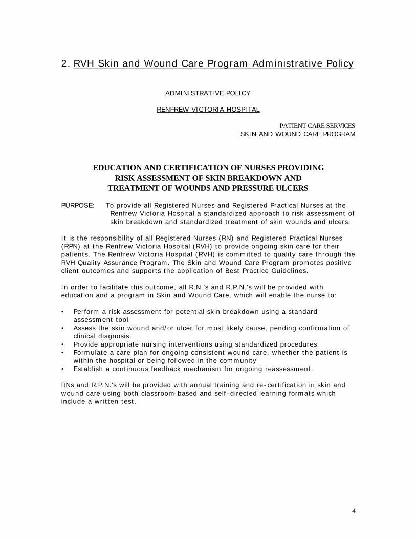
2. RVH Skin and Wound Care Program Administrative Policy
ADMINISTRATIVE POLICY
RENFREW VICTORIA HOSPITAL
PATIENT CARE SERVICES
SKIN AND WOUND CARE PROGRAM
EDUCATION AND CERTIFICATION OF NURSES PROVIDING RISK ASSESSMENT OF SKIN BREAKDOWN AND
TREATMENT OF WOUNDS AND PRESSURE ULCERS
PURPOSE: To provide all Registered Nurses and Registered Practical Nurses at the Renfrew Victoria Hospital a standardized approach to risk assessment of skin breakdown and standardized treatment of skin wounds and ulcers.
It is the responsibility of all Registered Nurses (RN) and Registered Practical Nurses (RPN) at the Renfrew Victoria Hospital (RVH) to provide ongoing skin care for their patients. The Renfrew Victoria Hospital (RVH) is committed to quality care through the RVH Quality Assurance Program. The Skin and Wound Care Program promotes positive client outcomes and supports the application of Best Practice Guidelines. In order to facilitate this outcome, all R.N.’s and R.P.N.’s will be provided with education and a program in Skin and Wound Care, which will enable the nurse to: • Perform a risk assessment for potential skin breakdown using a standard
assessment tool • Assess the skin wound and/or ulcer for most likely cause, pending confirmation of
clinical diagnosis, • Provide appropriate nursing interventions using standardized procedures, • Formulate a care plan for ongoing consistent wound care, whether the patient is
within the hospital or being followed in the community • Establish a continuous feedback mechanism for ongoing reassessment. RNs and R.P.N.'s will be provided with annual training and re-certification in skin and wound care using both classroom-based and self-directed learning formats which include a written test.

5
3. Predisposing Factors to Skin Breakdown • MECHANICAL FACTORS: Pressure, Friction & Shearing • MOBILITY • NUTRITIONAL STATUS • BODY MASS • CHRONIC DISEASES • NEOPLASMS • NEUROLOGICAL DISORDERS • MEDICATIONS • SKIN STATUS • SMOKING • EXPOSURE TO MOISTURE • HYDRATION
4. Braden Scale for Predicting Pressure Sore Risk Client's Name ________________________ Evaluator's Name__________________________________ Date of assessment ______________________
Sensory perception Ability to respond meaningfully to pressure-related discomfort
1. Completely limited: Unresponsive (does not moan, flinch, or grasp) to painful stimuli, due to diminished level of consciousness or sedation.
OR Limited ability to feel pain over most of body surface.
2. Very limited: Responds to only painful stimuli. Cannot communicate discomfort except by moaning or restlessness.
OR Has a sensory impairment, which limits the ability to feel pain or discomfort over ½ of body.
3. Slightly limited: Responds to verbal commands but cannot always communicate discomfort or need to be turned.
OR Has some sensory impairment, which limits ability to feel pain or discomfort in 1 or 2 extremities.
4. No impairment: Responds to verbal commands. Has no sensory deficit, which would limit ability to feel or voice pain or discomfort.
Moisture Degree to which skin is exposed to moisture.
1. Constantly moist: Skin is kept moist almost constantly by perspiration, urine, etc. Dampness is detected every time patient is
2. Moist: Skin is often but not always moist. Linen must be changed at least once a shift.
3. Occasionally moist: Skin is occasionally moist requiring an extra linen change approximately once a day.
4. Rarely moist: Skin is usually dry; linen requires changing only at routine intervals.

6
moved or turned.
Activity Degree of physical activity
1. Bedfast: Confined to bed.
2. Chairfast: Ability to walk severely limited or nonexistent. Cannot bear down weight and/or must be assisted into chair or wheelchair.
3. Walks occasionally: Walks occasionally during day but for very short distances, with or without assistance. Spends majority of each shift in bed or chair.
4. Walks frequently: Walks outside the room at least twice a day and inside room at least once every 2 hours during waking hours.
Mobility Ability to change and control body position.
1. Completely immobile: Does not make even slight changes in body or extremity position without assistance.
2. Very limited: Makes occasional slight changes in body or extremity position but unable to make frequent or significant changes independently.
3. Slightly limited: Makes frequent though slight changes in body or extremity position independently.
4. No limitations: Makes major and frequent changes in position without assistance.
Nutrition Usual food intake pattern.
1. Very poor: Never eats a complete meal. Rarely eats more than 1/3 of any food offered. Eats 2 servings or less of protein (meat or dairy products) per day. Takes fluids poorly. Does not take a liquid dietary supplement.
OR Is NPO1 and/or maintained on clear liquids or IV2 for more than 5 days.
2. Probably inadequate: Rarely eats a complete meal and generally eats only ½ of any food offered. Protein intake includes only 3 servings of meat or dairy products per day. Occasionally will take a dietary supplement.
OR Receives less than optimum amount of liquid diet or
3. Adequate: Eats over half of most meals. Eats a total of 4 servings of protein (meat or dairy products) each day. Occasionally refuses a meal, but will usually take a supplement if offered.
OR Is on a tube feeding or TPN3 regimen, which probably meets most of nutritional
4. Excellent: Eats most of every meal. Never refuses a meal. Usually eats a total of 4 or more servings of meat and dairy products. Occasionally eats between meals. Does not require supplementation.

7
tube feeding. needs. Friction and shear
1. Problem: Requires moderate to maximum assistance in moving. Complete lifting without sliding against sheets is impossible. Frequently slides down in bed or chair, requiring frequent repositioning with maximum assistance. Spasticity, contractures, or agitation leads to almost constant friction.
2. Potential problem: Moves feebly or requires minimum assistance. During a move, skin probably slides to some extent against sheets, chair, restraints, or other devices. Maintains relatively good position in chair or bed most of the time but occasionally slides down.
3. No apparent problem: Moves in bed and in chair independently and has sufficient muscle strength to lift up completely during move. Maintains good position in bed or chair at all times.
1 NPO: Nothing by mouth. 2 IV: Intravenously. 3 TPN: Total parenteral nutrition.
Scoring: The maximum possible score is 23. A lower score reflects higher risk for pressure ulceration. The cutoff score to denote risk for pressure ulceration is <16. Source: Reprinted with permission, Braden B, Bergstrom N. Predictive validity of the Braden Scale for pressure sore risk in a nursing home population. Res Nurs Health. 1994;17:459-470.

8
5. Cardinal Rules of Prevening Skin Breakdown
• No wet to dry dressings, as this pulls off dead and live cells and tissue. • If the dressing sticks, change the type of dressing or increase the time between
dressing changes. • Do not rub directly on phase alert (stage 1) areas. This increases the damage to
cells • Do not use doughnuts or IV bags to cover ulcers. This can decrease blood supply
to the ulcer area. • Do not position pillows under knees, place the pillows under the legs from the
knees to ankles, leaving the heels hang over • For stasis ulcers - compression, compression, compression (ted's or TENSORS) • Always cleanse with sea clens or normal saline DO NOT USE HYDROGEN PEROXIDE - IT DESTROYS NEW CELLS
6. Classification of Pressure Ulcers and Wounds Stage 1 Non-blanchable erythema of intact skin, the heralding lesion of skin
ulceration. In individuals with darker skin, discoloration of the skin, warmth, edema, induration, or hardness may also be indicators.
Stage 2 Partial-thickness skin loss involving epidermis, dermis, or both.
Stage 3 Full-thickness skin loss involving damage to or necrosis of subcutaneous tissue that may extend down to, but not through, underlying fascia. The ulcer presents clinically as a deep crater with or without undermining adjacent tissue.
Stage 4 Full-thickness skin loss with extensive destruction, tissue necrosis, or damage to muscle, bone, or supporting structures, i.e. tendon or joint capsule.
9
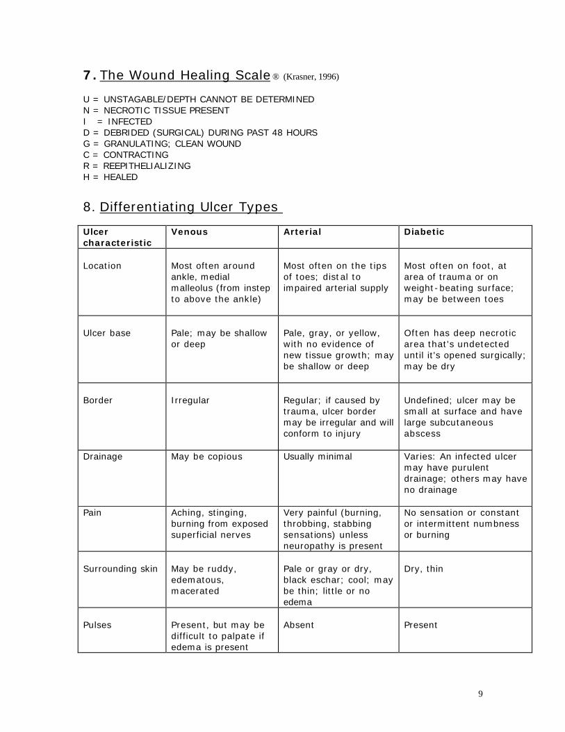
7. The Wound Healing Scale ® (Krasner, 1996)
U = UNSTAGABLE/DEPTH CANNOT BE DETERMINED N = NECROTIC TISSUE PRESENT I = INFECTED D = DEBRIDED (SURGICAL) DURING PAST 48 HOURS G = GRANULATING; CLEAN WOUND C = CONTRACTING R = REEPITHELIALIZING H = HEALED
8. Differentiating Ulcer Types Ulcer characteristic
Venous Arterial
Diabetic
Location
Most often around ankle, medial malleolus (from instep to above the ankle)
Most often on the tips of toes; distal to impaired arterial supply
Most often on foot, at area of trauma or on weight-beating surface; may be between toes
Ulcer base
Pale; may be shallow or deep
Pale, gray, or yellow, with no evidence of new tissue growth; may be shallow or deep
Often has deep necrotic area that's undetected until it's opened surgically; may be dry
Border
Irregular
Regular; if caused by trauma, ulcer border may be irregular and will conform to injury
Undefined; ulcer may be small at surface and have large subcutaneous abscess
Drainage May be copious Usually minimal Varies: An infected ulcer may have purulent drainage; others may have no drainage
Pain Aching, stinging, burning from exposed superficial nerves
Very painful (burning, throbbing, stabbing sensations) unless neuropathy is present
No sensation or constant or intermittent numbness or burning
Surrounding skin
May be ruddy, edematous, macerated
Pale or gray or dry, black eschar; cool; may be thin; little or no edema
Dry, thin
Pulses
Present, but may be difficult to palpate if edema is present
Absent
Present

10
9. Infection versus Inflammation What to look for when suspecting infection of the wound: All wounds contain bacteria. Differentiation between wound colonization/bacteria and infection can be difficult. Features that suggest infection is playing a role are new pain, ulcer extension and change in nature and amount of exudate (Pierscianowski, 1995). What to look for when differentiating inflammation from infection:
INFLAMMATION
• Promotes healing by initiating the subsequent phases of healing
• Usually lasts up to 2 weeks • If prolonged, can slow healing since
there is no collagen synthesis
INFECTION
• Prolongs the inflammation and delays healing
• Induces tissue damage • Exudate can macerate surrounding
skin and dilute wound healing factors at the wound surface.
Classic signs of inflammation include: • Pain • Heat • Redness • Swelling A percentage of yellow slough can indicate inflammation in a wound.
Suspect infection is there is: • New pain • Increase in redness or increase in
area of redness • Change in nature and amount of
drainage/exudate • Change in odor
Nursing intervention for signs of infections: swab the wound AFTER cleansing with Sea Clens or Sterile Saline (see recommended pattern). Remember that COLONIZATION OF WOUND IS NORMAL, INFECTION IS NOT since INFECTION WILL PROLONG OR ARREST HEALING.
10. The Ideal Dressing (Eisenbud, 1999). Dressings are applied after wound debriding and/or cleaning, and are chosen according to the stage and color classification of the wound and whether the wound is dry or wet. There is no one perfect dressing that is applicable to all clinical situations. The ideal dressing is EASY TO APPLY. As well, the ideal dressing:
• Can be removed easily and completely • Lessens wound pain • Is acceptable to patient

11
• Maintains a moist wound environment • Is impermeable to external contamination • Absorbs excess wound fluid • Helps to prevent external injury to the wound bed • Avoids damaging the wound edges • Avoids maceration of surrounding tissue • Requires infrequent changes • Reduces scarring • Is readily available • Is affordable (within the means of the client and caregiver for continuity of care)
CHOOSING THE APPROPRIATE DRESSING IS A BALANCE BETWEEN MAINTAINING A MOIST ENVIRONMENT AND AVOIDING MACERATION OR DRYING OUT THE WOUND. The choice of dressing will change as the wound progresses towards healing.

12
11. Wound Dressings by Categories
CATEGORY
PRODUCT* PURPOSE
CLEANSER
SEA CLENS® OR STERILE SALINE
Cleanse wound without destroying new cells
DEBRIDING AGENTS
Triad®
To breakdown necrotic tissue and promote healing
TRANSPARENT DRESSINGS
Tegaderm®
To protect wound and maintain moist environment
HYDROCOLLOID AND GELS (MOISTURISERS)
Comfeel® Curafil gel®
To provide moisture to a dry wound
ABSORPTIVE DRESSING
Fibracol® Sea sorb® Mesalt®
To absorb exudate from wet wounds and provide collagen
NON-ADHERENT DRESSING
Vaseline Gauze Telfa® Jelonet®
To protect wound and avoid sticking
∗ NOTE: the products may change depending on best client outcomes and product
improvements. These are some of the products used at Renfrew Victoria Hospital (May 2000).

13
12. Dressings and Topical Agents used in the Treatment of Wounds and Pressure Ulcers
Category Indications for use
Comments
Cleanser
All wounds, all stages
Apply enough pressure to cleanse, light pat with gauze
Debriding agent
Stages 2, 3 & 4
May require secondary dressing
Transparent dressings
Stages 1,2
Manages light exudate only, may tear fragile skin
Gel and Hydrocolloids (brings moisture to a dry wound)
Stage 2 and 3
May macerate surrounding tissue, may require multiple dressing changes
Absorptive dressing (absorbs exudate from wet wounds)
Stages 3 & 4
permeable to fluids and bacteria, may be difficult to remove
Non-adherent moisturizing dressing
Stages 1 & 2 and 3 & 4 if appropriate
no absorption, contact layer with wound to prevent outer dressing from sticking

14
13. Suggested Pressure Ulcer Treatment
Ulcer Stage
Suggested Treatment
Stage 1 ulcer • skin care, reduce pressure, reduce friction, prevent shear
Dressings: transparent dressings
Stage 2 ulcer • skin care, local pressure elimination, cleansing with normal saline, inspect for secondary infection
Dressings: transparent dressings, hydrocolloids for dry ulcer; absorptive dressings for wet ulcer
Stage 3 ulcer • skin care, local pressure elimination, cleansing with normal saline, antibiotics if needed, debridement
Dressings: absorption dressings for wet ulcer; hydrocolloids for dry ulcer
Stage 4 ulcer • skin care, local pressure elimination, antibiotics, debridement
Dressings: absorptive dressings for wet ulcer; hydrocolloids for dry ulcer.

15
14. Nursing Care Plan
SKIN CARE AND RISK ASSESSMENT RISK ASSESSMENT (BRADEN)
ONGOING CARE PLAN OF ACTION
Calculated Braden Score= The maximum possible score is 23. A lower score reflects higher risk for pressure ulceration. The cutoff score to denote risk for pressure ulceration is <16.
• Change position q 2 hours • Encourage water intake
min.1500cc • reduce potential for
mechanical injury (pressure, friction, shearing)
• apply Cardinal rules for preventing skin breakdown
• Reassess risk assessment if situation of patient changes (ex. becomes incontinent, immobile).
WOUND CARE
ASSESS WOUND BY: STAGE: 1,2,3 or 4 COLOUR: black, yellow or red (with percentages) and whether wet or dry SIZE DEPTH EXUDATE OR ODOUR PRESENCE OF PAIN
NURSING INTERVENTION
• cleanse • debride • hydrate for dry wound or
absorb for wet • protect with secondary
dressing as needed
REASSESS
set change of dressing date to allow for maximum moist healing without macerating or drying wound edge.
example: May 27, 2000 o- Stage 2, yellow (100%)
dry ulcer on rt. forearm Pain: pain 0 on scale 0 (no pain) to 10 (extreme pain) Size: 5cm by 3 cm Depth: 0.5 cm
example: • cleansed with normal saline
or Sea Clens, • applied debriding product
(Triad) with non-adherent dressing (Jelonet), 4x4s and Tegaderm.
example: REASSESS May 30, 2000 • remove dressing,
reassess wound for stage, color, exudate, odor, pain and healing status, and dress wound as needed.

16
15. References
Braden B, Bergstrom N. Predictive validity of the Braden Scale for pressure sore risk in a nursing home population. Res Nurs Health. 1994;17:459-470. Eisenbud, D.E. (1999). Modern Wound Management. Columbus Ohio: Anadem Publishing Inc. Krasner, D. (1996). Wound Healing Scale, in Chronic Wound Care, Second Edition, from the Wound Care Symposium, Toronto, November 1999. Ottawa General Hospital (1996). Drug Administration Notes for Nurses, Drug Consultation Services. No.3. Perricone, N. (1999). How to approach acute and chronic wound healing in the elderly. Wounds, 11(6), 145-151. Pierscianowski, T.A. (1995). Why won't that leg ulcer heal? The Canadian Symposium on Wound Management. Toronto. Renfrew Victoria Hospital (1998). General Procedure Manual: Nursing Services. Visinski, P. (2000). Personal communication. Wound Care Course material and slides from clinical practice Renfrew Victoria Hospital.