simple and robust whole blood staining procedure using ... · simple and robust whole blood...
TRANSCRIPT
Simple and robust whole blood staining procedure using PerFix-nc reagentsgenerates unprecedented FOXP3 signal/noise ratioFabrice Malergue1, Laeticia Khemici1, Tewfik Miloud1, Carlos A. Garcia2, Félix A. Montero-Julian1
Beckman Coulter Life Science, Inc., 1Global Assay and Applications Development, Marseille, France, 2Life Science Research, Miami, USA
INTRODUCTIONFOXP3 is a nuclear antigen expressed specifically in regulatory T cells (Tregs). Tregs play a critical role in the maintenance of the dominant self-tolerance and have been subject of extensive research efforts for the last two decades. Today the detection of FOXP3 staining is laborious, time consuming (3-4 hours) and poorly reproducible.
A new commercially available method, named PerFix-nc*, allows simultaneous intra- and extra-cellular staining without any wash step. This new permeabiliza-tion and fixation procedure allows a reduction of the workflow to 45 min. In these conditions, most common intracellular antigens are easily and efficiently stained.
In this study we aimed at the optimization of the PerFix-nc protocol for the detection of FOXP3, an antigen that is difficult to detect by currently available techniques. We show that a smart modification of PerFix-nc staining procedure allows a clean, efficient and reproducible detection of FOXP3 in whole blood samples.
MATERIALS & METHODSIllustration of the straight PerFix-nc procedure:
EDTA blood from healthy individuals was stained with FOXP3 antibody con-jugated to Alexa Fluor 647 (BioLegend), CD4, CD25, and CD127 conjugates (Beckman Coulter) were also used. As a starting point, we used the com-mercially available reagents provided in the PerFix-nc kit (Beckman Coulter), following the manufacturer instructions. Then, different reagent volumes and kinetics were tested and optimized. Reagents for blocking non-specific staining as well as additional washing steps were also investigated. Optimized method was compared with the reference procedure (eBioscience), considered as the current gold standard.
Samples were acquired on a Gallios* cytometer and data were analyzed with Kaluza* software (Beckman Coulter). All comparisons between methods were run on the same blood samples, with the same cytometer settings, using FITC + Alexa 647 conjugates which did not require any compensation.
RESULTS(RMFI=Relative Mean Fluorescence Intensity = S/N ratio)Figure 1. Comparison between the reference procedure (eBioscience, without the addition of normal human serum) and the PerFix-nc procedure (with the optional final wash).
Both methods are providing comparable results ( 8% of CD4+ cells are stained by the anti-FoxP3 conjugate, with a signal/noise ratio around 4 ).
Scatter characteristics of the cells are very different: With PerFix-nc a clear separation of lymphocytes / monocytes / granulocytes is observed, whereas monocytes and granulocytes are undistinguishable with the eBioscience pro-cedure. Discrimination between debris and lymphocytes is also more accurate with the Perfix-nc procedure.
CD4-FITC staining is reduced in the PerFix-nc procedure but still clearly detectable, indicating that any other conjugate with a brighter dye than FITC will be easily detectable (see CD4-PC7 on Figure 7).
Figure 2. eBioscience procedure : Comparison of two anti-FoxP3 clones (206D and 259D), with addition of normal serum to block non-specific back-ground.
Results are significantly improved by the addition of human serum (not provided with eBioscience kit), and clone 206D displays an enhanced S/N ratio (around 7) in this procedure.
Figure 3. Optimization of the FoxP3 staining in the PerFix-nc procedure (clone 259D): Increase of the incubation time and addition of a second wash step:
• Results are significantly improved when the staining time is increased from 30 min. to 60 min.
• A second wash also improves the S/N in all conditions (data not shown), and is even more beneficial if the amount of conjugate is increased from the recommended 5µL to 10µL.
• The combination of these 3 modifications produces a significant improvement of the S/N, from 4 to 8-9 (right histogram).
Figure 4. Optimization of the FoxP3 staining in the PerFix-nc procedure: Replacement of the final R3 solution for the additional wash step.
The amount of final R3 provided in the kit does not allow to perform 2 washes at the end of the procedure. Other common buffers available in most laboratories (PBS, PBS-BSA 0.5%, PBS-FCS 0.5%, and PBS-formaldehyde 0.5%) have thus been evaluated to replace one of the two washes in all possible combinations.
Surprisingly, PBS (or PBS-proteins) induced a dramatic signal improvement when used as the first wash buffer. No other combination reached similar results, indicating that the first wash buffer must be devoid of fixative or detergent, whereas the second wash buffer must be the final R3 Reagent.
Figure 5. Confirmation of the staining improvement on additional samples, & stability of the sample staining:
Three other blood samples from normal donors have then been analyzed with this new procedure to confirm the results, and the stained samples have been stored for 6 hours at room temperature to evaluate their stability. S/N of 10 to 15 are again obtained, and are stable.
Figure 6. Comparison of the two FoxP3 clones in this new procedure:
*Gallios, Perfix-nc and Kaluza are for research use only. Not for use in diagnostic procedures. Gallios and Kaluza are trademarks of Beckman Coulter, Inc. The stylized logo are registered trademarks of Beckman Coulter, Inc and are registered in the USPTO. Alexa Fluor is a regis-tered trademark of Molecular Probes, Inc.
It is clear from this experiment that the first wash with PBS plays a critical role in the quality of the staining for both clones.
Nevertheless, and in contrast to the eBioscience procedure (see Figure 2), the clone 259D is more efficient for FoxP3 staining in the optimized PerFix-nc procedure. Figure 4. Fluorescent protein detection with 488 nm and 561 nm excitation.Figure 7. Questionable role of CD25 for the gating of Tregs:
Optimized PerFix-nc procedure allows an easy separation of FoxP3+ cells from CD4+FoxP3- cells demonstrating then no benefit in using a dim CD25 marker for Treg gating. It thus offers the possibility to use more markers for FoxP3+ cells characterization.Figure 8. Repeatability studies:
Eight replicates were performed on the same blood sample using either PerFix-nc or eBioscience procedures.In order to analyze the variability of the results mean % of positive cells and S/N ratios, and the corresponding standard deviations are shown:The discrimination improvement with PerFix-nc allows to gate an ad-ditional 1% FoxP3+CD4+ cells with an even lower variability. More strikingly
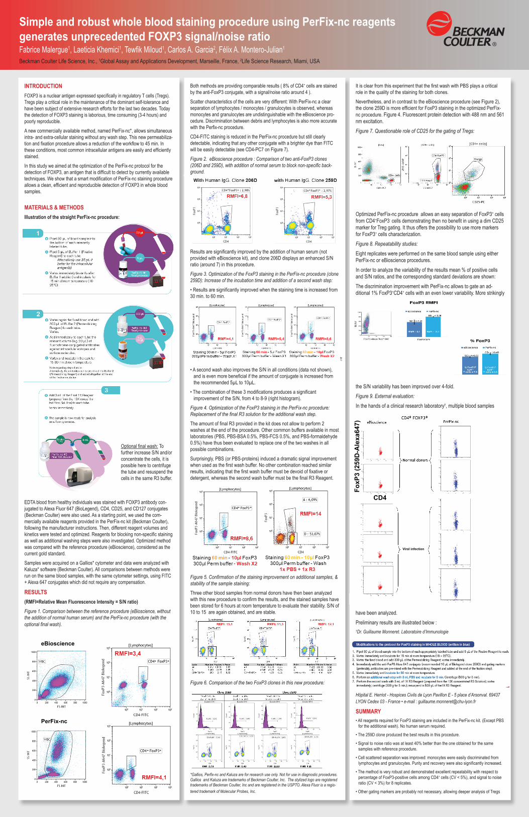
the S/N variability has been improved over 4-fold.Figure 9. External evaluation:
In the hands of a clinical research laboratory†, multiple blood samples
have been analyzed.Preliminary results are illustrated below :†Dr. Guillaume Monneret. Laboratoire d’Immunologie
Hôpital E. Herriot - Hospices Civils de Lyon Pavillon E - 5 place d’Arsonval. 69437 LYON Cedex 03 - France • e-mail : [email protected]
SUMMARY• All reagents required for FoxP3 staining are included in the PerFix-nc kit. (Except PBS for the additional wash). No human serum required.
• The 259D clone produced the best results in this procedure.
• Signal to noise ratio was at least 40% better than the one obtained for the same samples with reference procedure.
• Cell scattered separation was improved: monocytes were easily discriminated from lymphocytes and granulocytes. Purity and recovery were also significantly increased.
• The method is very robust and demonstrated excellent repeatability with respect to percentage of FoxP3-positive cells among CD4+ cells (CV < 5%), and signal to noise ratio (CV < 3%) for 8 replicates.
• Other gating markers are probably not necessary, allowing deeper analysis of Tregs
eBioscience
PerFix-nc
CD4
FoxP
3 (2
59D
-Ale
xa64
7)
Viral infection
Optional final wash: To further increase S/N and/or concentrate the cells, it is possible here to centrifuge the tube and resuspend the cells in the same R3 buffer.
eBioscience
PerFix-nc
CD4
FoxP
3 (2
59D
-Ale
xa64
7)
Viral infection
eBioscience
PerFix-nc
CD4
FoxP
3 (2
59D
-Ale
xa64
7)
Viral infection
eBioscience
PerFix-nc
CD4
FoxP
3 (2
59D
-Ale
xa64
7)
Viral infection
eBioscience
PerFix-nc
CD4
FoxP
3 (2
59D
-Ale
xa64
7)
Viral infection
eBioscience
PerFix-nc
CD4
FoxP
3 (2
59D
-Ale
xa64
7)
Viral infection
eBioscience
PerFix-nc
CD4
FoxP
3 (2
59D
-Ale
xa64
7)
Viral infection
eBioscience
PerFix-nc
CD4
FoxP
3 (2
59D-
Alex
a647
)
Viral infection
eBioscience
PerFix-nc
CD4
FoxP
3 (2
59D-
Alex
a647
)
Viral infection
eBioscience
PerFix-nc
CD4
FoxP
3 (2
59D
-Ale
xa64
7)
Viral infection