signaling function of na,k-atpase induced by ouabain against lps
TRANSCRIPT
Signaling function of Na,K-ATPase induced byouabain against LPS as an inflammation model inhippocampusKinoshita et al.
Kinoshita et al. Journal of Neuroinflammation 2014, 11:218http://www.jneuroinflammation.com/content/11/1/218
JOURNAL OF NEUROINFLAMMATION
Kinoshita et al. Journal of Neuroinflammation (2014) 11:218 DOI 10.1186/s12974-014-0218-z
RESEARCH Open Access
Signaling function of Na,K-ATPase induced byouabain against LPS as an inflammation model inhippocampusPaula Fernanda Kinoshita1, Lidia Mitiko Yshii1, Andrea Rodrigues Vasconcelos1, Ana Maria Marques Orellana1,Larissa de Sá Lima1, Ana Paula Couto Davel2, Luciana Venturini Rossoni2, Elisa Mitiko Kawamoto1
and Cristoforo Scavone1*
Abstract
Background: Ouabain (OUA) is a newly recognized hormone that is synthesized in the adrenal cortex andhypothalamus. Low doses of OUA can activate a signaling pathway by interaction with Na,K-ATPase, which isprotective against a number of insults. OUA has central and peripheral anti-inflammatory effects. Lipopolysaccharide(LPS), via toll-like receptor 4 activation, is a widely used model to induce systemic inflammation. This study used alow OUA dose to evaluate its effects on inflammation induced by LPS injection in rats.
Methods: Adult male Wistar rats received acute intraperitoneal (ip) OUA (1.8 μg/kg) or saline 20 minutes before LPS(200 μg/kg, ip) or saline injection. Some of the animals had their femoral artery catheterized in order to assessarterial blood pressure values before and after OUA administration. Na,K-ATPase activity, cytokine mRNA levels,apoptosis-related proteins, NF-κB activation brain-derived neurotrophic factor BDNF, corticosterone and TNF-α levelswere measured.
Results: OUA pretreatment decreased mRNA levels of the pro-inflammatory cytokines, inducible nitric oxidesynthase (iNOS) and IL-1β, which are activated by LPS in the hippocampus, but with no effect on serum measures ofthese factors. None of these OUA effects were linked to Na,K-ATPase activity. The involvement of the inflammatorytranscription factor NF-κB in the OUA effect was indicated by its prevention of LPS-induced nuclear translocation of theNF-κB subunit, RELA (p65), as well as the decreased cytosol levels of the NF-κB inhibitor, IKB, in the hippocampus. OUApretreatment reversed the LPS-induced glial fibrillary acidic protein (GFAP) activation and associated inflammation inthe dentate gyrus. OUA also prevented LPS-induced increases in the hippocampal Bax/Bcl2 ratio suggesting ananti-apoptotic action in the brain.
Conclusion: Our results suggest that a low dose of OUA has an important anti-inflammatory effect in the rathippocampus. This effect was associated with decreased GFAP induction by LPS in the dentate gyrus, a brain arealinked to adult neurogenesis.
Keywords: Ouabain, Na,K-ATPase, TNF-α, NF-κB, Inflammation, LPS
* Correspondence: [email protected] Neuropharmacology Laboratory, Department of Pharmacology,Institute of Biomedical Science, University of São Paulo, 05508-900 São Paulo,BrazilFull list of author information is available at the end of the article
© 2014 Kinoshita et al.; licensee BioMed Central. This is an Open Access article distributed under the terms of the CreativeCommons Attribution License (http://creativecommons.org/licenses/by/4.0), which permits unrestricted use, distribution, andreproduction in any medium, provided the original work is properly credited. The Creative Commons Public DomainDedication waiver (http://creativecommons.org/publicdomain/zero/1.0/) applies to the data made available in this article,unless otherwise stated.
Kinoshita et al. Journal of Neuroinflammation (2014) 11:218 Page 2 of 12
IntroductionNa,K-ATPase (NKA) is a membrane protein that isessential for the survival of the organism. This enzyme isexpressed in all the cells of the human body, havingmany functions including the maintenance of osmoticbalance, cell volume, pH and membrane potential. Thisoccurs by the hydrolysis of an adenosine triphosphate(ATP) molecule leading to the export of three sodiumions and the import of two potassium ions into thecell, which is fundamental for neuronal excitabilityand cell maintenance [1,2].NKA is constituted of three subunits: α, β and γ [3],
with each subunit having a number of isoforms thatprovide functional versatility across different cell types, inturn highlighting the different roles and responsesproduced by NKA activation across cell types [4-8].However, the γ (gamma) subunit, is not present in allthe cells, with the other subunits being required forNKA to be functional [9].In the adult brain, α1 is expressed in all cells, with α2
being expressed primarily in astrocytes and α3 inneurons [10,11]. Mutations in the α2- and α3-isoformgenes are involved in neurological disorders, such asfamilial hemiplegic migraine type-2 [12], rapid-onsetdystonia [13], alternating hemiplegia of childhood[14] and cerebellar ataxia, areflexia, pes cavus, opticatrophy and sensorineural hearing loss (CAPOS) [15],with genetic variations in NKA also associating withbipolar disorder, suggesting a role for this enzyme inthe etiology of this disease [16]. The NKA α-isoformplays a critical role in the modulation of learning andmemory, in turn regulating susceptibility to Alzheimer’sdisease [17]. Several works show NKA to operate as areceptor and not only as a pump, with a number ofintracellular pathway activations driving its effects [18,19].Ouabain (OUA) is synthesized by the adrenal gland
and hypothalamus [20,21] and is likely to have importantphysiological roles in both the central and peripheralnervous systems [22,23]. OUA binds to NKA in hippocam-pal astrocytes, activating inositol trisphosphate receptor(InsP3R), which generates calcium oscillations, thereby acti-vating NF-κB [23]. Xie and Askari [24] also showed OUAto act as a signal transducer, by binding to NKA andthereby activating the Ras-Raf-MAPK signaling cascade bythe epidermal growth factor receptor (EGFR).OUA has a dual role, given its dose-dependent response
curve effects. A high concentration of OUA can cause celldeath, driving neuronal necrosis via NKA inhibition,leading to potassium ion depletion and thereby increasingintracellular sodium and calcium ions [25]. Conversely,low concentrations of OUA (0.01 nM) are protectiveagainst kainic acid-induced lesions in the rat striatum,where it reduces apoptosis by increasing Bcl-2 [26].Similarly, OUA affords protection in rat kidney primary
cultures against Shiga toxin [27]. As such, OUA can affordprotection both peripherally and centrally.NF-κB is a nuclear transcription factor, which is
commonly induced following danger or inflammatorysignaling, including by lipopolysaccharide (LPS) [28].NF-κB comprises homo- and heterodimers via thecombination of the subunits p65 (RELA), p50, p52, c-RELand REL of the REL/NF-κB family of proteins [28].Different dimer combinations can activate or blockdistinct gene transcriptions, exemplified by the inhibitoryhomodimers p50/p50 and p52/p52 [29]. This transcriptionfactor is evolutionarily conserved, driving a wide range ofeffects that depend on the specific activating stimulus,particular cell type and the cellular phenomenon that itregulates.When it is not stimulated, NF-κB resides in the cytoplasm
in association with the inhibitory protein, IκB (IKB) [30].When IKB is phosphorylated by IKK (IκB kinase), IKB isdegraded by proteasome 26S [31], freeing NF-κB, which canthen translocate to the nucleus and bind to the regulatorysequence close to the promotor region, thereby regulatingthe expression of target genes such as, the pro-apoptoticBax, anti-apoptotic Bcl2, inducible nitric oxide synthase(iNOS), several cytokines, manganese-superoxide dismutase(MnSOD) and brain-derived neurotrophic factor (BDNF)[29]. BDNF is an important trophic protein, including in thecentral nervous system (CNS), where it plays importantroles in brain development and plasticity, with its malfunc-tion being involved in many CNS diseases [32]. In the CNS,NF-κB plays a dual role in neurodegenerative diseases,enhancing survival in neurons, whilst driving pathologicalglial inflammatory processes [33].LPS is a bacterial Gram-negative membrane element
which is released in a free form or in aggregates. LPS isclassically utilized as a model of inflammation and sepsis,with effects via toll-like receptor 4 (TLR4) [34,35].Peripheral LPS administration induces NF-κB activationin different brain areas [36], as well as de novo synthesis ofIκb mRNA (an NF-κB activation index), in the brainparenchyma [37]. Peripheral LPS can, therefore, activatepro-inflammatory CNS genes [38]. The present work aimsto evaluate the alterations induced by a low dose of OUAin the rat hippocampus, in an inflammation modelinduced by intraperitoneal (ip) LPS injection.
MethodsAnimal and tissue preparationThree-month old male Wistar rats (Biomedical SciencesInstitute, University of São Paulo) were kept under12 hour light/dark cycle (lights on at 7:00 am) andallowed free access to food and water. Animals weretreated with OUA (1.8 μg/kg, ip) or saline 20 minutesbefore the injection LPS (200 μg/kg, ip) or saline andeuthanized 2 hours after the administration (between
Kinoshita et al. Journal of Neuroinflammation (2014) 11:218 Page 3 of 12
9:00 and 11:00 am) following procedures approved bythe Biomedical College of Animal Experimentation(COBEA). All procedures were also approved by theEthical Committee for Animal Research (CEEA) of theBiomedical Sciences Institute of the University of SãoPaulo. The brain was immediately removed and immersedin cold PBS. Each hippocampus was rapidly dissected,quickly immersed in liquid nitrogen, and stored at −80°Cfor later use.
Hemodynamic parametersMale Wistar rats (n = 4) were anesthetized with aketamine, xylazine and acepromazine mixture (64.9,3.2 and 0.78 mg/kg). The left femoral artery was cannulatedwith a polyethylene catheter (PE-10 connected to PE-50filled with heparinized saline, (BD,New Jersey, NY, USA)that was exteriorized in the mid-scapular region. After24 hours, arterial pressure and heart rate were measured inconscious animals by a pressure transducer (modelDT-100, Utah Medical Products, Midvale, UT, USA) andrecorded using an interface and software for computerdata acquisition (Power Lab 4/25, AD Instruments,Sidney, Australia). Heart rate was determined from theintra-beat intervals. The effects of OUA (1.8 μg/kg, ip) onthe arterial blood pressure and heart rate, was evaluatedbefore and 1 hour after OUA administration in consciousanimals. Results are expressed as mean ± SE.
Chemicals and kitsRoutine reagents, OUA and LPS from Escherichia coliO111:B4 were purchased from Sigma Chemicals (St. Louis,MO, USA); Bio-Rad protein assay kit was purchased fromBio-Rad (Hercules, CA, USA). Corticosterone, TNF-α andBDNF immunoassay kits were purchased from Enzo LifeSciences, Inc. (Farmingdale, NY, USA), eBioscience(San Diego, CA, USA) and Promega (Fitchburg, WI,USA), respectively. The kits were utilized according tomanufacturer instructions. All solutions were preparedimmediately before use.
Semiquantitative RT-PCR determination of inducible nitricoxide synthase (iNos), Il-1β, Bax and Bcl2 mRNA levelsThe effect of OUA on LPS-modulated gene expressionin the hippocampus of rats was measured. Total RNAwas isolated with Trizol reagent (Invitrogen, Carlsbad,CA, USA) from the hippocampus according to themanufacturer's instructions. Semiquantitative reversetranscription-PCR (RT-PCR) amplification was performedusing the ThermoScript RT kit (Invitrogen, Carlsbad, CA,USA) according to the manufacturer's instructions. Theprimer sequences were: iNos (GenBank access number012611.3, 651 bp) 5′-GTGCTAATGCGGAAGGTCATA-3′(sense) and 5′CCAAATGTGCTTGTCACCACA-3′ (anti-sense); Bax (GenBank access number 017059.1, 260 bp),
5′-TGAACTGGACAACAACATGGAGC-3′ (sense) and5′-GGTCTTGGATCCAGACAAACAGC-3′ (antisense);Bcl2 (GenBank access number 016993.1, 271 bp), 5-′GGAGGATTGTGGCCTTCTTTGAG-3′ (sense) and 5′-TATGCACCCAGAGTGATGCAGGC-3′(antisense); Il-1β (GenBank access number 031512.2, 1,339 bp), 5′-ATGCTCAGCAGTCAAGTGCC-3′ (sense) and 5′-AGCCTT CCTTCGTGTAACCC-3′ (antisense).The PCR conditions consisted of 5 minutes at 94°C,
33 cycles of 94°C for 45 seconds, 63 °C for 45 seconds, and72°C for 1 minute and 30 seconds and a final extension at72°C for 10 minutes. Glyceraldehyde-3-phosphate dehydro-genase (Gapdh; GenBank access number 017008.3, 264 bp)was also amplified as an internal PCR control using the fol-lowing primers: 5′-CGGGAAGCTTGTGATCAATGG-3′(sense) and 5′-GGCAGTGATGCCATGGACTG-3′ (anti-sense). In this case, the temperature cycling conditions wereas follows: 5 minutes at 94°C, 20 cycles of 94°C for45 seconds, 63°C for 45 seconds, and 72°C for 1 minuteand 30 seconds and a final extension at 72°C for 10 minutes.Gel electrophoresis of the PCR product was performedusing an ethidium bromide-containing agarose gel (2%),and resulting bands were visualized under UV light.The photographs were captured by photo documentation
system DP-001-FDC (VilberLourmat, Torcy, France), andthe optical density of the bands was determined usingNIH ImageJ software (http://rsb.info.nih.gov/ij). Resultswere expressed in relation to the intensity of GapdhmRNA levels.
Protein extractionNuclear extract of each hippocampus was prepared aspreviously described [39]. Briefly, hippocampal struc-tures were homogenized using a Dounce homogenizerin cold PBS supplemented with 0.5 mM dithiothreitol(DTT), 0.5 mM phenylmethylsulfonide fluoride (PMSF),2 μg/ml leupeptin, 2 μg/ml antipain, and 3 mM sodiumorthovanadate and centrifuged at 4°C for 30 seconds at12,000 g. Supernatant was discarded and pellet wasresuspended in lysis buffer (10 mM HEPES, pH 7.9,1.5 mM MgCl2, 10 mM KCl, 0.1 mM ethylenediamine-tetraacetic acid (EDTA), 0.5 mM DTT, 0.5 mM PMSF,2 μg/ml leupeptin, 2 μg/ml antipain, and 3 mM sodiumorthovanadate) and incubated on ice for 10 minutes.After addition of NP-40 (10%), samples were vigorouslymixed and centrifuged for 30 seconds at 12,000 g. Thesupernatant was reserved for Western blotting andenzymatic assays (cytosolic fraction), and the pellet wasresuspended in lysis buffer (10 mM HEPES, pH 7.9,1.5 mM MgCl2, 10 mM KCl, 0.1 mM EDTA, 0.5 mMDTT, 0.5 mM PMSF, 2 μg/ml leupeptin, 2 μg/mlantipain, and 3 mM sodium orthovanadate) and incubatedon ice for 10 minutes. After addition of NP-40 (10%),samples were vigorously mixed and centrifuged for
Kinoshita et al. Journal of Neuroinflammation (2014) 11:218 Page 4 of 12
30 seconds at 12,000 g. Supernatant was discarded,and the pellet was resuspended in extraction buffer(20 mM HEPES, pH 7.9, 25% glycerol, 1.5 mMMgCl2, 300 mM NaCl, 0.25 mM EDTA, 0.5 mMDTT, 0.5 mM PMSF, 2 lg/ml leupeptin, 2 lg/ml antipain,and 3 mM sodium orthovanadate), incubated for 20 minuteson ice, and centrifuged for 20 minutes at 12,000 g at 4°C.The resulting supernatants containing nuclear proteinswere stored at −80°C and used to test the expression ofRELA. Protein concentration was determined using theBio-Rad protein reagent (Hercules, CA, USA).
Western blottingElectrophoresis was performed using 10% polyacrylamidegel and the Bio-Rad mini-Protean III apparatus (Bio-Rad,Hercules, CA, USA). In brief, the proteins present inthe hippocampal cytosolic and nuclear fractions weresize-separated in 10% SDS-PAGE (90 V). The proteinswere blotted onto a nitrocellulose membrane (Bio-Rad,Hercules, CA, USA) and incubated with the specific anti-body RELA (p65) (sc-0372; Santa Cruz Biotechnology,Santa Cruz, CA, USA) and IKB (sc-0371; Santa Cruz Bio-technology, Santa Cruz, CA, USA). The Ponceau methodto immunoblot was used to ensure equal protein loading[40]. Proteins recognized by antibodies were revealed byan electrochemiluminescence (ECL) technique, followingthe manufacturer's instructions (Amersham Biosciences,Amersham, UK). To standardize and quantify the immu-noblots, we used the photo documentation systemDP-001-FDC (VilberLourmat, Torcy, France) and NIHImageJ software (http://rsb.info.nih.gov/ij). Several exposuretimes were analyzed to ensure the linearity of theband intensities. β-ACTIN antibody (sc-1616; SantaCruz Biotechnology, Santa Cruz, CA, USA) was used asan internal experimental control, with results expressed inrelation to β-ACTIN intensity.
Measurement of Na, K-ATPase activityThe NKA activity was determined by assaying Pireleased from ATP hydrolysis. This inorganic compoundforms a complex with molybdate which can be read spec-trophotometrically at 700 nm [41]. For this colorimetricATP assay, hippocampus supernatant (S1) was centrifuged(12,000 g for 15 minutes at 4°C). The particulate fractionwas resuspended in a buffer containing: 0.32 M sucrose,20 mM HEPES, 1 mM EDTA, 1 mM DTT, and 1 mMPMSF, pH 7.4.Protein content was determined in particulate samples
by colorimetric assay (Bio-Rad, Hercules, CA, USA)[42]. NKA activity was tested by adding 10 μg of theparticulate fraction (in 40 μl of buffer) to 360 μl ofbuffer containing: 3 mM ATP, 120 mM NaCl, 2 mMKCl, 3 mM MgCl2, and 30 mM histidine (pH 7.2), with orwithout OUA (3 μM or 3 mM). After 20 minutes of
incubation at 37°C, NKA activity was measured. Thereaction was terminated by the addition of a quenchingsolution (0.6 ml) containing 1 N H2SO4 and 0.5% am-monium molybdate. Formation of a phosphomolyb-date complex was determined spectrophotometricallyat 700 nm [43].The total ATPase, Mg-ATPase, NKA α1- and α2/3-isoform
activities were linearly related up to 20 minutes. In rodents,the NKA α1-isoform is 1,000 times less sensitive tothe cardiac glycoside than the NKA α2/3isoform measuredfrom the difference between OUA-untreated andOUA-treated samples.The high-affinity α2/3-isoform fraction was calculated
by subtracting the activity obtained with 3 μM OUA fromtotal-ATPase activity. To determine the low-affinity fraction(α1 subunit-associated NKA activity), the values obtained inthe presence of 3 μM OUA were subtracted from thoseobtained in the presence of 3 mM OUA. The NKA activitywas expressed as nmol/mg protein x minutes.
Immunofluorescence in the hippocampusThe brain was fixed with paraformaldehyde and kept in30% sucrose for 48 hours. Hippocampal coronal sections(30 μm) were cut in the microtome and the tissuescollected were kept in 0.1 M PBS. The sections were incu-bated with 2% normal donkey serum for 2 hours. Forimmunofluorescence reactions, the sections were incubatedovernight with primary antibody (glial fibrillary acidicprotein (GFAP), Sigma-Aldrich, St. Louis, MO, USA) and4',6-diamidino-2-phenylindole; DAPI), followed by 2-hoursincubation with secondary antibody (Alexa fluor 488donkey anti-rabbit for GFAP). Six slices were placed in eachslide and mounted with a coverslip. The sections wereexamined by fluorescence microscopy (Nikon Eclipse 80iwith DXM 1200C digital camera; Nikon, Tokyo, Japan).
Statistical analysisResults are expressed as mean ± SEM of the indicatednumber of experiments. Statistical comparisons forOUA-induced changes in mRNA levels, protein expression,ELISA kits and NKA activities were performed byone-way analysis of variance (ANOVA), followed by theNewman-Keuls post-test. All analyses were performedusing a Prism 6 software package (GraphPad Software,San Diego, CA, USA). P-values < 0.05 were considered toreflect a statistically significant difference.
ResultsOUA (1.8 μg/kg) does not induce any effect in bloodpressure and NKA activityOne-hour OUA administration (1.8 μg/kg; ip) did notchange mean arterial pressure (Before: 113 ± 3.16 versusAfter OUA: 112 ± 4.71 mmHg; P > 0.05) or heart rate(Before: 323 ± 21.35 versus After OUA: 331 ± 15.46 bpm;
Kinoshita et al. Journal of Neuroinflammation (2014) 11:218 Page 5 of 12
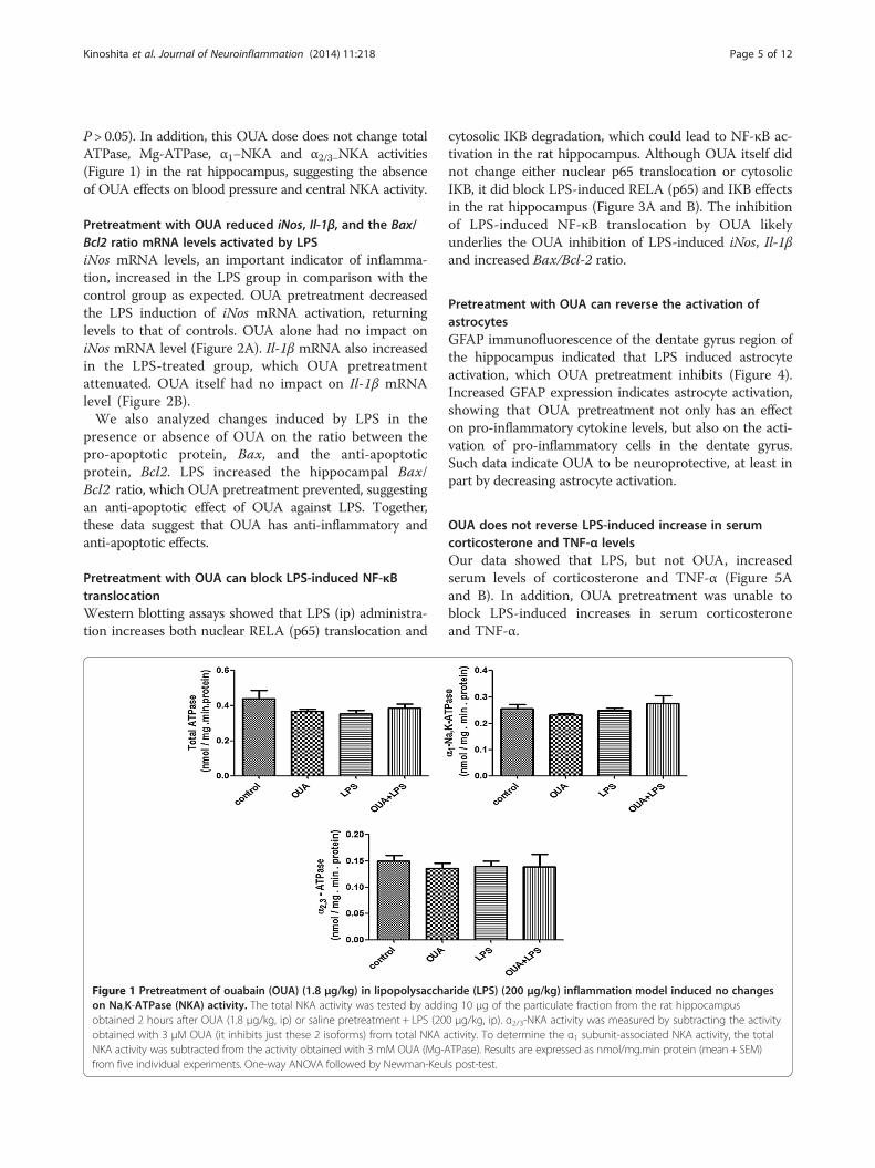
P > 0.05). In addition, this OUA dose does not change totalATPase, Mg-ATPase, α1−NKA and α2/3−NKA activities(Figure 1) in the rat hippocampus, suggesting the absenceof OUA effects on blood pressure and central NKA activity.
Pretreatment with OUA reduced iNos, Il-1β, and the Bax/Bcl2 ratio mRNA levels activated by LPSiNos mRNA levels, an important indicator of inflamma-tion, increased in the LPS group in comparison with thecontrol group as expected. OUA pretreatment decreasedthe LPS induction of iNos mRNA activation, returninglevels to that of controls. OUA alone had no impact oniNos mRNA level (Figure 2A). Il-1β mRNA also increasedin the LPS-treated group, which OUA pretreatmentattenuated. OUA itself had no impact on Il-1β mRNAlevel (Figure 2B).We also analyzed changes induced by LPS in the
presence or absence of OUA on the ratio between thepro-apoptotic protein, Bax, and the anti-apoptoticprotein, Bcl2. LPS increased the hippocampal Bax/Bcl2 ratio, which OUA pretreatment prevented, suggestingan anti-apoptotic effect of OUA against LPS. Together,these data suggest that OUA has anti-inflammatory andanti-apoptotic effects.
Pretreatment with OUA can block LPS-induced NF-κBtranslocationWestern blotting assays showed that LPS (ip) administra-tion increases both nuclear RELA (p65) translocation and
Figure 1 Pretreatment of ouabain (OUA) (1.8 μg/kg) in lipopolysacchaon Na,K-ATPase (NKA) activity. The total NKA activity was tested by addiobtained 2 hours after OUA (1.8 μg/kg, ip) or saline pretreatment + LPS (20obtained with 3 μM OUA (it inhibits just these 2 isoforms) from total NKA aNKA activity was subtracted from the activity obtained with 3 mM OUA (Mg-Afrom five individual experiments. One-way ANOVA followed by Newman-Keu
cytosolic IKB degradation, which could lead to NF-κB ac-tivation in the rat hippocampus. Although OUA itself didnot change either nuclear p65 translocation or cytosolicIKB, it did block LPS-induced RELA (p65) and IKB effectsin the rat hippocampus (Figure 3A and B). The inhibitionof LPS-induced NF-κB translocation by OUA likelyunderlies the OUA inhibition of LPS-induced iNos, Il-1βand increased Bax/Bcl-2 ratio.
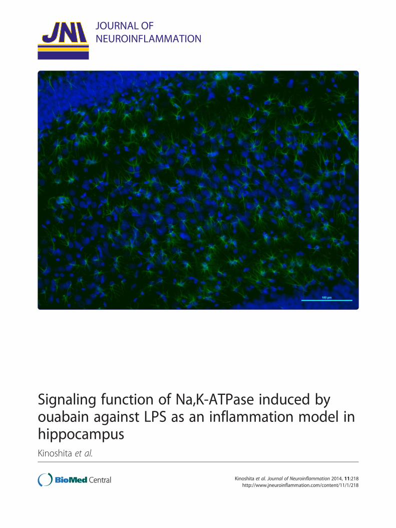
Pretreatment with OUA can reverse the activation ofastrocytesGFAP immunofluorescence of the dentate gyrus region ofthe hippocampus indicated that LPS induced astrocyteactivation, which OUA pretreatment inhibits (Figure 4).Increased GFAP expression indicates astrocyte activation,showing that OUA pretreatment not only has an effecton pro-inflammatory cytokine levels, but also on the acti-vation of pro-inflammatory cells in the dentate gyrus.Such data indicate OUA to be neuroprotective, at least inpart by decreasing astrocyte activation.
OUA does not reverse LPS-induced increase in serumcorticosterone and TNF-α levelsOur data showed that LPS, but not OUA, increasedserum levels of corticosterone and TNF-α (Figure 5Aand B). In addition, OUA pretreatment was unable toblock LPS-induced increases in serum corticosteroneand TNF-α.
ride (LPS) (200 μg/kg) inflammation model induced no changesng 10 μg of the particulate fraction from the rat hippocampus0 μg/kg, ip). α2/3-NKA activity was measured by subtracting the activityctivity. To determine the α1 subunit-associated NKA activity, the totalTPase). Results are expressed as nmol/mg.min protein (mean + SEM)ls post-test.
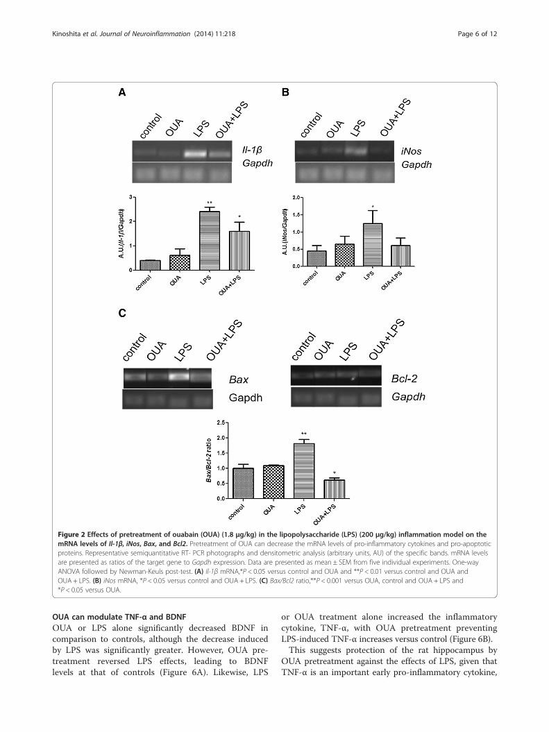
Figure 2 Effects of pretreatment of ouabain (OUA) (1.8 μg/kg) in the lipopolysaccharide (LPS) (200 μg/kg) inflammation model on themRNA levels of Il-1β, iNos, Bax, and Bcl2. Pretreatment of OUA can decrease the mRNA levels of pro-inflammatory cytokines and pro-apoptoticproteins. Representative semiquantitative RT- PCR photographs and densitometric analysis (arbitrary units, AU) of the specific bands. mRNA levelsare presented as ratios of the target gene to Gapdh expression. Data are presented as mean ± SEM from five individual experiments. One-wayANOVA followed by Newman-Keuls post-test. (A) Il-1β mRNA,*P < 0.05 versus control and OUA and **P < 0.01 versus control and OUA andOUA + LPS. (B) iNos mRNA, *P < 0.05 versus control and OUA + LPS. (C) Bax/Bcl2 ratio,**P < 0.001 versus OUA, control and OUA + LPS and*P < 0.05 versus OUA.
Kinoshita et al. Journal of Neuroinflammation (2014) 11:218 Page 6 of 12
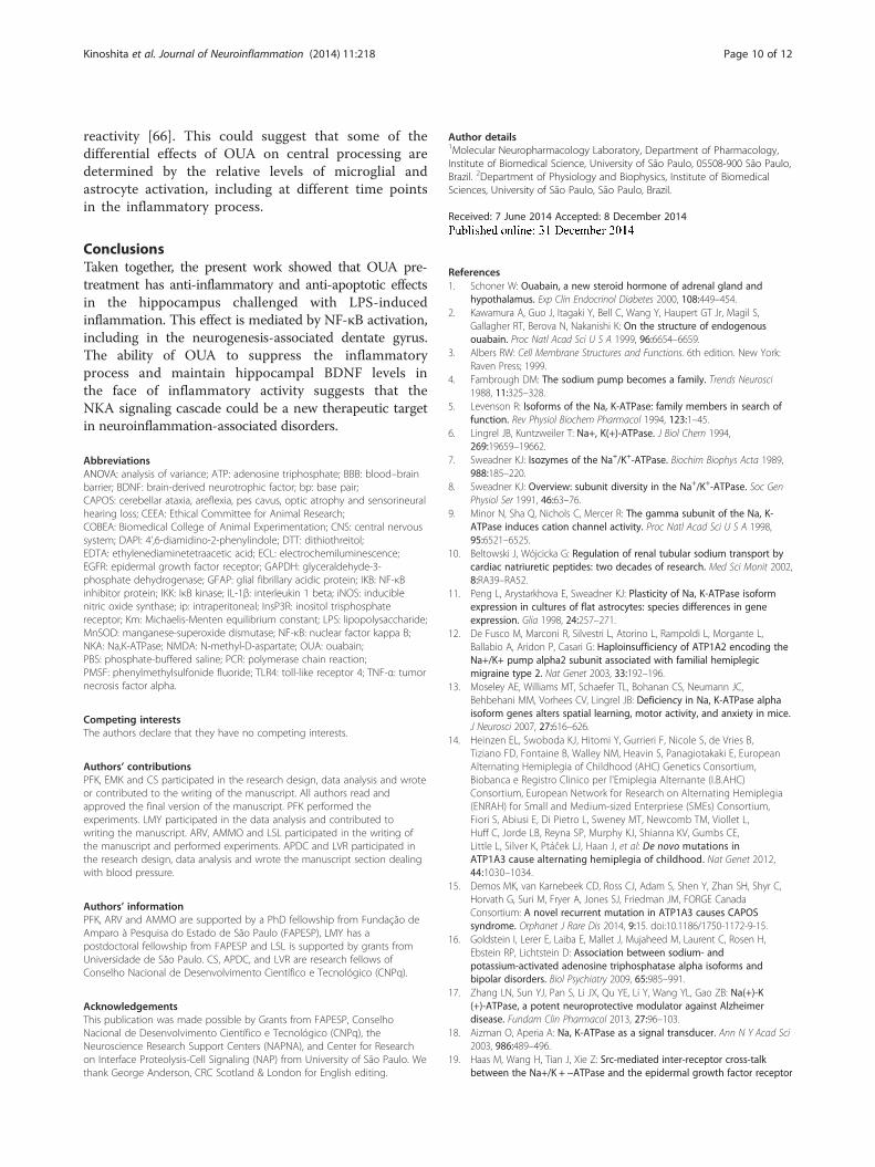
OUA can modulate TNF-α and BDNFOUA or LPS alone significantly decreased BDNF incomparison to controls, although the decrease inducedby LPS was significantly greater. However, OUA pre-treatment reversed LPS effects, leading to BDNFlevels at that of controls (Figure 6A). Likewise, LPS
or OUA treatment alone increased the inflammatorycytokine, TNF-α, with OUA pretreatment preventingLPS-induced TNF-α increases versus control (Figure 6B).This suggests protection of the rat hippocampus by
OUA pretreatment against the effects of LPS, given thatTNF-α is an important early pro-inflammatory cytokine,
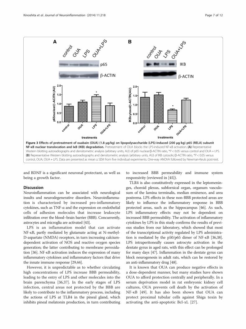
Figure 3 Effects of pretreatment of ouabain (OUA) (1.8 μg/kg) on lipopolysaccharide (LPS)-induced (200 μg/kg) p65 (RELA) subunitNF-κB nuclear translocation and IκB (IKB) degradation. Pretreatment of OUA blocks the LPS-induced NF-κB activation. (A) RepresentativeWestern blotting autoradiographs and densitometric analysis (arbitrary units, AU) of p65 nuclear/β-ACTIN ratio, *P < 0.05 versus control and OUA + LPS.(B) Representative Western blotting autoradiographs and densitometric analysis (arbitrary units, AU) of IKB cytosolic/β-ACTIN ratio, *P < 0.05 versuscontrol, OUA, OUA+ LPS. Data are presented as mean ± SEM from five individual experiments. One-way ANOVA followed by Newman-Keuls post-test.
Kinoshita et al. Journal of Neuroinflammation (2014) 11:218 Page 7 of 12
and BDNF is a significant neuronal protectant, as well asbeing a growth factor.
DiscussionNeuroinflammation can be associated with neurologicalinsults and neurodegenerative disorders. Neuroinflamma-tion is characterized by increased pro-inflammatorycytokines, such as TNF-α and the expression on endothelialcells of adhesion molecules that increase leukocyteinfiltration over the blood–brain barrier (BBB). Concurrently,astrocytes and microglia are activated [43].LPS is an inflammation model that can activate
NF-κB, partly mediated by glutamate acting at N-methyl-D-aspartate (NMDA) receptors, in turn increasing calcium-dependent activation of NOS and reactive oxygen speciesgeneration; the latter contributing to membrane peroxida-tion [36]. NF-κB activation induces the expression of manyinflammatory cytokines and inflammatory factors that drivethe innate immune response [29,44].However, it is unpredictable as to whether circulating
high concentrations of LPS increase BBB permeability,leading to the entry of LPS and other molecules into thebrain parenchyma [36,37]. In the early stages of LPSinfection, central areas not protected by the BBB arelikely to contribute to the inflammatory process, includingthe actions of LPS at TLR4 in the pineal gland, whichinhibits pineal melatonin production, in turn contributing
to increased BBB permeability and immune systemresponsivity (reviewed in [45]).TLR4 is also constitutively expressed in the leptomenin-
ges, choroid plexus, subfornical organ, organum vasculo-sum of the lamina terminalis, median eminence, and areapostrema. LPS effects in these non-BBB protected areas arelikely to influence the inflammatory response in BBBprotected areas, such as the hippocampus [46]. As such,LPS inflammatory effects may not be dependent onincreased BBB permeability. The activation of inflammatorycytokines by LPS in this study confirms the results of previ-ous studies from our laboratory, which showed that mostof the transcriptional activity regulated by LPS administra-tion is mediated by the p50/p65 dimer of NF-κB [36,38].LPS intraperitoneally causes astrocyte activation in thedentate gyrus in aged rats, with this effect can be prolongedfor many days [47]. Inflammation in the dentate gyrus canblock neurogenesis in adult rats, which can be restored byan anti-inflammatory drug [48].It is known that OUA can produce negative effects in
a dose-dependent manner, but many studies have shownOUA to afford protection centrally and peripherally. In aserum deprivation model in rat embryonic kidney cellcultures, OUA prevents cell death by the activation ofNF-κB [49]. It has also been shown that OUA canprotect proximal tubular cells against Shiga toxin byactivating the anti-apoptotic Bcl-xL [27].
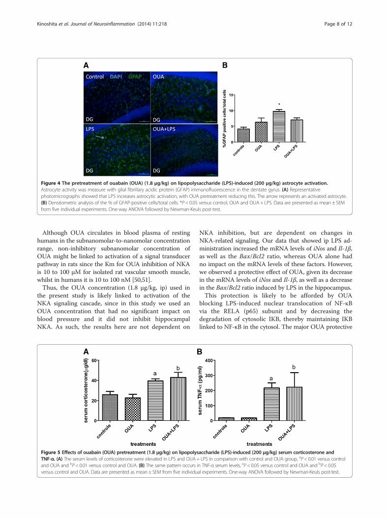
Figure 4 The pretreatment of ouabain (OUA) (1.8 μg/kg) on lipopolysaccharide (LPS)-induced (200 μg/kg) astrocyte activation.Astrocyte activity was measure with glial fibrillary acidic protein (GFAP) immunofluorescence in the dentate gyrus. (A) Representativephotomicrographs showed that LPS increases astrocytic activation, with OUA pretreatment reducing this. The arrow represents an activated astrocyte.(B) Densitometric analysis of the % of GFAP-positive cells/total cells. *P < 0.05 versus control, OUA and OUA+ LPS. Data are presented as mean ± SEMfrom five individual experiments. One-way ANOVA followed by Newman-Keuls post-test.
Kinoshita et al. Journal of Neuroinflammation (2014) 11:218 Page 8 of 12
Although OUA circulates in blood plasma of restinghumans in the subnanomolar-to-nanomolar concentrationrange, non-inhibitory subnanomolar concentration ofOUA might be linked to activation of a signal transducerpathway in rats since the Km for OUA inhibition of NKAis 10 to 100 μM for isolated rat vascular smooth muscle,whilst in humans it is 10 to 100 nM [50,51].Thus, the OUA concentration (1.8 μg/kg, ip) used in
the present study is likely linked to activation of theNKA signaling cascade, since in this study we used anOUA concentration that had no significant impact onblood pressure and it did not inhibit hippocampalNKA. As such, the results here are not dependent on
Figure 5 Effects of ouabain (OUA) pretreatment (1.8 μg/kg) on lipopolysTNF-α. (A) The serum levels of corticosterone were elevated in LPS and OUA+and OUA and bP< 0.01 versus control and OUA. (B) The same pattern occurs iversus control and OUA. Data are presented as mean ± SEM from five individua
NKA inhibition, but are dependent on changes inNKA-related signaling. Our data that showed ip LPS ad-ministration increased the mRNA levels of iNos and Il-1β,as well as the Bax/Bcl2 ratio, whereas OUA alone hadno impact on the mRNA levels of these factors. However,we observed a protective effect of OUA, given its decreasein the mRNA levels of iNos and Il-1β, as well as a decreasein the Bax/Bcl2 ratio induced by LPS in the hippocampus.This protection is likely to be afforded by OUA
blocking LPS-induced nuclear translocation of NF-κBvia the RELA (p65) subunit and by decreasing thedegradation of cytosolic IKB, thereby maintaining IKBlinked to NF-κB in the cytosol. The major OUA protective
accharide (LPS)-induced (200 μg/kg) serum corticosterone andLPS in comparison with control and OUA group, aP< 0.01 versus control
n TNF-α serum levels, aP< 0.05 versus control and OUA and bP< 0.05l experiments. One-way ANOVA followed by Newman-Keuls post-test.
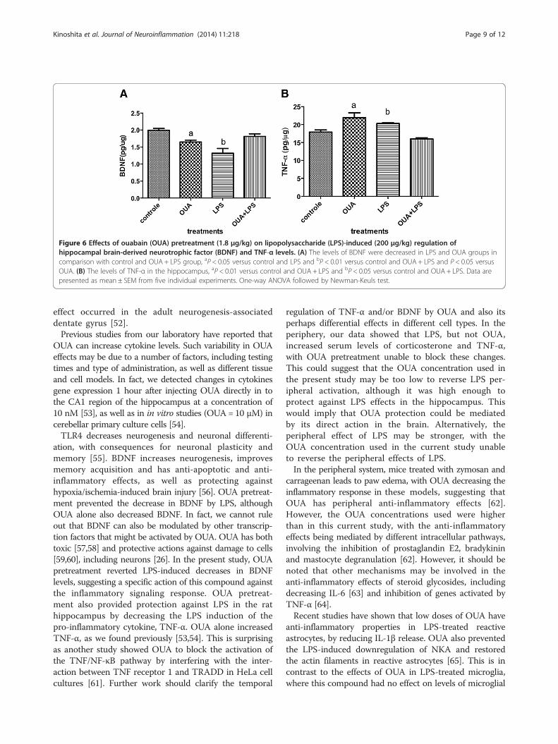
Figure 6 Effects of ouabain (OUA) pretreatment (1.8 μg/kg) on lipopolysaccharide (LPS)-induced (200 μg/kg) regulation ofhippocampal brain-derived neurotrophic factor (BDNF) and TNF-α levels. (A) The levels of BDNF were decreased in LPS and OUA groups incomparison with control and OUA + LPS group, aP < 0.05 versus control and LPS and bP < 0.01 versus control and OUA + LPS and P < 0.05 versusOUA. (B) The levels of TNF-α in the hippocampus, aP < 0.01 versus control and OUA + LPS and bP < 0.05 versus control and OUA + LPS. Data arepresented as mean ± SEM from five individual experiments. One-way ANOVA followed by Newman-Keuls test.
Kinoshita et al. Journal of Neuroinflammation (2014) 11:218 Page 9 of 12
effect occurred in the adult neurogenesis-associateddentate gyrus [52].Previous studies from our laboratory have reported that
OUA can increase cytokine levels. Such variability in OUAeffects may be due to a number of factors, including testingtimes and type of administration, as well as different tissueand cell models. In fact, we detected changes in cytokinesgene expression 1 hour after injecting OUA directly in tothe CA1 region of the hippocampus at a concentration of10 nM [53], as well as in in vitro studies (OUA= 10 μM) incerebellar primary culture cells [54].TLR4 decreases neurogenesis and neuronal differenti-
ation, with consequences for neuronal plasticity andmemory [55]. BDNF increases neurogenesis, improvesmemory acquisition and has anti-apoptotic and anti-inflammatory effects, as well as protecting againsthypoxia/ischemia-induced brain injury [56]. OUA pretreat-ment prevented the decrease in BDNF by LPS, althoughOUA alone also decreased BDNF. In fact, we cannot ruleout that BDNF can also be modulated by other transcrip-tion factors that might be activated by OUA. OUA has bothtoxic [57,58] and protective actions against damage to cells[59,60], including neurons [26]. In the present study, OUApretreatment reverted LPS-induced decreases in BDNFlevels, suggesting a specific action of this compound againstthe inflammatory signaling response. OUA pretreat-ment also provided protection against LPS in the rathippocampus by decreasing the LPS induction of thepro-inflammatory cytokine, TNF-α. OUA alone increasedTNF-α, as we found previously [53,54]. This is surprisingas another study showed OUA to block the activation ofthe TNF/NF-κB pathway by interfering with the inter-action between TNF receptor 1 and TRADD in HeLa cellcultures [61]. Further work should clarify the temporal
regulation of TNF-α and/or BDNF by OUA and also itsperhaps differential effects in different cell types. In theperiphery, our data showed that LPS, but not OUA,increased serum levels of corticosterone and TNF-α,with OUA pretreatment unable to block these changes.This could suggest that the OUA concentration used inthe present study may be too low to reverse LPS per-ipheral activation, although it was high enough toprotect against LPS effects in the hippocampus. Thiswould imply that OUA protection could be mediatedby its direct action in the brain. Alternatively, theperipheral effect of LPS may be stronger, with theOUA concentration used in the current study unableto reverse the peripheral effects of LPS.In the peripheral system, mice treated with zymosan and
carrageenan leads to paw edema, with OUA decreasing theinflammatory response in these models, suggesting thatOUA has peripheral anti-inflammatory effects [62].However, the OUA concentrations used were higherthan in this current study, with the anti-inflammatoryeffects being mediated by different intracellular pathways,involving the inhibition of prostaglandin E2, bradykininand mastocyte degranulation [62]. However, it should benoted that other mechanisms may be involved in theanti-inflammatory effects of steroid glycosides, includingdecreasing IL-6 [63] and inhibition of genes activated byTNF-α [64].Recent studies have shown that low doses of OUA have
anti-inflammatory properties in LPS-treated reactiveastrocytes, by reducing IL-1β release. OUA also preventedthe LPS-induced downregulation of NKA and restoredthe actin filaments in reactive astrocytes [65]. This is incontrast to the effects of OUA in LPS-treated microglia,where this compound had no effect on levels of microglial
Kinoshita et al. Journal of Neuroinflammation (2014) 11:218 Page 10 of 12
reactivity [66]. This could suggest that some of thedifferential effects of OUA on central processing aredetermined by the relative levels of microglial andastrocyte activation, including at different time pointsin the inflammatory process.
ConclusionsTaken together, the present work showed that OUA pre-treatment has anti-inflammatory and anti-apoptotic effectsin the hippocampus challenged with LPS-inducedinflammation. This effect is mediated by NF-κB activation,including in the neurogenesis-associated dentate gyrus.The ability of OUA to suppress the inflammatoryprocess and maintain hippocampal BDNF levels inthe face of inflammatory activity suggests that theNKA signaling cascade could be a new therapeutic targetin neuroinflammation-associated disorders.
AbbreviationsANOVA: analysis of variance; ATP: adenosine triphosphate; BBB: blood–brainbarrier; BDNF: brain-derived neurotrophic factor; bp: base pair;CAPOS: cerebellar ataxia, areflexia, pes cavus, optic atrophy and sensorineuralhearing loss; CEEA: Ethical Committee for Animal Research;COBEA: Biomedical College of Animal Experimentation; CNS: central nervoussystem; DAPI: 4',6-diamidino-2-phenylindole; DTT: dithiothreitol;EDTA: ethylenediaminetetraacetic acid; ECL: electrochemiluminescence;EGFR: epidermal growth factor receptor; GAPDH: glyceraldehyde-3-phosphate dehydrogenase; GFAP: glial fibrillary acidic protein; IKB: NF-κBinhibitor protein; IKK: IκB kinase; IL-1β: interleukin 1 beta; iNOS: induciblenitric oxide synthase; ip: intraperitoneal; InsP3R: inositol trisphosphatereceptor; Km: Michaelis-Menten equilibrium constant; LPS: lipopolysaccharide;MnSOD: manganese-superoxide dismutase; NF-κB: nuclear factor kappa B;NKA: Na,K-ATPase; NMDA: N-methyl-D-aspartate; OUA: ouabain;PBS: phosphate-buffered saline; PCR: polymerase chain reaction;PMSF: phenylmethylsulfonide fluoride; TLR4: toll-like receptor 4; TNF-α: tumornecrosis factor alpha.
Competing interestsThe authors declare that they have no competing interests.
Authors’ contributionsPFK, EMK and CS participated in the research design, data analysis and wroteor contributed to the writing of the manuscript. All authors read andapproved the final version of the manuscript. PFK performed theexperiments. LMY participated in the data analysis and contributed towriting the manuscript. ARV, AMMO and LSL participated in the writing ofthe manuscript and performed experiments. APDC and LVR participated inthe research design, data analysis and wrote the manuscript section dealingwith blood pressure.
Authors’ informationPFK, ARV and AMMO are supported by a PhD fellowship from Fundação deAmparo à Pesquisa do Estado de São Paulo (FAPESP), LMY has apostdoctoral fellowship from FAPESP and LSL is supported by grants fromUniversidade de São Paulo. CS, APDC, and LVR are research fellows ofConselho Nacional de Desenvolvimento Científico e Tecnológico (CNPq).
AcknowledgementsThis publication was made possible by Grants from FAPESP, ConselhoNacional de Desenvolvimento Científico e Tecnológico (CNPq), theNeuroscience Research Support Centers (NAPNA), and Center for Researchon Interface Proteolysis-Cell Signaling (NAP) from University of São Paulo. Wethank George Anderson, CRC Scotland & London for English editing.
Author details1Molecular Neuropharmacology Laboratory, Department of Pharmacology,Institute of Biomedical Science, University of São Paulo, 05508-900 São Paulo,Brazil. 2Department of Physiology and Biophysics, Institute of BiomedicalSciences, University of São Paulo, São Paulo, Brazil.
Received: 7 June 2014 Accepted: 8 December 2014
References1. Schoner W: Ouabain, a new steroid hormone of adrenal gland and
hypothalamus. Exp Clin Endocrinol Diabetes 2000, 108:449–454.2. Kawamura A, Guo J, Itagaki Y, Bell C, Wang Y, Haupert GT Jr, Magil S,
Gallagher RT, Berova N, Nakanishi K: On the structure of endogenousouabain. Proc Natl Acad Sci U S A 1999, 96:6654–6659.
3. Albers RW: Cell Membrane Structures and Functions. 6th edition. New York:Raven Press; 1999.
4. Fambrough DM: The sodium pump becomes a family. Trends Neurosci1988, 11:325–328.
5. Levenson R: Isoforms of the Na, K-ATPase: family members in search offunction. Rev Physiol Biochem Pharmacol 1994, 123:1–45.
6. Lingrel JB, Kuntzweiler T: Na+, K(+)-ATPase. J Biol Chem 1994,269:19659–19662.
7. Sweadner KJ: Isozymes of the Na+/K+-ATPase. Biochim Biophys Acta 1989,988:185–220.
8. Sweadner KJ: Overview: subunit diversity in the Na+/K+-ATPase. Soc GenPhysiol Ser 1991, 46:63–76.
9. Minor N, Sha Q, Nichols C, Mercer R: The gamma subunit of the Na, K-ATPase induces cation channel activity. Proc Natl Acad Sci U S A 1998,95:6521–6525.
10. Beltowski J, Wójcicka G: Regulation of renal tubular sodium transport bycardiac natriuretic peptides: two decades of research. Med Sci Monit 2002,8:RA39–RA52.
11. Peng L, Arystarkhova E, Sweadner KJ: Plasticity of Na, K-ATPase isoformexpression in cultures of flat astrocytes: species differences in geneexpression. Glia 1998, 24:257–271.
12. De Fusco M, Marconi R, Silvestri L, Atorino L, Rampoldi L, Morgante L,Ballabio A, Aridon P, Casari G: Haploinsufficiency of ATP1A2 encoding theNa+/K+ pump alpha2 subunit associated with familial hemiplegicmigraine type 2. Nat Genet 2003, 33:192–196.
13. Moseley AE, Williams MT, Schaefer TL, Bohanan CS, Neumann JC,Behbehani MM, Vorhees CV, Lingrel JB: Deficiency in Na, K-ATPase alphaisoform genes alters spatial learning, motor activity, and anxiety in mice.J Neurosci 2007, 27:616–626.
14. Heinzen EL, Swoboda KJ, Hitomi Y, Gurrieri F, Nicole S, de Vries B,Tiziano FD, Fontaine B, Walley NM, Heavin S, Panagiotakaki E, EuropeanAlternating Hemiplegia of Childhood (AHC) Genetics Consortium,Biobanca e Registro Clinico per l'Emiplegia Alternante (I.B.AHC)Consortium, European Network for Research on Alternating Hemiplegia(ENRAH) for Small and Medium-sized Enterpriese (SMEs) Consortium,Fiori S, Abiusi E, Di Pietro L, Sweney MT, Newcomb TM, Viollet L,Huff C, Jorde LB, Reyna SP, Murphy KJ, Shianna KV, Gumbs CE,Little L, Silver K, Ptáček LJ, Haan J, et al: De novo mutations inATP1A3 cause alternating hemiplegia of childhood. Nat Genet 2012,44:1030–1034.
15. Demos MK, van Karnebeek CD, Ross CJ, Adam S, Shen Y, Zhan SH, Shyr C,Horvath G, Suri M, Fryer A, Jones SJ, Friedman JM, FORGE CanadaConsortium: A novel recurrent mutation in ATP1A3 causes CAPOSsyndrome. Orphanet J Rare Dis 2014, 9:15. doi:10.1186/1750-1172-9-15.
16. Goldstein I, Lerer E, Laiba E, Mallet J, Mujaheed M, Laurent C, Rosen H,Ebstein RP, Lichtstein D: Association between sodium- andpotassium-activated adenosine triphosphatase alpha isoforms andbipolar disorders. Biol Psychiatry 2009, 65:985–991.
17. Zhang LN, Sun YJ, Pan S, Li JX, Qu YE, Li Y, Wang YL, Gao ZB: Na(+)-K(+)-ATPase, a potent neuroprotective modulator against Alzheimerdisease. Fundam Clin Pharmacol 2013, 27:96–103.
18. Aizman O, Aperia A: Na, K-ATPase as a signal transducer. Ann N Y Acad Sci2003, 986:489–496.
19. Haas M, Wang H, Tian J, Xie Z: Src-mediated inter-receptor cross-talkbetween the Na+/K + −ATPase and the epidermal growth factor receptor
Kinoshita et al. Journal of Neuroinflammation (2014) 11:218 Page 11 of 12
relays the signal from ouabain to mitogen-activated protein kinases.J Biol Chem 2002, 277:18694–18702.
20. Murrell JR, Randall JD, Rosoff J, Zhao JL, Jensen RV, Gullans SR, Haupert GTJr: Endogenous ouabain: upregulation of steroidogenic genes inhypertensive hypothalamus but not adrenal. Circulation 2005,112:1301–1308.
21. El-Masri MA, Clark BJ, Qazzaz HM, Valdes R Jr: Human adrenal cells inculture produce both ouabain-like and dihydroouabain-like factors.Clin Chem 2002, 48:1720–1730.
22. Liu X, Spicarova Z, Rydholm S, Li J, Brismar H, Aperia A: Ankyrin Bmodulates the function of Na, K-ATPase/inositol 1,4,5-trisphosphatereceptor signaling microdomain. J Biol Chem 2008, 283:11461–11468.
23. Liu XL, Miyakawa A, Aperia A, Krieger P: Na, K-ATPase generates calciumoscillations in hippocampal astrocytes. Neuroreport 2007,18:597–600.
24. Xie Z, Askari A: Na(+)/K(+)-ATPase as a signal transducer. Eur J Biochem2002, 269:2434–2439.
25. Xiao AY, Wei L, Xia S, Rothman S, Yu SP: Ionic mechanism ofouabain-induced concurrent apoptosis and necrosis in individualcultured cortical neurons. J Neurosci 2002, 22:1350–1362.
26. Golden WC, Martin LJ: Low-dose ouabain protects against excitotoxicapoptosis and up-regulates nuclear BLC-2 in vivo. Neuroscience 2006,37:133–144.
27. Burlaka I, Liu XL, Rebetz J, Arvidsson I, Yang L, Brismar H, Karpman D,Aperia A: Ouabain protects against Shiga toxin-triggered apoptosis byreversing the imbalance between Bax and Bcl-xL. J Am Soc Nephrol 2013,24:1413–1423.
28. Sen R, Baltimore D: Multiple nuclear factors interact with theimmunoglobulin enhancer sequences. Cell 1986, 46:705–716.
29. Ghosh S, May MJ, Kopp EB: NF-kappa B and Rel proteins: evolutionarilyconserved mediators of immune responses. Annu Rev Immunol 1998,16:225–260.
30. Baeuerle PA, Baltimore D: NF-kappa B: ten years after. Cell 1996, 87:13–20.31. Zandi E, Rothwarf DM, Delhase M, Hayakawa M, Karin M: The IkappaB
kinase complex (IKK) contains two kinase subunits, IKKalpha andIKKbeta, necessary for IkappaB phosphorylation and NF-kappaBactivation. Cell 1997, 91:243–252.
32. Mattson MP: Glutamate and neurotrophic factors in neuronal plasticityand disease. Ann N Y Acad Sci 2008, 1144:97–112.
33. Camandola S, Mattson MP: NF-kappa B as a therapeutic target inneurodegenerative diseases. Expert Opin Ther Targets 2007, 11:123–132.
34. Glaros TG, Chang S, Gilliam EA, Maitra U, Deng H, Li L: Causes andconsequences of low grade endotoxemia and inflammatory diseases.Front Biosci (Schol Ed) 2013, 5:754–765.
35. Kawamoto EM, Scavone C, Mattson MP, Camandola S: Curcumin requirestumor necrosis factor alpha signaling to alleviate cognitive impairmentelicited by lipopolysaccharide. Neurosignals 2013, 21:75–88.
36. Glezer I, Munhoz CD, Kawamoto EM, Marcourakis T, Avellar MC, Scavone C:MK-801 and 7-Ni attenuate the activation of brain NF-kappa B inducedby LPS. Neuropharmacology 2003, 45:1120–1129.
37. Laflamme N, Rivest S: Effects of systemic immunogenic insults andcirculating proinflammatory cytokines on the transcription of theinhibitory factor kappaB alpha within specific cellular populations of therat brain. J Neurochem 1999, 73:309–321.
38. Munhoz CD, Lepsch LB, Kawamoto EM, Malta MB, Lima Lde S, Avellar MC,Sapolsky RM, Scavone C: Chronic unpredictable stress exacerbateslipopolysaccharide-induced activation of nuclear factor-kappaB in thefrontal cortex and hippocampus via glucocorticoid secretion. J Neurosci2006, 26:3813–3820.
39. Rong Y, Baudry M: Seizure activity results in a rapid induction of nuclearfactor-kappa B in adult but not juvenile rat limbic structures. J Neurochem.1996, 67:662–668.
40. Salinovich O, Montelaro R: Reversible staining and peptide mapping ofprotein transferred to nitrocellulose after separation by SDS-PAGE.Anal Biochem 1986, 156:341–347.
41. Esmann M: ATPase and phosphatase activity of Na+, K + −ATPase: molarand specific activity, protein determination. Methods Enzymol 1988,156:105–115.
42. Bradford MM: A rapid and sensitive method for the quantitation ofmicrogram quantities of protein utilizing the principle of protein-dyebinding. Anal Biochem 1976, 72:248–254.
43. Gonzales-Scarano F, Baltuch G: Microglia as mediators of inflammatoryand degenerative diseases. Annu Rev Neurosci 1999, 22:219–240.
44. Heese K, Fiebich B, Bauer J, Otten U: NF-kappaB modulateslipopolysaccharide-induced microglial nerve growth factor expression.Glia 1998, 22:401–407.
45. Markus RP, Cecon E, Pires-Lapa MA: Immune-pineal axis: nuclear factor κB(NF-kB) mediates the shift in the melatonin source from pinealocytes toimmune competent cells. Int J Mol Sci 2013, 14:10979–10997.
46. Laflamme N, Soucy G, Rivest S: Circulating cell wall components derived fromGram-negative, not Gram-positive, bacteria cause a profound induction ofthe gene-encoding Toll-like receptor 2 in the CNS. J Neurochem 2001,79:648–657.
47. Fu H, Yang T, Xiao W, Fan L, Wu Y, Terrando N, Wang T: Prolongedneuroinflammation after lipopolysaccharide exposure in aged rats. PlosOne 2014, 9:e106331.
48. Monje M, Toda H, Palmer T: Inflammatory blockade restores adulthippocampal neurogenesis. Science 2003, 302:1760–1765.
49. Li J, Khodus GR, Kruusmagi M, Kamali-Zare P, Liu XL, Eklof AC, Zelenin S,Brismar H, Aperia A: Ouabain protects against adverse developmentalprogramming of the kidney. Nat Commun 2010, 1:1–7.
50. Orlov SN, Taurin S, Hamet P: The alpha1-Na/K pump does not mediatethe involvement of ouabain in the development of hypertension in rats.Blood Press 2002, 11:56–62.
51. Schoner W, Scheiner-Bobis G: Endogenous and exogenous cardiacglycosides: their roles in hypertension, salt metabolism, and cell growth.Am J Physiol Cell Physiol 2007, 293:C509–C536.
52. Cameron H, McKay R: Adult neurogenesis produces a large pool of newgranule cells in the dentate gyrus. J Comp Neurol 2001, 435:406–417.
53. Kawamoto EM, Lima LS, Munhoz CD, Yshii LM, Kinoshita PF, Amara FG,Pestana RR, Orellana AM, Cipolla-Neto J, Britto LR, Avellar MC, Rossoni LV,Scavone C: Influence of N-methyl-D-aspartate receptors on ouabainactivation of nuclear factor-kappaB in the rat hippocampus. J NeurosciRes 2012, 90:213–228.
54. de Sa LL, Kawamoto EM, Munhoz CD, Kinoshita PF, Orellana AM, Curi R,Rossoni LV, Avellar MC, Scavone C: Ouabain activates NFkappaB throughan NMDA signaling pathway in cultured cerebellar cells.Neuropharmacology 2013, 73:327–336.
55. Rolls A, Shechter R, London A, Ziv Y, Ronen A, Levy R, Schwartz M: Toll-likereceptors modulate adult hippocampal neurogenesis. Nat Cell Biol 2007,9:1081–1088.
56. Chen A, Xiong L, Tong Y, Mao M: The neuroprotective roles of BDNF inhypoxic ischemic brain injury. Biomed Rep 2013, 1:167–176.
57. Valente RC, Capella LS, Monteiro RQ, Rumjanek VM, Lopes AG, Capella MA:Mechanisms of ouabain toxicity. FASEB J 2003, 17:1700–1702.
58. Orlov SN, Thorin-Trescases N, Pchejetski D, Taurin S, Farhat N, Tremblay J,Thorin E, Hamet P: Na/K pump and endothelial cell survival:[Na ]i/[K ]i-independent necrosis triggered by ouabain, andprotection against apoptosis mediated by elevation of [Na]i.Pflugers Arch 2004, 448:335–345.
59. Li J, Zelenin S, Aperia A, Aizman O: Low doses of ouabain protect from serumdeprivation-triggered apoptosis and stimulate kidney cell proliferation viaactivation of NF-kappaB. J Am Soc Nephrol 2006, 17:1848–1857.
60. Pasdois P, Quinlan CL, Rissa A, Tariosse L, Vinassa B, Costa AD, Pierre SV,Dos Santos P, Garlid KD: Ouabain protects rat hearts againstischemia-reperfusion injury via pathway involving src kinase, mitoKATP,and ROS. Am J Physiol Heart Circ Physiol 2007, 292:H1470–H1478.
61. Yang Q, Huang W, Jozwik C, Lin Y, Glasman M, Caohuy H, Srivastava M,Esposito D, Gillette W, Hartley J, Pollard HB: Cardiac glycosides inhibitTNF-alpha/NF-kappaB signaling by blocking recruitment of TNFreceptor-associated death domain to the TNF receptor. Proc Natl AcadSci U S A 2005, 102:9631–9636.
62. de Vasconcelos DI, Leite JA, Carneiro LT, Piuvezam MR, de Lima MR,de Morais LC, Rumjanek VM, Rodrigues-Mascarenhas S: Anti-inflammatoryand antinociceptive activity of ouabain in mice. Mediators Inflamm 2011,2011:912925.
63. Matsumori A, Ono K, Nishio R, Igata H, Shioi T, Matsui S, Furukawa Y,Iwasaki A, Nose Y, Sasayama S: Modulation of cytokine production andprotection against lethal endotoxemia by the cardiac glycoside ouabain.Circulation 1997, 96:1501–1506.
64. Ye J, Chen S, Maniatis T: Cardiac glycosides are potent inhibitors ofinterferon-beta gene expression. Nat Chem Biol 2011, 7:25–33.
Kinoshita et al. Journal of Neuroinflammation (2014) 11:218 Page 12 of 12
65. Forshammar J, Block L, Lundborg C, Biber B, Hansson E: Naloxone andouabain in ultralow concentrations restore Na+/K + −ATPase andcytoskeleton in lipopolysaccharide-treated astrocytes. J Biol Chem 2011,286:31586–31597.
66. Forshammar J, Jörneberg P, Björklund U, Westerlund A, Lundborg C, Biber B,Hansson E: Anti-inflammatory substances can influence some glial celltypes but not others. Brain Res 2013, 1539:34–40.
Submit your next manuscript to BioMed Centraland take full advantage of:
• Convenient online submission
• Thorough peer review
• No space constraints or color figure charges
• Immediate publication on acceptance
• Inclusion in PubMed, CAS, Scopus and Google Scholar
• Research which is freely available for redistribution
Submit your manuscript at www.biomedcentral.com/submit