signal transduction: ep2 receptor signaling pathways ... quan, jianxiong jiang and ray dingledine...
TRANSCRIPT

Yi Quan, Jianxiong Jiang and Ray Dingledine Classical Activation of MicrogliaEP2 Receptor Signaling Pathways RegulateSignal Transduction:
doi: 10.1074/jbc.M113.455816 originally published online February 12, 20132013, 288:9293-9302.J. Biol. Chem.
10.1074/jbc.M113.455816Access the most updated version of this article at doi:
.JBC Affinity SitesFind articles, minireviews, Reflections and Classics on similar topics on the
Alerts:
When a correction for this article is posted•
When this article is cited•
to choose from all of JBC's e-mail alertsClick here
Supplemental material:
http://www.jbc.org/content/suppl/2013/02/12/M113.455816.DC1.html
http://www.jbc.org/content/288/13/9293.full.html#ref-list-1
This article cites 58 references, 18 of which can be accessed free at
at Em
ory University on M
arch 25, 2014http://w
ww
.jbc.org/D
ownloaded from
at E
mory U
niversity on March 25, 2014
http://ww
w.jbc.org/
Dow
nloaded from

EP2 Receptor Signaling Pathways Regulate ClassicalActivation of Microglia*□S
Received for publication, January 22, 2013 Published, JBC Papers in Press, February 12, 2013, DOI 10.1074/jbc.M113.455816
Yi Quan, Jianxiong Jiang, and Ray Dingledine1
From the Department of Pharmacology, Emory University, Atlanta, Georgia 30322
Background:Microglial activation contributes strongly to brain inflammation.Results: The modulation of microglial cyclooxygenase-2, iNOS, and cytokine production by EP2 (PTGER2) activation is notblocked by a protein kinase A antagonist but is mimicked by an Epac agonist.Conclusion: Epac signaling pathways prominently contribute to the modulation of microglial activation by EP2.Significance: EP2 and Epac represent potential immunomodulatory targets during brain inflammation.
Activation of EP2 receptors by prostaglandin E2 (PGE2) pro-motes brain inflammation in neurodegenerative diseases, butthe pathways responsible are unclear. EP2 receptors couple toG�s and increase cAMP, which associates with protein kinase A(PKA) and cAMP-regulated guanine nucleotide exchange fac-tors (Epacs). Here, we studied EP2 function and its signalingpathways in rat microglia in their resting state or undergoingclassical activation in vitro following treatment with low con-centrations of lipopolysaccharide and interferon-�. Real timePCR showed that PGE2 had no effect on expression of CXCL10,TGF-�1, and IL-11 and exacerbated the rapid up-regulation ofmRNAs encoding cyclooxygenase-2, inducible NOS, IL-6, andIL-1� but blunted the production of mRNAs encoding TNF-�,IL-10, CCL3, and CCL4. These effects were mimicked fully bythe EP2 agonist butaprost but only weakly by the EP1/EP3 ago-nist 17-phenyl trinor PGE2 or the EP4 agonist CAY10598 andnot at all by the EP3/EP1 agonist sulprostone and confirmed byprotein measurements of cyclooxygenase-2, IL-6, IL-10, andTNF-�. In resting microglia, butaprost induced cAMP forma-tion and altered the mRNA expression of inflammatory media-tors, but protein expression was unchanged. The PKA inhibitorH89 had little or no effect on inflammatory mediators modu-lated by EP2, whereas the Epac activator 8-(4-chlorophenyl-thio)-2�-O-methyladenosine 3�,5�-cyclic monophosphate ace-toxymethyl ester mimicked all butaprost effects. These resultsindicate that EP2 activation plays a complex immune regulatoryrole during classical activation of microglia and that Epac path-ways are prominent in this role.
As the tissue macrophages of the central nervous system(CNS), microglia play the key role in the innate immunity sys-tem of the brain, actively monitoring their environment and
reacting quickly to local damage with a nuanced set of “activa-tion responses” (1). About 10–20% of the total glial cells in thebrain aremicroglia (2). Under resting conditions, they are char-acterized by a small cell body with fine, ramified processes andlow expression of surface antigens (3). In response to braininjury, ischemia, or inflammatory stimuli, microglia rapidlytransform into an activated phenotype associatedwith prolifer-ation, migration to the site of injury, elaboration of a host ofboth neurotoxic and neurotrophic cytokines and chemokines,and phagocytosis of cellular debris (1, 4, 5). Activatedmicrogliahave enlarged cell bodies attached to a few short, thick pro-cesses and play key roles in neuroinflammation and neurode-generative diseases.Cyclooxygenase-2 (COX-2;2 PTGS2) is dramatically up-reg-
ulated in neurons following injury or seizures that engage theN-methyl-D-aspartate system (6, 7). Both COX-2 and PGE2 arefunctionally implicated in brain inflammation. Cytosolic pros-taglandin E synthase and microsomal prostaglandin E syn-thase-2 are constitutively expressed, whereasmicrosomal pros-taglandin E synthase-1 is inducible and is often coupled toCOX-2, leading to PGE2 synthesis during inflammation (8–10).PGE2 activates four different G protein-coupled receptors des-ignated EP1, EP2, EP3, and EP4 (11). Previous studies and ourreal time PCR results show that only EP1, EP2, and EP4 areexpressed by rat microglia (12–14), and together they influencemicroglial function in complex ways. For example, PGE2decreases microglial expression of IL-12 and B7-2 (necessaryfor antigen presentation) produced by high concentrations oflipopolysaccharide (LPS) and interferon-� (IFN-�) (15, 16).Moreover, PGE2 is reported to down-regulate iNOS, IL-1�, andTNF-� expression in LPS-activated microglia (14, 17) and atthe same time to up-regulate the expression of anti-inflamma-tory IL-10 (18). Based on these findings, PGE2 appears to bothinterfere with inflammatory pathways and promote anti-in-flammatory pathways in activated microglial cells.EP2 receptors (PTGER2) are seven-transmembrane recep-
tors that couple to G�s and increase cAMP concentration. Pre-
* This work was supported, in whole or in part, by National Institutes of HealthGrants U01NS058158 and R21NS074169 (both to R. D.) from the NINDSand the Countermeasures against Chemical Threats (CounterACT) Pro-gram, National Institutes of Health Office of the Director. This work was alsosupported by the Epilepsy Foundation (to J. J.).
□S This article contains supplemental Table 1.1 To whom correspondence should be addressed: Dept. of Pharmacology,
Emory University School of Medicine, 1510 Clifton Rd., Atlanta, GA 30322.Tel.: 404-727-5626; Fax: 404-727-0365; E-mail: [email protected].
2 The abbreviations used are: COX-2, cyclooxygenase-2; PGE2, prostaglandinE2; Epac, cAMP-regulated guanine nucleotide exchange factor; iNOS,inducible NOS; 8-pCPT-2�-O-Me-cAMP-AM, 8-(4-chlorophenylthio)-2�-O-methyladenosine 3�,5�-cyclic monophosphate acetoxymethyl ester.
THE JOURNAL OF BIOLOGICAL CHEMISTRY VOL. 288, NO. 13, pp. 9293–9302, March 29, 2013© 2013 by The American Society for Biochemistry and Molecular Biology, Inc. Published in the U.S.A.
MARCH 29, 2013 • VOLUME 288 • NUMBER 13 JOURNAL OF BIOLOGICAL CHEMISTRY 9293
at Em
ory University on M
arch 25, 2014http://w
ww
.jbc.org/D
ownloaded from

viously, cAMP signaling had only been known to signal throughPKA (19). Epac1 (RAPGEF3) and Epac2 (RAPGEF4) have beenrecently identified as alternative cAMPmediators that regulatephysiological processes either alone and/or in concert withPKA (20, 21). Recent evidence based on EP2 gene ablation indi-cates that activation of EP2 receptors might promote inflam-mation in several mouse models of neurodegenerative diseasesincluding Alzheimer disease (22), Parkinson disease (23), andamyotrophic lateral sclerosis (24). EP2 gene ablation also abro-gates LPS-induced, microglially mediated neurotoxicity, andinduction of COX-2 and iNOS (25). A selective and brain-per-meant EP2 antagonist has recently been shown to blunt inflam-mation in a pilocarpine epilepsy model (26). Based on theseresults, we asked whether modulation of innate immunity byEP2 activation inmicroglia proceeds throughPKAor Epac.Ourfindings establish an immunomodulatory role for EP2 that pro-ceeds largely through Epac pathways.
EXPERIMENTAL PROCEDURES
Reagents and Solutions—Recombinant rat IFN-� and granu-locyte/macrophage colony-stimulating factor (GM-CSF) werepurchased from R&D Systems. LPS was obtained from Sigma.Heat-inactivated fetal bovine serum (FBS), Dulbecco’s modi-fied Eagle’s medium (high glucose; DMEM), and macrophageserum-free medium were from Invitrogen. PGE2, butaprost,and H89 were from Cayman Chemical. 8-pCPT-2�-O-Me-cAMP-AMwas from BioLog Life Science Institute. The novelEP2 potentiator TG3-95-1 (referred to as compound 1 in Ref.27) and EP2 antagonist TG4-155 were synthesized in ourlaboratory (27–29). All plasticware and reagents wereendotoxin-free.Animals and Microglial Cell Culture—Pregnant Sprague-
Dawley rats were from Charles River Laboratories. Primarymicroglia were prepared from the cortex of newborn (p1–3)Sprague-Dawley rats as described previously (30, 31). In brief,cortical tissue was carefully freed from blood vessels andmeninges, triturated, and washed. Cortical cells were culturedin DMEM, 10% FBS with penicillin/streptomycin plus 2 ng/mlGM-CSF for 11–21 days (medium was changed every 2–3days). Microglia were separated from the underlying astrocyticmonolayer by gentle agitation and spun down (300 � g for 10min). The cell pellet was resuspended in DMEM, 10% FBS withpenicillin/streptomycin plus 0.2 ng/ml GM-CSF and plated onPrimaria culture dishes or plates (BD Biosciences). Non-adher-ent cells were removed after 30–60 min by changing themedium, and then adherent microglia were incubated for 24 hin culture medium before being serum-starved in macrophageserum-free medium plus 0.2 ng/ml GM-CSF for 24 h. Suchcultures consist of �95% Ox42-positive microglia (29).RNA Isolation, Reverse Transcription, and Quantitative Real
Time PCR—RNA isolation (including on-columnDNase diges-tion) and cDNA synthesis were done by using the PureLinkRNA minikit and Superscript II reverse transcriptase fromInvitrogen, and then simplex quantitative real time polymerasechain reaction (PCR) was performed using the iQTM5 Multi-color real time PCR system (Bio-Rad). The iQ SYBR GreenSuperMix kit was used to amplify transcripts of interest andendogenous controls HPRT1, �-actin, and GAPDH. Normal-
ization of quantitative real time PCR data was performed bysubtracting the geometric average of these three internal con-trol genes from the measured cycle threshold of each gene ofinterest (32). The following components were combined per20-�l reaction volume: cDNA, 10 �l of SYBR Green SuperMix,and 400 nMmouse forward primer and reverse primer. Cyclingconditions were 95 °C for 3 min followed by 40 cycles of 95 °Cfor 15 s and 60 °C for 1 min. Melting curve analysis was used toverify a single species PCR product. Fluorescence data wereacquired at the 60 °C step. All experiments had a “no template”negative control, and most primers used were intron-spanning(supplemental Table 1). Data were analyzed by a relative quan-tification method as described previously (33, 34).Time-resolved FRET cAMP Assay—cAMP was measured
with a homogeneous time-resolved FRET method (Cisbio Bio-assays). The assay is based on generation of a strong FRET sig-nal upon the interaction of two molecules: an anti-cAMP anti-body coupled to a FRET donor (cryptate) and cAMP coupled toa FRET acceptor (d2). Endogenous cAMP produced by cellscompeteswith labeled cAMP for binding to the cAMPantibodyand thus reduces the FRET signal. Briefly, microglia wereseeded into 384-well plates in 30�l of completemedium (4,000cells/well) and grown overnight. The medium was thoroughlywithdrawn, and 10�l ofHanks’ buffered salt solution (Hyclone)plus 20�M rolipramwas added into thewells to block phospho-diesterase. The cells were incubated at room temperature for 30min and then treatedwith vehicle or TG4-155 for 30min beforeaddition of butaprost for 2 h. The cells were lysed in 10 �l oflysis buffer containing the FRET acceptor cAMP-d2, and 1 minlater another 10 �l of lysis buffer with anti-cAMP-cryptate wasadded. After a 60–90-min incubation at room temperature, thetime-resolved FRET signal was detected by an Envision 2103multilabel plate reader (PerkinElmer Life Sciences) with laserexcitation at 337 nm and dual emissions at 665 and 590 nm ford2 and cryptate, respectively. The FRET signal is expressed asF665/F590 � 104.ELISA and Western Blot—After being seeded and then
serum-starved, rat microglia received various treatments for24 h, and then supernatants were collected and frozen at�80 °C. The levels of IL-6 and TNF-� were measured withcommercial ELISAkits fromR&DSystems. The cellswere lysedin radioimmune precipitation assay buffer with proteinase andphosphatase inhibitors (Thermo Scientific). The lysate wascleared by centrifugation at 14,000 � g for 15min and stored at�80 °C. The protein level of COX-2 was measured byWesternblot. The polyclonal COX-2 antibodywas fromCaymanChem-ical, and polyclonal iNOS antibody was from Abcam.Statistical Analysis—Statistical evaluation was carried out
using PRISM software (GraphPad, San Diego, CA). Multiplecomparisons were made using one-way analysis of variancewith Bonferroni post-test. Data are presented as mean � S.E.,and statistical significance was assumed if p � 0.05.
RESULTS
EP2ActivationModulates Expression of InflammatoryMedi-ators in RatMicroglia—Resting statemicrogliawere stimulatedwith 100 nM or 1 �M PGE2, 200 nM or 2 �M butaprost, or 10ng/ml each LPS and IFN-� for 2 h, and then the levels of
EP2 Receptor Signaling in Microglial Activation
9294 JOURNAL OF BIOLOGICAL CHEMISTRY VOLUME 288 • NUMBER 13 • MARCH 29, 2013
at Em
ory University on M
arch 25, 2014http://w
ww
.jbc.org/D
ownloaded from

mRNAs encoding inflammation-related genes were measuredby RT-PCR.We selected 14 inflammatorymodulators to study.COX-2; iNOS; the cytokines IL-1�, IL-6, IL-10, IL-11, andTNF-�; and the chemokines CXCL10, CCL3, and CCL4 are allimportant inflammatory mediators in the brain. Ablation ofCOX-2 in forebrain neurons dampens brain inflammation afterstatus epilepticus in part by reducing the induction of CCL3,CCL4, CXCL10, IL-11, and TNF-� (6). For the remaining pro-teins, EP1 and EP2 are important prostanoid receptors that canbe activated by PGE2, TGF-�1 appears to be neuroprotective inischemic brain and stroke, and BDNF supports neuronal sur-vival after injury and encourages the growth and differentiationof new neurons and synapses. Following treatment, the genesfell into four groups (Fig. 1): those that were induced by all threeagents (COX-2, iNOS, IL-6, and IL-1�), those induced by LPS/IFN-� but suppressed by PGE2 and butaprost (TNF-�, IL-10(see Fig. 3), CCL3, and CCL4), one induced only by LPS/IFN-�(CXCL10), and those that were affected little or were unaf-fected by any of the three agents (EP1, EP2, TGF-�1, BDNF, andIL-11). Based on these results, we selectedCOX-2, iNOS, IL-1�,IL-6, IL-10, TNF-�, CCL3, and CCL4 for additional study.In rat microglia stimulated with 10 ng/ml each LPS and
IFN-� for 2 h to trigger classical activation (35), mRNA levelsencoding all eight of these inflammatory mediators were
increased as described above. Pretreatment with 1 �M PGE2 for30 min dose-dependently potentiated the induction of COX-2,iNOS, IL-1�, and IL-6 mRNA and suppressed the LPS/IFN-�-induced expression of TNF-�, CCL3, and CCL4 mRNA. PGE2had little or no effect on expression of CXCL10 and EP2mRNAin activated microglia (not shown). The EP2 agonist butaprost(2 �M) mimicked each PGE2 effect in microglia undergoingclassical activation (Figs. 2 and 4). Agonists selective for theother PGE2 receptors were then used to determine whetherother PGE2 receptors modulate microglial activation. 17-Phe-nyl trinor PGE2 activates EP1 and EP3 (36) and increases intra-cellular [Ca2�] in rat astrocytes with an EC50 of 69 nM.3 Sulpr-ostone activates EP3 receptors with an EC50 �1 nM (36) and hasa weaker effect on EP1. Finally, CAY10598 is a selective EP4agonist (37)with anEC50 of 18 pM for elevating cAMP inhumanEP4-expressingHEK293 cells, whereas 10�MCAY10598 elicitsonly 10% of the maximum cAMP signal in microglia producedby PGE2 (not shown). At saturating concentrations, 17-phenyltrinor PGE2 (2 �M) and CAY10598 (100 nM) were much lesseffective than butaprost in modulating expression of theseinflammatory mediators, and sulprostone (2 �M) had no effect(Fig. 2). These data taken together indicate that EP2 receptorscontribute strongly to the modulatory effects of PGE2 in classi-cally activated rat microglia with EP1 and EP4 playing a minorrole if any. The two other G�s-coupled prostanoid receptors,DP1 and IP, appear to play no role in microglia because sat-urating concentrations of BW245C (1 �M) and iloprost (10nM) had no effect on cAMP levels (not shown). We alsotested the effect of forskolin, a direct adenylate cyclase acti-vator, on expression of these inflammatory mediators inclassically activatedmicroglia. The EC50 of forskolin is 10 �M
for elevating cAMP in primary rat microglia (not shown). Asubsaturating concentration of forskolin (50 �M) mimicked
3 S. J. Myers and R. Dingledine, unpublished data.
FIGURE 1. Effect of PGE2, butaprost, and LPS/IFN-� on the mRNA expres-sion of inflammatory mediators in resting state rat microglia. Rat micro-glia were incubated with vehicle, 200 nM butaprost, 2 �M butaprost, 100 nM
PGE2, 1 �M PGE2, or 10 ng/ml LPS/IFN-� for 2 h, and mRNA levels were meas-ured by quantitative real time PCR. The mRNA changes were normalized tothe mean of the control group. Data are expressed as mean � S.E. (error bars),n � 3.
FIGURE 2. Effect of PGE2 and its receptors on the mRNA expression ofinflammatory mediators in classically activated rat microglia. Rat micro-glia pretreated with vehicle, 2 �M butaprost, 17-phenyl trinor PGE2, sulpros-tone,100 nM CAY 10598, 50 �M forskolin, or 1 �M PGE2 for 30 min were incu-bated with 10 ng/ml LPS/IFN-� (L/I) for 2 h. The mRNA changes werenormalized to the mean of the LPS/IFN-� group. Data are expressed asmean � S.E. (error bars), n � 3.
EP2 Receptor Signaling in Microglial Activation
MARCH 29, 2013 • VOLUME 288 • NUMBER 13 JOURNAL OF BIOLOGICAL CHEMISTRY 9295
at Em
ory University on M
arch 25, 2014http://w
ww
.jbc.org/D
ownloaded from

the effect of butaprost and PGE2 on the three EP2-inducedinflammatory mediators and partially mimicked their effecton the EP2-suppressed cytokines (Fig. 2). The lower efficacyof forskolin on the down-regulated cytokines suggestsinvolvement of G proteins other than G�s or perhaps a �-ar-restin signaling pathway.ELISA and Western blot were then performed to determine
whether secreted cytokine protein levels were also altered byEP2 activation. Induction of COX-2 protein by butaprostrequired LPS/IFN-� treatment (Fig. 3B). The EP2 agonistbutaprost (200 nM) increased IL-6 secretion in rat microgliastimulated with 10 ng/ml LPS/IFN-� for 24 h (Fig. 3C) butdecreased TNF-� secretion (Fig. 3D), and the EP2 inhibitorTG4-155 (1�M; Fig. 3A) prevented these effects. EP2 activationby butaprost in the absence of LPS/IFN-�, however, had noeffect on 24-h inflammatory protein expression (Fig. 3, B–D)even though 2 h of butaprost treatment alone altered mRNAexpression of these cytokines in resting microglia (Fig. 1). Sim-ilar effects were obtained when cytokine and COX-2 protein
levels were measured 8 h after stimulation with butaprost (notshown), making it unlikely that a transient protein inductionescaped notice. The EP2 antagonist did not alter inflammatorymediator levels beyond that of LPS/IFN-� treatment itself (Fig.3, C and D), suggesting that classical activation of microgliadoes not require EP2 receptor activation. Similarly, LPS/IFN-�(10 ng/ml for 72 h) induced IL-10 mRNA and protein levels,and butaprost (200 nM) opposed IL-10 induction (Fig. 3E).These results indicate that EP2 activation rapidlymodulates themRNA expression of inflammatory mediators in both restingand activated microglia, but these effects are translated tochanges in protein expression only in microglia undergoingclassical activation.EP2 Receptor Pharmacology in Rat Microglia—The results
described above indicate that, of the four PGE2 receptors, EP2 isthe most prominent modulator of inflammatory mediators inresting or activatedmicroglia. To explore EP2 pharmacology inmicroglia, we examined the effects of an EP2 receptor antago-nist (28) and allosteric potentiator (27).
FIGURE 3. Effect of EP2 activation on secreted cytokines and COX-2 expression in resting or classically activated rat microglia. After treatments asshown below each graph (1 �M TG4-155, 200 nM butaprost, or 10 ng/ml LPS/IFN-� (L/I)), cell culture medium was harvested for cytokine ELISA, or cells werelysed to obtain total protein samples for Western blot. A, the structure of TG4-155. B, the changes in COX-2 protein induced by different treatments. The datashown are representative of three independent experiments. Different treatments induced changes in IL-6 (C) and TNF-� (D) secretion. Data were analyzed byanalysis of variance followed by comparison of selected pairs with Bonferroni correction. Data are expressed as mean � S.E. (error bars), n � 4. E, changes inIL-10 mRNA and protein under different treatments. Protein was tested by ELISA after treatments for 72 h. *, p � 0.05; **, p � 0.01; ***, p � 0.001.
EP2 Receptor Signaling in Microglial Activation
9296 JOURNAL OF BIOLOGICAL CHEMISTRY VOLUME 288 • NUMBER 13 • MARCH 29, 2013
at Em
ory University on M
arch 25, 2014http://w
ww
.jbc.org/D
ownloaded from
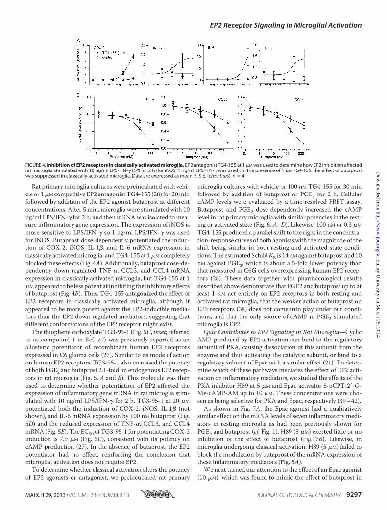
Rat primary microglia cultures were preincubated with vehi-cle or 1�Mcompetitive EP2 antagonist TG4-155 (28) for 20minfollowed by addition of the EP2 agonist butaprost at differentconcentrations. After 5 min, microglia were stimulated with 10ng/ml LPS/IFN-� for 2 h, and thenmRNAwas isolated to mea-sure inflammatory gene expression. The expression of iNOS ismore sensitive to LPS/IFN-� so 1 ng/ml LPS/IFN-� was usedfor iNOS. Butaprost dose-dependently potentiated the induc-tion of COX-2, iNOS, IL-1�, and IL-6 mRNA expression inclassically activatedmicroglia, andTG4-155 at 1�Mcompletelyblocked these effects (Fig. 4A). Additionally, butaprost dose-de-pendently down-regulated TNF-�, CCL3, and CCL4 mRNAexpression in classically activated microglia, but TG4-155 at 1�Mappeared to be less potent at inhibiting the inhibitory effectsof butaprost (Fig. 4B). Thus, TG4-155 antagonized the effect ofEP2 receptors in classically activated microglia, although itappeared to be more potent against the EP2-inducible media-tors than the EP2-down-regulated mediators, suggesting thatdifferent conformations of the EP2 receptor might exist.The thiophene carboxylate TG3-95-1 (Fig. 5C, inset; referred
to as compound 1 in Ref. 27) was previously reported as anallosteric potentiator of recombinant human EP2 receptorsexpressed in C6 glioma cells (27). Similar to its mode of actionon human EP2 receptors, TG3-95-1 also increased the potencyof both PGE2 and butaprost 2.1-fold on endogenous EP2 recep-tors in rat microglia (Fig. 5, A and B). This molecule was thenused to determine whether potentiation of EP2 affected theexpression of inflammatory gene mRNA in rat microglia stim-ulated with 10 ng/ml LPS/IFN-� for 2 h. TG3-95-1 at 20 �M
potentiated both the induction of COX-2, iNOS, IL-1� (notshown), and IL-6 mRNA expression by 100 nM butaprost (Fig.5D) and the reduced expression of TNF-�, CCL3, and CCL4mRNA (Fig. 5E). The EC50 ofTG3-95-1 for potentiatingCOX-2induction is 7.9 �M (Fig. 5C), consistent with its potency oncAMP production (27). In the absence of butaprost, the EP2potentiator had no effect, reinforcing the conclusion thatmicroglial activation does not require EP2.To determine whether classical activation alters the potency
of EP2 agonists or antagonist, we preincubated rat primary
microglia cultures with vehicle or 100 nM TG4-155 for 30 minfollowed by addition of butaprost or PGE2 for 2 h. CellularcAMP levels were evaluated by a time-resolved FRET assay.Butaprost and PGE2 dose-dependently increased the cAMPlevel in rat primarymicroglia with similar potencies in the rest-ing or activated state (Fig. 6, A–D). Likewise, 100 nM or 0.3 �M
TG4-155 produced a parallel shift to the right in the concentra-tion-response curves of both agonistswith themagnitude of theshift being similar in both resting and activated state condi-tions. The estimated SchildKB is 14 nMagainst butaprost and 10nM against PGE2, which is about a 5-fold lower potency thanthat measured in C6G cells overexpressing human EP2 recep-tors (28). These data together with pharmacological resultsdescribed above demonstrate that PGE2 and butaprost up to atleast 1 �M act entirely on EP2 receptors in both resting andactivated rat microglia, that the weaker action of butaprost onEP3 receptors (38) does not come into play under our condi-tions, and that the only source of cAMP in PGE2-stimulatedmicroglia is EP2.Epac Contributes to EP2 Signaling in Rat Microglia—Cyclic
AMP produced by EP2 activation can bind to the regulatorysubunit of PKA, causing dissociation of this subunit from theenzyme and thus activating the catalytic subunit, or bind to aregulatory subunit of Epac with a similar effect (21). To deter-mine which of these pathways mediates the effect of EP2 acti-vation on inflammatorymediators, we studied the effects of thePKA inhibitor H89 at 5 �M and Epac activator 8-pCPT-2�-O-Me-cAMP-AM up to 10 �M. These concentrations were cho-sen as being selective for PKA and Epac, respectively (39–42).As shown in Fig. 7A, the Epac agonist had a qualitatively
similar effect on the mRNA levels of seven inflammatory medi-ators in resting microglia as had been previously shown forPGE2 and butaprost (cf. Fig. 1). H89 (5 �M) exerted little or noinhibition of the effect of butaprost (Fig. 7B). Likewise, inmicroglia undergoing classical activation, H89 (5 �M) failed toblock the modulation by butaprost of the mRNA expression ofthese inflammatory mediators (Fig. 8A).We next turned our attention to the effect of an Epac agonist
(10 �M), which was found to mimic the effect of butaprost in
FIGURE 4. Inhibition of EP2 receptors in classically activated microglia. EP2 antagonist TG4-155 at 1 �M was used to determine how EP2 inhibition affectedrat microglia stimulated with 10 ng/ml LPS/IFN-� (L/I) for 2 h (for iNOS, 1 ng/ml LPS/IFN-� was used). In the presence of 1 �M TG4-155, the effect of butaprostwas suppressed in classically activated microglia. Data are expressed as mean � S.E. (error bars), n � 4.
EP2 Receptor Signaling in Microglial Activation
MARCH 29, 2013 • VOLUME 288 • NUMBER 13 JOURNAL OF BIOLOGICAL CHEMISTRY 9297
at Em
ory University on M
arch 25, 2014http://w
ww
.jbc.org/D
ownloaded from

microglia undergoing activation (Fig. 8B). H89 exerted similarsmall effects on the response to the Epac agonist (Fig. 7C) inresting microglia, suggesting that PKA impinges on the Epac
pathway. An Epac antagonist has recently been reported (43,44), butwe found that this antagonist (10�M) had no consistenteffect on cytokine modulation by the Epac agonist in primarymicroglia, which precluded its testing against butaprost; wepresume the antagonist has poor cell penetration in microgliaunder our conditions. These data are consistentwith the notionthat cAMP-activated Epac, not PKA, mediates much of theeffect of the EP2 receptor on the mRNA expression of inflam-matory genes in both resting and classically activatedmicroglia.
DISCUSSION
We show that in classically activated or resting state micro-glia EP2 receptor activation by PGE2 or butaprost creates amixed immune state by exacerbating the rapid induction ofproinflammatory COX-2, iNOS, IL-1�, and IL-6 but bluntingthe induction of proinflammatory TNF-� and the chemotacticfactors CCL3 and CCL4. Selective activation of the other PGE2receptors, EP1, EP3, and EP4, has little or no effect on expres-sion of these inflammatory mediators. Based on the effects of aselective PKA antagonist and a selective Epac activator, we con-clude that cAMP-activated Epac, not PKA, appears to mediatethe effect of EP2 receptors on these inflammatory genes in clas-sically activated microglia.COX-2 is an important component of neuroinflammation.
In an Alzheimer disease model, a selective COX-2 inhibitordecreased inflammatory factors and attenuated astrogliosis andneuronal cell loss (45). Inhibition of COX-2 was able to prevent
FIGURE 5. Potentiation of EP2 receptors in classically activated microglia. A and B, EP2 potentiator TG3-95-1 at 20 �M was used to determine how EP2potentiation affected cAMP levels induced by PGE2 and butaprost in rat primary microglia. C, effect of EP2 potentiator on the COX-2 mRNA expression in thepresence of 100 nM butaprost in classically activated microglia. Data are expressed as mean � S.E. (error bars), n � 4. Changes in COX-2, iNOS, and IL-6 mRNAexpression (D) and TNF-�, CCL3, and CCL4 mRNA expression (E) caused by 20 �M EP2 potentiator TG3-95-1 in the presence of 100 nM butaprost in classicallyactivated microglia. *, p � 0.05; **, p � 0.01; ***, p � 0.001. L/I, LPS/IFN-�.
FIGURE 6. cAMP levels induced by EP2 activation in rat primary microglia.Rat microglia pretreated with vehicle or 0.1 or 0.3 �M TG4-155 for 30 min wereincubated with different concentrations of PGE2 or butaprost for 2 h. TG4-155caused a concentration-dependent shift to the right in the PGE2 or butaprostconcentration-response curve in resting state microglia (A) and classicallyactivated microglia (B). Data points represent mean � S.E. (error bars) from asingle experiment run in triplicate.
EP2 Receptor Signaling in Microglial Activation
9298 JOURNAL OF BIOLOGICAL CHEMISTRY VOLUME 288 • NUMBER 13 • MARCH 29, 2013
at Em
ory University on M
arch 25, 2014http://w
ww
.jbc.org/D
ownloaded from

or slow down dopamine neuron degeneration either directly orthrough inhibition of microglial activation (46). Likewise, con-ditional ablation of COX-2 from principal forebrain neuronswas neuroprotective and anti-inflammatory in a mouse modelof epilepsy (6). iNOS, induced after inflammatory insults in thebrain, mediates neurotoxicity because of the oxidative/nitrosa-tive effects produced by NO release and the production of per-oxynitrite. In stroke models, excessive glutamate receptor acti-vation induces neurotoxicity partly by iNOS and peroxynitriteproduction (47). Pharmacological inhibition of iNOS delayeddisease onset and extended survival in mutant SOD1 mice,showing that iNOS expression in amyotrophic lateral sclerosiscontributed to disease progression (48). IL-6 is a multifunc-
tional cytokine during neuroinflammation in the central nerv-ous system. Mice with IL-6 deficiency were fully resistant toexperimental allergic encephalomyelitis, and IL-6 is proposedto be a therapeutic target for autoimmune diseases (49). How-ever, in traumatic brain injury, mice deficient for IL-6 showedincreased oxidative stress, decreased cell survival, and delayedwound healing, indicating that IL-6 might also be protective(50). Here, we show that PGE2 and EP2 activation by butaprostexacerbates the induction of COX-2, iNOS, and IL-6, whichshould promote inflammation caused by microglial activation,in classically activated microglia.COX-2 is present in cytosolic vesicle-like structures in
macrophages (51), and its protein expression and PGE2 secre-
FIGURE 7. EP2 receptor signaling pathways in resting state microglia. Epac activator 8-pCPT-2�-O-Me-cAMP-AM (1–10 �M) caused changes in mRNAexpression of the inflammatory mediators (A) in resting state microglia that mimicked those of EP2 activation. Data are expressed as mean � S.E. (error bars),n � 5. In the presence of 5 �M H89, mRNA expression changes in the butaprost group (B) or 8-pCPT-2�-O-Me-cAMP-AM group (C) were normalized by settingthe control group as 0 and the butaprost or 8-pCPT-2�-O-Me-cAMP-AM group as �1. *, p � 0.05 versus butaprost or 8-pCPT-2�-O-Me-cAMP-AM group.
FIGURE 8. EP2 receptor signaling pathways in classically activated microglia. H89 at 5 �M had little or no effect on expression of inflammatory mediatormRNA. The H89 effects were normalized by setting the LPS/IFN-� (L/I) group as 0 and the butaprost with LPS/IFN-� group as �1 (A). Epac activator 8-pCPT-2�-O-Me-cAMP-AM at 10 �M induced changes in inflammatory mediator mRNA expression (B) in classically activated microglia that mimicked the effects of EP2activation. Data are expressed as mean � S.E. (error bars), n � 3. *, p � 0.05; **, p � 0.01 versus the LPS/IFN-� group.
EP2 Receptor Signaling in Microglial Activation
MARCH 29, 2013 • VOLUME 288 • NUMBER 13 JOURNAL OF BIOLOGICAL CHEMISTRY 9299
at Em
ory University on M
arch 25, 2014http://w
ww
.jbc.org/D
ownloaded from

tion are low in rat microglia even after treatment with 10 ng/mlLPS/IFN-� for 24 h. Butaprost, however, can increase COX-2expression at both the mRNA and protein levels (Figs. 1–3),confirming previous results in microglia (25) and suggestingthat EP2 activation on microglia by PGE2 released from anysource should produce a self-reinforcing proinflammatoryeffect. The major source of PGE2 in the early stages of inflam-mation is likely to be neurons (6), which would promote braininflammation in part by activating EP2 receptors on nearbymicroglia.Based on the effects of EP2 activation on activatedmicroglia,
the consequences of EP2 receptor activation at the systems levelin the inflamed brain are expected to be complex. Prolongedincubation of rat microglia with LPS/IFN-� induces delayedexpression of the anti-inflammatory cytokine IL-10, which lim-its inflammation and promotes survival of neurons and glialcells in the brain (56). Induced IL-10 byLPS/IFN-� is blunted byEP2 activation. The reduction in IL-10 expression is expectedto be a secondary consequence of reduced TNF-� expressionbecause TNF-� is a strong inducer of IL-10 (52). The cytokineTNF-� plays a multidimensional role in inflammation-inducedneuronal damage in brain diseases. TNF-� expression in sub-stantia nigra elicits progressive neurodegeneration, motorsymptoms, and microglial activation (53), and TNF-� receptoractivation enhances microglial phagocytic activity (54). Inhibi-tion of TNF-� prevents cognitive decline and �-amyloid accu-mulation in a mouse model of Alzheimer disease (55). Chemo-kines CCL3 and CCL4 function to attract macrophages fromblood in addition to microglia migrating to the injured site inthe brain and thereby magnify local inflammation in neurode-generative diseases (56). Our results show that EP2 activationblunts the induction of TNF-� and the chemotactic factorsCCL3 and CCL4 in classically activatedmicroglia, and this mayinhibit chemotaxis of inflammatory cells in the brain.Modulation of the immune response by cAMP inmonocytes
and macrophages had been attributed to PKA and PKA-medi-ated changes in protein expression and function (57). However,Epac can act as an alternative cAMP mediator in monocytesand macrophages (20). Cytokine and chemokine production inbone marrow-derived dendritic cells involves both Epac1 andPKA (58). It has also been reported that high concentrations ofPKA and cell-impermeable Epac agonists play differential rolesin the production of cytokines in primary cultured microgliastimulated with a high concentration of endotoxin (59). In ourstudy, the actions of selective concentrations of both H89 (5�M) and the cell-permeable Epac agonist (10 �M) suggest thatcAMP-activated Epac rather than PKA mediates the effect ofEP2 receptors on the expression of COX-2, iNOS, and inflam-matory cytokines in classically activated rat microglia.Our study demonstrates that EP2 activation promotes brain
inflammation by exacerbating COX-2, iNOS, IL-1�, and IL-6induction and by opposing IL-10 induction in classically acti-vated rat microglia. At the same time, PGE2 and EP2 activationalso plays a negative feedback role by decreasing TNF-�, CCL3,and CCL4 release to inhibit chemotaxis of inflammatory cells.In conclusion, PGE2-activated EP2 regulates innate immunityin the central nervous system in a nuanced manner by promot-ingmany aspects of inflammationwhile reducing chemotaxis of
macrophages andmicroglia to the inflamed area. Epac signalingpathways are engaged in these EP2 actions and may providenew drug targets for brain inflammation.
REFERENCES1. Hanisch, U. K., and Kettenmann, H. (2007) Microglia: active sensor and
versatile effector cells in the normal and pathologic brain. Nat. Neurosci.10, 1387–1394
2. Carson, M. J., Thrash, J. C., andWalter, B. (2006) The cellular response inneuroinflammation: the role of leukocytes, microglia and astrocytes inneuronal death and survival. Clin. Neurosci. Res. 6, 237–245
3. Kreutzberg, G.W. (1996)Microglia: a sensor for pathological events in theCNS. Trends Neurosci. 19, 312–318
4. Melchior, B., Puntambekar, S. S., and Carson, M. J. (2006) Microglia andthe control of autoreactive T cell responses.Neurochem. Int. 49, 145–153
5. Garden, G. A., and Möller, T. (2006) Microglia biology in health and dis-ease. J. Neuroimmune Pharmacol. 1, 127–137
6. Serrano, G. E., Lelutiu, N., Rojas, A., Cochi, S., Shaw, R., Makinson, C. D.,Wang, D., FitzGerald, G. A., and Dingledine, R. (2011) Ablation of cy-clooxygenase-2 in forebrain neurons is neuroprotective and dampensbrain inflammation after status epilepticus. J. Neurosci. 31, 14850–14860
7. Yamagata, K., Andreasson, K. I., Kaufmann, W. E., Barnes, C. A., andWorley, P. F. (1993) Expression of a mitogen-inducible cyclooxygenase inbrain neurons: regulation by synaptic activity and glucocorticoids.Neuron11, 371–386
8. Murakami, M., Naraba, H., Tanioka, T., Semmyo, N., Nakatani, Y., Ko-jima, F., Ikeda, T., Fueki, M., Ueno, A., Oh, S., and Kudo, I. (2000) Regu-lation of prostaglandin E2 biosynthesis by induciblemembrane-associatedprostaglandin E2 synthase that acts in concert with cyclooxygenase-2.J. Biol. Chem. 275, 32783–32792
9. Tanioka, T., Nakatani, Y., Semmyo, N.,Murakami,M., and Kudo, I. (2000)Molecular identification of cytosolic prostaglandin E2 synthase that isfunctionally coupled with cyclooxygenase-1 in immediate prostaglandinE2 biosynthesis. J. Biol. Chem. 275, 32775–32782
10. Murakami, M., Nakashima, K., Kamei, D., Masuda, S., Ishikawa, Y., Ishii,T., Ohmiya, Y., Watanabe, K., and Kudo, I. (2003) Cellular prostaglandinE2 production by membrane-bound prostaglandin E synthase-2 via bothcyclooxygenases-1 and -2. J. Biol. Chem. 278, 37937–37947
11. Narumiya, S., Sugimoto, Y., and Ushikubi, F. (1999) Prostanoid receptors:structures, properties, and functions. Physiol. Rev. 79, 1193–1226
12. Cimino, P. J., Keene, C. D., Breyer, R.M.,Montine, K. S., andMontine, T. J.(2008) Therapeutic targets in prostaglandin E2 signaling for neurologicdisease. Curr. Med. Chem. 15, 1863–1869
13. Shi, J., Johansson, J., Woodling, N. S., Wang, Q., Montine, T. J., and An-dreasson, K. (2010) The prostaglandin E2 E-prostanoid 4 receptor exertsanti-inflammatory effects in brain innate immunity. J. Immunol. 184,7207–7218
14. Caggiano, A. O., and Kraig, R. P. (1999) Prostaglandin E receptor subtypesin cultured rat microglia and their role in reducing lipopolysaccharide-induced interleukin-1� production. J. Neurochem. 72, 565–575
15. Aloisi, F., Penna, G., Cerase, J., Menéndez Iglesias, B., and Adorini, L.(1997) IL-12 production by central nervous system microglia is inhibitedby astrocytes. J. Immunol. 159, 1604–1612
16. Menèndez Iglesias, B., Cerase, J., Ceracchini, C., Levi, G., and Aloisi, F.(1997) Analysis of B7–1 and B7–2 costimulatory ligands in culturedmouse microglia: upregulation by interferon-� and lipopolysaccharideand downregulation by interleukin-10, prostaglandin E2 and cyclic AMP-elevating agents. J. Neuroimmunol. 72, 83–93
17. Minghetti, L., Nicolini, A., Polazzi, E., Créminon, C., Maclouf, J., and Levi,G. (1997) Inducible nitric oxide synthase expression in activated rat mi-croglial cultures is downregulated by exogenous prostaglandin E2 and bycyclooxygenase inhibitors. Glia 19, 152–160
18. Aloisi, F., De Simone, R., Columba-Cabezas, S., and Levi, G. (1999) Oppo-site effects of interferon-� and prostaglandin E2 on tumor necrosis factorand interleukin-10 production in microglia: a regulatory loop controllingmicroglia pro- and anti-inflammatory activities. J. Neurosci. Res. 56,571–580
EP2 Receptor Signaling in Microglial Activation
9300 JOURNAL OF BIOLOGICAL CHEMISTRY VOLUME 288 • NUMBER 13 • MARCH 29, 2013
at Em
ory University on M
arch 25, 2014http://w
ww
.jbc.org/D
ownloaded from

19. Cohen, P. (2002) Protein kinases—the major drug targets of the twenty-first century? Nat. Rev. Drug Discov. 1, 309–315
20. Serezani, C. H., Ballinger, M. N., Aronoff, D. M., and Peters-Golden, M.(2008) Cyclic AMP: master regulator of innate immune cell function.Am. J. Respir. Cell Mol. Biol. 39, 127–132
21. Gloerich, M., and Bos, J. L. (2010) Epac: defining a new mechanism forcAMP action. Annu. Rev. Pharmacol. Toxicol. 50, 355–375
22. Shie, F. S., Breyer, R. M., and Montine, T. J. (2005) Microglia lacking Eprostanoid receptor subtype 2 have enhanced A� phagocytosis yet lackA�-activated neurotoxicity. Am. J. Pathol. 166, 1163–1172
23. Jin, J., Shie, F. S., Liu, J., Wang, Y., Davis, J., Schantz, A. M., Montine, K. S.,Montine, T. J., and Zhang, J. (2007) Prostaglandin E2 receptor subtype 2(EP2) regulatesmicroglial activation and associated neurotoxicity inducedby aggregated �-synuclein. J. Neuroinflammation 4, 2
24. Liang, X., Wang, Q., Shi, J., Lokteva, L., Breyer, R. M., Montine, T. J., andAndreasson, K. (2008) The prostaglandin E2 EP2 receptor accelerates dis-ease progression and inflammation in amodel of amyotrophic lateral scle-rosis. Ann. Neurol. 64, 304–314
25. Shie, F. S., Montine, K. S., Breyer, R. M., and Montine, T. J. (2005) Micro-glial EP2 is critical to neurotoxicity from activated cerebral innate immu-nity. Glia 52, 70–77
26. Jiang, J., Quan, Y., Ganesh, T., Pouliot,W.A., Dudek, F. E., andDingledine,R. (February 11, 2013) Inhibition of the prostaglandin receptor EP2 fol-lowing status epilepticus reduces delayed mortality and brain inflamma-tion. Proc. Natl. Acad. Sci. U.S.A. 10.1073/pnas.1218498110
27. Jiang, J., Ganesh, T., Du, Y., Thepchatri, P., Rojas, A., Lewis, I., Kurtkaya, S.,Li, L., Qui, M., Serrano, G., Shaw, R., Sun, A., and Dingledine, R. (2010)Neuroprotection by selective allosteric potentiators of the EP2 prostaglan-din receptor. Proc. Natl. Acad. Sci. U.S.A. 107, 2307–2312
28. Jiang, J., Ganesh, T., Du, Y., Quan, Y., Serrano, G., Qui, M., Speigel, I.,Rojas, A., Lelutiu, N., and Dingledine, R. (2012) Small molecule antag-onist reveals seizure-induced mediation of neuronal injury by prosta-glandin E2 receptor subtype EP2. Proc. Natl. Acad. Sci. U.S.A. 109,3149–3154
29. Jiang, J., and Dingledine, R. (2013) Role of prostaglandin receptor EP2 inthe regulations of cancer cell proliferation, invasion, and inflammation.J. Pharmacol. Exp. Ther. 344, 360–367
30. Quan, Y., Möller, T., and Weinstein, J. R. (2009) Regulation of Fc� recep-tors and immunoglobulin G-mediated phagocytosis in mouse microglia.Neurosci. Lett. 464, 29–33
31. Quan, Y., Jiang, C. T., Xue, B., Zhu, S. G., and Wang, X. (2011) Highglucose stimulates TNF� andMCP-1 expression in rat microglia via ROSand NF-�B pathways. Acta Pharmacol. Sin. 32, 188–193
32. Vandesompele, J., De Preter, K., Pattyn, F., Poppe, B., Van Roy, N., DePaepe, A., and Speleman, F. (2002) Accurate normalization of real-timequantitative RT-PCR data by geometric averaging of multiple internalcontrol genes. Genome Biol. 3, RESEARCH0034
33. Weinstein, J. R., Zhang, M., Kutlubaev, M., Lee, R., Bishop, C., Andersen,H., Hanisch, U. K., andMöller, T. (2009) Thrombin-induced regulation ofCD95(Fas) expression in the N9 microglial cell line: evidence for involve-ment of proteinase-activated receptor(1) and extracellular signal-regu-lated kinase 1/2. Neurochem. Res. 34, 445–452
34. Quan, Y., Du, J., andWang, X. (2007) High glucose stimulates GRO secre-tion from rat microglia via ROS, PKC, and NF-�B pathways. J. Neurosci.Res. 85, 3150–3159
35. Martinez, F. O., Helming, L., and Gordon, S. (2009) Alternative activationof macrophages: an immunologic functional perspective. Annu. Rev. Im-munol. 27, 451–483
36. Kiriyama, M., Ushikubi, F., Kobayashi, T., Hirata, M., Sugimoto, Y., andNarumiya, S. (1997) Ligand binding specificities of the eight types andsubtypes of themouse prostanoid receptors expressed inChinese hamsterovary cells. Br. J. Pharmacol. 122, 217–224
37. Billot, X., Chateauneuf, A., Chauret, N., Denis, D., Greig, G., Mathieu,M. C., Metters, K.M., Slipetz, D.M., and Young, R. N. (2003) Discovery ofa potent and selective agonist of the prostaglandin EP4 receptor. Bioorg.Med. Chem. Lett. 13, 1129–1132
38. Abramovitz, M., Adam,M., Boie, Y., Carrière, M., Denis, D., Godbout, C.,Lamontagne, S., Rochette, C., Sawyer, N., Tremblay, N. M., Belley, M.,
Gallant, M., Dufresne, C., Gareau, Y., Ruel, R., Juteau, H., Labelle, M.,Ouimet, N., and Metters, K. M. (2000) The utilization of recombinantprostanoid receptors to determine the affinities and selectivities of pros-taglandins and related analogs. Biochim. Biophys. Acta 1483, 285–293
39. Engh, R. A., Girod, A., Kinzel, V., Huber, R., and Bossemeyer, D. (1996)Crystal structures of catalytic subunit of cAMP-dependent protein kinasein complex with isoquinolinesulfonyl protein kinase inhibitors H7, H8,and H89. Structural implications for selectivity. J. Biol. Chem. 271,26157–26164
40. Davies, S. P., Reddy, H., Caivano, M., and Cohen, P. (2000) Specificity andmechanism of action of some commonly used protein kinase inhibitors.Biochem. J. 351, 95–105
41. Vliem, M. J., Ponsioen, B., Schwede, F., Pannekoek, W. J., Riedl, J., Koois-tra, M. R., Jalink, K., Genieser, H. G., Bos, J. L., and Rehmann, H. (2008)8-pCPT-2�-O-Me-cAMP-AM: an improved Epac-selective cAMP ana-logue. Chembiochem 9, 2052–2054
42. Chepurny, O. G., Leech, C. A., Kelley, G. G., Dzhura, I., Dzhura, E., Li, X.,Rindler, M. J., Schwede, F., Genieser, H. G., and Holz, G. G. (2009) En-hanced Rap1 activation and insulin secretagogue properties of an ace-toxymethyl ester of an Epac-selective cyclic AMP analog in rat INS-1 cells:studies with 8-pCPT-2�-O-Me-cAMP-AM. J. Biol. Chem. 284,10728–10736
43. Chen, H., Tsalkova, T., Mei, F. C., Hu, Y., Cheng, X., and Zhou, J. (2012)5-Cyano-6-oxo-1,6-dihydro-pyrimidines as potent antagonists targetingexchange proteins directly activated by cAMP. Bioorg. Med. Chem. Lett.22, 4038–4043
44. Almahariq, M., Tsalkova, T., Mei, F. C., Chen, H., Zhou, J., Sastry, S. K.,Schwede, F., and Cheng, X. (2013) A novel EPAC specific inhibitor sup-presses pancreatic cancer cell migration and invasion. Mol. Pharmacol.83, 122–128
45. Dargahi, L., Nasiraei-Moghadam, S., Abdi, A., Khalaj, L., Moradi, F., andAhmadiani, A. (2011) Cyclooxygenase (COX)-1 activity precedes theCOX-2 induction inA�-inducedneuroinflammation. J.Mol. Neurosci.45,10–21
46. Sánchez-Pernaute, R., Ferree, A., Cooper, O., Yu, M., Brownell, A. L., andIsacson, O. (2004) Selective COX-2 inhibition prevents progressive dop-amine neuron degeneration in a rat model of Parkinson’s disease. J. Neu-roinflammation 1, 6
47. Kumar, A., Singh, R. L., and Babu, G. N. (2010) Cell death mechanisms inthe early stages of acute glutamate neurotoxicity. Neurosci. Res. 66,271–278
48. Chen, K., Northington, F. J., andMartin, L. J. (2010) Inducible nitric oxidesynthase is present in motor neuronmitochondria and Schwann cells andcontributes to disease mechanisms in ALSmice. Brain Struct. Funct. 214,219–234
49. Tanaka, T., Katada, Y., Higa, S., Fujiwara, H., Wang, W., Saeki, Y.,Ohshima, S., Okuda, Y., Suemura, M., and Kishimoto, T. (2001) Enhance-ment of T helper2 response in the absence of interleukin (IL-)6; an inhi-bition of IL-4-mediated T helper2 cell differentiation by IL-6.Cytokine 13,193–201
50. Penkowa, M., Giralt, M., Carrasco, J., Hadberg, H., and Hidalgo, J. (2000)Impaired inflammatory response and increased oxidative stress and neu-rodegeneration after brain injury in interleukin-6-deficient mice.Glia 32,271–285
51. Yamashita, M., Tsuji, S., Nishiyama, A., Myrvik, Q. N., Henriksen, R. A.,and Shibata, Y. (2007) Differential subcellular localization of COX-2 inmacrophages phagocytosing heat-killedMycobacteriumbovisBCG.Am. J.Physiol. Cell Physiol. 293, C184–C190
52. Foey, A. D., Parry, S. L.,Williams, L.M., Feldmann,M., Foxwell, B.M., andBrennan, F. M. (1998) Regulation of monocyte IL-10 synthesis by endog-enous IL-1 and TNF-�: role of the p38 and p42/44 mitogen-activatedprotein kinases. J. Immunol. 160, 920–928
53. De Lella Ezcurra, A. L., Chertoff, M., Ferrari, C., Graciarena, M., andPitossi, F. (2010) Chronic expression of low levels of tumor necrosis fac-tor-� in the substantia nigra elicits progressive neurodegeneration, de-layed motor symptoms and microglia/macrophage activation. Neurobiol.Dis. 37, 630–640
54. von Zahn, J., Möller, T., Kettenmann, H., and Nolte, C. (1997) Microglial
EP2 Receptor Signaling in Microglial Activation
MARCH 29, 2013 • VOLUME 288 • NUMBER 13 JOURNAL OF BIOLOGICAL CHEMISTRY 9301
at Em
ory University on M
arch 25, 2014http://w
ww
.jbc.org/D
ownloaded from

phagocytosis is modulated by pro- and anti-inflammatory cytokines.Neu-roreport 8, 3851–3856
55. Chavant, F., Deguil, J., Pain, S., Ingrand, I., Milin, S., Fauconneau, B.,Pérault-Pochat, M. C., and Lafay-Chebassier, C. (2010) Imipramine, inpart through tumor necrosis factor � inhibition, prevents cognitive de-cline and �-amyloid accumulation in a mouse model of Alzheimer’s dis-ease. J. Pharmacol. Exp. Ther. 332, 505–514
56. Ransohoff, R. M. (2009) Chemokines and chemokine receptors: standingat the crossroads of immunobiology and neurobiology. Immunity 31,711–721
57. Zambon, A. C., Zhang, L., Minovitsky, S., Kanter, J. R., Prabhakar, S.,Salomonis, N., Vranizan, K., Dubchak, I., Conklin, B. R., and Insel, P. A.
(2005) Gene expression patterns define key transcriptional events in cell-cycle regulation by cAMP and protein kinase A. Proc. Natl. Acad. Sci.U.S.A. 102, 8561–8566
58. Aronoff, D. M., Carstens, J. K., Chen, G. H., Toews, G. B., and Peters-Golden, M. (2006) Short communication: differences between macro-phages and dendritic cells in the cyclic AMP-dependent regulation oflipopolysaccharide-induced cytokine and chemokine synthesis. J. Inter-feron Cytokine Res. 26, 827–833
59. Liu, J., Zhao, X., Cao, J., Xue, Q., Feng, X., Liu, X., Zhang, F., and Yu, B.(2011) Differential roles of PKA and epac on the production of cytokinesin the endotoxin-stimulated primary cultured microglia. J. Mol. Neurosci.45, 186–193
EP2 Receptor Signaling in Microglial Activation
9302 JOURNAL OF BIOLOGICAL CHEMISTRY VOLUME 288 • NUMBER 13 • MARCH 29, 2013
at Em
ory University on M
arch 25, 2014http://w
ww
.jbc.org/D
ownloaded from