setting up an andrology laboratory - clevelandclinic.org · this chapter serves as a practical...
TRANSCRIPT
Seediscussions,stats,andauthorprofilesforthispublicationat:https://www.researchgate.net/publication/259687687
SettingUpanAndrologyLaboratory
Chapter·January2014
CITATIONS
0
READS
735
1author:
Someoftheauthorsofthispublicationarealsoworkingontheserelatedprojects:
AntioxidanttherapyinidiopathicoligoasthenoteratozoospermiaViewproject
SpermDNAFragmentationViewproject
AshokAgarwal
ClevelandClinic
1,844PUBLICATIONS31,897CITATIONS
SEEPROFILE
AllcontentfollowingthispagewasuploadedbyAshokAgarwalon14January2014.
Theuserhasrequestedenhancementofthedownloadedfile.
ABSTRACT
Male factor infertility is a sole or contributing factor inapproximately 40% of the infertile couples and thereforeinvestigating the cause of the male infertility constitutes amajor part of the diagnosis and treatment of infertile couples.The andrology laboratory supports and conducts clinicalassessment of the semen and helps in the diagnosis ofvarious diseases and malfunctions related to male infertility.
Development of advanced assisted reproductivetechniques in the last decade has increased the scope oftoday’s andrology laboratories which are also involved incryopreservation of gametes and reproductive tissuespreserving the reproductive capacities of couples for variousreasons.
This chapter serves as a practical guide for setting-up anandrology laboratory for diagnostic and therapeuticpurposes.
INTRODUCTION
“Andrology” is the field of medicine that deals with
matters affecting the male reproductive system. The
earliest use of this term appeared in 1891 in the Journal
of American Medical Association when it reported on
the formation of the American Andrological Association.1
Because male infertility is the primary or contributing
factor in more than 40% of infertile couples 2, this field
has been the object of constant research and has therefore
seen advances in diagnostic tests and tools used for the
investigation of male infertility.
The Andrology Laboratory encompasses clinical
laboratories under both Pathology and Laboratory
Medicine. It is entrusted with supporting clinical
assessments of semen that may help diagnose potential
Kelly S Athayde, Ashok Agarwal
16
Setting Up an Andrology Laboratory
diseases or malfunctions related to male fertility. Utilizing
both simple and complex techniques, its evaluation
provides useful information for the infertility specialist
in the management of the subfertile man. Besides
diagnostic activities, it should provide therapeutic
procedures for patients requiring assistance in achieving
pregnancy such as Assisted Reproduction techniques:
Intrauterine Insemination (IUI) and In Vitro Fertilization
(IVF). The Andrology laboratory also works in parallel
with endocrinology, genetic, and in vitro fertilization
laboratories to diagnose and assist in the treatment of
patients facing difficulties conceiving or seeking
preservation of their reproductive capacity by
cryopreserving their gametes or reproductive tissue.
This chapter will provide readers, in a very practical
way, with information on the basic requirements for
setting up and operating an Andrology laboratory for
diagnostic and therapeutic activities.
LABORATORY ASPECTS AND PROCEDURES
Numerous procedures can be performed in the
Andrology Laboratory. Due to different applications
(clinical and research) and clinical correlations in the
current scientific literature, some of these procedures may
not be included in this chapter.
Semen analysis is the most common test to be
conducted in the Andrology Laboratory. It reveals how
many spermatozoa are in the ejaculate, the vigor with
which they move (quantitative motility), and how normal
these cells look morphologically. Complementary
assessments should include viability, the presence of
ANDROLOGY LABORATORY MANUAL158
round cells and their identification (i.e. leukocytes or
immature sperm cells), and detection of antibodies.
Additional tests can be performed based on the results
obtained from this initial analysis. Such tests may
complement the initial analysis and provide more
information about sperm cell functionality and structure.
Biochemical markers such as Reactive Oxygen Species
(ROS) and tests that assess sperm DNA damage and the
inducibility of the Acrosome Reaction (AR) can provide
a more specific functional assessment of these gametes.
Bioassays that assess gamete interaction, such as the
sperm-hamster egg penetration assay (SPA) and the
human sperm-zona pellucida binding test (hemizona
assay, HZA), evaluate the occurrence of spontaneous
sperm AR and the capacity for penetration, respectively.
They have strong diagnostic power but, like the AR test,
still require better standardization before they can be
introduced as clinical tools. 3 Moreover, the bioassays
require hamster eggs and non-fertilizable human oocytes,
which can impose extra practical limitations for their
introduction into routine laboratory practice.
The most commonly performed tests are listed below.
They can be classified in two groups: diagnostic and
therapeutic.
Diagnostic Procedures
Complete Semen Analysis
Macroscopic parameters (color, volume, pH, viscosity,
liquefaction, and agglutination)
Microscopic parameters (sperm concentration, motility,
morphology, presence of round cells, and different types
of debris)
Detection of Leukocytes in Semen
Myeloperoxidase test (Endtz)
Sperm Vitality and Membrane Viability
Eosin-Nigrosin test
Hypo-osmotic Swelling test (HOS)
Biochemical
Fructose qualitative and quantitative
Oxidative Stress
Reactive Oxygen Species (ROS)
Total Antioxidant Capacity (TAC)
Azoospermia Screen Procedure
Antibody Assessment
Test for anti-sperm antibodies on the sperm surface
Test for free antibodies, directed against the sperm surface
(seminal plasma, serum or cervical mucus)
Sperm DNA Damage Test
Sperm Chromatin Structure Assay (SCSA)
Terminal deoxynucleotidyl transferase dUTP nick end
labeling (TUNEL)
COMET, also called single cell gel electrophoresis (SCGE).
Therapeutic
Sperm Preparation for Intrauterine Insemination (IUI)
or In vitro Fertilization (IVF)
Preparation of cryopreserved semen for IUI or IVF
Techniques
Gradient method
Swim-up method
Simple wash (concentration technique)
Cryopreservation
Semen
Epididymal aspirate
Testicular Tissue
LABORATORY DESIGN
Before working on any laboratory design, local
regulations must be carefully studied. These regulations
may differ between countries or even between states, and
compliance is not optional.
Ideally, the Andrology Laboratory should contain
distinct areas for diagnostic and therapeutic procedures
(Figure 16.1). Therapeutic procedures should be
performed in a sterile environment, which can be
achieved by using a laminar flow hood. The physical area
should be determined according to a pre-estimation of
the number of tests to be offered by the service and the
volume of procedures expected to be performed. This will
determine the number of technicians that must be
employed and the different types of equipment that must
be made available such as microscopes, cell counters,
incubators, etc. New laboratories may not have an
established range of workload due to regional and market
variations and thus should always keep in mind plans
for future expansion.
SETTING UP AN ANDROLOGY LABORATORY 159
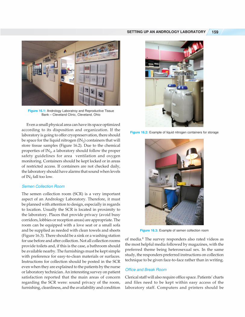
Even a small physical area can have its space optimized
according to its disposition and organization. If the
laboratory is going to offer cryopreservation, there should
be space for the liquid nitrogen (lN2) containers that will
store tissue samples (Figure 16.2). Due to the chemical
properties of lN2, a laboratory should follow the proper
safety guidelines for area ventilation and oxygen
monitoring. Containers should be kept locked or in areas
of restricted access. If containers are not checked daily,
the laboratory should have alarms that sound when levels
of lN2 fall too low.
Semen Collection Room
The semen collection room (SCR) is a very important
aspect of an Andrology Laboratory. Therefore, it must
be planned with attention to design, especially in regards
to location. Usually the SCR is located in proximity to
the laboratory. Places that provide privacy (avoid busy
corridors, lobbies or reception areas) are appropriate. The
room can be equipped with a love seat or a small sofa
and be supplied as needed with clean towels and sheets
(Figure 16.3). There should be a sink or a washing station
for use before and after collection. Not all collection rooms
provide toilets and, if this is the case, a bathroom should
be available nearby. The furnishings must be kept simple
with preference for easy-to-clean materials or surfaces.
Instructions for collection should be posted in the SCR
even when they are explained to the patients by the nurse
or laboratory technician. An interesting survey on patient
satisfaction reported that the main areas of concern
regarding the SCR were: sound privacy of the room,
furnishing, cleanliness, and the availability and condition
of media.4 The survey responders also rated videos as
the most helpful media followed by magazines, with the
preferred theme being heterosexual sex. In the same
study, the responders preferred instructions on collection
technique to be given face-to-face rather than in writing.
Office and Break Room
Clerical staff will also require office space. Patients’ charts
and files need to be kept within easy access of the
laboratory staff. Computers and printers should be
Figure 16.1: Andrology Laboratroy and Reproductive Tissue
Bank – Cleveland Clinic, Cleveland, Ohio
Figure 16.2: Example of liquid nitrogen containers for storage
Figure 16.3: Example of semen collection room
ANDROLOGY LABORATORY MANUAL160
Workstation 3: Biochemical and antibody tests. Due to
the nature of the fructose determination, a safety hood is
recommended. Reagents for these procedures are usually
kept refrigerated.
Workstation 4: Semen preparation and cryopreservation
procedures. As mentioned previously, these procedures
should be performed in a sterile environment
(Figure 16.7). The same laminar flow hood, for example,
could be used for both procedures; this could be an option
if the lab cannot support two separate workstations
logistically or financially. When optimizing the semen
preparation procedures, a CO2 incubator can be used for
sample incubation in both the pre- and post-processing
periods.
available for entering results and work-related issues. A
very important aspect is the break room. A resting area
for employees must be included in the plan. Restrooms
for employees should be close by.
Workstations
For practical purposes, workstations can be pre-defined.
For example:
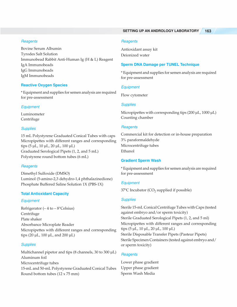
Workstation 1: Semen wet preparation (macroscopic
parameters on seminal analysis, sperm count and
motility, Endtz test, HOS and Eosin-Nigrosin
preparations). This area can be equipped with
microscopes, cell counters, incubators, water baths or
warm stage and the necessary supplies and reagents
(Figure 16.4). Controlled temperature at 37°C is needed
for liquefaction, count and motility assessment, and HOS
incubation. This workstation can also be equipped with
an automated semen analyzer, which generates a more
complete assessment of sperm motion characteristics. It
is important to emphasize that the automated evaluation
cannot replace a manual evaluation. It is still
recommended that practitioners manually count semen
samples to validate the computer results.
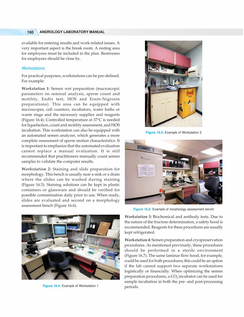
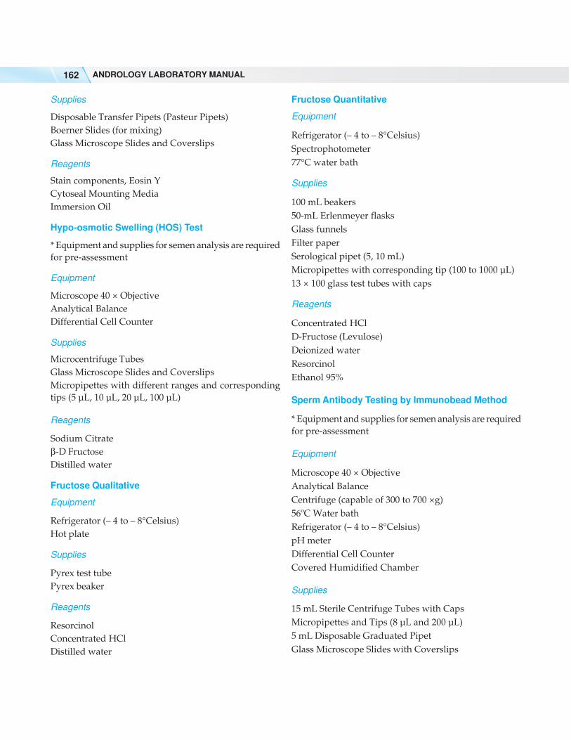
Workstation 2: Staining and slide preparation for
morphology. This bench is usually near a sink or a drain
where the slides can be washed during staining
(Figure 16.5). Staining solutions can be kept in plastic
containers or glassware and should be verified for
possible contamination daily prior to use. When ready,
slides are evaluated and second on a morphology
assessment bench (Figure 16.6).
Figure 16.4: Example of Workstation 1
Figure 16.5: Example of Workstation 2
Figure 16.6: Example of morphology assessment bench

SETTING UP AN ANDROLOGY LABORATORY 161
EQUIPMENT, SUPPLIES AND REAGENTS
The equipment, supplies, and reagents needed for each
procedure vary according to the protocols to be followed.
The following list suggests what each of the following
tests may require:
Semen Analysis
Equipment
Phase Contrast Microscope with 10 × and 20 × Objective.
Cell Counter
Counting Chamber
Centrifuge (~1600 rpm)
Refrigerator (– 4 to – 8°Celsius)
Vortex mixer
Supplies
Micropipettes with different ranges and corresponding
tips (5 µL, 10 µL, 20 µL, 100 µL)
Automatic Pipetor (rechargeable)
Graduated Serological Pipets (1, 2 and 5 ml)
Test Tube rack for 15 mL tubes
15 mL Polystyrene Graduated Conical Centrifuge Tubes
Specimen Cups
Glass Microscope Slides and Coverslips
pH paper Range 6-8
Disposable Transfer Pipets (Pasteur Pipets)
Microcentrifuge tubes for dilution
Reagents
Tyrode’s Salt Solution
Stain for Morphology
Equipment
Microscope with 100 × Objective
Differential cell Counter
Supplies
Micropipettes with different ranges and corresponding
tips (5 µL, 10 µL, 20 µL, 100 µL)
Glass Microscope Slides and Coverslips
Reagents
Cytoseal Mounting Media
Papanicolaou stain or Diff-Quik (rapid staining method)
or Shorr stain
Endtz Test (Peroxidase Staining)
* Equipment and supplies for semen analysis are required
for pre-assessment
Equipment
Analytical Balance
Microscope 10 × objective
Supplies
Makler Chamber
Aluminum Foil
Dark Colored Microcentrifuge Tubes
Micropipettes with different ranges and corresponding
tips (5 µL, 10 µL, 20 µL, 100 µL)
Reagents
Ethanol, 96%
Benzidine
3% H2O2
Tyrodes Salt Solution
Eosin-Nigrosin Staining
* Equipment and supplies for semen analysis are required
for pre-assessment
Equipment
Microscope 100 × objective
Differential cell counter
Figure 16.7: Example of Workstation 4
ANDROLOGY LABORATORY MANUAL162
Supplies
Disposable Transfer Pipets (Pasteur Pipets)
Boerner Slides (for mixing)
Glass Microscope Slides and Coverslips
Reagents
Stain components, Eosin Y
Cytoseal Mounting Media
Immersion Oil
Hypo-osmotic Swelling (HOS) Test
* Equipment and supplies for semen analysis are required
for pre-assessment
Equipment
Microscope 40 × Objective
Analytical Balance
Differential Cell Counter
Supplies
Microcentrifuge Tubes
Glass Microscope Slides and Coverslips
Micropipettes with different ranges and corresponding
tips (5 µL, 10 µL, 20 µL, 100 µL)
Reagents
Sodium Citrate
β-D Fructose
Distilled water
Fructose Qualitative
Equipment
Refrigerator (– 4 to – 8°Celsius)
Hot plate
Supplies
Pyrex test tube
Pyrex beaker
Reagents
Resorcinol
Concentrated HCl
Distilled water
Fructose Quantitative
Equipment
Refrigerator (– 4 to – 8°Celsius)
Spectrophotometer
77°C water bath
Supplies
100 mL beakers
50-mL Erlenmeyer flasks
Glass funnels
Filter paper
Serological pipet (5, 10 mL)
Micropipettes with corresponding tip (100 to 1000 µL)
13 × 100 glass test tubes with caps
Reagents
Concentrated HCl
D-Fructose (Levulose)
Deionized water
Resorcinol
Ethanol 95%
Sperm Antibody Testing by Immunobead Method
* Equipment and supplies for semen analysis are required
for pre-assessment
Equipment
Microscope 40 × Objective
Analytical Balance
Centrifuge (capable of 300 to 700 ×g)
56ºC Water bath
Refrigerator (– 4 to – 8°Celsius)
pH meter
Differential Cell Counter
Covered Humidified Chamber
Supplies
15 mL Sterile Centrifuge Tubes with Caps
Micropipettes and Tips (8 µL and 200 µL)
5 mL Disposable Graduated Pipet
Glass Microscope Slides with Coverslips
SETTING UP AN ANDROLOGY LABORATORY 163
Reagents
Bovine Serum Albumin
Tyrodes Salt Solution
Immunobead Rabbit Anti-Human Ig (H & L) Reagent
IgA Immunobeads
IgG Immunobeads
IgM Immunobeads
Reactive Oxygen Species
* Equipment and supplies for semen analysis are required
for pre-assessment
Equipment
Luminometer
Centrifuge
Supplies
15 mL Polystyrene Graduated Conical Tubes with caps
Micropipettes with different ranges and corresponding
tips (5 µL, 10 µL, 20 µL, 100 µL)
Graduated Serological Pipets (1, 2, and 5 mL)
Polystyrene round bottom tubes (6 mL)
Reagents
Dimethyl Sulfoxide (DMSO)
Luminol (5-amino-2,3 dehydro-1,4 phthalazinedione)
Phosphate Buffered Saline Solution 1X (PBS-1X)
Total Antioxidant Capacity
Equipment
Refrigerator (– 4 to – 8°Celsius)
Centrifuge
Plate shaker
Absorbance Microplate Reader
Micropipettes with different ranges and corresponding
tips (20 µL, 100 µL, and 200 µL)
Supplies
Multichannel pipetor and tips (8 channels, 30 to 300 µL)
Aluminum foil
Microcentrifuge tubes
15-mL and 50-mL Polystyrene Graduated Conical Tubes
Round bottom tubes (12 x 75 mm)
Reagents
Antioxidant assay kit
Deionized water
Sperm DNA Damage per TUNEL Technique
* Equipment and supplies for semen analysis are required
for pre-assessment
Equipment
Flow cytometer
Supplies
Micropipettes with corresponding tips (200 µL, 1000 µL)
Counting chamber
Reagents
Commercial kit for detection or in-house preparation
3% paraformaldehyde
Microcentrifuge tubes
Ethanol
Gradient Sperm Wash
* Equipment and supplies for semen analysis are required
for pre-assessment
Equipment
37ºC Incubator (CO2 supplied if possible)
Supplies
Sterile 15-mL Conical Centrifuge Tubes with Caps (tested
against embryo and/or sperm toxicity)
Sterile Graduated Serological Pipets (1, 2, and 5 ml)
Micropipettes with different ranges and corresponding
tips (5 µL, 10 µL, 20 µL, 100 µL)
Sterile Disposable Transfer Pipets (Pasteur Pipets)
Sterile Specimen Containers (tested against embryo and/
or sperm toxicity)
Reagents
Lower phase gradient
Upper phase gradient
Sperm Wash Media
ANDROLOGY LABORATORY MANUAL164
Semen, Epididymal Aspiration, and Testicular TissueCryopreservation
* Equipment and supplies for semen analysis are required
for pre-assessment
Equipment
Aliquot Mixer/Barnstead/Thermolyne
37 ºC Incubator (CO2 supplied if possible)
– 20 ºC Freezer
Liquid nitrogen container
Liquid nitrogen container alarm
Oxygen monitor
Supplies
Sterile 15 mL Conical Centrifuge Tubes with Caps
Sterile Graduated Serological Pipets (1, 2, and 5 mL)
Micropipettes with different ranges and corresponding
tips (5 µL, 10 µL, 20 µL, 100 µL)
Sterile Graduated Serological Pipets (1, 2, and 5 mL)
Sterile Specimen Containers (tested against embryo and/
or sperm toxicity)
Test Tube rack for 15-mL tubes
Plastic Cryosleeves
Cryovials – 2 mL
Cryocanes
Laboratory Cryomarkers (non-toxic)
Liquid Nitrogen
Cryogenic Protective Gloves
Protective Eye Goggles
Reagents
Freezing Medium
Additional for Testicular Tissue
Equipment
Dissecting Microscope
Inverted Microscope
Supplies
Kontes Pellet Pestle
Sterile Tissue Culture Plates, 60 x 15 mm
Reagent
Sterile Mineral Oil
SAFETY GUIDELINES
The safety guidelines for the Andrology Laboratory
include use of protective gloves and appropriate
laboratory coats for all procedures. All samples should
be treated as potentially contaminated and harmful. The
World Health Organization (WHO) laboratory manual5,
in its fourth edition, has described these guidelines.
REFERENCES
1. Andrology The American Society of. A Handbook ofAndrology. Lawrence: The American Society ofAndrology; 1995.
2. Jeyendran RS. Protocols for Semen Analysis in ClinicalDiagnosis. New York: The Parthenon Publishing Group;2003.
3. Oehninger S, Franken D, Kruger T. Approaching the nextmillennium: How should we manage andrologydiagnosis in the intracytoplasmic sperm injection era?Fertil Steril. 1997;67(3):434-6.
4. Levy BMCS, editor. The men’s lounge improvementproject: What do men really want? American Society forReproductive Medicine; September, 2007. Elsevier Inc.
5. Kruger TF, Coetzee K. The role of sperm morphology inassisted reproduction. Hum Reprod Update.1999;5(2):172-8.
View publication statsView publication stats