senescence marker protein 30 functions as gluconolactonase ... · senescence marker protein 30...
TRANSCRIPT
Senescence marker protein 30 functions asgluconolactonase in L-ascorbic acid biosynthesis,and its knockout mice are prone to scurvyYoshitaka Kondo*†‡, Yoko Inai‡§, Yasunori Sato*‡¶, Setsuko Handa*, Sachiho Kubo*, Kentaro Shimokado†,Sataro Goto¶, Morimitsu Nishikimi§�, Naoki Maruyama*, and Akihito Ishigami*,**
*Department of Molecular Pathology, Tokyo Metropolitan Institute of Gerontology, Tokyo 173-0015, Japan; †Vascular Medicine and Geriatrics, TokyoMedical and Dental University, Tokyo 113-8510, Japan; §Department of Biochemistry, Wakayama Medical University, Wakayama 641-0012, Japan;and ¶Department of Biochemistry, Toho University, Chiba 274-8510, Japan
Edited by John E. Halver, University of Washington, Seattle, WA, and approved February 27, 2006 (received for review December 30, 2005)
We originally identified senescence marker protein 30 (SMP30) asa distinctive protein whose expression decreases in an androgen-independent manner with aging. Here, we report its sequencehomology found in two kinds of bacterial gluconolactonases(GNLs) by using the BLAST search. Then, through a biochemicalstudy, we identify SMP30 as the lactone-hydrolyzing enzyme GNLof animal species. SMP30 purified from the rat liver had lactonaseactivity toward various aldonolactones, such as D- and L-glucono-�-lactone, D- and L-gulono-�-lactone, and D- and L-galactono-�-lactone, with a requirement for Zn2� or Mn2� as a cofactor.Furthermore, in SMP30 knockout mice, no GNL activity was de-tectable in the liver. Thus, we conclude that SMP30 is a unique GNLin the liver. The lactonase reaction with L-gulono-�-lactone is thepenultimate step in L-ascorbic acid (AA) biosynthesis, and theessential role of SMP30 in this synthetic process was verified hereby a nutritional study using SMP30 knockout mice. These knockoutmice (n � 6), fed a vitamin C-deficient diet, did not thrive; i.e., theydisplayed symptoms of scurvy such as bone fracture and rachiticrosary and then died by 135 days after the start of receiving thedeficient diet. The AA levels in their livers and kidneys at the timeof death were <1.6% of those in WT control mice. In addition, byusing the SMP30 knockout mouse, we demonstrate that thealternative pathway of AA synthesis involving D-glucurono-�-lactone operates in vivo, although its flux is fairly small.
aging � osteogenic disorder � vitamin C
Senescence marker protein 30 (SMP30) is a 34-kDa proteinwhose tissue levels in the liver, kidney, and lung decrease
with aging (1, 2). To examine the physiological function ofSMP30, we established SMP30 knockout mice (3) and found thatthey were viable and fertile, although they were lower in bodyweight and shorter in life span than WT mice (4). Their liverswere also far more susceptible to TNF-�- and Fas-mediatedapoptosis than those of WT mice, indicating that SMP30 may actto protect cells from apoptosis (3). The livers of SMP30 knock-out mice showed abnormal accumulations of triglycerides, cho-lesterol, and phospholipids (4). In addition, the lungs of theseknockout mice had enlarged alveolar airspaces during their firstto sixth month of life (2). However, the molecular mechanism ofSMP30 function has remained obscure.
Recently, we reported that SMP30 acts as a hydrolase fordiisopropyl phosphorofluoridate (5), a compound resemblingchemical warfare nerve agents such as sarine, soman, and tabun.However, a physiological substrate for SMP30 must be present,because this compound is an artificial chemical. Our recentsearch for amino acid sequences resembling SMP30 was accom-plished by using the BLAST program, which revealed that ratSMP30 is homologous with gluconolactonase (GNL) [EC3.1.1.17], a lactone-hydrolyzing enzyme, of Nostoc punctiformeand Zymomonas mobilis (6). Therefore, we suspected thatSMP30 is a GNL of animal species. In mammalian metabolism,
GNL is involved in L-ascorbic acid (AA) biosynthesis, catalyzingthe lactonization of L-gulonic acid, the reverse reaction oflactone hydrolysis (7). The product L-gulono-�-lactone is oxi-dized to AA (8–10). In this study, to further investigate theidentity of SMP30, we have unequivocally proven that it is aGNL. Purified rat liver SMP30 had GNL activity, as did arecombinant rat SMP30 produced in Escherichia coli. Further-more, SMP30 knockout mice developed symptoms of scurvywhen fed a vitamin C-deficient diet, verifying the pivotal role ofSMP30 in AA biosynthesis.
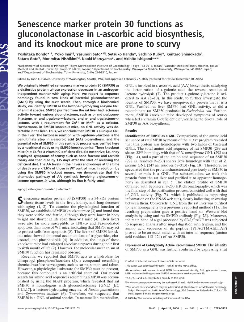
ResultsIdentification of SMP30 as a GNL. Comparisons of the amino acidsequence of rat SMP30 by means of the BLAST program revealedthat this protein was homologous with two kinds of bacterialGNLs. The total amino acid sequence of rat SMP30 (299 aa)shares 32% homology with that of N. punctiforme GNL (292 aa)(Fig. 1A), and a part of the amino acid sequence of rat SMP30(222 aa, residues 9–230) shares 26% homology with that of Z.mobilis GNL (247 aa, residues 67–313) (Fig. 1B). Therefore, wespeculated that the protein characterized previously as SMP30 inseveral animals is a GNL. For substantiation, we took thisprotein from the rat liver and purified it to apparent homoge-neity as described in ref. 5. The elution profile of SMP30obtained with Sephacryl S-200 HR chromatography, which wasthe final step of the purification process, coincided well with thatof GNL activity (Fig. 7A, which is published as supportinginformation on the PNAS web site), clearly indicating an overlapbetween them. Conversely, GNL from the rat liver was purifiedto near homogeneity by a previously reported method (11). Theresulting preparation gave a positive band on Western blotanalysis by using anti-rat SMP30 antibody (Fig. 7B). Moreover,the main band of a gel processed by SDS�PAGE was subjectedto sequence analysis after in-gel digestion with trypsin, and theamino acid sequence of its peptide (YFAGTMAEETAP)proved to be an exact match with an internal sequence (aminoacid residues 113–124) of rat SMP30.
Expression of Catalytically Active Recombinant SMP30. The identityof SMP30 as a GNL was further confirmed by expressing a rat
Conflict of interest statement: No conflicts declared.
This paper was submitted directly (Track II) to the PNAS office.
Abbreviations: AA, L-ascorbic acid; BMD, bone mineral density; GNL, gluconolactonase;MBP, maltose-binding protein; SMP30, senescence marker protein 30.
‡Y.K., Y.I., and Y.S. contributed equally to this work.
�To whom correspondence may be addressed. E-mail: [email protected].
**To whom correspondence may be addressed at: Department of Molecular Pathology,Tokyo Metropolitan Institute of Gerontology, 35-2 Sakae-cho, Itabashi-ku, Tokyo 173-0015, Japan. E-mail: [email protected].
© 2006 by The National Academy of Sciences of the USA
www.pnas.org�cgi�doi�10.1073�pnas.0511225103 PNAS � April 11, 2006 � vol. 103 � no. 15 � 5723–5728
BIO
CHEM
ISTR
Y
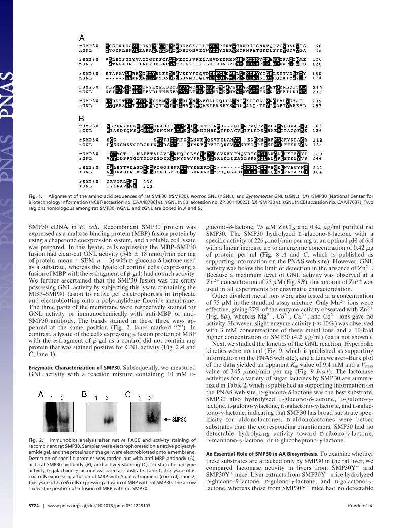
SMP30 cDNA in E. coli. Recombinant SMP30 protein wasexpressed as a maltose-binding protein (MBP) fusion protein byusing a chaperone coexpression system, and a soluble cell lysatewas prepared. In this lysate, cells expressing the MBP–SMP30fusion had clear-cut GNL activity (546 � 18 nmol�min per mgof protein, mean � SEM, n � 3) with D-glucono-�-lactone usedas a substrate, whereas the lysate of control cells (expressing afusion of MBP with the �-fragment of �-gal) had no such activity.We further ascertained that the SMP30 fusion was the entitypossessing GNL activity by subjecting this lysate containing theMBP–SMP30 fusion to native gel electrophoresis in triplicateand electroblotting onto a polyvinylidene fluoride membrane.The three parts of the membrane were respectively stained forGNL activity or immunochemically with anti-MBP or anti-SMP30 antibody. The bands stained in these three ways ap-peared at the same position (Fig. 2, lanes marked ‘‘2’’). Incontrast, a lysate of the cells expressing a fusion protein of MBPwith the �-fragment of �-gal as a control did not contain anyprotein that was stained positive for GNL activity (Fig. 2 A andC, lane 1).
Enzymatic Characterization of SMP30. Subsequently, we measuredGNL activity with a reaction mixture containing 10 mM D-
glucono-�-lactone, 75 �M ZnCl2, and 0.42 �g�ml purified ratSMP30. The SMP30 hydrolyzed D-glucono-�-lactone with aspecific activity of 226 �mol�min per mg at an optimal pH of 6.4with a linear increase up to an enzyme concentration of 0.42 �gof protein per ml (Fig. 8 A and C, which is published assupporting information on the PNAS web site). However, GNLactivity was below the limit of detection in the absence of Zn2�.Because a maximum level of GNL activity was observed at aZn2� concentration of 75 �M (Fig. 8B), this amount of Zn2� wasused in all experiments for enzymatic characterization.
Other divalent metal ions were also tested at a concentrationof 75 �M in the standard assay mixture. Only Mn2� ions wereeffective, giving 27% of the enzyme activity observed with Zn2�
(Fig. 8B), whereas Mg2�, Co2�, Ca2�, and Cd2� ions gave noactivity. However, slight enzyme activity (��10%) was observedwith 3 mM concentrations of these metal ions and a 10-foldhigher concentration of SMP30 (4.2 �g�ml) (data not shown).
Next, we studied the kinetics of the GNL reaction. Hyperbolickinetics were normal (Fig. 9, which is published as supportinginformation on the PNAS web site), and a Lineweaver–Burk plotof the data yielded an apparent Km value of 9.4 mM and a Vmaxvalue of 345 �mol�min per mg (Fig. 9 Inset). The lactonaseactivities for a variety of sugar lactones by SMP30 are summa-rized in Table 2, which is published as supporting information onthe PNAS web site. D-glucono-�-lactone was the best substrate.SMP30 also hydrolyzed L-glucono-�-lactone, D-gulono-�-lactone, L-gulono-�-lactone, D-galactono-�-lactone, and L-galac-tono-�-lactone, indicating that SMP30 has broad substrate spec-ificity for aldonolactones. D-aldonolactones were bettersubstrates than the corresponding enantiomers. SMP30 had nodetectable hydrolyzing activity toward D-ribono-�-lactone,D-mannono-�-lactone, or D-glucoheptono-�-lactone.
An Essential Role of SMP30 in AA Biosynthesis. To examine whetherthese substrates are attacked only by SMP30 in the rat liver, wecompared lactonase activity in livers from SMP30Y� andSMP30Y� mice. Liver extracts from SMP30Y� mice hydrolyzedD-glucono-�-lactone, D-gulono-�-lactone, and D-galactono-�-lactone, whereas those from SMP30Y� mice had no detectable
Fig. 1. Alignment of the amino acid sequences of rat SMP30 (rSMP30), Nostoc GNL (nGNL), and Zymomonas GNL (zGNL). (A) rSMP30 [National Center forBiotechnology Information (NCBI) accession no. CAA48786] vs. nGNL (NCBI accession no. ZP�00110023). (B) rSMP30 vs. zGNL (NCBI accession no. CAA47637). Tworegions homologous among rat SMP30, nGNL, and zGNL are boxed in A and B.
Fig. 2. Immunoblot analysis after native PAGE and activity staining ofrecombinant rat SMP30. Samples were electrophoresed on a native polyacryl-amide gel, and the proteins on the gel were electroblotted onto a membrane.Detection of specific proteins was carried out with anti-MBP antibody (A),anti-rat SMP30 antibody (B), and activity staining (C). To stain for enzymeactivity, D-galactono-�-lactone was used as substrate. Lane 1, the lysate of E.coli cells expressing a fusion of MBP with �-gal �-fragment (control); lane 2,the lysate of E. coli cells expressing a fusion of MBP with rat SMP30. The arrowshows the position of a fusion of MBP with rat SMP30.
5724 � www.pnas.org�cgi�doi�10.1073�pnas.0511225103 Kondo et al.
hydrolyzing activity toward these lactones (Table 1). Clearly,therefore, SMP30 is a unique enzyme that effectively hydrolyzesvarious aldonolactones in the liver. Because the lactonization ofL-gulonic acid to L-gulono-�-lactone, the reverse reaction of thelactonase reaction, is the penultimate step of AA synthesis, weinvestigated the role of SMP30 in this metabolic process by usingSMP30 knockout mice. Ten days after weaning at the age of 30days, SMP30Y� and SMP30Y� mice, six each, were fed a vitaminC-deficient diet. One of the knockout mice started to lose weightafter 25 days of consuming this diet (at an age of 65 days), walkedwith an abnormal gait after 31 days, and died after 37 days. Theaverage body weight of the other five knockout mice started todecrease after 56 days of the vitamin C-deficient diet (at an ageof 96 days) (Fig. 3A); their gait became abnormal at that time,and they died by the 106th to 135th days of this dietarydeficiency. The AA level in plasma of SMP30Y� mice after 106 days of consuming the deficient diet was �1% of that in
SMP30Y� mice (Fig. 3B). The livers and kidneys of SMP30Y�
mice at the time of death contained �1.6% of the AA levels inthe SMP30Y� mice (Fig. 3 C and D).
For the previous studies, our SMP30Y� mice were fed auto-claved mouse chow after weaning, and this food contains �55mg�kg of vitamin C. When we assessed the vitamin C status ofsuch mice after 80 days of eating autoclaved chows, their plasma,livers, and kidneys contained only 6–8% of the AA values in theWT mice (Fig. 4). Thus, the knockout mice that were fedautoclaved mouse chow proved to be severely vitamin C-deficient.
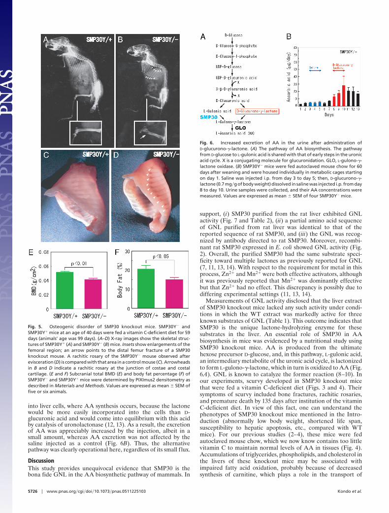
Osteogenic Disorder of SMP30 Knockout Mice. Because thin, brittlebones with a tendency to fracture are known as characteristicmanifestations of scurvy, we checked the skeletal structure ofSMP30Y� and SMP30Y� mice by x-ray examination after theyhad consumed the vitamin C-deficient diet for 59 days (at an ageof 99 days) (Fig. 5 A and B). Fracture at the distal end of theirfemurs (Fig. 5B Inset) and rachitic rosaries at the junction ofcostae and costal cartilages (Fig. 5B) were prominent in aSMP30Y� mouse (Fig. 5D), but not a SMP30Y� mouse (Fig.5C). Moreover, subcranial total bone mineral density (BMD)and body fat were significantly decreased in SMP30Y� micecompared with SMP30Y� mice (Fig. 5 E and F).
Occurrence of an Alternative Pathway of AA Synthesis. Two pathwayswere proposed as forming the last part of the AA syntheticpathway (12). Obviously, the main pathway of this process dealtwith here includes the steps from D-glucose to L-gulonic acid(detailed on the left side of Fig. 6A). However, for the formationof L-gulono-�-lactone, the immediate precursor to AA, anotherpathway branches from D-glucuronic acid (Fig. 6A, right side).To clarify whether the latter pathway exists in the mammalianmetabolism, we injected D-glucurono-�-lactone i.p. intoSMP30Y� mice and measured the amount of AA excreted intheir urine. We used the lactone for delivery of D-glucuronic acid
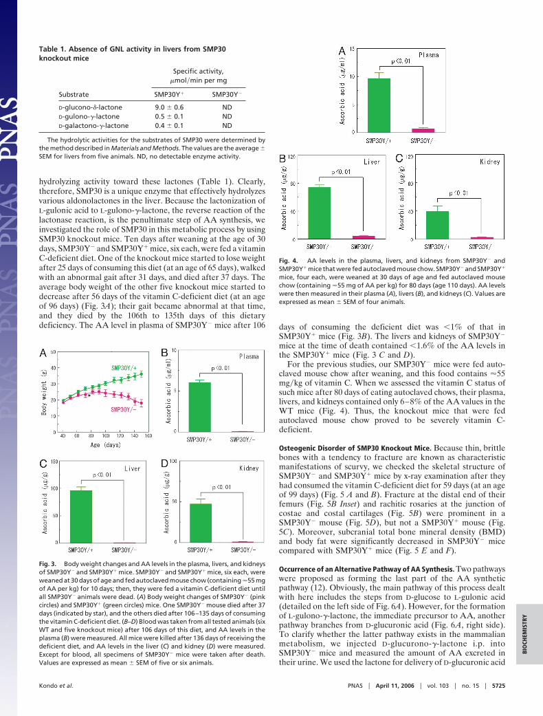
Table 1. Absence of GNL activity in livers from SMP30knockout mice
Substrate
Specific activity,�mol�min per mg
SMP30Y� SMP30Y�
D-glucono-�-lactone 9.0 � 0.6 NDD-gulono-�-lactone 0.5 � 0.1 NDD-galactono-�-lactone 0.4 � 0.1 ND
The hydrolytic activities for the substrates of SMP30 were determined bythe method described in Materials and Methods. The values are the average �SEM for livers from five animals. ND, no detectable enzyme activity.
Fig. 3. Body weight changes and AA levels in the plasma, livers, and kidneysof SMP30Y� and SMP30Y� mice. SMP30Y� and SMP30Y� mice, six each, wereweaned at 30 days of age and fed autoclaved mouse chow (containing �55 mgof AA per kg) for 10 days; then, they were fed a vitamin C-deficient diet untilall SMP30Y� animals were dead. (A) Body weight changes of SMP30Y� (pinkcircles) and SMP30Y� (green circles) mice. One SMP30Y� mouse died after 37days (indicated by star), and the others died after 106–135 days of consumingthe vitamin C-deficient diet. (B–D) Blood was taken from all tested animals (sixWT and five knockout mice) after 106 days of this diet, and AA levels in theplasma (B) were measured. All mice were killed after 136 days of receiving thedeficient diet, and AA levels in the liver (C) and kidney (D) were measured.Except for blood, all specimens of SMP30Y� mice were taken after death.Values are expressed as mean � SEM of five or six animals.
Fig. 4. AA levels in the plasma, livers, and kidneys from SMP30Y� andSMP30Y� mice that were fed autoclaved mouse chow. SMP30Y� and SMP30Y�
mice, four each, were weaned at 30 days of age and fed autoclaved mousechow (containing �55 mg of AA per kg) for 80 days (age 110 days). AA levelswere then measured in their plasma (A), livers (B), and kidneys (C). Values areexpressed as mean � SEM of four animals.
Kondo et al. PNAS � April 11, 2006 � vol. 103 � no. 15 � 5725
BIO
CHEM
ISTR
Y
into liver cells, where AA synthesis occurs, because the lactonewould be more easily incorporated into the cells than D-glucuronic acid and would come into equilibrium with this acidby catalysis of uronolactonase (12, 13). As a result, the excretionof AA was appreciably increased by the injection, albeit in asmall amount, whereas AA excretion was not affected by thesaline injected as a control (Fig. 6B). Thus, the alternativepathway was clearly operational here, regardless of its small f lux.
DiscussionThis study provides unequivocal evidence that SMP30 is thebona fide GNL in the AA biosynthetic pathway of mammals. In
support, (i) SMP30 purified from the rat liver exhibited GNLactivity (Fig. 7 and Table 2), (ii) a partial amino acid sequenceof GNL purified from rat liver was identical to that of thereported sequence of rat SMP30, and (iii) the GNL was recog-nized by antibody directed to rat SMP30. Moreover, recombi-nant rat SMP30 expressed in E. coli showed GNL activity (Fig.2). Overall, the purified SMP30 had the same substrate speci-ficity toward multiple lactones as previously reported for GNL(7, 11, 13, 14). With respect to the requirement for metal in thisprocess, Zn2� and Mn2� were both effective activators, althoughit was previously reported that Mn2� was dominantly effectivebut that Zn2� had no effect. This discrepancy is possibly due todiffering experimental settings (11, 13, 14).
Measurements of GNL activity disclosed that the liver extractof SMP30 knockout mice lacked any such activity under condi-tions in which the WT extract was markedly active for threeknown substrates of GNL (Table 1). This outcome indicates thatSMP30 is the unique lactone-hydrolyzing enzyme for thesesubstrates in the liver. An essential role of SMP30 in AAbiosynthesis in mice was evidenced by a nutritional study usingSMP30 knockout mice. AA is produced from the ultimatehexose precursor D-glucose, and, in this pathway, L-gulonic acid,an intermediary metabolite of the uronic acid cycle, is lactonizedto form L-gulono-�-lactone, which in turn is oxidized to AA (Fig.6A). GNL is known to catalyze the former reaction (8–10). Inour experiments, scurvy developed in SMP30 knockout micethat were fed a vitamin C-deficient diet (Figs. 3 and 4). Theirsymptoms of scurvy included bone fractures, rachitic rosaries,and premature death by 135 days after institution of the vitaminC-deficient diet. In view of this fact, one can understand thephenotypes of SMP30 knockout mice mentioned in the Intro-duction (abnormally low body weight, shortened life span,susceptibility to hepatic apoptosis, etc., compared with WTmice). For our previous studies (2–4), these mice were fedautoclaved mouse chow, which we now know contains too littlevitamin C to maintain normal levels of AA in tissues (Fig. 4).Accumulations of triglycerides, phospholipids, and cholesterol inthe livers of these knockout mice may be associated withimpaired fatty acid oxidation, probably because of decreasedsynthesis of carnitine, which plays a role in the transport of
Fig. 5. Osteogenic disorder of SMP30 knockout mice. SMP30Y� andSMP30Y� mice at an age of 40 days were fed a vitamin C-deficient diet for 59days (animals’ age was 99 days). (A–D) X-ray images show the skeletal struc-tures of SMP30Y� (A) and SMP30Y� (B) mice. Insets show enlargements of thefemoral region; an arrow points to the distal femur fracture of a SMP30knockout mouse. A rachitic rosary of the SMP30Y� mouse observed afterevisceration (D) is compared with that area in a control mouse (C). Arrowheadsin B and D indicate a rachitic rosary at the junction of costae and costalcartilage. (E and F) Subcranial total BMD (E) and body fat percentage (F) ofSMP30Y� and SMP30Y� mice were determined by PIXImus2 densitometry asdescribed in Materials and Methods. Values are expressed as mean � SEM offive or six animals.
Fig. 6. Increased excretion of AA in the urine after administration ofD-glucurono-�-lactone. (A) The pathway of AA biosynthesis. The pathwayfrom D-glucose to L-gulonic acid is shared with that of early steps in the uronicacid cycle. X is a conjugating molecule for glucuronidation. GLO, L-gulono-�-lactone oxidase. (B) SMP30Y� mice were fed autoclaved mouse chow for 60days after weaning and were housed individually in metabolic cages startingon day 1. Saline was injected i.p. from day 3 to day 5; then, D-glucurono-�-lactone (0.7 mg�g of body weight) dissolved in saline was injected i.p. from day8 to day 10. Urine samples were collected, and their AA concentrations weremeasured. Values are expressed as mean � SEM of four SMP30Y� mice.
5726 � www.pnas.org�cgi�doi�10.1073�pnas.0511225103 Kondo et al.
long-chain fatty acids into mitochondria. AA is required as acofactor in two hydroxylation reactions in carnitine biosynthesis(15). In fact, AA-deficient guinea pigs were shown to haveabnormalities of lipid metabolism, such as hyperlipidemia andhypercholesterolemia (16). Another clinical feature of youngSMP30 knockout mice is enlargement of alveolar airspacescompared with those of WT mice (2). A similar change wasreported in ODS (osteogenic disorder Shionogi) rats that sur-vived for a long time on a vitamin C-deficient diet (17). However,never before did we observe scurvy symptoms in the knockoutmice that were fed autoclaved mouse chow, despite their smallerbody size and shorter life span (3, 4). Assuming that an averageweight of mice is 25 g and that the amount of chow taken per dayis 4 g (18), SMP30 knockout mice ingest �0.01 mg�g of bodyweight per day. Based on the observation that 0.02 mg�g of bodyweight per day cannot sustain normal body function in ascurvy-prone mouse whose L-gulono-�-lactone oxidase gene wasdeleted (19), the SMP30 knockout mice seem not to haveingested enough vitamin C. Probably, a small amount of AA maybe synthesized through the alternative pathway (Fig. 6A) pre-viously proposed based on an enzymatic study (20) and dem-onstrated here in SMP30 knockout mice. Furthermore, theabsence of GNL may lead to decreased degradation of AA,because GNL can hydrolyze the lactone ring of dehydroascorbicacid, the oxidation product of AA, to 2,3-dioxo-L-gulonic acid,as formerly reported (20).
Because SMP30 is abundant in the kidney and also present,although in lesser amounts, in other organs (21), this proteinmust have some function other than AA synthesis, which doesnot occur at all these sites. SMP30 may prevent the productionof glycated proteins by hydrolyzing D-glucono-�-lactone. It ispossible that this lactone is formed from D-glucose by glucosedehydrogenase in vivo, and it may be involved in glycation ofproteins. In fact, human and rat hemoglobin was shown to beglycated with D-glucono-�-lactone (22). Glycated proteins, es-pecially advanced glycation end products, are known to cause thedeterioration of cellular functions; therefore, SMP30 may pro-tect cells from such an effect.
Materials and MethodsChemicals. L-glucono-�-lactone, D-gulono-�-lactone, L-gulono-�-lactone, L-galactono-�-lactone, D-ribono-�-lactone, D-glucohep-tono-�-lactone, and D-mannono-�-lactone were purchased fromSigma-Aldrich. D-glucono-�-lactone, D-galactono-�-lactone, andother reagents were purchased from Wako Pure Chemical(Osaka).
Animals. Male Wister rats, 3–9 months of age, were obtainedfrom the Animal Facility at Tokyo Metropolitan Institute ofGerontology, and their livers were used as a source of purifiedSMP30. For purification of GNL, 10-week-old male Wister ratspurchased from Kiwa Laboratory Animals (Misato-cho, Japan)were used. SMP30 knockout mice were previously generatedwith the gene-targeting technique (3), and heterozygous femalemice (SMP30�/�) were mated with male knockout mice(SMP30Y�) to produce knockout and WT (SMP30Y�) litter-mates. These littermates were fed autoclaved mouse chow(CRF-1; Charles River Breeding Laboratories) ad libitum withfree access to water and used for measurement of GNL activityin the liver when the animals were 6 months old. The autoclavedchow contained �55 mg of AA per kg as determined at the timeof experimentation.
In a nutritional study, SMP30Y� and SMP30Y� mice wereweaned at 30 days of age and fed autoclaved mouse chow for 10days, followed by a vitamin C-deficient diet (CL-2; CLEA Japan,Tokyo). Throughout the experiments, animals were maintainedon 12-h light�dark cycles in a controlled environment. Allexperimental procedures using laboratory animals were ap-
proved by the Animal Care and Use Committee of TokyoMetropolitan Institute of Gerontology.
Purification of SMP30 from Rat Liver. SMP30 was purified from asoluble fraction of rat livers as described in ref. 5. Briefly, liverhomogenate was fractionated by ammonium sulfate precipita-tion followed by a successive series of chromatographies onDEAE-Sephacel, Phenyl Sepharose CL-4B, and Sephacryl S-200HR columns (all from Amersham Pharmacia Biosciences). Theelution of SMP30 was followed by the dot-blot immunoassaydescribed in ref. 5. The purified SMP30 was stored at �70°Cuntil use.
Purification of GNL and Sequence Analysis of Its Peptide. GNL waspurified from rat livers according to the method of Grossmanand Axelrod (11), with slight modifications. Briefly, a solublefraction of the liver homogenate was heated at 50°C for 30 min,and the resulting soluble fraction was fractionated with ammo-nium sulfate. The fraction with GNL activity was further purifiedby successive chromatographies on columns of Sephadex G-150(Amersham Pharmacia Fine Chemicals), Resource Q (Amer-sham Pharmacia Biosciences), and Bio-Gel HTP (Bio-Rad).After the final preparation was subjected to SDS�PAGE, themain band was excised, and the protein in the gel was digestedwith trypsin. One of the peptides produced was sequencedthrough the custom service of APRO Life Science Institute(Naruto, Japan).
GNL and Related Activities. GNL activity was measured by thechange in absorbance of the pH indicator p-nitrophenol causedby free acid formation from the lactone (23). The standardmixture contained 10 mM D-glucono-�-lactone, 10 mM Pipes(pH 6.4), 0.25 mM p-nitrophenol, 75 �M ZnCl2, and an enzymein a total volume of 1 ml. The substrate solution was freshlyprepared immediately before the assay. The reaction was fol-lowed by monitoring a decrease in absorbance at 405 nm, and theacid production rate was determined with a calibration curveobtained by using known amounts of HCl. The rate of sponta-neous hydrolysis of the lactone was subtracted from the totalrate. To assess whether a divalent metal ion was required forGNL activity, we tested ZnCl2, MnCl2, MgCl2, CoCl2, CaCl2, andCdCl2 in this regard and then determined the hydrolyzingactivity for other lactones with the same procedure, except forthe substrate.
Lactonase activity was then analyzed in livers removed frommice and homogenized with ice-cold homogenization buffer (10mM Tris�HCl, pH 8.0�1 mM phenyl methanesulfonyl f luoride)for 30 s at high speed with a Polytron homogenizer. Thehomogenate was centrifuged at 10,000 � g for 10 min. Theprotein concentration of the sample was determined by BCAprotein assay (Pierce) using BSA as a standard. The lactone-hydrolyzing activity was assayed by using various lactones underthe standard conditions described above. In the purification ofrat GNL, lactonase activity was measured by recording pHchange with a pH meter, essentially as described in ref. 24.
Expression of Recombinant Rat SMP30. Total RNA was preparedfrom a rat liver and used to produce a single-strand cDNA withSuperScript II RNase H� reverse transcriptase (Life Technol-ogies, Rockville, MD) following the manufacturer’s protocol. ASMP30 cDNA was amplified by PCR from this cDNA such thatan EcoRI site would be produced at the both ends of the product.The PCR was carried out by using PfuUltra DNA polymerase(Stratagene) with a sense primer (5�-GAATTCATGTCTTC-CATCAAGATTG-3�) and an antisense primer (5�-GAATTCT-TACCCTGCATAGGAATATG-3�). The amplified DNA wasdigested with EcoRI and inserted into the EcoRI site of the E.coli expression vector pMAL-c2x (New England Biolabs). The
Kondo et al. PNAS � April 11, 2006 � vol. 103 � no. 15 � 5727
BIO
CHEM
ISTR
Y
constructed plasmid pMAL-c2x-rSMP30 was used to transformE. coli BL21 that had previously been transformed with pGro7(Takara Bio, Tokyo) for expression of two chaperone proteins:GroEL and GroES. The resulting clone was used to express ratSMP30 as a fusion protein with MBP as follows. It was culturedat 37°C overnight in LB containing 0.2% glucose, 100 �g�mlampicillin, and 20 �g�ml chloramphenicol. After the cell sus-pension was diluted 100-fold with the same culture medium,D-arabinose (4 mg�ml) was added for expression of the chap-erone proteins and cultured for 1.6 h at 37°C. Then, theexpression of MBP–SMP30 fusion protein was induced by addingisopropyl �-D-thiogalactoside to a final concentration of 0.3 mMand culturing for 3 h at 30°C. For control experiments, pMAL-c2x was used instead of pMAL-c2x-rSMP30, and a fusion proteinof MBP with the �-fragment of �-gal was induced in the sameway as above. The cells were washed with Dulbecco’s phosphatebuffered saline, resuspended in 20 mM Tris�HCl buffer (pH 7.4)containing 200 mM NaCl, and stored at �30°C until use.
Activity Staining and Western Blotting for Recombinant MBP–SMP30Fusion Protein. Suspensions of the test and control cells weresonicated and centrifuged at 9,000 � g for 30 min at 4°C. Eachresulting supernatant (60 �g of protein) was electrophoresed on a7% polyacrylamide gel by the method of Davis (25), with somemodifications. Proteins in the gel were transferred onto a polyvi-nylidene fluoride membrane with 25 mM Tris�192 mM glycine asa transfer buffer. Subsequent staining for GNL activity involvedimmersing the membrane successively in 5 mM Tris�HCl buffer (pH6.8) containing 1 mM DTT and 1 mM MnCl2 (buffer A), then ina mixture of buffer A and 0.04% methyl red in 60% ethanol (9:1),and finally in buffer A containing 80 mM D-galactono-�-lactoneuntil the red color appeared.
For Western blotting, the supernatant (20 �g of protein) waselectrophoresed in duplicate, and proteins were transferred ontoa polyvinylidene fluoride membrane in the same way. Themembrane was blocked overnight with a mixture of 2% BSA and7% skim milk and cut into halves. One half was incubated withanti-MBP rabbit antibody (1:3,000 dilution; New England Bio-labs), and the other half was incubated with anti-rat SMP30rabbit antibody (1:3,000 dilution; Cosmo Bio, Tokyo) (3). Then,both half-membranes were incubated with horseradish peroxi-dase-conjugated anti-rabbit IgG antibody (1:5,000 dilution; Cap-pel). MBP and SMP30 were visualized with an ECL (enhancedchemiluminescence) detection kit (Amersham PharmaciaBiosciences).
Measurement of AA. Plasma was mixed with nine volumes of 20%metaphosphate containing 1% SnCl2, and the mixture wascentrifuged at 10,000 � g for 10 min at 4°C. Livers and kidneyswere homogenized in 14 volumes of 5.4% metaphosphate, andthe homogenate was centrifuged as above. For measurement ofAA excreted into urine, a mouse was housed in a metabolic cage,and urine was collected in a bottle with 10% metaphosphate for24 h. The amount of 10% metaphosphate had been adjusted tokeep its concentration 5% after dilution with urine. Thevolume of the urine–metaphosphate mixture was recorded, andthe mixture was centrifuged as above. All samples obtained aftercentrifugation were kept at �80°C until use. AA in samples wasderivatized with dinitrophenylhydrazine and analyzed by HPLCwith a Shodex-5SIL-4E column (4.6 � 250 mm; Showa Denko,Tokyo). The mobile phase was hexane�ethylacetate�acetic acid(5:4:1) at a flow rate of 1 ml�min, and the absorbance at 495 nmwas recorded (26, 27). AA in mouse chow was determined by thesame method after extraction into 15% metaphosphate.
Quantification of BMD and Body Fat Percentage. Subcranial totalBMD and body fat percentage were determined by densitometrywith the PIXImus2 imager (General Electric�Lunar, Madison,WI). Field calibration and calibration vs. the quality-controlphantom were performed each day before imaging. Each mousewas positioned reproducibly in a prone position on the imagingtray and scanned three times. The coefficients of variance forBMD and body fat percentage were 0.9% and 2.2%, respectively,for in vitro measurements.
Statistical Analysis. The results are expressed as mean � SEM. Theprobability of statistical differences between experimental groupswas determined by Student’s t test or ANOVA as appropriate. Oneand two-way ANOVAs were performed by using KALEIDAGRAPHsoftware (Synergy Software, Reading, PA).
We thank Ms. P. Minick for excellent assistance in the review of Englishand Mr. H. Hosoi and Ms. Y. Takenaka for participation in thepurification of GNL in the training course on basic medicine atWakayama Medical University. This work was supported by a Grant-in-Aid for Scientific Research (to S.H. and S.K.) and a Grant-in-Aid forYoung Scientists (B) (to Y.I.) from the Japanese Ministry of Education,Culture, Sports, Science, and Technology and grants from HealthScience Research Grants for Comprehensive Research on Aging andHealth supported by the Ministry of Health, Labor, and Welfare ofJapan (to A.I.), the Smoking Science Foundation (to N.M.), and theVitamin C Research Committee of Japan (to M.N.).
1. Fujita, T., Uchida, K. & Maruyama, N. (1992) Biochim. Biophys. Acta 1116,122–128.
2. Mori, T., Ishigami, A., Seyama, K., Onai, R., Kubo, S., Shimizu, K., Maruyama,N. & Fukuchi, Y. (2004) Pathol. Int. 54, 167–173.
3. Ishigami, A., Fujita, T., Handa, S., Shirasawa, T., Koseki, H., Kitamura, T.,Enomoto, N., Sato, N., Shimosawa, T. & Maruyama, N. (2002) Am. J. Pathol.161, 1273–1281.
4. Ishigami, A., Kondo, Y., Nanba, R., Ohsawa, T., Handa, S., Kubo, S., Akita,M. & Maruyama, N. (2004) Biochem. Biophys. Res. Commun. 315, 575–580.
5. Kondo, Y., Ishigami, A., Kubo, S., Handa, S., Gomi, K., Hirokawa, K.,Kajiyama, N., Chiba, T., Shimokado, K. & Maruyama, N. (2004) FEBS Lett.570, 57–62.
6. Kanagasundaram, V. & Scopes, R. (1992) Biochim. Biophys. Acta 1171,198–200.
7. Bublitz, C. & Lehninger, A. L. (1961) Biochim. Biophys. Acta 47, 288–297.8. Burns, J. J. (1960) in Metabolic Pathway, ed. Greenberg, D. M. (Academic, New
York), Vol. 1, pp. 341–356.9. Nishikimi, M. & Yagi, K. (1996) in Subcellular Biochemistry–Ascorbic Acid:
Biochemistry and Biomedical Cell Biology, ed. Harris, J. R. (Plenum, New York),Vol. 25, pp. 17–39.
10. Nishikimi, M., Okamura, M. & Ohta, Y. (2003) in Recent Research Develop-ments in Biophysics and Biochemistry, ed. Pandalai, S. G. (Research Signpost,Kerala, India), Vol. 3, Part 1, pp. 531–545.
11. Grossman, S. H. & Axelrod, B. (1973) J. Biol. Chem. 248, 4846–4851.12. Shimazono, N. & Mano, Y. (1961) Ann. N.Y. Acad. Sci. 92, 91–104.
13. Winkelman, J. & Lehninger, A. L. (1958) J. Biol. Chem. 233, 794–799.14. Kawada, M., Takiguchi, H., Kagawa, Y., Suzuki, K. & Shimazono, N. (1962)
J. Biochem. (Tokyo) 51, 405–415.15. Rebouche, C. J. (1991) Am. J. Clin. Nutr. 54, 1147S–1152S.16. Ha, T. Y., Otsuka, M. & Arakawa, N. (1990) J. Nutr. Sci. Vitaminol. (Tokyo)
36, 227–234.17. Kono, K., Asai, K., Kuzuya, F. & Harada, T. (1990) in Vitamin C and the
Scurvy-Prone ODS Rat, eds. Fujita, T., Fukase, M. & Konishi, T. (Elsevier,Amsterdam), pp. 147–155.
18. Bernstein, S. E. (1966) in Biology of the Laboratory Mouse, ed. Green, E. L.(Dover, New York), 2nd Ed., pp. 337–350.
19. Maeda, N., Hagihara, H., Nakata, Y., Hiller, S., Wilder, J. & Reddick, R. (2000)Proc. Natl. Acad. Sci. USA 97, 841–846.
20. Kagawa, Y. & Takiguchi, H. (1962) J. Biochem. (Tokyo) 51, 197–203.21. Fujita, T., Shirasawa, T., Uchida, K. & Maruyama, N. (1992) Biochim. Biophys.
Acta 1132, 297–305.22. Lindsay, R. M., Smith, W., Lee, W. K., Dominiczak, M. H. & Baird, J. D. (1997)
Clin. Chim. Acta 263, 239–247.23. Hucho, F. & Wallenfels, K. (1972) Biochim. Biophys. Acta 276, 176–179.24. Nishikimi, M., Koshizaka, T., Mochizuki, H., Iwata, H., Makino, S., Hayashi,
Y., Ozawa, T. & Yagi, K. (1988) Biochem. Int. 16, 615–621.25. Davis, B. J. (1964) Ann. N.Y. Acad. Sci. 121, 404–427.26. Kishida, E., Nishimoto, Y. & Kojo, S. (1992) Anal. Chem. 64, 1505–1507.27. Kodaka, K., Inagaki, S., Ujie, T., Ueno, T. & Suda, H. (1985) Vitamin 59,
451–455.
5728 � www.pnas.org�cgi�doi�10.1073�pnas.0511225103 Kondo et al.