selective cadmium regulation mediated by a cooperative ...selective cadmium regulation mediated by a...
TRANSCRIPT
Selective cadmium regulation mediated by acooperative binding mechanism in CadRXichun Liua, Qingyuan Hua, Jinmei Yangb, Shanqing Huanga, Tianbiao Weia, Weizhong Chena, Yafeng Hea, Dan Wanga,Zhijun Liuc, Kang Wangb, Jianhua Gand, and Hao Chena,1
aState Key Laboratory of Coordination Chemistry, School of Chemistry and Chemical Engineering, Collaborative Innovation Center of Chemistry for LifeSciences, Nanjing University, 210023 Nanjing, China; bState Key Laboratory of Analytical Chemistry for Life Science, School of Chemistry and ChemicalEngineering, Collaborative Innovation Center of Chemistry for Life Sciences, Nanjing University, 210023 Nanjing, China; cNational Facility for Protein Sciencein Shanghai, Zhangjiang Laboratory, Shanghai Advanced Research Institute, Chinese Academy of Sciences, 201210 Shanghai, China; and dState KeyLaboratory of Genetic Engineering, Collaborative Innovation Center of Genetics and Development, Shanghai Public Health Clinical Center, School of LifeSciences, Fudan University, 200438 Shanghai, China
Edited by Thomas V. O’Halloran, Northwestern University, Evanston, IL, and accepted by Editorial Board Member Angela M. Gronenborn August 28, 2019(received for review May 21, 2019)
Detoxification of the highly toxic cadmium element is essential forthe survival of living organisms. Pseudomonas putida CadR, a MerRfamily transcriptional regulator, has been reported to exhibit anultraspecific response to the cadmium ion. Our crystallographicand spectroscopic studies reveal that the extra cadmium selectivityof CadR is mediated by the unexpected cooperation of thiolate-richsite I and histidine-rich site II. Cadmium binding in site I mediates thereorientation of protein domains and facilitates the assembly of siteII. Subsequently, site II bridge-links 2 DNA binding domains throughligands His140/His145 in the C-terminal histidine-rich tail. With dy-namic transit between 2 conformational states, this bridge couldstabilize the regulator into an optimal conformation that is criticalfor enhancing the transcriptional activity of the cadmium detoxifi-cation system. Our results provide dynamic insight into how natureutilizes the unique cooperative binding mechanism in multisite pro-teins to recognize cadmium ions specifically.
cadmium transcriptional regulator | cooperative binding | MerR family |protein crystallography | NMR spectroscopy
Cadmium pollution in the food chain has become a monu-mental public health concern since the emergence of the first
serious cadmium-related disease (Itai-Itai disease) in Japan (1–4).The toxicity of cadmium is due mainly to its interference with themetabolism of zinc and other essential metal ions in the organism(5–7). Examination of a rare biological function of cadmiumshowed that the cadmium ion acts as a catalysis center of carbonicanhydrase in marine diatoms at low zinc levels (8, 9). Organismshave evolved multiple detoxification systems to eliminate thetoxicity of cadmium. Metallothionein (MT), widely present in or-ganisms, binds multiple cadmium ions in clusters, which can bedivided into an α-domain Cd4S11 cluster and a β-domain Cd3S9cluster in mammalian ΜΤ subtypes (10–12). In bacteria, 2 ArsR/SmtB family homologs, CadC and CmtR, with functional CdS4 orCdS3O allosteric sites have been reported to regulate the cadmiumefflux system (13–16). These detoxification systems can detoxifyother heavy metal ions besides cadmium, such as lead or mercury.In addition, MT is involved in the metabolism of zinc and copper(17, 18). Therefore, this indicates that the thiolate-rich sites in thereported cadmium-binding metalloproteins cannot readily distin-guish cadmium ion from other heavy metals.Pseudomonas putida CadR belongs to the MerR family of
transcriptional regulators, which regulates its own transcriptionand the transcription of a cadmium efflux P-type ATPase CadA(19). It is the most specific natural cadmium-binding proteinreported to date, as the cadR promoter is induced only by cad-mium, and the induction of the cadA promoter by lead and zinc ismuch weaker than that by cadmium (19). The molecular basisunderlying the specific cadmium selectivity of CadR remains elu-sive, however. The previously reported MerR family homologspossess remarkable metal sensitivity and selectivity; for instance,
CueR can respond to free Cu(I) at zeptomolar concentrations(20). These metal-binding properties are typically achieved by theunique coordination geometry in a binding site, such as the linearCuS2 in CueR (20), the planer triangle HgS3 in MerR (21, 22), andthe triangular pyramid PbS3 in PbrR691 (23, 24). Along with itsoutstanding metal-binding ability, the MerR family protein has aunique allosteric transcriptional regulation mechanism. Thesedual-function, σ70-dependent regulators control transcription verytightly by changing the overall topology of the promoter with anelongated 19- to 20-bp spacer between the −10 element and the −35element, and they act as a repressor in the effector-free form oras an activator in the effector-binding form (25–27).In the present study, we characterized the CadR protein by
crystallography and spectroscopy. These methods provide dynamicinsight into the specific cadmium selectivity of the CadR protein.The crystal structures of CadR in 3 different states—apo-state,metal-free DNA-bound state, and cadmium-substituted DNA-bound state—show that CadR has 2 distinct types of functionalsites. Both the thiolate-rich allosteric site I, which is commonly
Significance
The highly toxic cadmium ion can cause destructive hazards toliving systems by nonspecific and tight binding on functionalmacromolecules. However, most of the developed cadmiumdetoxification systems are not sufficient to recognize or detoxifycadmium ions, specifically due to the similar coordination be-havior of heavy metal ions in thiolate-rich sites. Here we reportthat the ultraspecific cadmium regulator CadR has evolved 2distinct types of functional recognition sites rather than a mono-type thiolate-rich site to achieve outstanding selectivity. Thethiolate-rich site I and the adjacent histidine-rich recognition siteII are highly associated with transcription activation. This co-operative binding mechanism could improve our understandingof the relationship between the structural dynamics and bi-ological function of metalloregulators.
Author contributions: X.L. and H.C. designed research; X.L., Q.H., J.Y., S.H., T.W., W.C.,Y.H., D.W., Z.L., and J.G. performed research; X.L., Z.L., K.W., J.G., and H.C. analyzed data;and X.L. and H.C. wrote the paper.
The authors declare no conflict of interest.
This article is a PNAS Direct Submission. T.V.O. is a guest editor invited by theEditorial Board.
This open access article is distributed under Creative Commons Attribution-NonCommercial-NoDerivatives License 4.0 (CC BY-NC-ND).
Data deposition: The atomic coordinates and structure factors have been deposited in theProtein Data Bank, http://www.wwpdb.org/ (PDB ID codes 6JGF, 6JGV, 6JGW, 6JGX,and 6JNI).1To whom correspondence may be addressed. Email: [email protected].
This article contains supporting information online at www.pnas.org/lookup/suppl/doi:10.1073/pnas.1908610116/-/DCSupplemental.
First Published September 23, 2019.
20398–20403 | PNAS | October 8, 2019 | vol. 116 | no. 41 www.pnas.org/cgi/doi/10.1073/pnas.1908610116
Dow
nloa
ded
by g
uest
on
Apr
il 11
, 202
0
used by heavy-metal ion metalloregulators, and the additionalrecognition site II are critical to enhance the transcriptional ac-tivity of the cadmium detoxification system. The unique coopera-tion of both metal-binding sites is the key factor for elucidating theextremely specific cadmium selectivity of CadR.
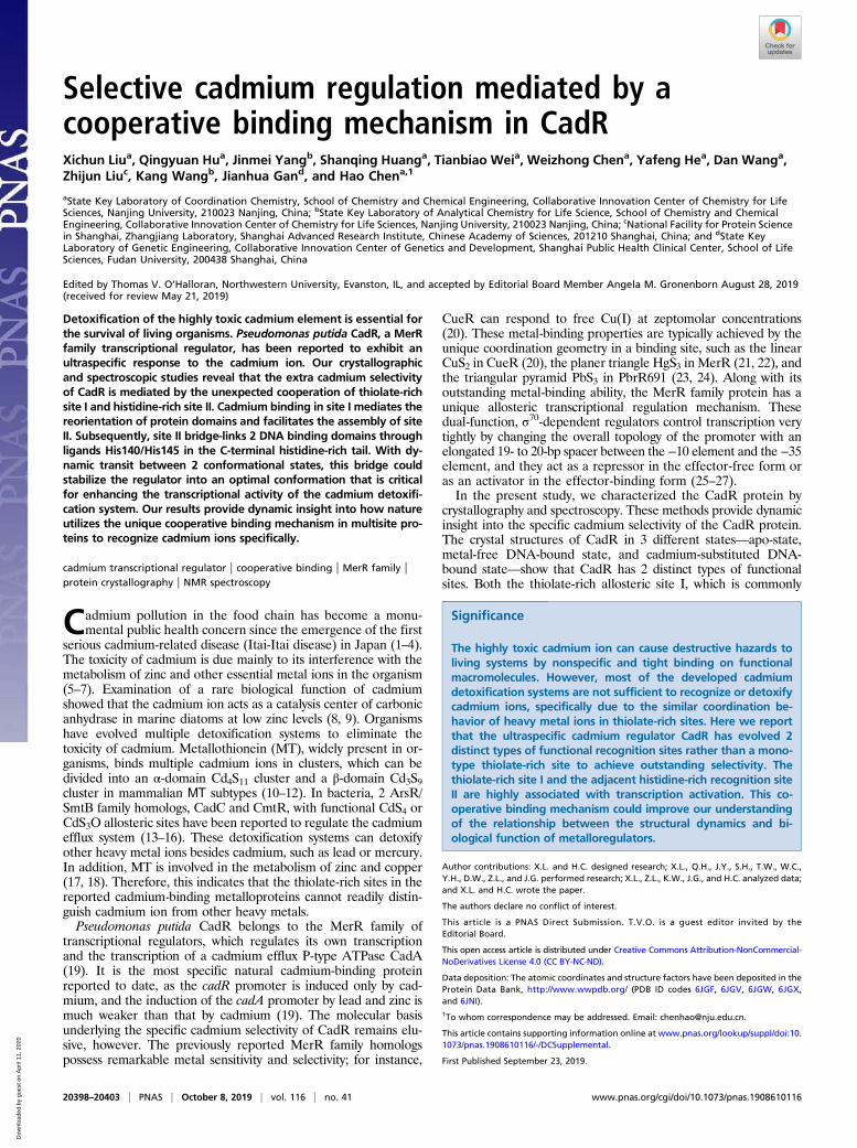
ResultsOverall Structure. To reveal the structural basis underlying thecadmium selectivity of CadR, 3 different states of CadR structurewere resolved: CadR in the apo form (apo-CadR), metal-freeCadR and promoter binary complex (CadR/DNA), and cadmium-bound CadR/DNA ternary complex (Cd/CadR/DNA) (Fig. 1Aand SI Appendix, Fig. S1). The structures show that CadR contains2 major domains as other MerR homologs: the N-terminal DNA-binding domain (DBD; residues 1 to 71) and the C-terminal metal-binding domain (MBD; residues 76 to 147), which are connectedby the flexible hinge (residues 72 to 75). In addition, CadR has aunique extended C-terminal histidine-rich region (His-tail; resi-dues 138 to 147) containing His138, His140, His145, and His147(SI Appendix, Fig. S2A). Although the MerR family proteins sharea similar topological arrangement, the C-terminal MBD is flexible,and the orientation between the MBD and DBD differs (SI Ap-pendix, Fig. S2B).The DBD of CadR is composed of 4 helices (α1 to α4) with
loops connecting them and is involved in the specific recognitionof promoter DNA, which was mapped by a footprint and elec-trophoretic mobility shift assay (EMSA) (SI Appendix, Fig. S3).There are 3 motifs involved in protein–DNA interactions (SI Ap-pendix, Fig. S4): the helix-turn-helix (HTH) motif (α1-turn-α2),wing 1 motif (loop region between α2 and α3), and wing 2 motif(loop region between α3 and α4). The rmsd values of Cα atoms ofDBD in the 3 different states are <0.525 Å, implying that the DBDbackbone is rigid (Fig. 1B). The DBD is involved in interdomainmotion with the MBD on DNA and cadmium binding. In contrast,the MBD of CadR can adopt flexible conformations depending on
the states of the structures. Residues 79 to 126 form a long-extended helix (α5) in apo-CadR, which bends approximately 9°and becomes more flexible in the CadR-DNA complex. Whenbound to metal ions, residues 111 to 119 transfer from helix to coil,and residues 120 to 126 form an independent α6 helix. Meanwhile,the remaining residues 76 to 110 of α5 helix bends another 42°.Importantly, the His-tail crosses over the molecular center andcontributes to metal binding in the Cd/CadR/DNA complex, whileit is disordered in other structures (Fig. 1B).
CadR Contains 2 Types of Functional Cadmium-Binding Sites. In-terestingly, different from other MerR family proteins, CadR has2 distinct types of metal-binding sites, and each CadR dimer bindsto 4 cadmium ions (Fig. 1A and SI Appendix, Fig. S5 A–C). Themetal-binding site I (S1 and S1′) is located at each end of thedimerization helices (α5 and α5′). The site is composed of 3 cys-teines (Cys77, Cys112′, and Cys119′) and 1 asparagine residue(Asn81). Cys77 comes from the same chain as Asn81, whileCys112′ and Cys119′ come from the partner chain. The cysteine-rich environment of site I is conserved in the MerR family pro-teins. The metal-binding site II (S2 and S2′) is located in themiddle of the molecule and formed by 2 histidines (His87 andHis90 in the α5 helix), 1 glutamic acid (Glu62 in the α4 helix), anda variable last ligand from the His-tail region. S2 and S2′ areasymmetric such that His140 coordinates to S2 and His145 coor-dinates to S2′. These metal-binding ligands are highly conservedamong CadR homologs (SI Appendix, Fig. S5D). Although thesequences of the extended C terminus are variable, a conservedHX4H motif is seen.To investigate the thermodynamics of these 2 cadmium-binding
sites, we performed a series of titration experiments. CadRWT bindscadmium ions with a stepwise-binding model, as the isothermaltitration calorimetry (ITC) result shows 2 steps clearly separated atN ∼2 and 4 (Fig. 1C). Moreover, the Raman spectrum shows thatthe peak of the sulfhydryl group (SH stretch; ∼2,563 cm−1) reaches
BA
S2 S2'
S1 S1'~ 42 °
α1α2
α3α4
α5
α6His-tail
DBD
MBD
hinge
metal-free
Cd(II)-bound
wing1
His-tail~ 9 °
wing2
C D 0.0
Stepwise
S1+S1'
S2+S2'
kcal
/mol
μcal
/sec
−20
−10
0
−1.0
−0.5
−1.5
0 2 4 6 8Molar ratio
01234
Molar ratio
2520 2560 2600
Nor
mal
ized
Ram
an in
tens
ity
cm−1
−SHC112'
C119'
N81
C77 H90
H140
H87
E62
α5
α4
α5'
α6'
S1
S2
Fig. 1. Two types of cadmium-binding sites and the allosteric effects triggered by cadmium binding. (A) Cartoon presentations of the Cd/CadR/DNA complex. DNAis colored in orange. The helical axis of DNA is represented as a solid gray line. Cadmium ions are shown as spheres. Close-up view of the first coordination shell ofmetal-binding sites is shown within the dashed box. (B) Superimposition of crystal structures based on the DBD of a CadR molecule of the metal-free CadR/DNAcomplex. For clarity, the partner CadR molecule and DNA are omitted. The 3 structures—apo-CadR, metal-free CadR/DNA complex, and Cd/CadR/DNA complex—are in wheat, magenta, and yellow, respectively. The allosteric movements triggered by cadmium binding are represented by dashed and solid arrows. (C) ITCcurves showing that CadR has a clear, stepwise bindingmodel toward the cadmium ion. (D) Raman peak of sulfhydryl group (∼2,563 cm−1) in CadR with a differentmolar ratio of Cd(II) ions.
Liu et al. PNAS | October 8, 2019 | vol. 116 | no. 41 | 20399
BIOCH
EMISTR
YCH
EMISTR
Y
Dow
nloa
ded
by g
uest
on
Apr
il 11
, 202
0
a minimum at a Cd(II)/CadRdimer ratio of ∼2 in the titration system(Fig. 1D). Three cysteine residues (Cys77, Cys112, and Cys119) areblocked with the metal binding in site I, while the other 2 (Cys12and Cys53) remain free. Based on these observations, we proposethat the cadmium ion first binds to the cysteine-rich site I (S1 +S1′) stoichiometrically and then binds to the histidine-rich site II(S2 + S2′). The binding affinity of site I is significantly stronger thanthat of site II, and the apparent binding affinities are fitted to K(Cd,site I)= 3.5± 0.1 × 10¹³M−1 and K(Cd, site II)= 1.0± 0.4 × 10⁷M−1,respectively (SI Appendix, Fig. S6).
Site I Is a Conserved Allosteric Site. As discussed above, the thiolate-rich binding site I is conserved among the MerR family metal-loproteins. The crystal structures show that the conformation ofMBD itself and the interaction network between MBD and DBDchanged dramatically on the binding of cadmium ions (SI Ap-pendix, Fig. S7 A and B). Residues of site I disperse from oneanother in the metal-free structures. Cys77 is disordered, and thedistance between Cys112′ and Cys119′ is >10 Å in the crystalstructure. Asn81 forms a hydrogen bond with Asn113′. On cad-mium ion binding to site I, Cys119′moves toward the coordinationcenter, and the newly formed α6′ helix bends toward and interactswith the DBD. Meanwhile, Cys77 becomes ordered and coordi-nates with the cadmium ion. Within the interface of the α3, α4, α5,α5′, and α6′ helices bundle, a vast network of hydrophobic inter-actions is formed to stabilize the protein conformation (SI Ap-pendix, Fig. S7C).To gain more insight into structural function of the metal-
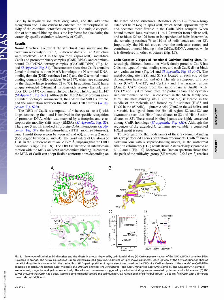
binding sites, we used 19F NMR spectroscopy (28). Fortunately, asole phenylalanine residue (Phe49) is located at the interface be-tween the DBD and the MBD that exhibits a significant confor-mational change on cadmium binding (Fig. 2A). In metal-freestructures, the DBD directly interacts with the α5′ helix of thepartner molecule, in which Arg70 of the α4 helix makes a hydrogenbond with Arg111′ of the α5′ helix. In the cadmium-bound struc-tures, Arg70 makes hydrogen bonds with Glu126′ in the α6′ helix.In addition, Asn52 in the α3 helix forms 2 hydrogen bonds withLeu125′ and Thr127′ in the α6′ helix. Thus, 19F-labeled Phe49 is anideal probe for detecting the allosteric motion of CadR. Threeclearly separated peaks—δ = −114.3, −112.4, and −111.9 ppm—
are observed with the titration of cadmium ions (Fig. 2B). Com-pared with the ITC result, which shows a stepwise-binding model(Fig. 1C), these peaks can be assigned to the metal-free state, site Ibound state in Cd2(CadR)2 and the sites I and II bound state inCd4(CadR)2, respectively. Cadmium binding in site I triggers a1.9-ppm downfield shift. Therefore, NMR spectroscopy also sug-gests that site I is an allosteric site.
Site II Bridging 2 DBDs by the Extended C-Terminal His-Tail. Theforegoing 19F NMR data also show that further cadmiumbinding in site II triggers a 0.5-ppm downfield shift of Phe49 (Fig.2B). This suggests that site II acts as an additional allosteric site tofurther alter the conformation of the DBD. The orientations of theα4 and α5 helices are stabilized by the strong coordination bondamong cadmium, Glu62 of the α4 helix, and His87/His90 of the α5helix. In the second coordination shell, residues Glu62 and His87also form hydrogen bonds with Thr59 and Leu56 in wing2, re-spectively (SI Appendix, Fig. S7 A and B). Part of the His-tail re-gion (residues 139 to 145) is ordered in such a way that His140coordinates with cadmium ions at site S2 and Ser139 contributeshydrogen bonds with His90 and Arg94.In particular, the cadmium ion of site S2′ coordinates with
His145 instead of His140′ from the partner molecule (Fig. 2C).The Cd/CadR/DNA complex is asymmetrical, in which residues141 to 145 cross over the molecular center once His140 partici-pates in coordination. Various interactions form during this pro-cess. Hydrogen bonds occur between Val141 and Arg94 andbetween Gly142 and Arg94′, polar contact occurs between Asp57′
and Ser144, and hydrophobic interactions include those amongVal141, Val91, and Leu98′. The residue His145 will reach the vi-cinity of site S2′ and coordinate to the cadmium ion. Conse-quently, the 2 DBDs in the Cd/CadR/DNA complex are bridge-linked through the His-tail (Fig. 2D). The length of this bridge is23.6 Å as measured by the distance between the cadmium ions atsites S2 and S2′.
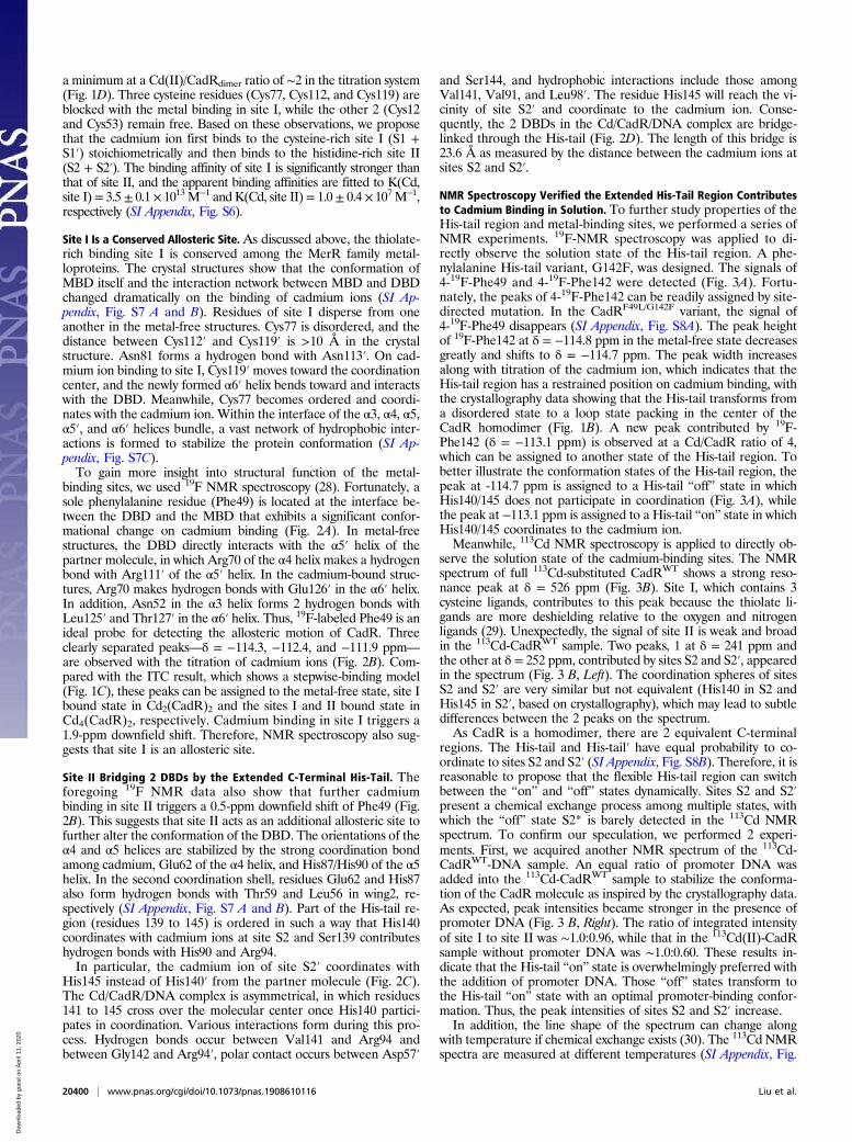
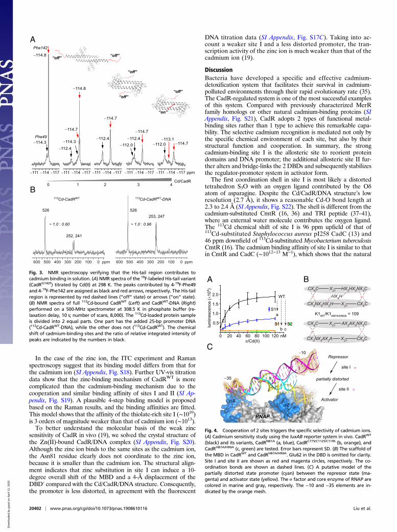
NMR Spectroscopy Verified the Extended His-Tail Region Contributesto Cadmium Binding in Solution. To further study properties of theHis-tail region and metal-binding sites, we performed a series ofNMR experiments. 19F-NMR spectroscopy was applied to di-rectly observe the solution state of the His-tail region. A phe-nylalanine His-tail variant, G142F, was designed. The signals of4-19F-Phe49 and 4-19F-Phe142 were detected (Fig. 3A). Fortu-nately, the peaks of 4-19F-Phe142 can be readily assigned by site-directed mutation. In the CadRF49L/G142F variant, the signal of4-19F-Phe49 disappears (SI Appendix, Fig. S8A). The peak heightof 19F-Phe142 at δ = −114.8 ppm in the metal-free state decreasesgreatly and shifts to δ = −114.7 ppm. The peak width increasesalong with titration of the cadmium ion, which indicates that theHis-tail region has a restrained position on cadmium binding, withthe crystallography data showing that the His-tail transforms froma disordered state to a loop state packing in the center of theCadR homodimer (Fig. 1B). A new peak contributed by 19F-Phe142 (δ = −113.1 ppm) is observed at a Cd/CadR ratio of 4,which can be assigned to another state of the His-tail region. Tobetter illustrate the conformation states of the His-tail region, thepeak at -114.7 ppm is assigned to a His-tail “off” state in whichHis140/145 does not participate in coordination (Fig. 3A), whilethe peak at −113.1 ppm is assigned to a His-tail “on” state in whichHis140/145 coordinates to the cadmium ion.Meanwhile, 113Cd NMR spectroscopy is applied to directly ob-
serve the solution state of the cadmium-binding sites. The NMRspectrum of full 113Cd-substituted CadRWT shows a strong reso-nance peak at δ = 526 ppm (Fig. 3B). Site I, which contains 3cysteine ligands, contributes to this peak because the thiolate li-gands are more deshielding relative to the oxygen and nitrogenligands (29). Unexpectedly, the signal of site II is weak and broadin the 113Cd-CadRWT sample. Two peaks, 1 at δ = 241 ppm andthe other at δ = 252 ppm, contributed by sites S2 and S2′, appearedin the spectrum (Fig. 3 B, Left). The coordination spheres of sitesS2 and S2′ are very similar but not equivalent (His140 in S2 andHis145 in S2′, based on crystallography), which may lead to subtledifferences between the 2 peaks on the spectrum.As CadR is a homodimer, there are 2 equivalent C-terminal
regions. The His-tail and His-tail′ have equal probability to co-ordinate to sites S2 and S2′ (SI Appendix, Fig. S8B). Therefore, it isreasonable to propose that the flexible His-tail region can switchbetween the “on” and “off” states dynamically. Sites S2 and S2′present a chemical exchange process among multiple states, withwhich the “off” state S2* is barely detected in the 113Cd NMRspectrum. To confirm our speculation, we performed 2 experi-ments. First, we acquired another NMR spectrum of the 113Cd-CadRWT-DNA sample. An equal ratio of promoter DNA wasadded into the 113Cd-CadRWT sample to stabilize the conforma-tion of the CadR molecule as inspired by the crystallography data.As expected, peak intensities became stronger in the presence ofpromoter DNA (Fig. 3 B, Right). The ratio of integrated intensityof site I to site II was ∼1.0:0.96, while that in the 113Cd(II)-CadRsample without promoter DNA was ∼1.0:0.60. These results in-dicate that the His-tail “on” state is overwhelmingly preferred withthe addition of promoter DNA. Those “off” states transform tothe His-tail “on” state with an optimal promoter-binding confor-mation. Thus, the peak intensities of sites S2 and S2′ increase.In addition, the line shape of the spectrum can change along
with temperature if chemical exchange exists (30). The 113Cd NMRspectra are measured at different temperatures (SI Appendix, Fig.
20400 | www.pnas.org/cgi/doi/10.1073/pnas.1908610116 Liu et al.
Dow
nloa
ded
by g
uest
on
Apr
il 11
, 202
0
S8C). The faster the exchange rate (i.e., at higher temperature), thesmaller the chemical shift difference between sites S2 and S2′. Theobserved difference is in agreement with the exchange speculation,increasing from 1.2 kHz to 2.1 kHz when temperature decreasesfrom 308.5 K to 278.5 K. Moreover, the 19F NMR spectra showthat the peak of Phe142 shifts at lower temperature, which alsoindicates the exchange of the His-tail (SI Appendix, Fig. S8A).Taken together, these data verify that the flexible His-tail regioncontributes to cadmium binding on site II in solution.
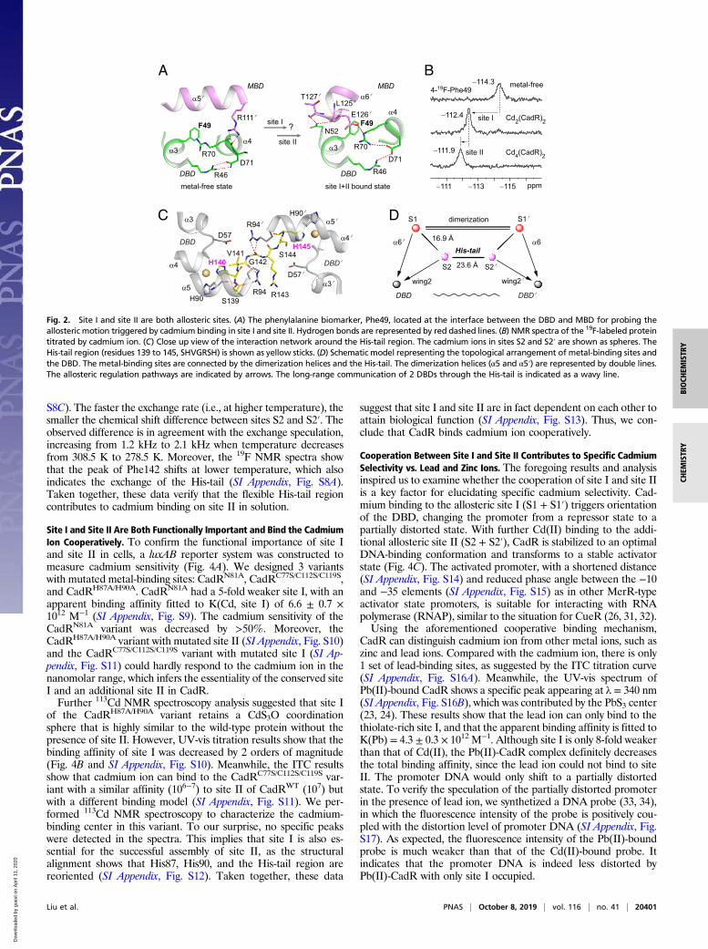
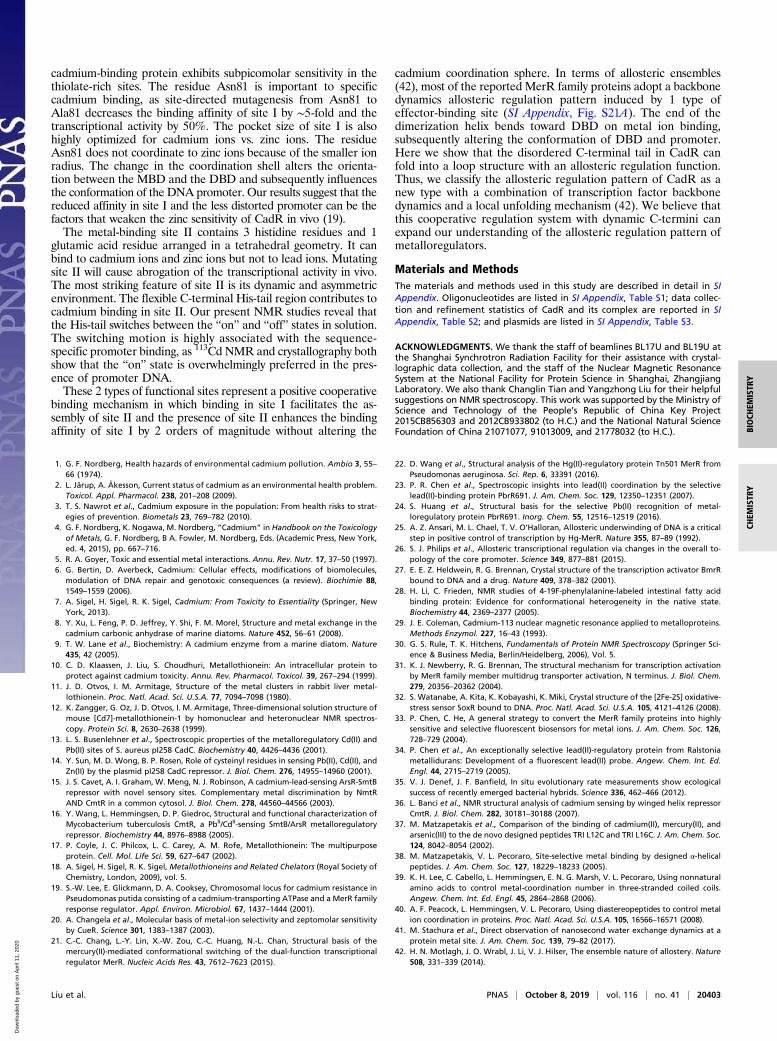
Site I and Site II Are Both Functionally Important and Bind the CadmiumIon Cooperatively. To confirm the functional importance of site Iand site II in cells, a luxAB reporter system was constructed tomeasure cadmium sensitivity (Fig. 4A). We designed 3 variantswith mutated metal-binding sites: CadRN81A, CadRC77S/C112S/C119S,and CadRH87A/H90A. CadRN81A had a 5-fold weaker site I, with anapparent binding affinity fitted to K(Cd, site I) of 6.6 ± 0.7 ×10¹² M−1 (SI Appendix, Fig. S9). The cadmium sensitivity of theCadRN81A variant was decreased by >50%. Moreover, theCadRH87A/H90A variant with mutated site II (SI Appendix, Fig. S10)and the CadRC77S/C112S/C119S variant with mutated site I (SI Ap-pendix, Fig. S11) could hardly respond to the cadmium ion in thenanomolar range, which infers the essentiality of the conserved siteI and an additional site II in CadR.Further 113Cd NMR spectroscopy analysis suggested that site I
of the CadRH87A/H90A variant retains a CdS3O coordinationsphere that is highly similar to the wild-type protein without thepresence of site II. However, UV-vis titration results show that thebinding affinity of site I was decreased by 2 orders of magnitude(Fig. 4B and SI Appendix, Fig. S10). Meanwhile, the ITC resultsshow that cadmium ion can bind to the CadRC77S/C112S/C119S var-iant with a similar affinity (106−7) to site II of CadRWT (107) butwith a different binding model (SI Appendix, Fig. S11). We per-formed 113Cd NMR spectroscopy to characterize the cadmium-binding center in this variant. To our surprise, no specific peakswere detected in the spectra. This implies that site I is also es-sential for the successful assembly of site II, as the structuralalignment shows that His87, His90, and the His-tail region arereoriented (SI Appendix, Fig. S12). Taken together, these data
suggest that site I and site II are in fact dependent on each other toattain biological function (SI Appendix, Fig. S13). Thus, we con-clude that CadR binds cadmium ion cooperatively.
Cooperation Between Site I and Site II Contributes to Specific CadmiumSelectivity vs. Lead and Zinc Ions. The foregoing results and analysisinspired us to examine whether the cooperation of site I and site IIis a key factor for elucidating specific cadmium selectivity. Cad-mium binding to the allosteric site I (S1 + S1′) triggers orientationof the DBD, changing the promoter from a repressor state to apartially distorted state. With further Cd(II) binding to the addi-tional allosteric site II (S2 + S2′), CadR is stabilized to an optimalDNA-binding conformation and transforms to a stable activatorstate (Fig. 4C). The activated promoter, with a shortened distance(SI Appendix, Fig. S14) and reduced phase angle between the −10and −35 elements (SI Appendix, Fig. S15) as in other MerR-typeactivator state promoters, is suitable for interacting with RNApolymerase (RNAP), similar to the situation for CueR (26, 31, 32).Using the aforementioned cooperative binding mechanism,
CadR can distinguish cadmium ion from other metal ions, such aszinc and lead ions. Compared with the cadmium ion, there is only1 set of lead-binding sites, as suggested by the ITC titration curve(SI Appendix, Fig. S16A). Meanwhile, the UV-vis spectrum ofPb(II)-bound CadR shows a specific peak appearing at λ = 340 nm(SI Appendix, Fig. S16B), which was contributed by the PbS3 center(23, 24). These results show that the lead ion can only bind to thethiolate-rich site I, and that the apparent binding affinity is fitted toK(Pb) = 4.3 ± 0.3 × 10¹² M−1. Although site I is only 8-fold weakerthan that of Cd(II), the Pb(II)-CadR complex definitely decreasesthe total binding affinity, since the lead ion could not bind to siteII. The promoter DNA would only shift to a partially distortedstate. To verify the speculation of the partially distorted promoterin the presence of lead ion, we synthetized a DNA probe (33, 34),in which the fluorescence intensity of the probe is positively cou-pled with the distortion level of promoter DNA (SI Appendix, Fig.S17). As expected, the fluorescence intensity of the Pb(II)-boundprobe is much weaker than that of the Cd(II)-bound probe. Itindicates that the promoter DNA is indeed less distorted byPb(II)-CadR with only site I occupied.
A
DBD'DBD
S2 S2'
S1 S1'
α6'His-tail
α6
23.6 Å
wing2 wing2'
dimerizationC
−111 −113 −115 ppm
−114.3
−112.4
−111.9
4-19F-Phe49
site I
site II
metal-free
Cd2(CadR)2
Cd4(CadR)2
H140
S139
V141
R143
S144H145
D57'
G142
R94H90
R94'D57
H90'α3
α4
α5 α3'
α4'
α5'
DBD
DBD'
DBD
E126'
T127'L125'
R70
N52
R46D71
α3
α4
α6'MBD
F49
B
R111'
R70
R46D71
α3α4
α5'
DBD
F49
MBD
site II
D
site I ?
16.9 Å
metal-free state site I+II bound state
Fig. 2. Site I and site II are both allosteric sites. (A) The phenylalanine biomarker, Phe49, located at the interface between the DBD and MBD for probing theallosteric motion triggered by cadmium binding in site I and site II. Hydrogen bonds are represented by red dashed lines. (B) NMR spectra of the 19F-labeled proteintitrated by cadmium ion. (C) Close up view of the interaction network around the His-tail region. The cadmium ions in sites S2 and S2′ are shown as spheres. TheHis-tail region (residues 139 to 145, SHVGRSH) is shown as yellow sticks. (D) Schematic model representing the topological arrangement of metal-binding sites andthe DBD. The metal-binding sites are connected by the dimerization helices and the His-tail. The dimerization helices (α5 and α5′) are represented by double lines.The allosteric regulation pathways are indicated by arrows. The long-range communication of 2 DBDs through the His-tail is indicated as a wavy line.
Liu et al. PNAS | October 8, 2019 | vol. 116 | no. 41 | 20401
BIOCH
EMISTR
YCH
EMISTR
Y
Dow
nloa
ded
by g
uest
on
Apr
il 11
, 202
0
In the case of the zinc ion, the ITC experiment and Ramanspectroscopy suggest that its binding model differs from that forthe cadmium ion (SI Appendix, Fig. S18). Further UV-vis titrationdata show that the zinc-binding mechanism of CadRWT is morecomplicated than the cadmium-binding mechanism due to thecooperation and similar binding affinity of sites I and II (SI Ap-pendix, Fig. S19). A plausible 4-step binding model is proposedbased on the Raman results, and the binding affinities are fitted.This model shows that the affinity of the thiolate-rich site I (∼1010)is 3 orders of magnitude weaker than that of cadmium ion (∼1013).To better understand the molecular basis of the weak zinc
sensitivity of CadR in vivo (19), we solved the crystal structure ofthe Zn(II)-bound CadR/DNA complex (SI Appendix, Fig. S20).Although the zinc ion binds to the same sites as the cadmium ion,the Asn81 residue clearly does not coordinate to the zinc ion,because it is smaller than the cadmium ion. The structural align-ment indicates that zinc substitution in site I can induce a 10-degree overall shift of the MBD and a 4-Å displacement of theDBD′ compared with the Cd/CadR/DNA structure. Consequently,the promoter is less distorted, in agreement with the fluorescent
DNA titration data (SI Appendix, Fig. S17C). Taking into ac-count a weaker site I and a less distorted promoter, the tran-scription activity of the zinc ion is much weaker than that of thecadmium ion (19).
DiscussionBacteria have developed a specific and effective cadmium-detoxification system that facilitates their survival in cadmium-polluted environments through their rapid evolutionary rate (35).The CadR-regulated system is one of the most successful examplesof this system. Compared with previously characterized MerRfamily homologs or other natural cadmium-binding proteins (SIAppendix, Fig. S21), CadR adopts 2 types of functional metal-binding sites rather than 1 type to achieve this remarkable capa-bility. The selective cadmium recognition is mediated not only bythe specific chemical environment of each site, but also by theirstructural function and cooperation. In summary, the strongcadmium-binding site I is the allosteric site to reorient proteindomains and DNA promoter; the additional allosteric site II fur-ther alters and bridge-links the 2 DBDs and subsequently stabilizesthe regulator-promoter system in activator form.The first coordination shell in site I is most likely a distorted
tetrahedron S3O with an oxygen ligand contributed by the Oδatom of asparagine. Despite the Cd/CadR/DNA structure’s lowresolution (2.7 Å), it shows a reasonable Cd-O bond length at2.3 to 2.4 Å (SI Appendix, Fig. S22). The shell is different from thecadmium-substituted CmtR (16, 36) and TRI peptide (37–41),where an external water molecule contributes the oxygen ligand.The 113Cd chemical shift of site I is 96 ppm upfield of that of113Cd-substituted Staphylococcus aureus pI258 CadC (13) and46 ppm downfield of 113Cd-substituted Mycobacterium tuberculosisCmtR (16). The cadmium binding affinity of site I is similar to thatin CmtR and CadC (∼1012−13 M−1), which shows that the natural
B113Cd-CadRWT
526
252, 241
~ 1.0 : 0.60
ppm600 300500 400 200 100 0
113Cd-CadRWT-DNA
526253, 247
~ 1.0 : 0.96
ppm600 400 300500 100200 0
−114.7
−112.4−112.0
−112.4
−114.7
−111 −114 −117 −111 −114 −117 −111 −114 −117 −111 −114 −117 −111 −114 −117
Phe49
−114.8
−114.3
Phe142
A
−114.7−113.1
−112.0
ppm
Cd/CadR0 1 2 3 4
−112.4
−114.8
−114.7
−114.3
“off” “off”
“off”
“off”
“on”
“off”
Fig. 3. NMR spectroscopy verifying that the His-tail region contributes tocadmium binding in solution. (A) NMR spectra of the 19F-labeled His-tail variant(CadRG142F) titrated by Cd(II) at 298 K. The peaks contributed by 4-19F-Phe49and 4-19F-Phe142 are assigned as black and red arrows, respectively. The His-tailregion is represented by red dashed lines (“off” state) or arrows (“on” state).(B) NMR spectra of full 113Cd-bound CadRWT (Left) and CadRWT-DNA (Right)performed on a 500-MHz spectrometer at 308.5 K in phosphate buffer (re-laxation delay, 10 s; number of scans, 8,000). The 113Cd-loaded protein sampleis divided into 2 equal parts. One part has the added 25-bp promoter DNA(113Cd-CadRWT-DNA), while the other does not (113Cd-CadRWT). The chemicalshift of cadmium-binding sites and the ratio of relative integrated intensity ofpeaks are indicated by the numbers in black.
×S1
a
WT
b c×S2
A B
C
0 20 40 60 80 100 120
1.0
2.0
nM
0.5
1.5
lum
ines
cenc
e (×
104 )
c/Cd(II)
S1
HX4H
X21
X21CX3NX5HX2H
HX2HX5NX3CCX6C
CX6C
X21
X21CX3NX5AX2A
AX2AX5NX3CCX6C
CX6C
Repressor
σ4σ2
RNAP
Activator
partially distorted−35
−10
site I
site II
K1WT/K1H87A/H90A = 109
Fig. 4. Cooperation of 2 sites triggers the specific selectivity of cadmium ions.(A) Cadmium sensitivity study using the luxAB reporter system in vivo. CadRWT
(black) and its variants, CadRN81A (a, blue), CadRC77S/C112S/C119S (b, orange), andCadRH87A/H90A (c, green) are tested. Error bars represent SD. (B) The scaffold ofthe MBD in CadRWT and CadRH87A/H90A. Glu62 in the DBD is omitted for clarity.Site I and site II are shown as red and magenta circles, respectively. The co-ordination bonds are shown as dashed lines. (C) A putative model of thepartially distorted state promoter (cyan) between the repressor state (ma-genta) and activator state (yellow). The σ factor and core enzyme of RNAP arecolored in marine and gray, respectively. The −10 and −35 elements are in-dicated by the orange mesh.
20402 | www.pnas.org/cgi/doi/10.1073/pnas.1908610116 Liu et al.
Dow
nloa
ded
by g
uest
on
Apr
il 11
, 202
0
cadmium-binding protein exhibits subpicomolar sensitivity in thethiolate-rich sites. The residue Asn81 is important to specificcadmium binding, as site-directed mutagenesis from Asn81 toAla81 decreases the binding affinity of site I by ∼5-fold and thetranscriptional activity by 50%. The pocket size of site I is alsohighly optimized for cadmium ions vs. zinc ions. The residueAsn81 does not coordinate to zinc ions because of the smaller ionradius. The change in the coordination shell alters the orienta-tion between the MBD and the DBD and subsequently influencesthe conformation of the DNA promoter. Our results suggest that thereduced affinity in site I and the less distorted promoter can be thefactors that weaken the zinc sensitivity of CadR in vivo (19).The metal-binding site II contains 3 histidine residues and 1
glutamic acid residue arranged in a tetrahedral geometry. It canbind to cadmium ions and zinc ions but not to lead ions. Mutatingsite II will cause abrogation of the transcriptional activity in vivo.The most striking feature of site II is its dynamic and asymmetricenvironment. The flexible C-terminal His-tail region contributes tocadmium binding in site II. Our present NMR studies reveal thatthe His-tail switches between the “on” and “off” states in solution.The switching motion is highly associated with the sequence-specific promoter binding, as 113Cd NMR and crystallography bothshow that the “on” state is overwhelmingly preferred in the pres-ence of promoter DNA.These 2 types of functional sites represent a positive cooperative
binding mechanism in which binding in site I facilitates the as-sembly of site II and the presence of site II enhances the bindingaffinity of site I by 2 orders of magnitude without altering the
cadmium coordination sphere. In terms of allosteric ensembles(42), most of the reported MerR family proteins adopt a backbonedynamics allosteric regulation pattern induced by 1 type ofeffector-binding site (SI Appendix, Fig. S21A). The end of thedimerization helix bends toward DBD on metal ion binding,subsequently altering the conformation of DBD and promoter.Here we show that the disordered C-terminal tail in CadR canfold into a loop structure with an allosteric regulation function.Thus, we classify the allosteric regulation pattern of CadR as anew type with a combination of transcription factor backbonedynamics and a local unfolding mechanism (42). We believe thatthis cooperative regulation system with dynamic C-termini canexpand our understanding of the allosteric regulation pattern ofmetalloregulators.
Materials and MethodsThe materials and methods used in this study are described in detail in SIAppendix. Oligonucleotides are listed in SI Appendix, Table S1; data collec-tion and refinement statistics of CadR and its complex are reported in SIAppendix, Table S2; and plasmids are listed in SI Appendix, Table S3.
ACKNOWLEDGMENTS. We thank the staff of beamlines BL17U and BL19U atthe Shanghai Synchrotron Radiation Facility for their assistance with crystal-lographic data collection, and the staff of the Nuclear Magnetic ResonanceSystem at the National Facility for Protein Science in Shanghai, ZhangjiangLaboratory. We also thank Changlin Tian and Yangzhong Liu for their helpfulsuggestions on NMR spectroscopy. This work was supported by the Ministry ofScience and Technology of the People’s Republic of China Key Project2015CB856303 and 2012CB933802 (to H.C.) and the National Natural ScienceFoundation of China 21071077, 91013009, and 21778032 (to H.C.).
1. G. F. Nordberg, Health hazards of environmental cadmium pollution. Ambio 3, 55–66 (1974).
2. L. Järup, A. Åkesson, Current status of cadmium as an environmental health problem.Toxicol. Appl. Pharmacol. 238, 201–208 (2009).
3. T. S. Nawrot et al., Cadmium exposure in the population: From health risks to strat-egies of prevention. Biometals 23, 769–782 (2010).
4. G. F. Nordberg, K. Nogawa, M. Nordberg, “Cadmium” in Handbook on the Toxicologyof Metals, G. F. Nordberg, B A. Fowler, M. Nordberg, Eds. (Academic Press, New York,ed. 4, 2015), pp. 667–716.
5. R. A. Goyer, Toxic and essential metal interactions. Annu. Rev. Nutr. 17, 37–50 (1997).6. G. Bertin, D. Averbeck, Cadmium: Cellular effects, modifications of biomolecules,
modulation of DNA repair and genotoxic consequences (a review). Biochimie 88,1549–1559 (2006).
7. A. Sigel, H. Sigel, R. K. Sigel, Cadmium: From Toxicity to Essentiality (Springer, NewYork, 2013).
8. Y. Xu, L. Feng, P. D. Jeffrey, Y. Shi, F. M. Morel, Structure and metal exchange in thecadmium carbonic anhydrase of marine diatoms. Nature 452, 56–61 (2008).
9. T. W. Lane et al., Biochemistry: A cadmium enzyme from a marine diatom. Nature435, 42 (2005).
10. C. D. Klaassen, J. Liu, S. Choudhuri, Metallothionein: An intracellular protein toprotect against cadmium toxicity. Annu. Rev. Pharmacol. Toxicol. 39, 267–294 (1999).
11. J. D. Otvos, I. M. Armitage, Structure of the metal clusters in rabbit liver metal-lothionein. Proc. Natl. Acad. Sci. U.S.A. 77, 7094–7098 (1980).
12. K. Zangger, G. Oz, J. D. Otvos, I. M. Armitage, Three-dimensional solution structure ofmouse [Cd7]-metallothionein-1 by homonuclear and heteronuclear NMR spectros-copy. Protein Sci. 8, 2630–2638 (1999).
13. L. S. Busenlehner et al., Spectroscopic properties of the metalloregulatory Cd(II) andPb(II) sites of S. aureus pI258 CadC. Biochemistry 40, 4426–4436 (2001).
14. Y. Sun, M. D. Wong, B. P. Rosen, Role of cysteinyl residues in sensing Pb(II), Cd(II), andZn(II) by the plasmid pI258 CadC repressor. J. Biol. Chem. 276, 14955–14960 (2001).
15. J. S. Cavet, A. I. Graham, W. Meng, N. J. Robinson, A cadmium-lead-sensing ArsR-SmtBrepressor with novel sensory sites. Complementary metal discrimination by NmtRAND CmtR in a common cytosol. J. Biol. Chem. 278, 44560–44566 (2003).
16. Y. Wang, L. Hemmingsen, D. P. Giedroc, Structural and functional characterization ofMycobacterium tuberculosis CmtR, a PbII/CdII-sensing SmtB/ArsR metalloregulatoryrepressor. Biochemistry 44, 8976–8988 (2005).
17. P. Coyle, J. C. Philcox, L. C. Carey, A. M. Rofe, Metallothionein: The multipurposeprotein. Cell. Mol. Life Sci. 59, 627–647 (2002).
18. A. Sigel, H. Sigel, R. K. Sigel, Metallothioneins and Related Chelators (Royal Society ofChemistry, London, 2009), vol. 5.
19. S.-W. Lee, E. Glickmann, D. A. Cooksey, Chromosomal locus for cadmium resistance inPseudomonas putida consisting of a cadmium-transporting ATPase and a MerR familyresponse regulator. Appl. Environ. Microbiol. 67, 1437–1444 (2001).
20. A. Changela et al., Molecular basis of metal-ion selectivity and zeptomolar sensitivityby CueR. Science 301, 1383–1387 (2003).
21. C.-C. Chang, L.-Y. Lin, X.-W. Zou, C.-C. Huang, N.-L. Chan, Structural basis of themercury(II)-mediated conformational switching of the dual-function transcriptionalregulator MerR. Nucleic Acids Res. 43, 7612–7623 (2015).
22. D. Wang et al., Structural analysis of the Hg(II)-regulatory protein Tn501 MerR fromPseudomonas aeruginosa. Sci. Rep. 6, 33391 (2016).
23. P. R. Chen et al., Spectroscopic insights into lead(II) coordination by the selectivelead(II)-binding protein PbrR691. J. Am. Chem. Soc. 129, 12350–12351 (2007).
24. S. Huang et al., Structural basis for the selective Pb(II) recognition of metal-loregulatory protein PbrR691. Inorg. Chem. 55, 12516–12519 (2016).
25. A. Z. Ansari, M. L. Chael, T. V. O’Halloran, Allosteric underwinding of DNA is a criticalstep in positive control of transcription by Hg-MerR. Nature 355, 87–89 (1992).
26. S. J. Philips et al., Allosteric transcriptional regulation via changes in the overall to-pology of the core promoter. Science 349, 877–881 (2015).
27. E. E. Z. Heldwein, R. G. Brennan, Crystal structure of the transcription activator BmrRbound to DNA and a drug. Nature 409, 378–382 (2001).
28. H. Li, C. Frieden, NMR studies of 4-19F-phenylalanine-labeled intestinal fatty acidbinding protein: Evidence for conformational heterogeneity in the native state.Biochemistry 44, 2369–2377 (2005).
29. J. E. Coleman, Cadmium-113 nuclear magnetic resonance applied to metalloproteins.Methods Enzymol. 227, 16–43 (1993).
30. G. S. Rule, T. K. Hitchens, Fundamentals of Protein NMR Spectroscopy (Springer Sci-ence & Business Media, Berlin/Heidelberg, 2006), Vol. 5.
31. K. J. Newberry, R. G. Brennan, The structural mechanism for transcription activationby MerR family member multidrug transporter activation, N terminus. J. Biol. Chem.279, 20356–20362 (2004).
32. S. Watanabe, A. Kita, K. Kobayashi, K. Miki, Crystal structure of the [2Fe-2S] oxidative-stress sensor SoxR bound to DNA. Proc. Natl. Acad. Sci. U.S.A. 105, 4121–4126 (2008).
33. P. Chen, C. He, A general strategy to convert the MerR family proteins into highlysensitive and selective fluorescent biosensors for metal ions. J. Am. Chem. Soc. 126,728–729 (2004).
34. P. Chen et al., An exceptionally selective lead(II)-regulatory protein from Ralstoniametallidurans: Development of a fluorescent lead(II) probe. Angew. Chem. Int. Ed.Engl. 44, 2715–2719 (2005).
35. V. J. Denef, J. F. Banfield, In situ evolutionary rate measurements show ecologicalsuccess of recently emerged bacterial hybrids. Science 336, 462–466 (2012).
36. L. Banci et al., NMR structural analysis of cadmium sensing by winged helix repressorCmtR. J. Biol. Chem. 282, 30181–30188 (2007).
37. M. Matzapetakis et al., Comparison of the binding of cadmium(II), mercury(II), andarsenic(III) to the de novo designed peptides TRI L12C and TRI L16C. J. Am. Chem. Soc.124, 8042–8054 (2002).
38. M. Matzapetakis, V. L. Pecoraro, Site-selective metal binding by designed α-helicalpeptides. J. Am. Chem. Soc. 127, 18229–18233 (2005).
39. K. H. Lee, C. Cabello, L. Hemmingsen, E. N. G. Marsh, V. L. Pecoraro, Using nonnaturalamino acids to control metal-coordination number in three-stranded coiled coils.Angew. Chem. Int. Ed. Engl. 45, 2864–2868 (2006).
40. A. F. Peacock, L. Hemmingsen, V. L. Pecoraro, Using diastereopeptides to control metalion coordination in proteins. Proc. Natl. Acad. Sci. U.S.A. 105, 16566–16571 (2008).
41. M. Stachura et al., Direct observation of nanosecond water exchange dynamics at aprotein metal site. J. Am. Chem. Soc. 139, 79–82 (2017).
42. H. N. Motlagh, J. O. Wrabl, J. Li, V. J. Hilser, The ensemble nature of allostery. Nature508, 331–339 (2014).
Liu et al. PNAS | October 8, 2019 | vol. 116 | no. 41 | 20403
BIOCH
EMISTR
YCH
EMISTR
Y
Dow
nloa
ded
by g
uest
on
Apr
il 11
, 202
0