secondary damage after traumatic brain injury ... doortje caroline.pdf · secondary damage after...
TRANSCRIPT

Secondary Damage after Traumatic Brain Injury: Epidemiology, Pathophysiology and Therapy
Sedondaire schade na traumatisch schedelhersenletsel: epidemiologie, pathofysiologie en therapie
Proefschrift
ter verkrijging van de graad van doctor aan de Erasmus Universiteit Rotterdam
op gezag van de rector magnificus Prof.dr. S.W.J. Lamberts en volgens besluit
van het College voor Promoties
De openbare verdediging zal plaatsvinden op woensdag 23 april 2008 om 9:45 uur
door
Doortje Caroline Engel
geboren te Rotterdam
Promotoren: Overigeleden:prof. dr. C.I. De Zeeuw prof. dr. C.M.F. Dirven prof. dr. N. Plesnila prof. dr. M.A. Frens prof. dr. E.W. Steyerberg
This thesis contains previously published manuscripts:
Engel DC, Slemmer JE, Vlug AS, Maas AIR, Weber JT (2005). Combined effects of mechanical and ischemic injury to cortical cells: secondary ischemia increases damage and decreases effects of neuro-protective agents. Neuropharmacology. 2005 Dec;49(7):985-95.
Zweckberger K, Erös C, Zimmermann R, Kim SW, Engel DC, Plesnila N. Effect of Early and De-layed Decompression Craniotomy on Secondary Brain Damage after Controlled Cortical Impact in Mice. JNeurotrauma. 2006 Jul;23(7):1083-93.
Mushkudiani N, Engel DC, Steyerberg EW, Butcher I, Lu J, Marmarou A, Slieker F, McHugh GS, Murray GD, Maas AI. Prognostic value of demographic characteristics in traumatic brain injury: results from the IMPACT study. JNeurotrauma. 2007 Feb;24(2):259-69.
McHugh GS, Engel DC, Butcher I, Steyerberg EW, Lu J, Mushkudiani N, Hernandez AV, Marmarou A, Maas AI, Murray GD. Prognostic value of secondary insults in traumatic brain injury: results from the IMPACT study. JNeurotrauma. 2007 Feb;24(2):287-93.
Maegele M, Engel DC, Bouillon B, Lefering R, Fach H, Raum M, Buchheister B, Schaefer U, Klug N, Neugebauer. The incidence and outcome of traumatic brain injury in an urban area in western Europe over 10 years. EurSurgRes. 2007;39(6):372-9.
ISBN 978-90-9022809-9© Doortje C. Engel, Rotterdam 2008All rights reserved. Save exceptions stated by the law, no part of this publicatino may be reproduced, stored in a retrieval system of any nature, or tranmitted in any form or by any means, electronic, me-chanical, photocopying, recording or otherwhise, included a complete or partial transcription, without the prior written permission of the author.
Printed by: Gildeprint, EnschedeDesign cover: Mitch Thijssen ([email protected])
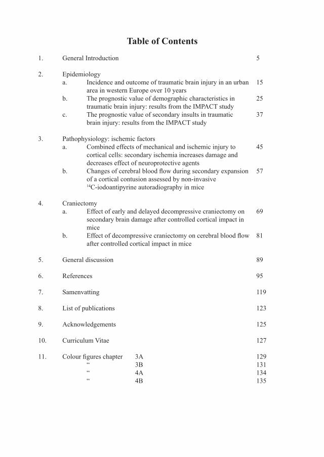
Table of Contents
General Introduction 5
Epidemiology a. Incidence and outcome of traumatic brain injury in an urban 15 area in western Europe over 10 years b. The prognostic value of demographic characteristics in 25 traumatic brain injury: results from the IMPACT study c. The prognostic value of secondary insults in traumatic 37 brain injury: results from the IMPACT study
Pathophysiology: ischemic factors a. Combined effects of mechanical and ischemic injury to 45 cortical cells: secondary ischemia increases damage and decreases effect of neuroprotective agents b. Changesofcerebralbloodflowduringsecondaryexpansion 57 of a cortical contusion assessed by non-invasive 14C-iodoantipyrine autoradiography in mice
Craniectomy a. Effect of early and delayed decompressive craniectomy on 69 secondary brain damage after controlled cortical impact in mice b. Effectofdecompressivecraniectomyoncerebralbloodflow 81 after controlled cortical impact in mice
General discussion 89
References 95
Samenvatting 119
List of publications 123
Acknowledgements 125
Curriculum Vitae 127
Colourfigureschapter 3A 129 “ 3B 131 “ 4A 134 “ 4B 135
1.
2.
3.
4.
5.
6.
7.
8.
9.
10.
11.


5
CHAPTER 1. GENERAL INTRODUCTION
Traumatic brain injuryTraumaticbraininjury(TBI)isdefinedasamicroscopicormacroscopicinjurytothebraincausedbyexternalphysicalforces.Roadtrafficaccidents,falls,sportsinjuries(i.e.boxing),recreationalaccidents(i.e.parachutejumping),theuseoffirearms,assault,childabuse,andseveral rare causes e.g. the use of nail guns or lawn mowers have all been described as causes ofTBI.ThepathologyofTBIcanbeclassifiedbymechanism(closedversuspenetrating);clinical severity (Glasgow Coma Scale) and structural damage (imaging e.g. CT-examination). In most cases TBI is graded according to injury severity assessing the level of consciousness of the patient by, most frequently, the Glasgow Coma Scale (GCS). The GCS scores patients based on their ability to open their eyes, perform limb movements and respond adequately to simple questions (Teasdale and Jennett 1974) (see table 1). Mild TBI, e.g. a light concussion, isdefinedasapatientwithaGCSof13-15possiblysufferingfromshort-termmemoryandconcentrationdeficits(Rimeletal.,1981;Mosenthaletal.,2004).ModerateTBIisscoredbya GCS of 9-12, e.g. a lethargic and stuporous patient. A comatose patient, unable to open eyes or follow commands has been severely injured and has a GCS of 3-8.
Table 1. Glasgow Coma Scale (GCS) Score
EYEOPENING MOTORRESPONSE VERBALRESPONSE
4.Spontaneous3.ToVoice2.ToPain1.None
6.ObeysCommands5.LocalizesPain4.Withdraws(Pain)3.Flexion(Pain)2.Extension(Pain)1.None
5.Oriented4.Confused3.InappropiateWords2.IncomprehensiveWords1.NonetIntubated
EpidemiologyMany articles and books concerning TBI start out with the following, or a similar, sentence: TBI is a leading cause of death and disability in young people in Western industrialised soci-eties. In Europe the one year prevalence of TBI was estimated to be 708,954 (Andlin-Sobocki et al., 2005). A systematic review published in 2006 by Tagliaferri et al. showed that epide-miologicalstudiesonTBIinEuropediffergreatlyinstudydesign,whichmakesitdifficulttocompare. An incidence of about 235 per 100,000 and a mortality rate of about 15 per 100,000 were derived. These facts implicate that TBI represents a highly relevant medical and socio-economic burden for modern societies (Murray et al., 1997, Ghajar 2000). Another approach to assessing incidence and mortality of TBI was used in Cologne between the years 1990 and 2000 (Bouillon et al., 1999; see chapter 2A). The total prevalence of brain disorders in Europe is127 ,012,482 (Andlin-Sobocki et al., 2005). Thus less than 0.5% of all brain disorders (in-cluding depression and stroke, Alzheimer’s disease etc.) is caused by TBI. Polytrauma is ranked 4th in death rates in ICD-9 chapters, after circulatory, neoplastic, and respiratory dis-eases. Data on the prevalence of different injuries in polytraumatised patients showed that in 34% of all patients TBI was present (Gennarelli et al., 1994). Polytraumatised patients suffer-ing from additional TBI, however, account for over 50% of all polytrauma-related mortality. This indicates that TBI is one of the most important prognostic factors for polytraumatised

�
individuals (Caldwell and McGovern 1993, Acosta et al., 1998, Dereeper et al., 1998, Jennett et al., 1998, Hodgson et al., 2000). Patients that survive TBI suffer from short or long term minor to major disabilities (Sosin et al., 1996, Thornhill et al., 2000, Bruns et al., 2003). Both moderate and severe TBI are associated with a higher lifetime risk to develop Alzheimer’s disease (2.3 and 4.5 times respectively; Langlois et al., 2006). Besides theseverityof theprimary impactother factorshavebeen identified to influenceoutcome.Thesefactorscanprovidepredictiveinformationbecausetheyinfluencetheviciouscircle (described below). The predictive information can provide assistance in decision mak-ingduringtreatment.Theinfluenceofpredictivefactorsismeasuredbyoutcomeparameters.A common outcome score is the Glasgow Outcome Scale (GOS) (Jennett et al., 1981) (see table 2).
Table 2. Glasgow Outcome Scale (GOS) Score1 Goodrecovery normalsociallifeandabilitytoreturntowork.
2 Moderatedisability indepent,butdisabled:abilitytolookafterthemselves,butnoreturntopre-injurylevelofdailylife.
3 Severedisability conscious,butdependent:needofanotherpersonforsome–allactivitiesduringtheday.
4 Vegetativestate non-sentientsurvival(JennettandPlum1972),nocommunica-tionpossibleandevensimplecommandscannotbeobeyed.
Many possible predictive factors have been investigated prospectively and retrospectively in reasonably small datasets in the past few years (Tiret et al., 1990; Chesnut et al., 1993; Asi-kainenetal.,1998;Onoetal.,2001;Hoofienetal.,2002;Servadeietal.,2002;Jeremitskyetal., 2003). Some factors, i.e. gender, race and hypothermia were proven to not correlate with better or poorer outcome. For other factors, i.e. age and hypotension, different consequences of presence or absence of the factors have been published in small datasets (Chesnut 1993; Manley et al., 2001; Walia and Sutcliffe, 2002; Hukkelhoven et al., 2003). This led to dis-agreement amongst researchers. The IMPACT study investigated these factors in a pooled set of individual data of 9205 patients (see chapters 2B and C). Again, these factors are important for treatment decision-making.
PathophysiologyTraumatic brain injury can be focal, multifocal or diffuse. In this thesis mainly focal/multifo-cal injury will be discussed. This kind of injury leads to a contusion, or an epidural or subdural haemorrhage caused by mechanical impact of trauma. The direct physical impact can injure neurons, glia and blood vessels throughout the brain in one or several regions. The patho-physiology of contusions is complex. Several processes after the primary brain damage lead to secondary brain damage, causing the contusion to expand over time. The more braintissue is lost, the larger the chance of function loss. Figure 1 illustrates processes leading to second-ary damage in detail. A number of factors are thought to contribute to contusion expansion, i.e. brain oedema formation leading to capillary compression and hypoperfusion (Bullock et al., 1991, Katayama et al., 1998), microthrombosis (Lafuente et al., 1999, Schwarzmaier et al., 2007), progressive bleeding causing microvasospasm (Bullock et al., 1992), glutamate excitotoxicity (Nilsson et al., 1990, Tanaka et al., 1994), or activation of intracellular cell
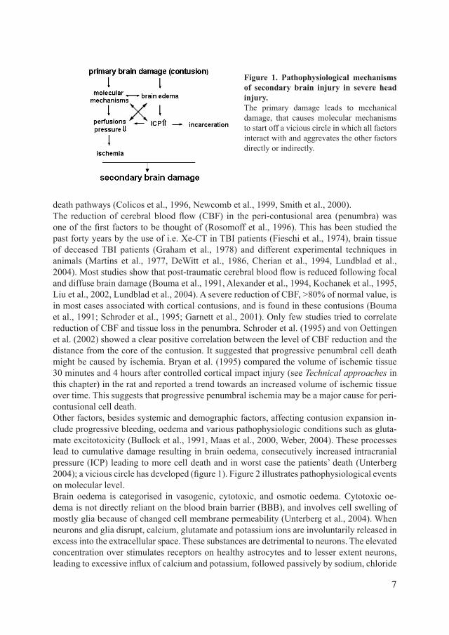
�
death pathways (Colicos et al., 1996, Newcomb et al., 1999, Smith et al., 2000). The reductionof cerebral bloodflow (CBF) in theperi-contusional area (penumbra)wasoneofthefirstfactorstobethoughtof(Rosomoffetal.,1996).Thishasbeenstudiedthepast forty years by the use of i.e. Xe-CT in TBI patients (Fieschi et al., 1974), brain tissue of deceased TBI patients (Graham et al., 1978) and different experimental techniques in animals (Martins et al., 1977, DeWitt et al., 1986, Cherian et al., 1994, Lundblad et al., 2004).Moststudiesshowthatpost-traumaticcerebralbloodflowisreducedfollowingfocaland diffuse brain damage (Bouma et al., 1991, Alexander et al., 1994, Kochanek et al., 1995, Liu et al., 2002, Lundblad et al., 2004). A severe reduction of CBF, >80% of normal value, is in most cases associated with cortical contusions, and is found in these contusions (Bouma et al., 1991; Schroder et al., 1995; Garnett et al., 2001). Only few studies tried to correlate reduction of CBF and tissue loss in the penumbra. Schroder et al. (1995) and von Oettingen et al. (2002) showed a clear positive correlation between the level of CBF reduction and the distance from the core of the contusion. It suggested that progressive penumbral cell death might be caused by ischemia. Bryan et al. (1995) compared the volume of ischemic tissue 30 minutes and 4 hours after controlled cortical impact injury (see Technicalapproachesin this chapter) in the rat and reported a trend towards an increased volume of ischemic tissue over time. This suggests that progressive penumbral ischemia may be a major cause for peri-contusional cell death. Other factors, besides systemic and demographic factors, affecting contusion expansion in-clude progressive bleeding, oedema and various pathophysiologic conditions such as gluta-mate excitotoxicity (Bullock et al., 1991, Maas et al., 2000, Weber, 2004). These processes lead to cumulative damage resulting in brain oedema, consecutively increased intracranial pressure (ICP) leading to more cell death and in worst case the patients’ death (Unterberg 2004);aviciouscirclehasdeveloped(figure1).Figure2illustratespathophysiologicaleventson molecular level.Brain oedema is categorised in vasogenic, cytotoxic, and osmotic oedema. Cytotoxic oe-dema is not directly reliant on the blood brain barrier (BBB), and involves cell swelling of mostly glia because of changed cell membrane permeability (Unterberg et al., 2004). When neurons and glia disrupt, calcium, glutamate and potassium ions are involuntarily released in excess into the extracellular space. These substances are detrimental to neurons. The elevated concentration over stimulates receptors on healthy astrocytes and to lesser extent neurons, leadingtoexcessiveinfluxofcalciumandpotassium,followedpassivelybysodium,chloride
Figure 1. Pathophysiological mechanisms of secondary brain injury in severe head injury.The primary damage leads to mechanical damage, that causes molecular mechanisms to start off a vicious circle in which all factors interact with and aggrevates the other factors directly or indirectly.

8
and water (Weber 1999). As a result of this osmotic load water will accumulate intracellular. Simultaneously degradation enzymes causing organelle swelling, plasma membrane swell-ing (Choi et al., 1995), necrosis (Wyllie et al., 1980) and apoptosis (Zipfel et al., 2000). The elevated intracellular calcium also leads to changes in the mitochondria (Muizelaar 1989, Sutton et al., 1994, Katayama et al., 1998), leading to release of free radicals. Free radicals are always present in organisms as a normal by-product of oxidative metabolism within mi-tochondria (Kontos, 1985, Siesjo, 1992a (II)). Yet, following trauma free radical activity is increased (Hall and Braughler, 1989, Siesjo, 1992 a,b), leading to cellular damage by peroxi-dation of proteins, DNA and lipid membranes and thereby necrosis and apoptosis (Kermer et al., 1999, Leker and Shohami, 2002). Thereby several reactive oxygen species (ROS) are generated excessively, such as superoxide and hydroxyl radicals (Leker and Shohami, 2002). Nitric oxide (NO) appears to be increased as well, which is generated by the enzyme nitric oxide synthase (NOS) (Samdani et al., 1997). NO contributes to several important physiological processes, such as cerebral vasodilation and neurotransmission (Dawson and Dawson, 1996). However, excess NO can interact with the ROS superoxide radical, resulting in the generation of peroxynitrite. Peroxynitrite is postulated to be one of the most damaging free radical species (Dawson, 1999, Liu et al., 2002). The idea exists that the application of a NOS-inhibitor protects cells from peroxidation.Osmotic oedema develops almost immediately after injury by high osmolarity in the necrotic tissue caused by increased substances in the extracellular space leading to increased intercel-lular water (Katayama and Kawamata, 2003). Vasogenic oedema results from disruption of the BBB, an increased permeability of the capillary endothelial cells for proteins caused by
Figure 2. Molecular events in TBI.Hypothetical scheme to depict post-traumatic events, with opening of ion-channels and uptake of po-tassium by astrocytes which jeopardizes the microcirculation. From: Head injury, editors: Reilly and Bullock (2005).

9
ischemiaor traumaitself,causing increasedextracellularfluidaswell (Betzetal.,1989).Katayama et al. (1998) emphasize the importance of the osmotic gradients caused by the pro-cesses described above within contusions, causing oedema, and thereby increased ICP. ICP raisesthemostwithinthefirst24hoursafterinjuryinhuman(Boumaetal.,1991,1992)andanimal studies (Zweckberger et al., 2003). In these 24 hours the brain is most vulnerable and susceptible to secondary injury insults by i.e. hypoperfusion, leading to progressive cell death (Jenkins, 1989). Ischemic changes have been demonstrated in over 90% of patients who died from head injury (Graham et al., 1978, Ross et al., 1993). As mentioned earlier decreased CBF is related to worse outcome (Graham et al., 1978, Jaggi et al., 1991, Garnett et al., 2001, Hlatky et al., 2004). The exact role of CBF, hypoperfusion and ischemia for secondary brain damage following mechanical injury is not clear. In different experimental settings the effect of CBF and ischemia was investigated in relation to cell death or tissue loss (see Chapter 3).
Current therapeutic optionsPrevention is the best cure. In the past 30 years preventive measures have resulted in a great reduction ofTBI inwestern societies.Themajor profitwasmade in road trafficTBI byseveral changes; i.e. road construction (Evans, 1991, Kraus, 1993), speed control (Brindle, 1992), motor vehicle design, airbags (Zador and Ciccone, 1991, Stewart et al., 2003), seat belts (Orsay 1990, Bradbury and Robertson 1993) motorcycle helmets (Sosin et al., 1990, McSwain and Belles, 1990, Kraus et al., 1994, Chiu et al., 2007), and, especially in the youngervictims,bicyclehelmets(Rivaraetal.,1994,Sosinetal.,1996,Shafietal.,1998,Kopjar et al., 2000, Thompson et al., 2000, Wesson et al., 2000). Occupational hazards have been prevented by strict regulations on the use of tools and wearing helmets at construction sites. Unfortunately, the prevention of falling of elderly from stairs or in the shower has not been accomplished yet. Often, TBI is accompanied by injuries of other parts of the body, leading to hypovolemic shock and multiple organ failure. The Advanced Trauma Life Support (ATLS) (Vestrup et al., 1988, Bell et al., 1999) has standardised the care of trauma patients as much as pos-sible. The implementation of ATLS and other changes within emergency medical systems (i.e. implementation of helicopters) have improved treatment (Klemen and Grmec, 2006, Chiu et al., 2007). However, injured patients in rural areas have worse outcome than patients closer to advanced medical care (Wald et al., 1993, Chiu et al., 2007, Tiesman et al., 2007). These patients cannot always reach optimal care within reasonable time. The Brain Trauma Foundation manages guidelines for the acute management of TBI patients from accident site to hospital discharge. First priority is to assess, stabilise and treat according to the basic resuscitation protocols, starting airway, breathing and circulation. Thereafter consciousness is assessed by the GCS. Patients with moderate to severe TBI (9-13 and 3-8, resp.) need to be transferred to a trauma centre. The appointed trauma centre should have:
24-hour CT scanning availability24-hour available operating room and neurosurgical care.ability to measure and treat intracranial pressure (ICP) promptly.
Pre-hospital the patient needs to be re-assessed every 5 minutes, maintaining an O2 satura-tion at >90% or PaO2 60 mmHg, systolic blood pressure > 90mmHg. These values should be maintained both pre-hospital and in-hospital.
•••

10
In-hospital ICP needs to be assessed, when GCS is 3-8, and an abnormal CT-scan (haemato-ma, contusions, swelling, herniations, or compressed basal cisterns). In patients aged over 40 years, uni- or bilateral motor posturing, or hypotension ICP measuring should be considered. The most accurate and cost effective way of ICP measuring is intraventricular, though other methods are available and secure as well. When ICP raises above 20-25 mmHg treatment should be started. To assess the presence of ischemia the cerebral perfusion pressure (CPP) is calculated (mean arterial pressure (MAP) – ICP). The threshold for CPP lays around 50-60 mmHg. When increased ICP is most likely to be caused by an intracranial haematoma or contusion operative treatment should be considered. When increased ICP is caused by other, non-operable, reasons, barbiturates or mannitol (hyperosmolar therapy) should be consid-ered. These options should only be used for severe increased ICP (signs of transtentorial herniation), special care is needed to maintain systolic blood pressure > 90 mmHg. Unfortunately the above-summarised guidelines often fail in treating raised ICP. Decompres-sive craniectomy (DC) is an old treatment that has been revived the past couple of years. DC can be considered when ICP is raised and is uncontrollable with conservative therapy. By opening the skull ICP reduces almost immediately; pathophysiological pathways leading to ischemia,apoptosisandnecrosisarediscontinued.Cerebralbloodflow(CBF)islikelytobealteredbyDCafterTBI.DCafterTBIwasfirstdescribedalreadyin1901byKocherandwasinvestigated in the 1960s&70s (Kjellberg and Prieto, 1971, Ransohoff, 1971, Venes and Col-lins 1975, Cooper 1976, Britt and Hamilton, 1978). The suggestion that DC would worsen brain swelling and induce herniation through the DC opening, made many neurosurgeons abandon this option of therapy. In the late 1980s new criteria for when and how to perform DC were introduced among other therapeutic strategies to reduce raised ICP (Alexander et al., 1987, Gaab et al., 1990, Marshall, 2000, Naredi et al., 1998,2001, Grande et al., 2002). Experimentalandclinicaldatashowpromisingbeneficialeffects(Polinetal.,1997,2003,Guerraetal.,1999,Yooetal.,1999,Tayloretal.,2001,Whitfieldetal.,2001,Hejazietal.,2002, Kontopoulos et al., 2002, Schneider et al., 2002, Simma et al., 2002, Albanese et al., 2003, Figaji et al., 2003, Zweckberger et al., 2003, Messing-Junger et al., 2003). DC, when performed early could be effective (Skoglund et al., 2006). In the paediatric population DC has been accepted as valid ‘second tier’ therapy for raised intracranial pressure (Rutigliano et al., 2006, Jagannathan et al., 2007). This is not (yet) the case in adult TBI patients. Clinics throughout Germany and the Netherlands seem to use different protocols and criteria (per-sonalcommunication).Instrokeearlydecompressivesurgeryhasbeenproventobebenefi-cial in a pooled analysis of three randomised controlled trials (RCTs) (Vahedi et al., 2007). Rosenfeld (2006) advocates early large, generous, craniectomy in war victims of cranial blast injuries. Prospective RCTs of early DC after TBI in adults injured outside war zones are currently running worldwide (RESCUEicp and DECRA) hopefully giving a last conclusive answer on clinical outcome. Why exactly DC works, besides taking off the pressure of the rigid box, is unknown. The hypothesis rose that CBF is improved after DC and thereby affect different pathophysiological pathways leading to secondary brain damage (see Chapter 5).
Technical ApproachesGreat effort has been made the past decades to investigate pathophysiological pathways and to test possible treatment strategies. As TBI itself and subsequent (patho)physiological pro-cessesoccurrapidlyaftereachother,itisdifficulttostudytheseprocessesinhumanbeings,let alone on a test dummy. The development of imaging by computer tomography (CT) and
11
magnetic resonance imaging (MRI) must be mentioned in the development of knowledge in science in general, but certainly in TBI research. CT enables a glance into the brain quickly after trauma and thereby the ability to diagnose rapidly and non-invasively (Marshall et al., 1992). Thereafter a more targeted treatment can be started. Images of both post-mortem and CT/MRI investigations have shown that there is great heterogeneity between patients and mechanisms of injury. This has led to different treatment protocols for i.e. diffuse injury versus brain contusions (Brain Trauma Foundation 2007 updated guidelines). However, not everything can be investigated that way. Animal models are able to model responses of body and brain after TBI. Therefore many studies have been and still are continued in the labora-tory invitro (in cell cultures) and invivo (laboratory animals) to study mechanisms of reac-tions to TBI from RNA level to systemic responses alongside post-mortem and clinical inves-tigations. Several methods are used to image brains (incl. pathophysiological mechanisms), brain structures, cells, and even the inside of cells by pathologic anatomy approaches (i.e. immunohistochemistry, immunocytochemistry), CT and MRI. Autoradiography, Laser Dop-plerandfMRIaremethodsusedtomeasurebloodflowormetabolismofglucoseindirectly.Metabolism and extracellular conditions can (in)directly be measured by microdialysis.
InvitroIn invitro studies cultured cells are utilized. For this thesis primary (embryonic) mouse cul-turesweregrownona silasticmembrane that,whenafluent layerhasbeen formedafter9-12 days invitro, can be mechanically injured by the invitro 94A Cell Injury Controller (CIC) (Bioengineering Facility, Virginia Commonwealth University, Richmond, VA, USA; seefigure3).Ellisetal.firstdescribedthisdevicein1995(Ellisetal.1995).Severalcellsinthefluentlayeraredisrupteddirectlybythemechanicalimpactofstretching(necrosis).Some of the remaining cells go into apoptosis because of extracellular changes; the human situation of primary and secondary damage alike. The amount of cell damage depends on the amount of stretch. This model is thought to represent the same mechanical impact on the brain when the brain is hit by physical impact or de-/acceleration, according to a mathemati-cal approach (Ljung et al., 1975; Schreiber et al., 1995). Cells can be dyed or stained after injury and looked at morphologically on the silastic membrane; i.e. by the use of propidium iodide and Hoechst, or Microtubulin Associated Protein 2 (MAP2) and 4’,6-Diamidine-2’-phenylindoldihydrochloride (DAPI) respectively. Substances released in the cell media can be determined by several techniques.
InvivoSeveral invivo models were designed to mimic a more realistic setting. In the 1960s research-ers started to develop experimental invivo head injury models (Terao 1963, Unterharnscheidt 1963, Ommaya et al., 1964). A precursor of an experimental TBI model that is still being used today, thoughwithsomeadjustments,wasfirstdescribed in1976bySullivanetal.:the Fluid Percussion Injury (FPI) model (Sullivan et al., 1976). This model mimics diffuse injury together with intraparenchymal haemorrhage or subarachnoid haemorrhage. It was used on opened or closed skulls. The use of the FPI model on closed skulls had various practical problems. On the other hand, the Closed Head Injury model of Marmarou et al. was able to induce more severe diffuse injury than the FPI model (Marmarou et al., 1994). Chen et al. (1996) made it possible to use a model alike in mice. As mice are to date the only rodentsthatcanbegeneticallyenhancedtheenhancementofthismodelenabledanewfield
12
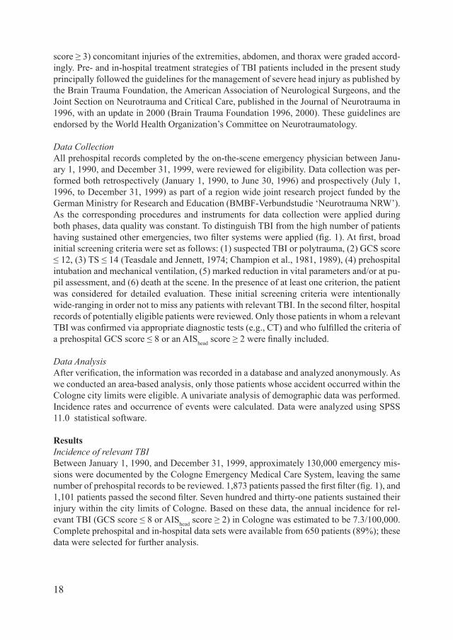
Figure 4. Control cortical impact model. The exact depth and speed of impact are set before placement of the mouse. The head of the anaes-thetisedmouseisfixedinastereotacticframe.Acraniotomyismadelocatedasshowninfigure5.Apneumatic pressure causes the impactor to move with the set speed directly onto the dura, a contusion is made. The severity can be regulated by alteration of depth and/or speed.
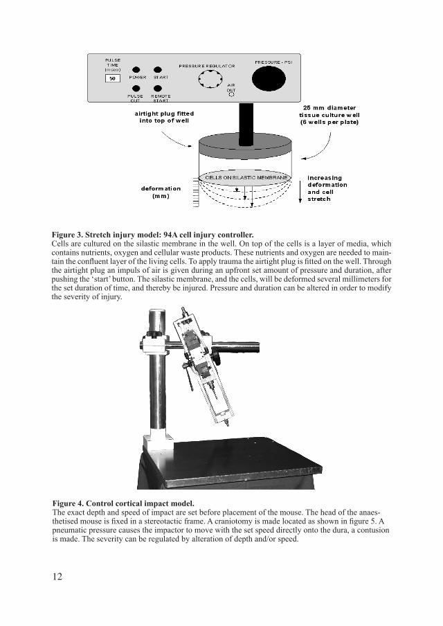
Figure 3. Stretch injury model: 94A cell injury controller. Cells are cultured on the silastic membrane in the well. On top of the cells is a layer of media, which contains nutrients, oxygen and cellular waste products. These nutrients and oxygen are needed to main-taintheconfluentlayerofthelivingcells.Toapplytraumatheairtightplugisfittedonthewell.Throughthe airtight plug an impuls of air is given during an upfront set amount of pressure and duration, after pushing the ‘start’ button. The silastic membrane, and the cells, will be deformed several millimeters for the set duration of time, and thereby be injured. Pressure and duration can be altered in order to modify the severity of injury.

13
of studies in closed skull experimental TBI as well as in open models. Lighthall et al. of the General Motors Research Laboratories introduced the Controlled Cortical Impact model in 1988(Lighthalletal.,1988).TheCCImodelisthemodelusedinthisthesis(figure4).Micehavetobecraniectomisedasdrawninfigure5.Thereaftermicereceivemainlyfocalinjuryapplied by a pneumatic impact tip (impactor), leading to a contusion and/or haemorrhage. Regulating depth, width and/or speed of the impactor can vary the injury severity. Acutely the most important aspect is the expansion of the primary contusion by several factors within thefirst24hours.ICP,CBF,BBB,oedemaandotherparameterscanbemeasuredatdifferenttime points after injury. The location of ICP and CBF by Laser Doppler measurements is also showninfigure5.Aftertheapplicationofinjurytheskullcanbeclosedagainbytissueglue,or left open (craniectomy instead of craniotomy) to investigate decompressive effects.
Scope of thesisThe overall aim of this thesis is to investigate the importance of ischemia in traumatic brain injury from different perspectives, ie. epidemiology, pathophysiology and therapeutic op-tions, hypothesizing that the role of ischemia is essential in secondary damage after TBI. Epidemiology, pathophysiology and therapeutic strategies will be discussed in the following chapters. In order to be able to examine an issue in depth, the issue should be clear. Epidemiology is the start of every medical query when searching for pathophysiological mechanisms and possibletherapeuticstrategies.Manyfactorsmightinfluence(patho)physiologicalconditionsand outcome. Some might be treatable, some might not. Chapter 2 describes the role of hy-potensionandhypoxia,whichareboththoughttoinfluenceCBF.Italsodescribesincidence,outcome and other potential predictive factors of TBI.Pathophysiology of TBI has been investigated to large extent the past decades, but certainly not everything is clear. Ischemia is one issue that is under constant investigation. Every year more pieces of the puzzle are put on the right spot. Chapter 3 considers the role of ischemia invitro and invivo, as to whether ischemia, as supposed, kills more cells when superimposed after mechanical injury invitro. The possible protective role of free radical scavengers in reducing secondary damage invitro is evaluated as well invitro. Invivo CBF is measured by autoradiography in mice, leading to a good comparison of CBF in and around the contu-sion.And lastly, by understanding further pathophysiological conditions and pathways future ther-apyisthefinalgoal.Asdescribedabove,manytherapeuticoptionshavebeeninvestigated,but not found effective. Decompressive craniectomy (DC) is a possible treatment currently under investigation. DC and correlations with pathophysiological factors, e.g. CBF, were investigated experimentally and shown and described in chapter 4.
Figure 5. Schematic drawing of a mouse skull. Placement of the following devices are depicted:
craniectomy (light grey),trauma impact,intracranial pressure (ICP) (through the skull),Laser Doppler (LD) (on the skull).
••••


15
CHAPTER 2. EPIDEMIOLOGY. A) Incidence and Outcome of Traumatic Brain injury in an Urban Area in Western Europe over 10 years
AbstractIntroduction: Valid epidemiological data on incidence and outcome of traumatic brain in-jury (TBI) show great variability. A study on incidence, severity and outcome of TBI was conducted in an urban area of one million inhabitants. MaterialsandMethods: 130,000 pre-hospital emergencies were screened for TBI. Inclusion criteria: Glasgow Coma Scale (GCS) score≤8and/orAbbreviatedInjuryScaleforheadinjuries(AIShead)score≥2withconfirmedTBI via appropriate diagnostics. Results: Annual incidence was 7.3/100,000. Overall mortal-ity rate was 45.8%: 182 (28%) were prehospital deaths, 116 (17.8%) patients died in hospital. Twohundredandfourteenof352(60.8%)survivingpatientsweresufficientlyrehabilitatedat discharge [Glasgow Outcome Scale (GOS) score = 1], but 138 patients (39.2%) survived withpersistingdeficits.GOSwasassociatedwithinitialGCSandAIShead. Conclusion: The incidence of TBI was lower compared to the literature. The overall mortality was high, espe-cially prehospital and early in-hospital mortality rates.
IntroductionTraumatic brain injury (TBI) is the leading cause of death and disability world-wide. In the United States an estimated number of 1.6 million people sustain TBI each year accounting for 52,000 deaths and 80,000 patients suffering from permanent neurological impairment (Sosin et al., 1996, Bruns and Hauser, 2003). TBI represents a highly relevant medical and socioeconomic burden for modern societies (Murray and Lopez, 1997, Ghajar, 2000).Despite its clinical relevance, valid epidemiological data on incidence, severity and out-come of TBI from Europe, in particular from Germany, are scarce and show great variation (Hartwig et al., 1993, Jennett, 1996; Lehr et al., 1997). A major problem with epidemiologi-calstudiesonTBIisstillassociatedwithinconsistenciesinTBIdefinitionandclassificationof injury severity (Jennett, 1996, Engberg, 1995, Kraus and McArthur, 1996, Bouillon et al., 1997, Fearnside and Simpson, 1997, Ingebrigtsen et al., 1998). Subsequently, the true impactofthisinjurywithrespecttoitsmagnitudeaswellastheeffectivenessandefficiencyoftherapeuticconceptsremainsdifficulttoestimate(Ghajaretal.,1995;Prough,andLang,1997; Tolias and Bullock, 2004).The value of a functioning emergency medical system for TBI patients has frequently been emphasized (Roy, 1987, Colohan et al., 1989, Sampalis et al., 1995, Mullins et al., 1996, Marshall, 2000). Major improvements in outcome and mortality can only be achieved by preventive strategies, early recognition of complications and stabilization of patients (for review see guidelines of the Brain Trauma Task Force and the Brain Trauma Foundation (BrainTraumaFoundation,2000).However,optimalcareofTBIpatientsrequiressufficientresourceswithrespecttologistics,management,organizationandfinances.Validdataonin-cidence, severity, and outcome are essential prerequisites to adequately consider and allocate these resources, and thus to improve emergency medical care (Jennett, 1996). We therefore conducted a 10 year epidemiological study to provide representative data on the significanceofTBIontheincidence,injuryseverity,andoutcomeinawesternEuropeanur-bancenterofonemillioninhabitants,namelyCologne(Germany),usingstrictlydefinedstan-

1�
dardized scoring systems for inclusion, injury severity and outcome at hospital discharge.
Materials and MethodsThe design of the present study has been previously published by Bouillon and colleagues (Bouillonetal.,1999).Briefly,certainconditionsencouragedustoinitiatethepresentepide-miological study in Cologne (Germany). First of all, the city of Cologne has a population of approximately one million inhabitants which meets the requirements of an urban area. Sub-sequently, Colognes pre-hospital emergency medical care system has a central organization in which all severely injured patients are seen by experienced physicians at the injury scene. Since 1987, all emergencies are prospectively documented by the physician involved on a standardized pre-hospital record (Bouillon et al., 1990). The central organization as well as the prospective documentation of all relevant data using standardized documentation sheets allowedtheidentificationofpotentiallybraininjuredpatientsatthesceneandensuredareli-able collection of all the necessary data for the study.
TheCologneEmergencyMedicalCareSystemThe treatment of trauma patients including TBI begins at the site of injury. The emergen-cy call reaches the central Cologne Emergency Dispatch Center, that coordinates medical emergencies. Emergency physicians are dispatched if the patient’s vital functions are pos-sibly impaired or endangered. Following stabilization at the scene by the emergency physi-cian patients are transferred to the most appropriate hospital that meets the diagnostic and therapeutic needs according to suspected injuries. In case of suspicion of TBI the patient is routinely admitted to one of the two designated trauma centres of the region with 24h avail-ability of computed tomography (CT) and neurosurgical care. These two centres are level I trauma centers according to the criteria of the American College of Surgeons (ACS). During the 10 year study period (January 1, 1990 – December 31, 1999), more than 100 emergency physicians (70% surgeons, 20% anesthesiologists, 10% others) and 1000 paramedics served thisemergencycare.Onehelicopter,fivephysician-staffedemergencyvehicles(NEF),and23 paramedic-only-staffed vehicles (RTW) cover a core area of 405 km2 with an average populationofapproximatelyonemillionpeople.ThefiveNEFsandthehelicopterservedapproximately 13,000 and 1,000 emergency missions per year, respectively. The Cologne Emergency Medical Care System works 24/7 a week all year round on a rendez-vous-system basis. The responsibility for further treatment was taken by the staff on duty in each trauma center.
DocumentationBased on preliminary studies, a standardized procedure has been implemented to precisely document the prehospital course of each patient beginning at the site of injury. Since Janu-ary 1, 1987, a standardized documentation sheet is mandatory for each emergency physician on duty; meanwhile, it has reached a high level of acceptance with a completion rate of > 90%. This documentation sheet is similar to the Germany-wide well-accepted DIVI emer-gency documentation sheet (Moecke et al., 1994) and includes two scoring systems: (1) the Glasgow Coma Scale (GCS) (Teasdale and Jennett, 1974) to assess the neurological state, and (2) the Trauma Score (TS) (Champion et al., 1981; 1989) to estimate the physiological response to severe trauma. It contains information on the patient’s age, gender, mechanism of injury, initial vital status, suspected diagnosis, prehospital monitoring and care, triage, and

1�
early time course after the injury (Bouillon et al., 1997). It further provides space to docu-mentspecificmeasuresincaseofcomplications.Thisprocedureensuresthatdatacollectionbegins as early as possible. One prehospital documentation sheet was added to the patient’s fileswhile a copywas archived at the emergency vehicle’s base.The latterwas used inthisstudyfortheidentificationofTBIcases.Informationaboutthehospitalcourse,includ-ing diagnostics, interventions, length of intubation, length of stay and outcome at discharge [mortality, Glasgow Outcome Scale (GOS)] (Jennett and Bond, 1975), was extracted from hospital records. All prehospital and hospital records were reviewed by two independent research assistants.
AssessmentofrelevanttraumaticbraininjuryTBI was assessed and graded using the GCS and the Abbreviated Injury Scale for head inju-ries (AIShead). A GCS score of 13–15 is considered as mild TBI, which means in most cases a concussion with full neurological recovery, although many patients present with short-term memoryandconcentrationdeficits(Rimeletal.,1981).PatientswithaGCSscoreof9–12,moderateTBI,are lethargicandstuporous.Comatose(GCSscore≤8)patients,unabletoopentheireyesortofollowspecificcommands,areregardedashavingsevereTBI(Teasdaleand Jennett, 1975). The AIShead is another commonly used instrument for the categorization of injury severity based on anatomical features using a 6-grade scale system (Walder et al., 1995). AISheadscores≥2areconsideredmoderatewhilescores≥3areseveretolife-threaten-ing. The AIS is further used to calculate other scoring systems, for example, the Injury Sever-ity Score (Osler et al., 1997). In the present study, relevant TBI was assumed when the initial GCSscorewas≤8ortheAISheadscorewas≥2.Relevant(AISscore≥2)andsevere(AIS
Figure 1. Filter criteria to identify patients with relevant TBI.
Figure 1
Cologne Area (1 million inhabitants)
1990 – 1999 130.000 emergency calls
Inclusion criteria
Suspected TBI or polytrauma GCS or TS Intubation and mechanical ventilation Vital parameters Alterations in pupilo-function Death at accident site
Review of patient´s hospital records/ data files for relevant TBI according to
appropriate diagnostic tests (e.g. CT scan) and verification of injury severity according
to GCS 8 and/or AIShead 2
731 patients with relevant TBI(650/731 patients with complete data sets)
1873 patients with potentially TBI
1st filter
2nd filter
18
score≥3)concomitantinjuriesoftheextremities,abdomen,andthoraxweregradedaccord-ingly. Pre- and in-hospital treatment strategies of TBI patients included in the present study principally followed the guidelines for the management of severe head injury as published by the Brain Trauma Foundation, the American Association of Neurological Surgeons, and the Joint Section on Neurotrauma and Critical Care, published in the Journal of Neurotrauma in 1996, with an update in 2000 (Brain Trauma Foundation 1996, 2000). These guidelines are endorsed by the World Health Organization’s Committee on Neurotraumatology.
DataCollectionAll prehospital records completed by the on-the-scene emergency physician between Janu-ary 1, 1990, and December 31, 1999, were reviewed for eligibility. Data collection was per-formed both retrospectively (January 1, 1990, to June 30, 1996) and prospectively (July 1, 1996, to December 31, 1999) as part of a region wide joint research project funded by the German Ministry for Research and Education (BMBF-Verbundstudie ‘Neurotrauma NRW’). As the corresponding procedures and instruments for data collection were applied during both phases, data quality was constant. To distinguish TBI from the high number of patients havingsustainedotheremergencies,twofiltersystemswereapplied(fig.1).Atfirst,broadinitial screening criteria were set as follows: (1) suspected TBI or polytrauma, (2) GCS score ≤12,(3)TS≤14(TeasdaleandJennett,1974;Championetal.,1981,1989),(4)prehospitalintubation and mechanical ventilation, (5) marked reduction in vital parameters and/or at pu-pil assessment, and (6) death at the scene. In the presence of at least one criterion, the patient was considered for detailed evaluation. These initial screening criteria were intentionally wide-ranginginordernottomissanypatientswithrelevantTBI.Inthesecondfilter,hospitalrecords of potentially eligible patients were reviewed. Only those patients in whom a relevant TBIwasconfirmedviaappropriatediagnostictests(e.g.,CT)andwhofulfilledthecriteriaofaprehospitalGCSscore≤8oranAISheadscore≥2werefinallyincluded.
DataAnalysisAfterverification,theinformationwasrecordedinadatabaseandanalyzedanonymously.Aswe conducted an area-based analysis, only those patients whose accident occurred within the Cologne city limits were eligible. A univariate analysis of demographic data was performed. Incidence rates and occurrence of events were calculated. Data were analyzed using SPSS 11.0 statistical software.
ResultsIncidenceofrelevantTBIBetween January 1, 1990, and December 31, 1999, approximately 130,000 emergency mis-sions were documented by the Cologne Emergency Medical Care System, leaving the same numberofprehospitalrecordstobereviewed.1,873patientspassedthefirstfilter(fig.1),and1,101patientspassedthesecondfilter.Sevenhundredandthirty-onepatientssustainedtheirinjury within the city limits of Cologne. Based on these data, the annual incidence for rel-evantTBI(GCSscore≤8orAISheadscore≥2)inColognewasestimatedtobe7.3/100,000.Complete prehospital and in-hospital data sets were available from 650 patients (89%); these data were selected for further analysis.

19
PatientCharacteristicsThe majority of patients who sustained TBI was male (73%, n = 473) and the average age was40.3years.Adetaileddistributionofageisshowninfigure2.Themeanageinthesec-ond half of the study (1995–1999) was 2 years higher (p < 0.001; t test). The percentage of patientsover60yearsofagewas16%duringthefirstyearofthestudyversus28%duringthe last year of the study.
MechanismofInjuryIn 617 cases (94.9%), the type of injury was blunt, while 33 cases (5.1%) sustained a pen-etrating trauma. Eighty cases (12.3%) resulted from suicide attempts. The predominant cause ofinjurywastraffic-related(55.3%),followedbyhigh(>3m,22.8%)andlowfalls(<3m,12.2%).Amongthe trafficvictims(358/650=100%),142(39.6%)werepedestrians,115(32.1%) car or truck drivers, 63 (17.6%) cyclists, and 38 (10.7%) motorcyclists. There wasno change with respect to mechanism of injury over the 10-year study period.
ScoresTBI patients transfered to a hospital had a mean GCS score of 8 ± 4; those who died at the scene (n = 182; 28%) had a GCS score of 4 ± 1. Among the 444 patients sustaining a TBI graded AIShead≥2,104(23.5%)patientsweregradedAIShead= 2. The remaining 340 (76.5%) patients sustained a severe TBI as graded AIShead≥3.
ConcomitantInjuriesAmongpatientsinwhomdiagnosticscouldsufficientlybecompleteduponhospitaladmis-sion(n=451,preclinicaldeathsexcluded),asignificantnumbersustainedconcomitantin-
Figure 2. Distribution of age and gender among the study population (n = 650).
<10 10-19 20-29 30-39 40-49 50-59 60-69 70-79 80-89 90+
Age
0
20
40
60
80
100
120
140
Num
ber o
f pat
ient
s
FemalesMales

20
juries. As expected, concomitant injuries were observed most frequently in the extremities (37%; AISext.score≥2);20%of theseinjurieswereconsideredsevere(AISext.score≥3).Twenty-four percent sustained relevant thoracic injuries, of which 20% were severe. Rel-evant concomitant injuries of the abdomen were observed in 11%, of which 8% were severe. Overall, in more than half of the patients (52%), at least one relevant concomitant injury was observed. Thirty-four percent sustained at least one concomitant injury graded as severe (AISscore≥3).
LengthofIntubationandIntensiveCareUnit/HospitalStayTBI patients who were admitted to a hospital remained intubated and mechanically ventilated for 5.8 ± 10 days. They stayed in the intensive care unit for 8.9 ± 13 days, and the overall length of stay in the hospital was 18.2 ± 22 days.
MortalityandOutcomeThe overall mortality rate for the study population was 45.8% (n = 298). Prehospital deaths occurred in 182 patients (61%), whereas 116 patients (39%) died in hospital. This corre-sponds toanoverall in-hospitalmortality rateof24.8%.Asignificantnumberofpatientsdied within 48 h upon admission (n = 68; 23% of all deaths), while 26 (9%) died within days 3–7afteradmission.Theremaining22nonsurvivors(7%)diedlater(fig.3).Amongthe352survivingpatients,214(60%)weresufficientlyrehabilitatedaccordingtotheGOSscoreatdischarge,while138patients(40%)survivedbutwithpersistingdeficitsofvariousdegrees(fig.3).
DiscussionWe present data over a 10-year study period from an urban area of approximately one million inhabitants in western Europe on the incidence, injury severity, and outcome of patients sus-taining relevant TBI. 130,000 prehospital emergency protocols from the Cologne Emergency Medical Care System (Germany) were screened. The number of patients we included into the present study per year was consistent over the
Figure 3. Mortality and outcome from TBI (n = 650). (A)Nonsurvivorsareclassifiedaccordingtotime until death. (B) Survivors are presented according to the GOS. PVS = Persistent vegetative state.
Figure 3
A non-survivors B survivors
died
n=29845,8%
survived
n=35254,2%
0%
10%
20%
30%
40%
50%
60%
70%
80%
90%
100%
goodrecovery
n=214; 60%moderate disability
n=109; 31%
severe disabilityn=23; 7%
PVS n=6; 2%
> 7d n=22; 7%
48h - 7d n=26; 9%
<48h n=68; 23% preclinical
182; 61%n=

21
entirestudyperiod.Fifty-onepercentofcasesoccurredduringthefirst5yearsofthestudy(1990–1994). The annual incidence of relevant TBI for the Cologne area, a typical urban area in western Europe of one million inhabitants, was found to be 7.3/100,000 inhabitants. This rate is low compared to other reports. In the literature, the annual incidence rates for TBI range between 13.6 and 300/100,000 (Annegers et al., 1980, MacKenzie et al., 1989, Tiret et al., 1990, Annoni et al., 1991, Kraus, 1992, Hartwig et al., 1993, Lehr et al., 1997, Tate et al., 1998, Bruns and Hauser 2003). Besides a general decrease in incidence, one reason for thesediscrepanciesmayhavebeenaproblemofTBIdefinition(Engberg,1995,Fearnsideand Simpson, 1995, Jennett, 1996, Kraus and McArthur, 1996, Bouillon et al., 1997, Inge-brigtsenetal.,1998).Lackofconsensusoninjuryclassificationledtoconsiderablevariabil-ityindefininginclusioncriteriaandsubsequentlytodifficultiesincomparingstudyresults.Nevertheless,itappearsthataccordingtoourdefinition(GCSscore≤8and/orAIShead score ≥2), the incidenceof relevantTBI is lower thanexpected.For further studiesandbettercomparabilityofresults,theuseofcommoncriteriaandscoringsystemstopreciselydefineTBI is highly recommended. Tagliaferri et al. (2006) noted critical differences in methods employed in the past 20 years across several reports on the epidemiology of TBI in Europe resulting in considerable difficulties to reach a consensus on all epidemiologicalfindingsacross the 23 published European studies to date. In conclusion, these authors highly recom-mendedthedevelopmentofresearchguidelinestostandardizedefinitional,casefinding,anddata reporting parameters to help establish a more precise and hence more useful description of the epidemiology of TBI in Europe.Another point of discussion with respect to the lowincidence reported here is related to the fact that the present study only included relevant TBI that occurred inside the 405 km2 Co-logne area. It might be possible that a relatively large number of Cologne citizens sustained a relevant TBI outside the area and thus may have been missed in the present study. TBI sus-tained outside the urban area will probably represent the majority of high-velocity injuries.Conversely, noninhabitants of this area if injured within the Cologne city limits were includ-ed. Therefore, it needs to be emphasized that the authors of the present study did not follow the urban population of one million inhabitants but the incidence of relevant TBI within a typical western European urban area of one million inhabitants. TBIispredominantlytheinjuryofyoungmales.Ourdataconfirmpreviousreportsregard-ing gender and age (Farin et al., 2003). This can be mainly attributed to a higher level of risk tolerance. Considerable differences in patient characteristics and case management between variousgeographicalregions,reflectingvariationsinsocial,cultural,andorganizationalas-pects have been reported (Hukkelhoven et al., 2002). Considering the distribution of age within our study population, a second peak was noted for the group between 50 and 59 years of age, but far less pronounced as compared to the younger age groups. In most other epi-demiological studies on TBI, a second peak in the elderly, mostly due to falls, is observed (Sundstrom et al., 2007). As our approach within the given study was to analyze relevant TBI based upon prehospital emergency system documentation, minor to moderate falls in the elderly may have been missed. Withrespecttocauseofinjury,itwasstrikingthatonly55%ofTBIwererelatedtotrafficaccidents,whereas45%hadothercauses.Percentrangesof48–57%fortraffic-relatedTBIhavebeenpublishedpreviously(Krausetal.,1984,Sosinetal.,1989).Thisisofgreatsignifi-canceasTBItodatehasmostlybeenconsideredandthusinvestigatedinthecontextoftrafficvictims. It is likely that only about half of the affected population has thus been recognized

22
and studied in other investigations.The overall mortality rate of 45.8% reported in our study appears to be higher as compared to the literature (Foulkes et al., 1991, Chesnut et al., 1993, Winchell and Hoyt, 1997), although there are reports of similar (Tiret et al., 1990) or even higher (Annegers et al., 1980; Kraus 1992) overall mortality rates associated with TBI. One aspect may be related to our very thorough and strict data inclusion from the scene of the accident until the in-hospital phase. An important difference to many other studies is the inclusion of all prehospital deaths. The possibility exists that some prehospital deaths might have involved brain injury but that TBI was not the primary cause of death. In our study, the occurrence of relevant concomitant injurieswas52%.Thisisinaccordancewithpreviousfindings(Bouillonetal.,1992;Gena-relli et al., 1994; Meixensberger and Roosen, 1998). In-hospital mortality ranges between 20 and 30% according to the literature (MacKenzie et al., 1989, Tiret et al., 1990, Annoni et al., 1991, Tate et al., 1998). In the present study, a similar in-hospital mortality rate was noted (24.8%). In view of prehospital versus in-hospital mortality, we found a similar ratio to Tiret et al. (1990) who applied a comparable approach. In both studies, the prehospital mortality rate was higher than the in-hospital rate.Early mortality is an important matter since the majority of all deaths occur within 48 h fol-lowing the accident (Tiret et al., 1990, Lieberman et al., 2003). A well-functioning emergency system, in which the time course of prehospital care is limited to < 45 min, was accountable for the fact that a substantial number of severely brain-injured patients reached the hospi-tal alive. An organized emergency medical services system has been shown to substantially improve outcome following TBI (Colohan et al., 1987, Roy, 1987, Sampalis et al., 1995, Mullins et al., 1996, Watts et al., 2004, Klemen et al., 2006). We have to emphasize that the present study was undertaken in an urban area in western Europe with a highly developed emergency medicine system; in rural areas, the situation may be different due to a longer prehospital period. Combining the annual incidence of severe TBI (7.3/100,000) with the overall mortality rate of 45.8% from severe TBI reported here, an annual mortality rate of 3.3/100,000 inhabitants can be calculated. This rate is low compared to other reports. For example, Kraus et al. (1984) reported a mortality rate of 13.5/100,000 from the San Diego area (United States) while Sundstrom et al. (2007) reported median TBI death rates for the four Scandinavian countries to range between 9.5 and 21.2/100,000 per year. More recently, Tagliaferri et al. (2006) sys-tematically reviewed brain injury epidemiology from 23 European studies and reported an average mortality rate of approximately 15 per 100,000, but there were also considerable dif-ficultiestoreachconsensusonallepidemiologicalfindingsacrossthesestudiesduetocriticaldifferencesinmethodsandclassificationsemployed.ThelowannualmortalityfromrelevantTBI reported here may be attributed to our strict inclusion criteria as well as to the inclusionof brain injuries that had occurred inside the 405 km2 Cologne area only.Among surviving patients, the average length of intensive care unit and total in-hospital stay was 8.9 and 18.1 days, respectively. These data correspond to previous reports (Vitaz et al., 2003). More than half of the survivors were successfully rehabilitated at discharge, whereas one third remained disabled, while only a few patients remained in a persistent vegetative state (1.4%). Similar observations have previously been reported (Hoffmann et al., 2002, Pfenninger and Santi, 2002, Rudehill et al., 2002, Vitaz et al., 2003).Apart from age, mechanism of injury, and pathologies evolving from the impact (Gennarelli et al., 1982, Changaris et al., 1987, Mazaux et al., 1997, Asikainen et al., 1998, 1999a,b,
23
McCleary et al., 1998, Satz et al., 1998a,b, Geraldina et al., 2003, Mazzini et al., 2003, Cha-melian et al., 2004, Demetriades et al., 2004a,b, Levin et al., 2004), the initial GCS score on admission is a strong predictor for mortality and functional outcome (Changaris et al., 1987, Levin et al., 1991, Asikainen et al., 1996, 1999a, Hoffmann et al., 2002; Demetriades 2004a,b). For example, a GCS score of 3 due to head trauma is associated with a high mortal-ity rate (70%) (Liebermann et al., 2003); in our study, this was 92.5%. Comparable data exist for the AIS head (Walder et al., 1995; Demetriades et al., 2004a,b). Similarly, in the present study, both scores, the initial GCS and the AIS head scores, were associated with the GOS. Surprisingly, there is no good correlation between GCS and AIS head themselves (Demetria-des et al., 2004a), indicating that both measures will be useful in future studies conducted on therapeutic strategies and their outcome. Thepresentstudyisthefirstofitskindtoprovidesubstantialdataontheincidence,mag-nitude and outcome of TBI from an urban area of approximately one million inhabitants in western Europe over a 10-year period. The study was conducted in an environment with an organized emergency medical services system including designated trauma facilities, 24-hour availability of CT, full neurosurgical care including intracranial pressure monitoring, andexperiencedcriticalcaremanagement.Ourdataaresimilartopreviousfindingsinotherareas of the western industrialized society, except for the prehospital mortality rates, which is probably due to the inclusion criteria. The authors believe that these inclusion criteria demonstrate a more complete view on the epidemiology of TBI in urban areas. Still, optimiz-ing pre- and intrahospital care and management remains the major challenge in preventing secondary brain damage which affects outcome and prognosis.


25
CHAPTER 2. B) The Prognostic Value of Demographic Characteristics in Traumatic Brain Injury: Results from the IMPACT* Study
*International Mission on Prognosis and Analysis of Clinical Trials in TBI
AbstractOutcome following traumatic brain injury (TBI) is not only dependent on the nature and severity of injury and subsequent treatment, but also on constituent characteristics of in-jured individuals. We aimed to describe and quantify the relationship between demographic characteristics and six month outcome assessed by the Glasgow Outcome Scale (GOS) after TBI. Individual patient data on age (N=8719), gender (N=8720), race (N=5320) and educa-tion (N=2201) were extracted from eight therapeutic Phase III randomized clinical trials and three surveys in moderate or severe TBI, contained in the IMPACT database. The strength of prognostic effects was analyzed with binary and proportional odds regression analysis and expressed as an odds ratio. Age was analyzed as a continuous variable with spline functions, and the odds ratio calculated over the difference between the 75th and 25th percentiles. As-sociations with other predictors were explored. Increasing age was strongly related to poorer outcome (OR 2.14; 95% CI 2.00-2.28) in a continuous fashion that could be approximated by a linear function. No gender differences in outcome were found (OR: 1.01; CI 0.92-1.11), and exploratory analysis failed to show any gender/age interaction. The studies included predominantly Caucasians (83%); outcome in black patients was poorer relative to this group (OR 1.30; CI 1.09-1.56). This relationship was sustained on adjusted analyses, and requires further study into mediating factors. Higher levels of education were weakly related to a better outcome (OR: 0.70; CI 0.52-0.94). On multivariable analysis adjusting for age, motor score and pupils the prognostic effect of race and education were sustained. We conclude that outcome following TBI is dependent on age, race, to a lesser extent on education but not on gender.
IntroductionOutcome following traumatic brain injury (TBI) is dependent on many factors including demographic characteristics. Access to the IMPACT database (Marmarou et al., 2006) per-mitted us to analyze the prognostic value of age, gender, race and education. Age is one of the strongest and best documented predictors in TBI, with a large body of evidence demon-strating that outcome is poorer with increasing age. Uncertainty exists about the nature and shape of the association between age and outcome. Most studies categorize age or report on threshold values. (Hukkelhoven et al., 2003) investigated the relationship between age and outcome in more detail in a meta-analysis of the literature and of individual patient data. They found a continuous association and argued that reported threshold values might have been artefactual due to the typically skewed age distribution in TBI. Considerable preclinical and clinical interest exists in gender issues related to TBI, particularly in relation to hormonal influencesandpossibilitiesforhormonaltreatment.Thisinterestisinpartsociallymotivated(adequate representation, equal rights) and in part scientific.Anatomical dimorphismandhormonalinfluenceshavebeenimplicatedasfactorsinvolvedinpossiblegender-relateddif-ferences in outcome. Whether gender differences in outcome actually exist remains debat-

2�
able. (Groswasser et al., 1998) describe better overall responses to rehabilitation therapy in females when controlling for age and severity. Other authors (Klauber et al., 1981, Kraus and Nourjah, 1988) describe a higher mortality in females. A meta-analysis (Farace and Alves, 2000) found on average poorer outcome in females on primarily subjective outcome mea-sures, and concluded that adjusted outcome analysis with gender as a covariate should be conducted in clinical trials in TBI. This statement may be considered strong in relation to the uncertainties which exist on the effect of gender on outcome after TBI.Even less is known about associations between race and outcome after TBI. Within the USA, African Americans reportedly have a 35% higher incidence of TBI than Caucasians (Jager et al., 2000), but to our knowledge no studies have addressed the relationship between race and outcome assessed with the GOS, following severe and moderate TBI. Racial differences in outcome deserve consideration and are particularly relevant to clinical trials in traumatic brain injury, as metabolic pathways and consequently responses to injury and drug pharma-cokinetics may differ due to biological differences.A demographic factor that, unlike gender and race, is not intrinsic to the individual is the level of education. Educational level may be related to outcome after TBI due to interactions withcauseofinjuryanddifferencesinsocio-culturalandsocio-economicalstatusinfluencingaccess to health care facilities.The aim of our study was to describe and quantify the relationship between demograph-iccharacteristicsandsixmonthoutcomeafterTBI.Specificobjectiveswere todeterminewhether the use of threshold values for describing the relationship between age and outcome is appropriate, and to investigate whether any gender effects at different ages could be identi-fied.
MethodsPatientsanddatacollectionIndividual patient data on demographic characteristics were included in the eight randomized controlled trials and three unselected prospective surveys assembled in the IMPACT* database (Marmarou et al., 2006). We selected patients with complete data on outcome (N=8721). In six studies the actual date of birth was recorded, permitting calculation of age. In four studies (UK4, EBIC, HIT I, HIT II) age was recorded in years. For the purposes of analysis we calculated age in years, rounded to whole numbers for all studies. Gender was recorded inallstudiesasabinaryvariable.DataonracewereavailableinfiveRCTs(TIUS,TINT,PEG, SKB, SAP) and in one of the prospective series (TCDB). All studies recorded the main differentiation: Caucasian/ Black/Asian/other. Some studies included differentiation of the category “other”, for instance into American Indian or Alaskan. This differentiation was not included in our analysis. Data on education were available in three studies (TIUS, TINT and TCDB). The codings for education differed between the Tirilazad trials and the TCDB. We recoded education into the categories: zero to 8 years, 9 to 12 years, 13 years or more. The primary endpoint for prognostic analysis was the Glasgow Outcome Scale (GOS) as recorded in the studies. If the 6 month GOS was missing we imputed the three month GOS (N=1613, including 1510 patients from the PEGSOD trial).
StatisticalanalysisDescriptive analyses included the calculation of the median and interquartile ranges for age, and of the male/female ratio per study. The prevalence of racial distribution and educational

2�
categories was derived per study. The relationship between demographic characteristics and outcomewasfirstanalyzedwithcrosstabulation.Theshapeoftherelationshipofagetoout-come was further examined with restricted spline functions. These functions are smooth and flexible,allowingforanadequatedescriptionofnonlinearrelationships.Logistic regression models were applied to quantify the predictive strength of the demo-graphic characteristics. In these regression models the GOS was dichotomized in four dif-ferent ways: good recovery vs. less than good recovery, favorable outcome vs unfavorable outcome, conscious survival vs. death/vegetative state and survival vs. death. We also con-sideredanordinalanalysiswithproportionaloddsmethodologywhichreflectsprognosticeffects across the various GOS categories (McHugh et al, 2006). Data from all of the studies were pooled using a random effects model. The strength of the associations was expressed usingoddsratioswith95%confidenceintervals.Exploratory analyses of clinically meaningful associations with other predictors were per-formed, and included adjusted analysis for the association between race and outcome.Finally, multivariate analysis was performed, adjusting for the three main clinical predictors: age, motor score and pupillary reactivity. This analysis was restricted to patients > 14 years of age.
ResultsDescriptiveanalysisThe availability and distribution of demographic characteristics within and across studies is summarized in Tables 1 and 2. The median age between studies varied from 26 to 37 and was in general higher in the European studies compared to the primarily North American studies.
a) This study included 162 children; on exclusion of patients <14 years of age, the median age is 36 (IQR: 22–55), b) the summary data presented as overall are simple totals rather than any more sophis-ticated pooled estimate. IQR, Interquartile range.
Table 1. Descriptive statistics for age and gender
Table 1: Descriptive statistics for age and gender
Age (N=8719) Gender (N=8720)
Study N N (%) available
Median IQR N (%) Available
% Male
TCDB 604 604 (100) 26 (21-40) 604 (100) 77UK4* 986 986 (100) 29 (17-51) 986 (100) 75HIT I 350 350 (100) 34 (21-47) 350 (100) 84HIT II 819 819 (100) 33 (22-49) 819 (100) 77TIUS 1042 1041 (100) 30 (23-41) 1041 (100) 78TINT 1121 1121 (100) 30 (21-45) 1121 (100) 76PEG 1510 1510 (100) 27 (20-38) 1510 (100) 77EBIC 835 834 (100) 37 (23-58) 834 (100) 75SLIN 409 409 (100) 28 (21-43) 409 (100) 78SKB 126 126 (100) 27 (20-39) 126 (100) 76SAP 919 919 (100) 32 (23-47) 919 (100) 80
Overall^ 8721 100 30 (21-45) 100 77
IQR: Inter Quartile range * this study included 162 children; on exclusion of patients < 14 years of age the median age is 36 (IQR: 22-55) ^ the summary data presented as overall are simple totals rather than any more sophisticated pooled estimate.
Table 2: Descriptive statistics for race and education
Race (N=5320) Education in years (N=2201)
Study N N (%) available
CaucasianN (%)
BlackN (%)
Asian N (%)
Other N (%)
N (%) available <9 9-12 >12
TCDB 604 604 (100) 507 (84) 81 (13) 13 (2) 3 (<1) 381 (63) 19 (5) 271 (71) 91 (24) TIUS 1042 1040 (100) 687 (66) 149 (14) 35 (3) 169 (16) 872 (84) 59 (7) 525 (60) 288 (33) TINT 1121 1121 (100) 1071 (96) 12 (1) 29 (3) 9 (<1) 948 (85) 249 (26) 423 (45) 276 (29) PEG 1510 1510 (100) 1170 (77) 179 (12) 29 (2) 132 (9) NA - - -SKB 126 126 (100) 93 (74) 17 (13) 0 (0) 16 ( 13) NA - - -SAP 919 919 (100) 903 (98) 6 (<1) 10 (1) 0 (0) NA - - -
28
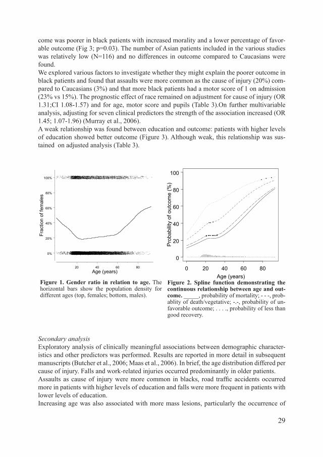
We found a clear male excess in the overall population (average 77% male; range 75-84%). However, this excess was mainly due to the contribution of younger patients and the male/fe-male ratio decreased to one above age 65 (Figure 1). Six studies reported the distribution of race, and within these studies data were available for virtually 100% of patients. MostpatientswereclassifiedasCaucasian(66-98%),withhigherpercentagesbeingobservedin the primarily European studies (TINT/SAP: 96-98%). Data on education were available for a total of 2201 patients over three datasets (TCDB, TIUS, TINT), but were missing in a considerable proportion of these patients (16-37%).
DemographiccharacteristicsandoutcomeThe relationship between demographic characteristics and outcome is summarized in Figures 2 and 3. Figure 2 shows that the relationship between age and outcome is continuous for dif-ferent points of dichotomization of the GOS. The spline functions can be well approximated by a linear function, but indicate a possible change point for the splits for mortality and death/vegetative at around age 35.Figure 3 demonstrates a similar distribution of GOS categories for each gender. We further explored a possible gender/age interaction with outcome. Figure 4 demonstrates the spline function for the probability of outcome differentiated by age for males and females separate-ly. This graph shows a very weak trend towards more favorable outcome in female patients atolderagesbutthedifferenceswerenotstatisticallysignificant,withwidelyoverlappingconfidenceintervals.Weconcludethatnoclearevidencewasfoundforagendereffectonoutcome in TBI.Table 3 summarizes the odds ratios calculated in binary and proportional odds analyses across the studies. We found a strong prognostic effect of age (odds ratio 2.14; 95% CI 2.00-2.28), a clear prognostic effect of race (odds ratio for black patients relative to caucasians: 1.30; 95% CI (1.09-1.56) and a weak effect of education (odds ratio for >12 years relative to 0 to 8 years 0.70; 95% CI 0.52-0.94).No gender effect was detected. The results were consistent across studies (Figure 5). On multivariate analysis, adjusting for age, motor score and pupillary reactivity, the prognostic effects observed in univariate analysis were sustained (Table 3). Significantdifferenceswereobservedintheoutcomedistributiondifferentiatedforrace:out-
Table 2. Descriptive statistics for Race and Education
Table 1: Descriptive statistics for age and gender
Age (N=8719) Gender (N=8720)
Study N N (%) available
Median IQR N (%) Available
% Male
TCDB 604 604 (100) 26 (21-40) 604 (100) 77UK4* 986 986 (100) 29 (17-51) 986 (100) 75HIT I 350 350 (100) 34 (21-47) 350 (100) 84HIT II 819 819 (100) 33 (22-49) 819 (100) 77TIUS 1042 1041 (100) 30 (23-41) 1041 (100) 78TINT 1121 1121 (100) 30 (21-45) 1121 (100) 76PEG 1510 1510 (100) 27 (20-38) 1510 (100) 77EBIC 835 834 (100) 37 (23-58) 834 (100) 75SLIN 409 409 (100) 28 (21-43) 409 (100) 78SKB 126 126 (100) 27 (20-39) 126 (100) 76SAP 919 919 (100) 32 (23-47) 919 (100) 80
Overall^ 8721 100 30 (21-45) 100 77
IQR: Inter Quartile range * this study included 162 children; on exclusion of patients < 14 years of age the median age is 36 (IQR: 22-55) ^ the summary data presented as overall are simple totals rather than any more sophisticated pooled estimate.
Table 2: Descriptive statistics for race and education
Race (N=5320) Education in years (N=2201)
Study N N (%) available
CaucasianN (%)
BlackN (%)
Asian N (%)
Other N (%)
N (%) available <9 9-12 >12
TCDB 604 604 (100) 507 (84) 81 (13) 13 (2) 3 (<1) 381 (63) 19 (5) 271 (71) 91 (24) TIUS 1042 1040 (100) 687 (66) 149 (14) 35 (3) 169 (16) 872 (84) 59 (7) 525 (60) 288 (33) TINT 1121 1121 (100) 1071 (96) 12 (1) 29 (3) 9 (<1) 948 (85) 249 (26) 423 (45) 276 (29) PEG 1510 1510 (100) 1170 (77) 179 (12) 29 (2) 132 (9) NA - - -SKB 126 126 (100) 93 (74) 17 (13) 0 (0) 16 ( 13) NA - - -SAP 919 919 (100) 903 (98) 6 (<1) 10 (1) 0 (0) NA - - -
29
come was poorer in black patients with increased morality and a lower percentage of favor-able outcome (Fig 3; p=0.03). The number of Asian patients included in the various studies was relatively low (N=116) and no differences in outcome compared to Caucasians were found. We explored various factors to investigate whether they might explain the poorer outcome in black patients and found that assaults were more common as the cause of injury (20%) com-pared to Caucasians (3%) and that more black patients had a motor score of 1 on admission (23% vs 15%). The prognostic effect of race remained on adjustment for cause of injury (OR 1.31;CI 1.08-1.57) and for age, motor score and pupils (Table 3).On further multivariable analysis, adjusting for seven clinical predictors the strength of the association increased (OR 1.45; 1.07-1.96) (Murray et al., 2006).A weak relationship was found between education and outcome: patients with higher levels of education showed better outcome (Figure 3). Although weak, this relationship was sus-tained on adjusted analysis (Table 3).
SecondaryanalysisExploratory analysis of clinically meaningful associations between demographic character-istics and other predictors was performed. Results are reported in more detail in subsequent manuscripts (Butcher et al., 2006; Maas et al., 2006). In brief, the age distribution differed per cause of injury. Falls and work-related injuries occurred predominantly in older patients.Assaults ascauseof injuryweremorecommon inblacks, road trafficaccidentsoccurredmore in patients with higher levels of education and falls were more frequent in patients with lower levels of education. Increasing age was also associated with more mass lesions, particularly the occurrence of
Figure 1. Gender ratio in relation to age. The horizontal bars show the population density for different ages (top, females; bottom, males).
Figure 2. Spline function demonstrating the continuous relationship between age and out-come._____, probability of mortality; - - -, prob-ablity of death/vegetative; -.-, probability of un-favorable outcome; . . . ., probability of less than good recovery.
20 40 60 80Age (years)
0%
20%
40%
60%
80%
100%
Frac
tion
of fe
mal
es
Fig. 1.
Age (years)
Pro
babi
lity
of o
utco
me
(%)
0 20 40 60 80
0
20
40
60
80
100
Fig. 2.
20 40 60 80Age (years)
0%
20%
40%
60%
80%
100%
Frac
tion
of fe
mal
esFig. 1.
Age (years)
Pro
babi
lity
of o
utco
me
(%)
0 20 40 60 80
0
20
40
60
80
100
Fig. 2.

30
Fig. 3.
20 40 60 80Age (years)
-2
-1
0
1
2
3
log
odds
of u
nfav
orab
le o
utco
me male
female
Fig. 4.
acute subdural hematomas. No age-related differences were found for patients with an epidu-ral hematoma or a contusion on the CT scan.
DiscussionWe found that age, race and education, but not gender are associated with outcome after moderateandsevereTBI.Thesefindingsemphasizetheimportanceofdemographiccharac-teristics in predicting outcome and underscore the opinion that each patient’s personal char-acteristics at the time of injury need to be taken into account when making statements about outcome (Wagner et al., 2000). Consideration of these variables is particularly relevant to trialsinthefieldofTBIasgender,raceandagemayalsoinfluencedrugmetabolism.
AgeOur study investigating the relationship between age and outcome in the largest database of TBIpatientswithsevereandmoderateheadinjuryassembledtodateconfirmsthatthisrela-tionship is continuous, in agreement with previous studies (Balestreri et al., 2004, Combes et al., 1996, Ellenberg et al., 1996, Gomez et al., 2000, Hukkelhoven et al., 2003, Lannoo et al., 2000). The strong relationship between age and outcome in TBI has been demonstrated in many prognostic studies. Most of these studies have documented threshold values varying from 30 to 60 years of age, whilst others described a stepwise categorical relationship (Table 4). We found that the continuous relationship holds across the different points of dichotomi-zationfortheGOSandisfurtherconfirmedbytheproportionaloddsanalysis.Therelation-ship can be well approximated by a linear function, which we consider more appropriate and
Figure 3. Bar charts illustrating the distribution of the 6-month Glasgow Outcome Scale (GOS) differentiated for gender, race, and education.

31
informative than the stepwise categorical approach adopted in previous studies. The spline function analysis indicated a change point at approximately 30 years of age for the relation-ship between age and mortality, and change point analysis showed a marginally better per-formancethanalinearfit.ThiscontradictsastudybySignorini(Signorinietal.,1999)whichreported a change point at age 50. The difference in change point can possibly be explained by the higher mean age (42 with a SD of 21) in the study reported by Signorini et al. The strength of the relationship between age and outcome found in our current studies (odds ratio 2.14) is greater than in the previous study from (Hukkelhoven et al., 2003) (odds ratio 1.49, 95% CI 1.43-1.56), although this study was based on four of the individual patient series included in the IMPACT database. This difference can be explained by different approaches to analysis. In our study the odds ratio was calculated for the shift in outcome between the 75th and 25th percentile age ranges (age 45/21) and in the study by Hukkelhoven per age decade. Consequently the age difference over which the odds ratio was calculated was larger in the present study, resulting in a higher odds ratio. A limitation of our study is that relatively few children are included in the IMPACT database, thus precluding strong statements on the association between age and outcome in the pediatric TBI population. For this reason also, we restricted the adjusted analyses to patients > 14 years of age.The strong association between age and mass lesions, in particular acute subdural hemato-mas, has been described previously (Gan et al., 2004, Gomez et al., 2000, Mosenthal et al., 2002,Munroetal.,2002,Onoetal.,2001)andconfirmedinourstudy.Thisobservationhasdirect consequences for healthcare planning, particularly in relation to the increasing age of the population and the increasing incidence of TBI in the elderly population (Kannus et al., 2001, Luukinen et al., 1999).
GenderOur studies show gender-related differences in the incidence of TBI, but not in outcome. Many studies have reported on the increased risk of males to sustain TBI (Bayir et al., 2004,
Figure 4. Spline function analysis demonstrating the relationship between age and unfavorable outcome differentiated for gender.
Fig. 3.
20 40 60 80Age (years)
-2
-1
0
1
2
3
log
odds
of u
nfav
orab
le o
utco
me male
female
Fig. 4.

32
Groswasser et al., 1998, Hukkelhoven et al., 2003, Kirkness et al., 2004, Kraus and Nourjah, 1988, Pentland et al., 1986). Our studies indicate that young adult males are particularly at risk for TBI but that the male/female ratio declines with increasing age, reaching an ap-proximateonetooneratioatagesover65.Similarfindingshavebeenreported(Pentlandetal., 1986) . The recent surge in interest in sex difference research has been stimulated by the realization that women had sometimes been excluded from drug studies because of fear that noveltreatmentscouldnegativelyaffecttheirfecundityandbecausefluctuationsinhormonalstatuscouldhaveundesirableinfluencesondrugpharmaco-kinetics(Stein,2005).Inourcur-rent study we found a slight difference in the percentage of females included in the observa-tionalstudies(24%)comparedtotheRCTs(22%),butthisdifferencewasnotsignificantandcan be explained by chance and by exclusion from trials for reasons of suspected pregnancy. Reports on gender-related differences in outcome after TBI have raised interest in hormonal
aAdjustedanalysisrestrictedtopatients≥14yearsofage.Odds ratios are calculated for the GOS dichotomized as less than good recovery (D/V/SD/MD/ vs. GR), unfavorable outcome (D/V/SD vs. MD/GR), death/vegetative state versus conscious survival (D/V vs. SD/MD/GR)anddeathversussurvival,aswellaswithaproportionaloddsmodel,withtheir95%confi-dence intervals. Reference categories: gender (male), race (Caucasian), education (< 9 years). The odds ratio for age was calculated on the shift in outcome between the 75th and 25th percentiles.
Table 3. Strength of the associationos of demographic characteristics with Glasgow Outcome Scale (GOS) at 6 months after Traumatic Brain Injury
Table 3: Strength of the associations of demographic characteristics with Glasgow Outcome Scale (GOS) at six months after Traumatic Brain Injury.
Age (n=8719)
Gender(n=8720)
Race(n=5320)
Education in years (n=2201)
Female Black Asian Other 9-12 > 12
Dichotomous OR OR(95% CI)
OR (95% CI)
OR (95% CI)
OR(95% CI)
OR(95% CI)
OR (95% CI)
OR(95% CI)
Less than good recovery
2.15(1.94-2.39)
0.98(0.88-1.09)
1.41(0.91-2.17)
1.01(0.67-1.51)
1.12(0.87-1.44)
0.88(0.67-1.15)
0.72(0.53-0.99)
Unfavorable vs favorable outcome
2.15(2.00-2.31)
1.04(0.93-1.16)
1.321.00-1.73)
1.15(0.79-1.69)
1.03(0.81-1.31
0.62(0.37-1.03)
0.59(0.39-0.91)
Death/vegetativeVs conscious
survival 2.06
(1.92-2.21)1.03
(0.93-1.15)1.20
(0.97-1.49)1.20
(0.75-1.91)1.08
(0.83-1.40)0.79
(0.58-1.07)0.81
(0.58-1.12)
Death vs alive 2.08(1.91-2.26)
1.01(0.90-1.13)
1.24(0.98-1.55)
1.42(0.94-2.15)
1.15(0.77-1.71)
0.92(0.56-1.54)
0.87(0.57 – 1.34)
Proportional OR
Unadjusted analysis 2.14(2.00-2.28)
1.01(0.92-1.11)
1.30(1.09-1.56)
1.09(0.78-1.51)
1.08(0.88-1.34)
0.78(0.57-1.07)
0.70(0.52-0.94)
Adjusted for age/ motor score/pupils* -
0.94 (0.85-1.04)
1.44(1.08-1.93)
1.22(0.84-1.78)`
1.11(0.89-1.40)
0.87(0.67-1.13)
0.74(0.56-0.97)
Odds ratios are calculated for the GOS dichotomized as less than good recovery (D/V/SD/MD/vs GR), unfavorable outcome (D/V/SD vs MD/GR), death/vegetative state versus conscious survival (D/V vs SD/MD/GR) and death versus survival, as well as with a proportional odds model, with their 95% confidence intervals. Reference categories: gender (male), race (Caucasian), education (< 9 years). The odds ratio for age was calculated on the shift in outcome between the 75th and 25th
percentiles.* Adjusted analysis restricted to patients > 14 years of age.

33
influencesandgeneratedresearchintoneuroprotectiveeffectsofestrogenandprogesterone.Higher levels of estrogen relative to progesterone were considered as a possible explanation for the poorer outcome observed by Farin (Farin et al., 2003) in females under 50 years of age. The sample size in the study was very small however, and the conclusions therefore open tocriticism.Wedidnotfindanyevidenceforagender/ageinteractiononoutcome.Onlyafew authors report better outcome in females (Groswasser et al., 1998). Most studies indicate poorer outcome in females (Bayir et al., 2004, Farace and Alves, 2000, Gan et al., 2004, Kirk-nessetal.,2004,Wagneretal.,2000).Ourstudiesdefinitivelyshowthatnogender-relateddifferences in outcome as assessed by the GOS exist in TBI. A limitation of our studies is that we could not analyze a possible relationship between the period of the menstrual cycle oruseoforalanticonceptioninfemalesonoutcome,butthepresenceofsuchaninfluenceisconsidered unlikely given the large number of patients included in our studies.
RaceAsurprisingfindinginourstudywasthehigherriskforpooreroutcomeinblackpatientswithTBIcomparedtoCaucasians.Thissignificanteffectevenincreasedinadjustedanalyses.Twoprevious studies have similarly shown poorer outcome in black patients (Jorge et al., 1994, Wagner et al., 2000). Other studies however (Burnett et al., 2003, Schreiber et al., 2002) and
Figure 5. Forest plots demonstrating the consistency of the associations between demographic characteristics and outcome across studies.
Fig 5.
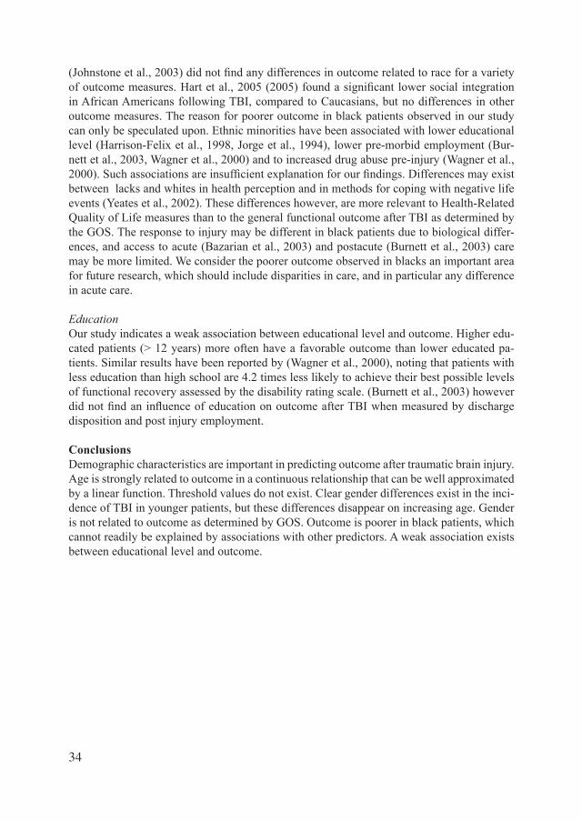
34
(Johnstoneetal.,2003)didnotfindanydifferencesinoutcomerelatedtoraceforavarietyofoutcomemeasures.Hartetal.,2005(2005)foundasignificantlowersocialintegrationin African Americans following TBI, compared to Caucasians, but no differences in other outcome measures. The reason for poorer outcome in black patients observed in our study can only be speculated upon. Ethnic minorities have been associated with lower educational level (Harrison-Felix et al., 1998, Jorge et al., 1994), lower pre-morbid employment (Bur-nett et al., 2003, Wagner et al., 2000) and to increased drug abuse pre-injury (Wagner et al., 2000).Suchassociationsareinsufficientexplanationforourfindings.Differencesmayexistbetween lacks and whites in health perception and in methods for coping with negative life events (Yeates et al., 2002). These differences however, are more relevant to Health-Related Quality of Life measures than to the general functional outcome after TBI as determined by the GOS. The response to injury may be different in black patients due to biological differ-ences, and access to acute (Bazarian et al., 2003) and postacute (Burnett et al., 2003) care may be more limited. We consider the poorer outcome observed in blacks an important area for future research, which should include disparities in care, and in particular any difference in acute care.
EducationOur study indicates a weak association between educational level and outcome. Higher edu-cated patients (> 12 years) more often have a favorable outcome than lower educated pa-tients. Similar results have been reported by (Wagner et al., 2000), noting that patients with less education than high school are 4.2 times less likely to achieve their best possible levels of functional recovery assessed by the disability rating scale. (Burnett et al., 2003) however didnotfindaninfluenceofeducationonoutcomeafterTBIwhenmeasuredbydischargedisposition and post injury employment.
ConclusionsDemographic characteristics are important in predicting outcome after traumatic brain injury. Age is strongly related to outcome in a continuous relationship that can be well approximated by a linear function. Threshold values do not exist. Clear gender differences exist in the inci-dence of TBI in younger patients, but these differences disappear on increasing age. Gender is not related to outcome as determined by GOS. Outcome is poorer in black patients, which cannot readily be explained by associations with other predictors. A weak association exists between educational level and outcome.


3�
CHAPTER 2. C) The Prognostic Value of Secondary Insults in TBI: Results from the IMPACT* Study *International Mission on Prognosis and Analysis of Clinical Trials in TBI
AbstractWe determined the relationship between secondary insults (hypoxia, hypotension and hy-pothermia) occurring prior - to or on admission to hospital and six month outcome after traumatic brain injury (TBI). A meta analysis of individual patient data, from seven Phase III randomized clinical trials (RCT) in moderate or severe TBI and three TBI population-based series, was performed to model outcome as measured by the Glasgow Outcome Scale (GOS). Proportional odds modeling was used to relate the probability of a poor outcome to hypoxia (N=5661), hypotension (N=6629) and hypothermia (N=4195) separately. We additionally analyzed the combined effects of hypoxia and hypotension and performed exploratory analy-sisofassociationswithCTclassificationandmonthofinjury.Havingapre-enrollmentinsultof hypoxia, hypotension or hypothermia is strongly associated with a poorer outcome (odds ratios of 2.1 95%CI (1.7-2.6), 2.7 95%CI (2.1-3.4) and 2.2 95%CI (1.6-3.2) respectively). Patients with both hypoxia and hypotension had poorer outcomes than those with either in-sult alone. Radiological signs of raised intracranial pressure (CT class III or IV) were more frequentinpatientswhohadsustainedhypoxiaorhypotension.Asignificantassociationwasobserved between month of injury and hypothermia. The occurrence of secondary insults prior-to or on admission to hospital in TBI patients is strongly related to poorer outcome and should therefore be a priority for emergency department personnel.
IntroductionFollowing an initial trauma the injured brain is vulnerable to secondary damage which may be exacerbated by secondary insults. For more than 20 years it has been reported that hypoxia and hypotension are associated with adverse outcome following traumatic brain injury (TBI) (Milleretal.,1978).Laterreportshaveconfirmedthisfinding(Manleyetal.,2001,WaliaandSutcliffe, 2002), and Chesnut (1993a) reported the combined effect of hypoxia and hypoten-sion as being substantially greater than the sum of their two effects. Hypothermia has also been shown to be associated with adverse outcome following TBI (Strachan et al., 1989).Many of the published reports of the impact of secondary insults on outcome have concen-trated on insults which develop while the patient is in the Intensive Care Unit. Indeed, one of the key principles of the modern management of severely brain injured patients is to reduce the risk of such insults occurring (EBIC/ABIC guidelines). The main focus of the IMPACT project (Maas et al., 2006b) is to improve the methodology for clinical trials in TBI, and so we have focused on insults which are present on the patient’s admission to ‘neuro’ care. Such insultsarestillofgreatclinicalrelevance,andiftheyareconfirmedtoberelatedtoadverseoutcome then there is the potential to improve outcome by intervening to correct the abnor-mality and/or to interrupt the processes which lead from secondary insult to secondary brain damage.We had the unique opportunity to perform analyses on data from 9205 patients in order to investigate the associations between hypoxia, hypotension and hypothermia and outcome measured on the Glasgow Outcome Scale (GOS) (Jennett and Bond, 1975).

38
MethodsData on at least one of the secondary insults were available for 10 of the 11 studies in the IM-PACT database (Marmarou et al., 2006). PEGSOD (Young et al., 1996) alone had no relevant data captured in the record forms. In general the insults were recorded as being absent, sus-pectordefiniteatorbeforeadmission.Theclassificationof‘suspect’wasbasedonaclinicalimpression,whereastheclassificationof‘definite’wasbasedonanobjectivemeasurementof oxygenation (hypoxia), blood pressure (hypotension) or temperature (hypothermia). The definitionfor“definite”insultswasconsistentacrossstudies:hypoxiawasdefinedasanarte-rial pO2 < 60 mmHg, hypotension as a systolic blood pressure < 90 mmHg and hypothermia as<35°C.Foralloftheformalanalysesthecategoriesofsuspectanddefinitewerepooled,so that each insult was reduced to a binary absent/present categorization. The outcome ex-amined in these analyses was the Glasgow Outcome Scale at six months. Where this was missing or systematically not recorded, the GOS at three months was substituted instead (102 cases). The strength of the association between each separate insult and outcome was analyzed univariately using binary and proportional odds regression models. For the binary models all four possible dichotomies were considered: death versus survival, death or veg-etative state versus better, death, vegetative state or severe disability (conventionally termed ‘unfavorable outcome’) versus better and worse than good recovery versus good recovery. For the proportional odds analysis the GOS was modeled as a four point scale with death and vegetative state categories combined. Results were expressed as odds ratios for an adverse
Table 1. Availability and prevalence of Hypoxia, Hypotension and Hypothermia at admission for each study
aThe summary data presented as “all” are simple totals rather than any more sophisticated pooled esti-mates. NR, not recorded in study.
Hypoxia Hypotension Hypothermia
Study N Available Prevalence Available Prevalence Available Prevalence N (% of total)
N (% of available)
N (% of total)
N (% of available)
N (% of total)
N (% of available)
604 604( 100) 109 (18.0) 604 (100) 143 (23.7) NR -TCDBUK4 986 968 (98.2) 227 (23.5) 974 (98.8) 241 (24.7) NR -
HIT I 350 NR - 342 (97.7) 17 (5.0) NR -
HIT II 819 NR - 815 (99.5) 81 (9.9) 552 (67.4) 50 (9.1)
TIUS 1042 923 (88.6) 266 (28.8) 1013 (97.2) 224 (22.1) 941 (90.3) 157 (16.7)
TINT 1121 989 (88.2) 149 (15.1) 1091 (97.3) 155 (14.2) 1018 (90.8) 39 (3.8)
EBIC 835 830 (99.4) 236 (28.4) 829 (99.3) 201 (24.2) 827 (99.0) 51 (6.2)
SLIN 409 409 (100) 24 (5.9) NR - NR -
SKB 126 84 (66.7) 29 (34.5) 104 (82.5) 21 (20.2) NR -
SAP 919 854 (92.9) 110 (12.9) 857 (93.3) 128 (14.9) 857 (93.3) 111 (13.0)
All * 8721 5661 1150 (20.3) 6629 1211 (18.3) 4195 408 (9.7)
NR - Not recorded in study.
* The summary data presented as ‘All’ are simple totals rather than any more sophisticated pooled estimates
Secondary insults version 07/02/06 Page 1 of 6
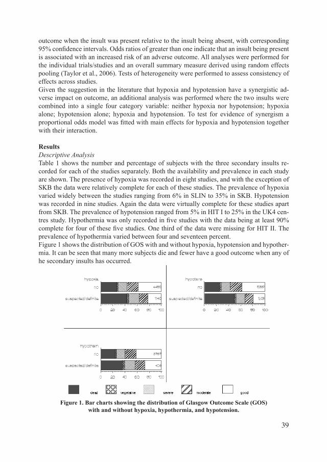
39
outcome when the insult was present relative to the insult being absent, with corresponding 95%confidenceintervals.Oddsratiosofgreaterthanoneindicatethataninsultbeingpresentis associated with an increased risk of an adverse outcome. All analyses were performed for the individual trials/studies and an overall summary measure derived using random effects pooling (Taylor et al., 2006). Tests of heterogeneity were performed to assess consistency of effects across studies.Given the suggestion in the literature that hypoxia and hypotension have a synergistic ad-verse impact on outcome, an additional analysis was performed where the two insults were combined into a single four category variable: neither hypoxia nor hypotension; hypoxia alone; hypotension alone; hypoxia and hypotension. To test for evidence of synergism a proportionaloddsmodelwasfittedwithmaineffectsforhypoxiaandhypotensiontogetherwith their interaction.
ResultsDescriptiveAnalysisTable 1 shows the number and percentage of subjects with the three secondary insults re-corded for each of the studies separately. Both the availability and prevalence in each study are shown. The presence of hypoxia was recorded in eight studies, and with the exception of SKB the data were relatively complete for each of these studies. The prevalence of hypoxia varied widely between the studies ranging from 6% in SLIN to 35% in SKB. Hypotension was recorded in nine studies. Again the data were virtually complete for these studies apart from SKB. The prevalence of hypotension ranged from 5% in HIT I to 25% in the UK4 cen-tresstudy.Hypothermiawasonlyrecordedinfivestudieswiththedatabeingatleast90%completeforfourofthesefivestudies.OnethirdofthedataweremissingforHITII.Theprevalence of hypothermia varied between four and seventeen percent. Figure 1 shows the distribution of GOS with and without hypoxia, hypotension and hypother-mia. It can be seen that many more subjects die and fewer have a good outcome when any of he secondary insults has occurred.
Figure 1. Bar charts showing the distribution of Glasgow Outcome Scale (GOS) with and without hypoxia, hypothermia, and hypotension.
Figure1: Bar charts showing the distribution of GOS with and without hypoxia, hypothermia and
hypotension
Secondary insults version 07/02/06 Page 5 of 6
Figure1: Bar charts showing the distribution of GOS with and without hypoxia, hypothermia and
hypotension
Secondary insults version 07/02/06 Page 5 of 6
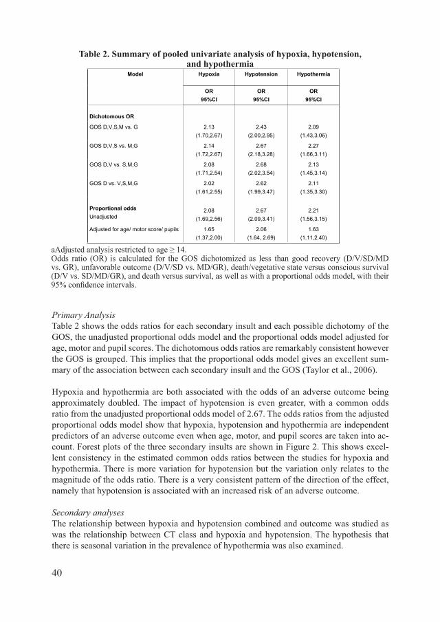
40
PrimaryAnalysisTable 2 shows the odds ratios for each secondary insult and each possible dichotomy of the GOS, the unadjusted proportional odds model and the proportional odds model adjusted for age, motor and pupil scores. The dichotomous odds ratios are remarkably consistent however the GOS is grouped. This implies that the proportional odds model gives an excellent sum-mary of the association between each secondary insult and the GOS (Taylor et al., 2006).
Hypoxia and hypothermia are both associated with the odds of an adverse outcome being approximately doubled. The impact of hypotension is even greater, with a common odds ratio from the unadjusted proportional odds model of 2.67. The odds ratios from the adjusted proportional odds model show that hypoxia, hypotension and hypothermia are independent predictors of an adverse outcome even when age, motor, and pupil scores are taken into ac-count. Forest plots of the three secondary insults are shown in Figure 2. This shows excel-lent consistency in the estimated common odds ratios between the studies for hypoxia and hypothermia. There is more variation for hypotension but the variation only relates to the magnitude of the odds ratio. There is a very consistent pattern of the direction of the effect, namely that hypotension is associated with an increased risk of an adverse outcome.
SecondaryanalysesThe relationship between hypoxia and hypotension combined and outcome was studied as was the relationship between CT class and hypoxia and hypotension. The hypothesis that there is seasonal variation in the prevalence of hypothermia was also examined.
Table 2: Summary of pooled univariate analysis of Hypoxia, Hypotension and Hypothermia
Model Hypoxia Hypotension Hypothermia
OR OR OR95%CI 95%CI 95%CI
Dichotomous OR
GOS D,V,S,M vs. G 2.13 2.43 2.09(1.70,2.67) (2.00,2.95) (1.43,3.06)
GOS D,V,S vs. M,G 2.14 2.67 2.27(1.72,2.67) (2.18,3.28) (1.66,3.11)
GOS D,V vs. S,M,G 2.08 2.68 2.13(1.71,2.54) (2.02,3.54) (1.45,3.14)
GOS D vs. V,S,M,G 2.02 2.62 2.11(1.61,2.55) (1.99,3.47) (1.35,3.30)
Proportional odds 2.212.672.08Unadjusted (1.56,3.15)(2.09,3.41)(1.69,2.56)
Adjusted for age/ motor score/ pupils 1.65 2.06 1.63(1.37,2.00) (1.64, 2.69) (1.11,2.40)
Secondary insults version 07/02/06 Page 2 of 6
aAdjustedanalysisrestrictedtoage≥14.Odds ratio (OR) is calculated for the GOS dichotomized as less than good recovery (D/V/SD/MD vs. GR), unfavorable outcome (D/V/SD vs. MD/GR), death/vegetative state versus conscious survival (D/V vs. SD/MD/GR), and death versus survival, as well as with a proportional odds model, with their 95%confidenceintervals.
Table 2. Summary of pooled univariate analysis of hypoxia, hypotension, and hypothermia
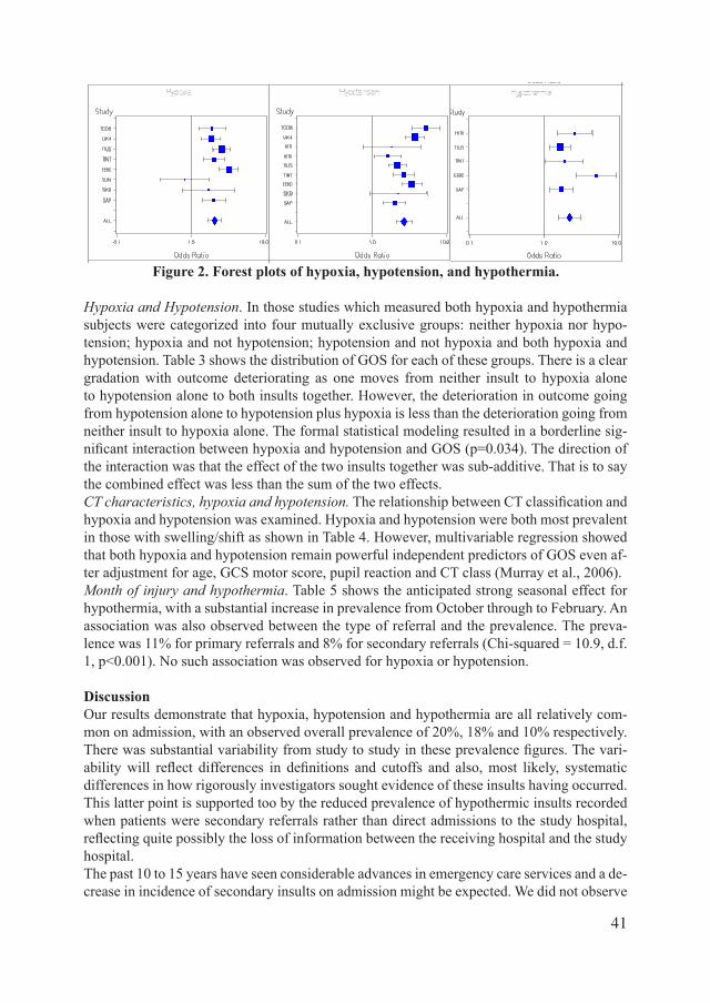
41
HypoxiaandHypotension. In those studies which measured both hypoxia and hypothermia subjects were categorized into four mutually exclusive groups: neither hypoxia nor hypo-tension; hypoxia and not hypotension; hypotension and not hypoxia and both hypoxia and hypotension. Table 3 shows the distribution of GOS for each of these groups. There is a clear gradation with outcome deteriorating as one moves from neither insult to hypoxia alone to hypotension alone to both insults together. However, the deterioration in outcome going from hypotension alone to hypotension plus hypoxia is less than the deterioration going from neither insult to hypoxia alone. The formal statistical modeling resulted in a borderline sig-nificantinteractionbetweenhypoxiaandhypotensionandGOS(p=0.034).Thedirectionofthe interaction was that the effect of the two insults together was sub-additive. That is to say the combined effect was less than the sum of the two effects.CTcharacteristics,hypoxiaandhypotension.TherelationshipbetweenCTclassificationandhypoxia and hypotension was examined. Hypoxia and hypotension were both most prevalent in those with swelling/shift as shown in Table 4. However, multivariable regression showed that both hypoxia and hypotension remain powerful independent predictors of GOS even af-ter adjustment for age, GCS motor score, pupil reaction and CT class (Murray et al., 2006).Monthofinjuryandhypothermia. Table 5 shows the anticipated strong seasonal effect for hypothermia, with a substantial increase in prevalence from October through to February. An association was also observed between the type of referral and the prevalence. The preva-lence was 11% for primary referrals and 8% for secondary referrals (Chi-squared = 10.9, d.f. 1, p<0.001). No such association was observed for hypoxia or hypotension.
DiscussionOur results demonstrate that hypoxia, hypotension and hypothermia are all relatively com-mon on admission, with an observed overall prevalence of 20%, 18% and 10% respectively. Therewassubstantialvariabilityfromstudytostudyintheseprevalencefigures.Thevari-abilitywill reflect differences in definitions and cutoffs and also,most likely, systematicdifferences in how rigorously investigators sought evidence of these insults having occurred. This latter point is supported too by the reduced prevalence of hypothermic insults recorded when patients were secondary referrals rather than direct admissions to the study hospital, reflectingquitepossiblythelossofinformationbetweenthereceivinghospitalandthestudyhospital.The past 10 to 15 years have seen considerable advances in emergency care services and a de-crease in incidence of secondary insults on admission might be expected. We did not observe
Figure 2: Forest plots of hypoxia, hypotension and hypothermia
Secondary insults version 07/02/06 Page 6 of 6
Figure 2. Forest plots of hypoxia, hypotension, and hypothermia.
Figure 2: Forest plots of hypoxia, hypotension and hypothermia
Secondary insults version 07/02/06 Page 6 of 6

42
this. It may be argued that the most recent dataset in the IMPACT database is nearly 10 years old. However, in the Pharmos dexanabinol study, conducted from 2001 to 2004, hypotension was observed in 15% of patients and hypoxia in 25% (Maas et al., 2006a).Allthreeinsultswereconfirmedaspowerfulmarkersofadverseoutcome,witheachinsultbeing associated with at least a doubling of the odds of adverse outcome. The strongest prognostic effects were noted for hypotension. The importance of hypoxia and hypotension as systemic secondary insults and the association between these insults and poorer outcome has been well documented (Chesnut et al., 1993b). We observed that hypoxia does add to hypotension although the effects are sub-additive rather than synergistic. Limitations of our analysisarethatwewererestrictedbythedefinitionsgivenbytheindividualstudiesasto
what constituted hypotension as a secondary insult and no information was available on the duration of insults. On analysis of the relation between the actual levels of blood pressure on admission and outcome we found a smooth U shaped relationship without any evidence of an abruptthresholdeffect(Butcheretal.,2006).Therelativelystrictdefinitionforhypotension(SBP < 90 mmHg) used during data collection may have been too restrictive and the actual incidence of hypotensive insults much greater.The strength of the prognostic effect of hypothermia is very similar to that of hypoxia. Few
NVP, no visible pathology
Table 3. Hypoxia and Hypotension (mutually exclusive categories) by derived Glasgow Outcome Scale (GOS)
Table 3: Hypoxia and Hypotension (mutually exclusive categories) by derived GOS
GOS
Variable Total Dead VegetativeState
SevereDisability
ModerateDisability
GoodRecovery
N % N % N % N % N %
Neither Hypoxia nor Hypotension
3506 803 22.9 107 3.1 502 14.3 651 18.6 1443 41.2
Hypoxia only 632 224 35.4 25 4.0 116 18.4 101 16.0 166 26.3
Hypotension only
581 282 48.5 18 3.1 77 13.3 80 13.8 124 21.3
Both Hypoxia andHypotension
465 254 54.6 20 4.3 68 14.6 55 11.8 68 14.6
Table 4: CT class and hypoxia and hypotension
Hypoxia Hypotension CT Class
N Prevalence N Prevalence
NVP 317 18.6% 402 15.9%
Diffuse injury 1540 16.9% 1736 16.1%
Swelling/shift 888 24.9% 938 23.3%
Mass 1573 19.8% 1864 14.4%
Secondary insults version 07/02/06 Page 3 of 6
Table 4. Computed Tomography (CT) class, and hypoxia and hypotension
Table 3: Hypoxia and Hypotension (mutually exclusive categories) by derived GOS
GOS
Variable Total Dead VegetativeState
SevereDisability
ModerateDisability
GoodRecovery
N % N % N % N % N %
Neither Hypoxia nor Hypotension
3506 803 22.9 107 3.1 502 14.3 651 18.6 1443 41.2
Hypoxia only 632 224 35.4 25 4.0 116 18.4 101 16.0 166 26.3
Hypotension only
581 282 48.5 18 3.1 77 13.3 80 13.8 124 21.3
Both Hypoxia andHypotension
465 254 54.6 20 4.3 68 14.6 55 11.8 68 14.6
Table 4: CT class and hypoxia and hypotension
Hypoxia Hypotension CT Class
N Prevalence N Prevalence
NVP 317 18.6% 402 15.9%
Diffuse injury 1540 16.9% 1736 16.1%
Swelling/shift 888 24.9% 938 23.3%
Mass 1573 19.8% 1864 14.4%
Secondary insults version 07/02/06 Page 3 of 6

43
previous studies have investigated the relationship between hypothermia on admission and finaloutcomeandresultsareconflicting.Zauner(Zauneretal.,1998)inarelativelysmallstudy of 60 patients with severe TBI describe an association between spontaneous hypother-miaandpooreroutcomebutthisassociationwasnotstatisticallysignificant.Jeremitski(Jer-emitskyetal.,2003)reportasignificantincreaseinmortalityinpatientswhowerehypother-miconadmission.However,inthisserieshypothermiawasdefinedasabraintemperaturebelow 35ºC, and it may be argued that the low brain temperature might have resulted from lowcerebralbloodflowandmaynothavebeen representativeofcorebody temperature.Steinemann (Steinemann et al., 1990) found no association between early post traumatic hy-pothermia and outcome in patients with multi trauma including head injury. The strong evi-dence of an adverse effect of hypothermia as a secondary insult seems paradoxical in the light of the strong experimental evidence that hypothermia is neuroprotective. This perhaps points to the mechanism of the adverse effect of hypothermia as a secondary insult being related to its management and in particular to an adverse effect of rewarming. This would be consistent with a secondary analysis of the pivotal Phase III trial of hypothermia in TBI (Clifton et al., 2001). Here patients who arrived hypothermic, were rewarmed and then subsequently ran-domized to the hypothermia treatment group had particularly poor outcomes.In summary, all three secondary insults examined have shown to be strongly associated with the risk of an adverse outcome. Although these results are purely observational, they do point totheneedforgreatvigilanceintheirearlyidentification,andthepotentialfortherapeuticresearch into their optimal management.
Table 5: Month of injury and hypothermia
Hypothermia
N Prevalence
January 258 13.6%
February 570 10.7%
March 589 8.0%
April 565 7.6%
May 374 9.1%
June 358 6.4%
July 338 8.3%
August 322 5.9%
September 292 8.2%
October 296 10.8%
November 274 17.1%
December 251 12.7%
Secondary insults version 07/02/06 Page 4 of 6
Table 5. Month of injury and hypothermia

45
CHAPTER 3. PATHOPHYSIOLOGY: ISCHEMIC FACTORS A) Combined Effects of Mechanical and Ischemic Injury to Cortical Cells: Secondary Ischemia Increases Damage and Decreases Effect of Neuroprotective Agents
AbstractTraumatic brain injury (TBI) involves direct mechanical damage, which may be aggravated by secondary insults such as ischemia. We utilized an invitro model of stretch-induced injury to investigate the effects of mechanical and combined mechanical/ischemic insults to cul-turedmousecorticalcells.Stretchinjuryalonecausedsignificantneuronallossandincreaseduptake of the dye, propidium iodide, suggesting cellular membrane damage to both glia and neurons. Exposure of cultures to ischemic conditions for 24 h, or a combination of stretch and 24 h of ischemia, caused greater neuronal loss compared to stretch injury alone. Next, we tested the neuroprotective effects of superoxide dismutase (SOD), and the nitric oxide (NO) synthase inhibitors 7-nitroindazole (7-NINA) and lubeluzole. In general, these agents decreased neuronal loss following stretch injury alone, but were relatively ineffective against the combined injury paradigm. A combination of SOD with 7-NINA or lubeluzole offered no additional protection than single drug treatment against stretch alone or combined injury. These results suggest that the effects of primary mechanical damage and secondary ischemia to cortical neurons are cumulative, and drugs that scavenge superoxide or reduce NO produc-tion may not be effective for treating the secondary ischemia that often accompanies TBI.
IntroductionTraumatic brain injury (TBI) is a leading cause of death and disability in Western industrial-ized societies (Jennett, 1996). Many factors contribute to the outcome of TBI besides the direct physical damage caused to neurons and glia by a mechanical insult. Secondary insults, such as glutamate excitotoxicity, calcium-mediated toxicity, and ischemia, play a large role in brain damage (Maas et al., 2000; Weber, 2004). For example, decreases in cerebral blood flow(CBF),andthusacorrespondingdecreaseofbraintissueoxygenpressure(PbrO2) as measured in both rats and humans (Clark et al., 1997, van Santbrink et al., 2002, van den Brink et al., 2000), can also aggravate cellular damage. In clinical studies, patients with decreased PbrO2,measuredwithinthefirst48hafterinjury,havealessfavourableGlasgowOutcome Scale score than patients without a decrease in PbrO2 (van Santbrink et al., 2003). This would implicate that a combination of the primary mechanical insult and secondary mechanisms, such as ischemia, contribute to cell damage and death following TBI. There-fore, experimental models that mimic both primary and secondary injury processes may pro-vide a more realistic assessment of trauma at the cellular level. Ourfirstgoalwastoestablishamodelofcombinedmechanicalandischemicinjuryinvitro using cultured mouse cortical cells. We utilized a well-described model of stretch-induced mechanical injury (Ellis et al., 1995), which has previously been characterized in rat cortical neurons and glia (Ellis et al., 1995, McKinney et al., 1996, Weber et al., 1999), rat septo-hip-pocampal cultures (Pike et al., 2000), and mouse hippocampal (Slemmer et al., 2002) and

4�
cerebellar cultures (Slemmer et al., 2004). We analyzed the effects of mechanical injury, ischemic injury, and a combination of these insults by measuring cortical cell damage and neuronal loss. Ischemia is accompanied by hypoxia, as well as by hypercapnia and acidosis (Hoffman et al., 1996; Raghupathi and McIntosh, 1998). In addition, during brain ischemia, several reactive oxygen species (ROS) are generated, such as superoxide and hydroxyl radicals (Leker and Shohami, 2002). There also appears to be an increase in nitric oxide (NO), which is gener-ated by the enzyme nitric oxide synthase (NOS) (Samdani et al., 1997). Three major isoforms ofNOShavebeenidentifiedinthebrain:theneuronalform(nNOS),theendothelialform(eNOS), and an inducible form (iNOS) (Dawson, 1999, Wada et al., 1999). NO contributes to several important physiological processes, such as cerebral vasodilation and neurotrans-mission (Dawson and Dawson, 1996). However, excess NO can interact with the superoxide radical (O2),resultinginthegenerationofperoxynitrite(ONOOˉ),postulatedtobeoneofthemost damaging free radical species (Dawson, 1999, Liu et al., 2002). Free radicals can cause cellular damage by peroxidation of proteins, DNA and lipid membranes (Kermer et al., 1999; Leker and Shohami, 2002). For this reason, decreasing the amount of NO and superoxide radicals, and thereby blocking the production of peroxynitrite, appears to be a possible pro-tective mechanism against cell death after mechanical and combined mechanical/ischemic injury. Hence, we evaluated the neuroprotective potential of superoxide dismutase (SOD) and inhibitors of NOS (7-nitroindazole and lubeluzole) separately, and in combination, in our invitro model of combined injury.
MethodsAnimalwelfareAll experiments were conducted in accordance with the European Communities Council Directive and were approved by the animal welfare committee of Erasmus Medical Center Rotterdam. Efforts were made throughout these studies to minimize animal suffering and the number of animals used.
CellculturePrimary cortical cell cultures (approximately 10% neurons and 90% glia) were prepared from E17/18 wildtype FVB/N mouse embryos similar to previously described methods for hippocampalcellcultures(Slemmeretal.,2002).Briefly,dissociatedcorticesweredilutedinserum-containing media [Basal Medium Eagles (GIBCO, Grand Island, NY) containing 10% horse serum (GIBCO), 10 µg/ml gentamycin (Sigma, St. Louis, MO), 0.5% glucose (Sigma), 1 mM sodium pyruvate (GIBCO) and 1% N2 supplements (GIBCO)] to a concentration of 500,000 cells per ml; cells were then plated in 1 ml aliquots onto collagen-coated sixwell FlexPlates (FlexCell, Hillsborough, NC) coated overnight with poly-L-ornithine (500 µg/ml; Sigma).Allculturesweremaintainedinahumidifiedincubator(5%CO2,37˚C).Neuronallyenhanced cultures were obtained by replacing half of the media 2 days after plating, and then twice per week, with serum-free media containing 2% B27 supplements (GIBCO). Glia formedaconfluentmonolayerthatadheredtothemembranesubstratewhileneuronsadheredto the underlying glia. Cells were used for experiments within 9-12 days invitro (DIV).
CellinjuryPrimary mouse cortical cultures were stretch-injured using a model 94A Cell Injury Control-

4�
ler (Bioengineering Facility, Virginia Commonwealth University, Richmond, VA, USA) as described previously (Ellis et al., 1995; Slemmer et al., 2002). In brief, the Silastic membrane of the FlexPlate well is rapidly and transiently deformed by a 50-ms pulse of compressed ni-trogen, which deforms the Silastic membrane and adherent cells to varying degrees controlled by pulse pressure. The extent of cell injury - produced by deforming the Silastic membrane on which the cells are grown - is dependent on the degree of deformation, or stretch. Based on previous work (Ellis et al., 1995), two levels of cells injury were chosen (5.5 and 6.5 mm deformations)anddefinedasmildandmoderate,respectively.Thesedegreesofmembranedeformation result in a biaxial strain or stretch of 31 and 38%, respectively. This range of cell stretch has been shown to be relevant to what would occur in humans after rotational accel-eration/deceleration injury (Schreiber et al., 1995). Uninjured control wells were contained in the same FlexPlates as injured wells, and thus underwent the same manipulations, except that they did not receive rapid deformation of the Silastic membrane. To simulate ischemia duetolowCBF,FlexPlatesofprimarymousecorticalcultureswereplacedinahumidifiedincubator set at 20% CO2and37˚C.Incubationofcellsforupto6hinischemicconditionscaused no measurable cell damage or death; however, 24 h of ischemia resulted in cell dam-age.Thecellswereincubatedfor24handthenfixedandstained,orreturnedtothenormalincubatorforanadditional24hbeforebeingfixedandstained(describedbelow).Incubationof cells for 24 h in the ischemic incubator resulted in a pH change from ~7.4 to ~7.0. For the combined mechanical and ischemic injury paradigm, cells were injured at 5.5 mm deforma-tion and immediately placed in the ischemic chamber for 24h.
CellviabilityThecellswerefixedandstainedat15min,24hor48haftermechanicalinjury,orat24hor 48 h after combined mechanical and ischemic injury. Cell injury was assessed in cultured cells using the dyes propidium iodide (PrI; Sigma) and Hoechst (Sigma), similar to previ-ously described methods (Slemmer et al., 2002; Zhao et al., 2000). Hoechst labels the nuclei of all cells, whereas PrI is normally excluded from intact cell membranes. If membranes are damaged,PrIwillentercellsandstain thenucleus, resulting inabright redfluorescence.CulturesweretreatedwithPrIandHoechstfor20minat4˚C,thenfixedandstainedwith4% paraformaldehyde for 20 min at room temperature. Images were captured using Texas RedandUVfilters(forPrIandHoechst,respectively)onaLeicaDMRBEfluorescencemi-croscope, equipped with a Hamamatsu C4880 CCD camera. PrI and Hoechst images were takeninfivecontiguous100Xor160Ximages,andwerecountedandaveragedperwell.Allimages were taken from the center portion of the well, as this region was previously shown to receive equal impact from the cell injury controller (Ellis et al., 1995). All PrI and Hoechst cell counting were performed by one of the authors blinded to experimental conditions. Data are expressed as the ratio of PrI positive cells to total cell number (as determined by Hoechst staining of cell nuclei). In control cultures, PrI staining was low, accounting for less than 5% of total cell number at all time points, consistent with previous reports (Slemmer et al., 2002; Zhao et al., 2000). Changes in total cell number are expressed as percentage of control values. All experiments were completed using at least two separate culture preparations at 9-12 DIV.
ImmunohistochemistryandneuronalcellcountsCultured cortical cells received either a mild (5.5 mm) or a moderate (6.5 mm) injury, or

48
remaineduninjured;ateither15min,24hor48hpost-injury,thecultureswerefixedfor20 min with 4% paraformaldehyde as previously described (Slemmer et al., 2004). For the ischemia and combined injury experiments, the cells were incubated for 24 h in the ischemic chamberandthenfixedandstained,orreturnedtothenormalincubatorforanadditional24hbeforebeingfixedandstained.Inbrief,cultureswereincubatedinprimaryantibody[anti-microtubule-associated protein 2 (MAP2, 1:500; Sigma)] for 1 h, followed by incubation with secondary antibody (Alexa-594-conjugated goat-antimouse, 1:300; Molecular Probes, Eugene, OR) for 1 h. Cultures were dehydrated with ethanol and mounted with 4’,6-diamid-ino-2-phenylindole (DAPI).MAP2 specifically labels thedendrites and somaofneurons,while DAPI labels the nuclei of all cells, similar to Hoechst. Images were captured using a LeicaDMRBEmicroscope(seeSection2.4).MAP2-positiveneuronswerecountedinfivecontiguousfieldsatamagnificationof100Xor160X.
DrugtreatmentThe drugs used for treatment were 7-nitroindazole monosodium salt (7-NINA, Tocris Cook-son, U.K.); lubeluzole (supplied by Janssen Pharmaceuticals, Belgium); and superoxide dis-mutase (SOD; Sigma). 7-NINA and lubeluzole were initially dissolved in dimethylsulfoxide (DMSO) and subsequently diluted in dH2O.ThemaximalfinalconcentrationofDMSOthatwas added to the culture wells was 0.1%. SOD was dissolved in dH2O and added to culture wellsinafinalamountof100units/ml.Theconcentrationrangesof7-NINA(1µMand10µM) and lubeluzole (10 nM and 100 nM), which were used in the current study, have been previously shown to protect cultured neurons against hypoxia (Huang et al., 2002), and glu-tamatemediated toxicity (Culmsee et al., 1998), respectively. An amount of 100 units/ml of SOD has been demonstrated to reduce oxygen free radical production invitro (Saito et al., 1997, Shanker and Aschner, 2003). Also, this amount of polyethylene glycol-conjugated SOD was utilized in a previous study using the same stretch-induced injury model (McKinney et al., 1996). The addition of the drugs alone to uninjured control wells (for 24 h) caused no significantdifferenceinPrIuptake,totalcellnumber,ornumberofMAP2-positiveneuronscomparedtountreatedwellsatthefinalconcentrationsusedinthisstudy.Higherlevelsof7-NINA (100 µM) or lubeluzole (1 µM) increased PrI uptake in uninjured controls, suggesting some toxicity at these concentrations. The drugs were either added to the media 15 min prior to mechanical injury, as pre-treatment, or 15 min after mechanical injury, as post-treatment. Unlike 7-NINA and lubeluzole, SOD is sensitive to proteolytic and oxidative inactivation. Therefore, in one set of experiments, SOD was added to injured cultures at two timepoints, 15 min and 12 h after injury. For ischemic injury only, or for combined injury, the plates were placed in the ischemic incubator immediately after the drugs were added.
DataanalysisThe data were analyzed using the statistical program GB Stat (Dynamic Microsystems, Sil-ver Spring, MD). Data were computed as meansGstandard error (SE) values. Statistical sig-nificancewasestablishedbyonewayanalysisofvariance(ANOVA)followedbyFisher’sprotectedleastsignificancedifferencetest.Dataareconsideredsignificantatp<0.05.
ResultsStretchinjuryincreasesPrIuptakeincorticalcellsIn uninjured control wells, there was little PrI staining (Fig. 1A and C, seepage129), indicat-

49
ing that most cells were healthy and viable, with intact cell membranes. Fifteen minutes after injury, however, cultures demonstrated an increased amount of PrI staining, the majority of whichoccurredintheunderlyinggliallayer(Fig.1BandC),similartoourpreviousfindingsin hippocampal cultures (Slemmer et al., 2002). PrI uptake decreased with time post-injury, butremainedsignificantlyelevatedthrough24hafter5.5mmdeformation(mildinjury)and48 h after 6.5 mm deformation (moderate injury). Mild injury also caused a drop in cell num-ber 24 h after injury (determined by the amount of Hoechst-positive nuclei), which returned to control levels at 48 h (Fig. 1D). Moderate injury caused a more dramatic effect on total cell number, which was decreased vs. control at 15 min and 24 h post-injury, and then elevated at 48 h post-injury, suggesting glial proliferation. We also observed an increased amount of condensed nuclei (see Fig. 1B) in cell cultures following both levels of injury, which is gener-ally an indication of delayed or apoptotic cell death (Toescu, 1998).
StretchinjurycausesalossofneuronsincorticalcellculturesTo evaluate more closely the effects of mechanical injury on neuronal cells, we counted the number of MAP2-positive neurons in injured cultures as a percentage of neurons in control wells (Fig. 2, seepage130). Uninjured neurons exhibited an intact soma and smooth neurites (shown in Fig. 2A). Injured neurons often displayed beaded neurites and condensed nuclei (showninFig.2B).CounterstainingwithDAPIfurthersupportedthefindingofcondensednuclei in both neuronal and glial cells, as previously observed with Hoechst staining. Mild injurycausedaslight,butnon-significantdecreaseinthenumberofneuronsto86.4±5.2%of control at 15 min, while moderate injury caused an even greater decrease to 40.6 ± 8.1% ofcontrol.TheamountofMAP2stainingdidnotchangesignificantlyovertimeaftermoder-ate injury, therefore the decrease in MAP2-positive neurons is an early event. However, after mildinjurytherewasasignificantdecreaseinneuronsat24hand48h,suggestingadelayedneuronal loss at this injury level.
Effectsofischemicandcombinedmechanical/ischemicinsultsTo evaluate the effects of secondary ischemic damage to cortical cells, we compared the effects of stretch injury alone to ischemic injury, and to a combined mechanical and isch-emic injury paradigm (as described in Section 2). Ischemic injury alone and combined injury caused an increase in PrI at 24 h, similar to that caused by stretch injury alone (Fig. 3A, also compare to Fig. 1C). PrI uptake remained elevated at 48 h after ischemic and combined injury, unlike stretch injury alone. However, there was no difference in PrI uptake between ischemia alone and combined injury at 48 h. Neither ischemia alone nor the combined in-juryparadigmcausedasignificantchangeintotalcellnumberateithertimepoint(datanotshown). Ischemia alone caused a reduction in MAP2-positive neurons to 48.0 ± 9.5% of con-trol at 24 h (Fig. 3B). The combined insult paradigm caused an even greater loss of neuronal cells to 32.9 ± 4.0% of control. At 48 h, neuronal cell counts were 47.9 ± 6.5% of control following ischemia alone and 25.5 ± 4.4% of control following the combination injury. These results suggest an additive effect of mechanical and ischemic damage to cortical neurons, and a delayed loss of neurons during ischemic conditions.
EvaluationofneuroprotectiveagentsagainstmechanicalandischemicinsultsWe evaluated the neuroprotective potential of several compounds using our invitro model of combined injury: 7-NINA, an inhibitor of neuronal NOS (Huang et al., 2002, Wada et al.,
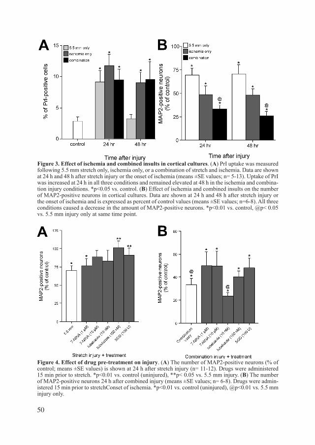
50
Figure 3. Effect of ischemia and combined insults in cortical cultures. (A) PrI uptake was measured following 5.5 mm stretch only, ischemia only, or a combination of stretch and ischemia. Data are shown at 24 h and 48 h after stretch injury or the onset of ischemia (means ±SE values; n= 5-13). Uptake of PrI was increased at 24 h in all three conditions and remained elevated at 48 h in the ischemia and combina-tion injury conditions. *p<0.05 vs. control. (B) Effect of ischemia and combined insults on the number of MAP2-positive neurons in cortical cultures. Data are shown at 24 h and 48 h after stretch injury or the onset of ischemia and is expressed as percent of control values (means ±SE values; n=6-8). All three conditions caused a decrease in the amount of MAP2-positive neurons. *p<0.01 vs. control, @p< 0.05 vs. 5.5 mm injury only at same time point.
Figure 4. Effect of drug pre-treatment on injury. (A) The number of MAP2-positive neurons (% of control; means ±SE values) is shown at 24 h after stretch injury (n= 11-12). Drugs were administered 15 min prior to stretch. *p<0.01 vs. control (uninjured), **p< 0.05 vs. 5.5 mm injury. (B) The number of MAP2-positive neurons 24 h after combined injury (means ±SE values; n= 6-8). Drugs were admin-istered 15 min prior to stretchConset of ischemia. *p<0.01 vs. control (uninjured), @p<0.01 vs. 5.5 mm injury only.

51
1999); lubeluzole, a NOS inhibitor, which in addition decreases glutamate release and blocks sodium and calcium channels (Maiese et al., 1997; Mueller et al., 2003); and SOD (Ikeda et al., 1994; Kamii et al., 1996; McKinney et al., 1996). Since the combination injury paradigm did not produce a profound effect on PrI uptake in cultures as compared to the dramatic neuronal loss, we decided to investigate the effects of these compounds only against MAP2 staining. The number of MAP2-positive neurons measured at 24 h after ischemic and combi-nationinjurieswasnotsignificantlydifferentfromthenumberofneuronsmeasuredfollow-ing these conditions at 48 h, suggesting no further loss of neurons after 24 h (Fig. 3B). We therefore chose to evaluate the effects of the neuroprotective agents at the 24 h timepoint.We evaluated the neuroprotective effects of both pretreatment (Fig. 4) and post-treatment (Fig. 5) with 7-NINA (1 µM, 10 µM), lubeluzole (10 nM, 100 nM) and SOD (100 U/ml). As shown in Fig. 4A, cultures pretreated with 10 µM 7-NINA or 10 nM lubeluzole no longer ex-hibitedasignificantreductioninneurons24hafter5.5mminjuryalone,suggestingaprotec-tive trend of these compounds vs. stretch-induced mechanical injury. Treatment with 100 nM lubeluzoleor100U/mlofSODshowedsignificantprotectionagainstneuronallossinducedby stretch injury, whereas 1 µM 7-NINA exhibited no measurable protective effect. In the combined injury paradigm, cultures treated with 1 µM or 10 µM 7-NINA, 100 nM lubelu-zole,or100U/mlSOD,nolongershowedasignificantreductionofMAP2-positiveneuronsin comparison to stretch injury alone. Cultures treated with 10 nM lubeluzole, however, did not exhibit a trend towards protection. For the post-treatment studies, we evaluated 7-NINA
Figure 5. Effect of drug post-treatment on injury. (A) The number of MAP2-positive neurons (% of control; means ±SE values) is shown at 24 h after stretch injury (n=6-10). Drugs were administered 15 min after stretch. *p! 0.01 vs. control (uninjured), **p<0.05 vs. 5.5 mm injury. (B) The number of MAP2-positive neurons 24 h after combined injury (means ±SE values; n=5-9). Drugs were adminis-tered 15 min after stretchConset of ischemia. *p<0.01 vs. control (uninjured), @p<0.05 vs. combina-tion injury.

52
(10 µM), lubeluzole (10 and 100 nM), single and repeated (15 min and 12 h) administration of SOD (100 U/ml), and combinations of these compounds for protection against stretch injury alone and combined stretch/ischemia. With stretch injury alone, 10 µM 7-NINA, the combination of 10 µM 7-NINA and 100 U/ml SOD, and the double addition of 100 U/ml SODshowedasignificantdecreaseinreductionofMAP2-positiveneurons(Fig.5A).Theother compounds, and combinations of compounds, demonstrated a protective trend against stretchinjuryalone,asthenumberofneuronswasnotsignificantlydecreasedat24hafterinjury compared to controls in these conditions. After combined stretch and ischemic injury, onlytherepeatedadditionof100U/mlSODdemonstratedasignificantprotectionasmea-sured by the number of MAP2-positive neurons (Fig. 5B). The other compounds and combi-nations showed no trend towards protection against combined injury.
DiscussionEvidence from both the clinic and experiments invivo demonstrates that TBI is often accom-panied by a reduction in CBF in the early post-traumatic period (Clark et al., 1997, van Sant-brink et al., 2002, van den Brink et al., 2000). This low CBF often produces ischemia, which is accompanied by hypoxia, hypercapnia and acidosis (Hoffman et al., 1996; Raghupathi and McIntosh, 1998). Therefore, experimental models that mimic both mechanical injury and secondary insults, such as ischemia, should prove useful for analyzing the mechanisms of cell damage and death following TBI. In the current study we utilized an invitro model of mechanical injury, which generates a highly reproducible amount of stretch to cultured cells. We also analyzed the effects of ischemic insults superimposed on stretch-induced injury in cultured mouse cortical cells.Stretch injury on its own increased uptake of PrI in cortical cells. This effect was dependent onthelevelofstretchandtimepost-injuryandwassimilartopreviousfindingsusingthesame injury model in cultured cortical cells from rat (McKinney et al., 1996, Weber et al., 1999). In addition, the total cell count was also affected following stretch injury alone. This was especially true after moderate injury where a decrease in cell number was evident at 15 min and 24 h after injury, and an elevated number of cells was observed at 48 h postinjury, suggestingthatglialcellshadproliferated.Thisfindingissimilartotheastrogliosisthatisoften reported following TBI invivo(McGrawetal.,2001).Wealsospecificallyanalyzedthe effects of stretch on neuronal viability. There were fewer MAP2-positive neurons at 24 h and 48 h after mild injury compared to control or 15 min post-injury, demonstrating some delayed neuronal death at this injury level. After moderate injury there was a dramatic neu-ronallossby15min,whichdidnotchangeovertime.Thisfindingsuggeststhattheneuronalloss following moderate injury occurs within the initial injury phase, and that cells die by necrotic means or that they lift from the membrane substrate, as no additional delayed cell death is apparent.Our invitro model of combined insults is similar to the model previously described by Glass et al. (2002). They also used the same stretch injury model originally described by Ellis et al. (1995), and superimposed hypoxic insults of various duration (1 h-24 h) following stretch injury of cultured rat cortical cells. We superimposed ischemic insults after stretch injury, which encompasses a drop in O2 levels, an increase in CO2, and a decrease in pH. Although we did not precisely control the level of O2 in our study, we believe that the hypercapnia and resulting acidosis that was induced may more closely mimic the overall effects of a global reductioninCBF,ratherthanspecificregionsofhypoxiaoccurringafterTBI.Inaddition,

53
unlike Glass et al. (2002) who measured trypan blue exclusion and lactate dehydrogenase releasefrominjuredcultures,weanalyzedtheeffectsofcombinedinsultsmorespecificallyon neurons.PrI uptake was similar following stretch alone, 24 h of ischemia, or both of these insults when measured at 24 h. Although PrI uptake returned to control levels at 48 h following stretch alone, it remained elevated following ischemia and combined injury. This suggested sustained damage to cells, but we were unable to discern whether this was due to glial or neuronaldamage.Whenwe investigatedMAP2-positiveneurons specifically,we foundaprofound difference between the three injury paradigms. Ischemia alone caused greater neu-ronal loss than stretch injury alone, while combined ischemia and stretch caused an even greaterreductioninneurons.AswiththefindingsofGlassetal.(2002),theeffectsofisch-emia and stretch appear to be additive in our invitro model. When cultures were placed back into a normal incubator for 24 h following 24 h of ischemia, there was no additional neuronal loss compared to measurements directly after 24 h of ischemia. Therefore, restoring normal levels of O2 and CO2inhibitedfurtherneuronaldeathinourculturesystem.Thisfindingalsosuggestedthatthefirst24haftertheinitiationofcombinedinsultswasmostappropriateasatarget window for drug treatment.Many free radical scavengers have been evaluated in experiments related to TBI. With our model, injuries are easily reproducible, providing an adequate means to screen possible ther-apeutic strategies. We decided to evaluate the protective effects of SOD and NOS inhibitors using our model, as cell damage due to TBI and ischemia are believed to involve excess production of both superoxide and NO (Hall et al., 2004, Ikeda et al., 1994, Kermer et al., 1999, Samdani et al., 1997, Wada et al., 1998). For protection against NO-mediated toxicity, we utilized the NOS inhibitors 7-NINA and lubeluzole. 7-NINA has demonstrated neuropro-tection invitro (Huang et al., 2002) and invivo (Wada et al., 1999). Lubeluzole has shown beneficialeffectsagainstexcitotoxicityandNOinvitro (Culmsee et al., 1998, Maiese et al., 1997) as well as invivo ischemia (Culmsee et al., 1998, Mueller et al., 2003).7-NINA, lubeluzole and SOD displayed some neuroprotective effects against stretch injury alone when administered either before or after injury. A differene between the NOS inhibitors wasmanifestwithpretreatmentofstretchinjury.Whereas7-NINAshowednosignificantpro-tective effect, 100 nM lubeluzole did show such an effect when administered before stretch injury. This effect appeared to be reversed with post-treatment. A possible explanation for thiscouldbefoundinthecharacteristicsofthecompounds.7-NINAisreportedtospecifical-ly decrease NO production, whereas lubeluzole also decreases glutamate release and blocks sodium and calcium channels (Maiese et al., 1997, Mueller et al., 2003). Thus, it is possible thatsolelydecreasingNOproductionbeforeinjuryisnotbeneficialtocells,andadrugwithmore broad effects, such as lubeluzole, leads to cell protection. However, lubeluzole showed nosignificantprotectiveeffectswhenused inposttreatment.Similarly, somestudieshaveshown that lubeluzole is not effective against TBI invivo (Kroppenstedt et al., 1999, O’Dell et al., 2000). Also, lubeluzole failed to demonstrate neurological improvement in recent clini-cal trials for stroke (Diener et al., 2000), which can be compared to our superimposed second-ary ischemia, where this compound was also ineffective. McKinney et al. (1996) had previously demonstrated that polyethylene glycol-conjugated SOD did not decrease PrI uptake after stretch injury of mixed cultures of neurons and glia. However,theeffectofSODpretreatmentspecificallyonneuronswasnotevaluated.Inourhands,SODdemonstratedsignificantneuroprotectiveeffectsagainstmechanicalinjurywith

54
bothpre-andpost-treatment.ThedifficultywithSOD,however,isthatitismetabolizedmorequickly than the NOS inhibitors. Thus, post-treatment against stretch injury required a more continuous higher concentration in order to obtain a substantial protective effect. Therefore, therepeatedadditionofSODdidhaveasignificanteffect,whereasthesingleadministrationdidnot.ThisrepeatedadministrationofSODalsoprovedefficaciousagainstthecombinedinjury paradigm, suggesting that scavenging of superoxide is pertinent for several hours, and not just in the initial time period after injury. The data with NOS inhibitors and SOD added alone to the cultures suggest that there is more damage occurring in combined mechanical and ischemic injury than these compounds can prevent. However, the trends towards protection with these drugs with pre-treatment against mechanical/ischemic injury, and against mechanical injury alone, do suggest that the pathways they are inhibiting are involved in the cell damaging processes. Consequently, it is likely that a combination of ischemia on top of the primary mechanical injury produces an increaseinbothsuperoxideandperoxynitrite,againstwhichasingledrugisnotefficacious.Therefore, we treated cells with a combination of SOD and either 7-NINA or lubeluzole, aimed at reducing superoxide and NO production (and thus excess peroxynitrite). A combina-tionofSODand10µM7-NINApost-injurywassignificantlyprotectiveagainstmechanicalinjury only. However, post-treatment with 10 µM 7-NINA only was equally as protective as the combination of SOD and 10 µM 7-NINA, suggesting that 7-NINA alone was responsible for this protective effect. These drug combinations also did not provide protection against neuronal loss in the combined injury paradigm.Overall, these results suggest that, although mechanical injury alone may be amenable to treatment with drugs that scavenge superoxide and reduce NO, this therapy may not be ef-fective in the presence of secondary ischemia, which often accompanies TBI in the clinic. It is possible that other mechanisms, such as glutamate or calcium-mediated toxicity, may be playing a more prominent role in cell death when ischemia is superimposed on mechanical injury. Also, because NO is a vasodilator (Dawson and Dawson, 1996), it may be impor-tant to block certain sources of NO and not others. For example, a recent study has shown that iNOS-derived NO is necessary for the phenomenon of ischemic preconditioning in the brain (Cho et al., 2005). In addition, although blockade of nNOS can protect neurons from ischemia-induced damage (Huang et al., 2002, Samdani et al., 1997), blockade of iNOS has been demonstrated to worsen CBF following in vivo TBI (Steiner et al., 2004). Our cur-rentfindingsfurtheremphasizetheimportanceofpreventingsecondaryischemicinsultsintheclinicalsituation,andindicatethatthelikelihoodofdemonstratingbeneficialeffectsofneuroprotectiveagentsinTBImaybesignificantlydecreasedinthepresenceofsecondaryischemic insults.


5�
CHAPTER 3. B) Changes of Cerebral Blood Flow during Secondary Expansion of a Cortical Contusion Assessed by Non-invasive 14C-iodoantipyrene Autoradiography in Mice
AbstractAlthoughchangesofcerebralbloodflow(CBF)inandaroundtraumaticcontusionsarewelldocumented, the role of CBF for the delayed death of neuronal cells in the traumatic pen-umbra ultimately resulting in secondary contusion expansion remains unclear. The aim of the current study was therefore to investigate the relationship between changes of CBF and progressive peri-contusional cell death following traumatic brain injury (TBI). CBF and con-tusion size were measured in C57/Bl6 mice under continuous on-line monitoring of ETpCO2 prior to trauma and 15 min or 24 hrs following controlled cortical impact by 14C-iodoantipy-rine autoradiography (IAP-AR; n=5-6 per group) and by Nissl staining, respectively. Con-tused and ischemic (CBF < 10%) tissue volumes were calculated based on eleven adjacent section pairs collected at equal distances throughout the contusion and compared over time. All physiological parameters were stable throughout the experiment. Cortical CBF in non injured mice varied between 69-93 ml/100mg/min depending on the anatomical location. Fifteen minutes after trauma CBF decreased in the whole brain by ~50% (p<0.05), except in contused tissue where it fell by more than 90% (p<0.001). Within 24 hours after TBI CBF recovered to normal values in all brain areas with the exception of the contusion where it remained reduced by more than 90% (p<0.001). Contusion volume expanded from 24.9 to 35.5 mm3 (p<0.01) from 15 minutes to 24 hours after trauma (+43%) whereas the area of se-vereischemia(CBF<10%)showedonlyaminimal(+13%)andnotsignificantincrease(22.3to 25.1 mm3). The current data therefore suggest that the delayed secondary expansion of a cortical contusion following traumatic brain injury is not caused by a reduction of cerebral bloodflowalone.
IntroductionContusions are the most frequent focal abnormalities on CT examination in head injured pa-tients and are associated with high mortality and unfavorable functional outcome (Leitgeb et al.,2007,Maasetal.,2007).Thepathophysiologyofcontusionsiscomplexanddifficulttoinvestigatesincemanycontusionstendtoincreaseinsizeovertime,afindingtermedsecond-ary contusion expansion and recently characterized in detail experimentally (Zweckberger et al., 2006). Several factors have been proposed to contribute to contusion expansion, e.g. brain edema formation leading to capillary compression and hypoperfusion (Bullock et al., 1991, Katayama et al., 1998), microthrombosis (Lafuente et al., 1999, Schwarzmaier et al., 2007), progressive bleeding causing microvasospasm (Bullock et al., 1992), glutamate exci-totoxicity (Nilsson et al., 1990, Tanaka et al., 1994), or activation of intracellular cell death pathways(Colicosetal.,1996,Newcombetal.,1999,Smithetal.,2000).Oneofthefirstfactors, however, postulated to be responsible for the expansion of cerebral contusions was thereductionofcerebralbloodflow(CBF)intheperi-contusionalarea(seeRosomoffetal.(1996) for review).
58
Starting with the pioneering work of Fieschi et al. using Xe-CT in TBI patients, Graham et al. using specimens of human TBI victims, and Martins et al. in experimental animals (Fieschi et al., 1974, Martins et al., 1977, Graham et al., 1978) changes of post-traumatic CBF have been studied extensively both in patients and animals during the past three decades. Most stud-ies show that post-traumatic CBF is reduced following focal and diffuse brain damage (see (Verweij et al., 2007, Werner et al., 2007) for review), however, there is general understand-ing that in man (Tenjin et al., 1990, Bouma et al., 1991, Bullock et al., 1992, Alexander et al., 1994, Garnett et al., 2001, von Oettingen et al., 2002) as well as in experimental animals (Nilsson et al., 1977, Pollay et al., 1980, Zhuang et al., 1992, Cherian et al., 1994, Bryan, Jr. et al., 1995, Kochanek et al., 1995, Maeda et al., 1997, Hendrich et al., 1999, Liu et al., 2002, Lundblad et al., 2004, Zweckberger et al., 2006) a severe reduction of CBF, i.e. cerebral isch-emia, is in most cases associated with cortical contusions. The most severe CBF reductions (>80% of normal CBF) are found in the contused tissue itself (Bouma et al., 1991, Bullock et al., 1992, Alexander et al., 1994, Bryan, Jr. et al., 1995, Kochanek et al., 1995, Schroder et al., 1995, Maeda et al., 1997, Hendrich et al., 1999, Garnett et al., 2001, von Oettingen et al., 2002, Zweckberger et al., 2006) and are observed almost immediately after TBI (Zweckber-ger et al., 2006). However, due to the dynamic nature of metabolic and structural changes in thepenumbraanditsrelativelysmallsizeatanygiventimepoint,knowledgeonbloodflowchanges in this crucial region is much less profound. In this crucial region delayed neuronal cell death results in gradual contusion expansion. Depending on the time of measurement, reductions (Bouma et al., 1991, Bryan Jr. et al., 1995, Kochanek et al., 1995, Schroder et al., 1995, Maeda et al., 1997, von Oettingen et al., 2002, Liu et al., 2002) as well as increases (Bullocketal.,1992,Garnettetal.,2001)ofpenumbralbloodflowwerereported.Onlyfewstudies tried to correlate reductions of CBF and tissue loss in the traumatic penumbra or to tracepenumbralbloodflowchangesovertime.Schroderetal.(1995)andvonOettingenetal. (2002) showed a clear positive correlation between the level of CBF reduction and the distance from the core of the contusion thereby suggesting that progressive penumbral cell death may be caused by ischemia (Schroder et al., 1995, von Oettingen et al., 2002). Bryan and colleagues compared the volume of ischemic tissue 30 minutes and 4 hours after con-trolled cortical impact injury (CCII) in the rat and reported a trend towards an increased vol-ume of ischemic tissue over time (Bryan, Jr. et al., 1995). The latter suggests that progressive penumbral ischemia may be the cause for pericontusional cell death. Despite thesefindings it still remainsunclearwhether tissue ischemia is the resultof theprimary lesionandremains thereforeconfined to theregionof initiallynecrotic tissue,orif CBF drops to ischemic levels in the traumatic penumbra over time and is responsible for pericontusional cell death. Therefore the aim of the current study was to measure the volumes of ischemic and non-viable tissue volumes immediately after experimental TBI and 24 hours later. Thus verifying the hypothesis that contusion volume will grow in parallel with the vol-ume of ischemic peri-contusional tissue.
Materials and Methods AnimalsMale C57/Bl6 mice (25 – 28 g, Charles River, Kisslegg, Germany) were used for this study. All efforts were made to minimize suffering and the number of animals according to the guidelines of the German animal protection law and derived guidelines on the ethical use of animals. Animals were kept under controlled light and environmental conditions (12 h

59
dark/light cycle, 23 ± 1ºC, 55 ± 5% relative humidity) and had free access to food (Altromin, Germany) and water at all times before and after the experiments.
AnesthesiaFor trauma application anesthesia was initialized in a halothane chamber (4%) and main-tained with a face mask using 30% O2, 69% N2O, and 1.2% halothane. Formeasurementsofabsolutecerebralbloodflowanesthesiawascarriedoutbyani.p.injec-tion of medetomidine (0.5 mg/kg b.w., Domitor®, Dr. E. Graeub AG, Basel, Switzerland), fentanyl (0.05 mg/kg b.w, Janssen-Cilag, Neuss, Germany), and midazolam (5 mg/kg b.w., Dormicum®,Roche,Basel,Switzerland),aprotocolwellknownnottoinfluencecerebralbloodflow.Micewerethenintubatedwithanoro-trachealtubeandmechanicallyventilatedusing 30% O2 in room air (MiniVent 845, Hugo Sachs Elektronik, March-Hungstetten, Ger-many). Physiological monitoring (blood pressure and pCO2) was performed non-invasively as previously reported in order to prevent disturbances of cardiovascular function by arterial orvenouscatheterizationandtominimizeanesthesiatime(Thaletal.,2007).Briefly,end-tidal pCO2, which closely correlates with pO2, was measured with a microcapnometer (Cl240, Columbus Instruments, Columbus, USA) and adjusted to 35-40 mmHg by respective ventila-tion. Blood pressure was measured at the tail using a non-invasive blood pressure monitoring system (RTBP 2000, Kent, USA). A thermostatically regulated, feedback-controlled heating
Figure 1 A)Localizationofregionsofinterest(ROI)usedformeasurementofcerebralbloodflowand,B)coronalplainsfromwherecoronalbrainsectionsweretakenforquantificationofcerebralbloodflowand volume of ischemic and contused brain tissue.
Figure 1
Quantification of cerebral blood flow
Volume measurements
6
54
3
1A
B
Trauma
2

�0
pad was used to maintain body temperature at 37°C (FHC, Bowdoinham, USA).
Controlledcorticalimpact(CCI)Traumatic brain injury was performed as described previously (Zweckberger et al., 2003b, Zweckbergeretal.,2006,Plesnilaetal.,2007).Briefly,afterinductionofanesthesiatheheadwasfixedinastereotacticframeandalargecraniectomywasperformedabovetherightpari-etal cortex using a high speed drill. Special attention was paid to leave the dura mater intact. CCI was performed perpendicular to the surface of the brain (diameter of the impactor tip: 3 mm, impact velocity: 8 m/s, impact duration: 150 ms, indentation: 1 mm). Thereafter, the craniectomywasclosedwiththeinitiallyremovedboneflapusingconventionaltissueglue(Histoacryl®, Braun-Melsungen, Melsungen, Germany). The skin over the craniotomy was carefully closed and animals were transferred to an incubator heated to 35°C until recovery of spontaneous motor activity.
Quantification of cerebral blood flow by 14C-iodoantipyrineautoradiographyAbsolutecerebralbloodflowwasmeasuredby14C-iodoantipyrine autoradiography 15 min and 24 hr after injury as described previously (Maeda et al., 2000, Zweckberger et al., 2006). Briefly,after inductionofanesthesiaandstabilizationofmeanarterialbloodpressureandend tidal pCO2 (35-40 mmHg) mice received 20 µCi 14C-iodoantipyrine by intraperitoneal injection.Thirtysecondsafterwards,animalsweresacrificedbywholebodysnapfreezing,and stored at - 80 ºC until further use. Coronal sections (20 µm) were collected every 500 µm from the brains on a cryostat (CryoStar HM 560, Microm, Walldorf, Germany) and ex-posedtoautoradiographyfilm(BiomaxMR,Kodak,Germany)for9-11dayswithrespec-tive radioactive standards (RPA504, Amersham, Piscataway, NJ, USA). A blood sample was taken from the frozen heart and the amount of 14C-radioactivity was measured by ß-count-ing as an internal standard. The values for blood 14C-radioactivity and the autoradiograms wereprocessedasdescribedpreviouslybyMaedaetal.(2000).Briefly,autoradiogramsweredigitized with a CCD camera connected to an image processing system (ImageMG, NIH, Bethesda,MD,USA).AbsoluteCBFwasquantifiedinsixregionsofinterest(ROIs)inthecortex of the injured (ROI 1-3) and non-injured hemisphere (ROI 4-6; Fig. 1A) on three coronal brain section obtained from the rostral to the occipital brain (bregma 1.7, -0.90, and -2.7 mm; Fig. 1B).
Quantification of cerebral blood flow laser Doppler fluxmetry (LDF)LDF measurements were performed as previously described (Plesnila et al., 2004, Groger etal.,2005,Traboldetal.,2006).Briefly,aflexiblelaserDopplerprobewasgluedontotheexposed left parietal skull over the territory of the middle cerebral artery (MCA) for con-tinuousmonitoringofregionalcerebralbloodflow(rCBF;Perimed4001Master,Perimed,Järfälla, Sweden). Data were digitized (Dasylab 5.0, measX, Germany) and analyzed off-line (Flexpro 6.0, Weisang, Germany). LDF values are expressed as % pre-traumatic recordings (baseline).
Quantification of contusion and ischemic tissue volumes Contused and severely ischemic (CBF < 10%) tissue volumes were calculated based on eleven adjacent section pairs collected at equal distances throughout the contusion (Fig. 1B).Contusedbraintissuewasidentifiedbycresylvioletstainingaspreviouslydescribed

�1
(Zweckberger et al., 2003, Zweckberger et al., 2006, Plesnila et al., 2007). The area of severe ischemia (CBF < 10%) was visualized on autoradiograms by allocating color codes only to CBFvaluesfrom10-90%ofnormalbloodflowtherebyleavingareaswithCBF<10%blank(Fig.3A).AreasofcontusedbrainandCBF<10%werequantifiedwithastandardimageanalysis software (Optimate 6.52, Media Cybernetics, Silver Spring, MD, USA) by an inves-tigator blinded to the treatment of the animals. Contusion and ischemic tissue volumes (Vi) were calculated based on the contused/ischemic areas (A) obtained from 11 sections by the equation: Vi = 0.5 x (A1+ A2+…+A11).
StatisticalanalysisStatistical analysis was performed using the SigmaStat 3.0 statistical software package (SPSS inc., Richmond, CA). Parametrical data were analyzed with independent-samples T test or one-way ANOVA, non-parametrical data with ANOVA on ranks procedures. Differ-enceswereconsideredsignificantatp<0.05.Resultsarepresentedasmeans±SEMifnotindicated otherwise.
ResultsAll animals used for the measurement of CBF had physiological blood pressure (90-130 mmHg systolic), endtidal pCO2 (35-40 mmHg), and body temperature (36.5-37.5 °C) during the whole duration of the experiment (Table 1). Due to the rapid set-up of the non-invasive monitoring protocol used in the current study CBF measurements were performed in each animal in less than 30 min after induction of anesthesia. CBF in control, non-injured, mice showed the highest values in the basal ganglia, while the lowest values were observed in the corpus callosum, i.e. in the white matter (Fig. 2A, seepage131).Inthecerebralcortexbloodflowwasatintermediatelevelsandvariedbetween69-93 ml/100mg/min (Fig. 2B-D; n=5-6). In all examined sections there was a trend towards higher CBF values in the lateral cortex and towards lower levels near the interhemispheric cleft(Fig.2B-D).Allmeasurementsofcerebralbloodflowshowedasatisfactorysmallinter-individual variability (Fig. 2, 4, and 5). Fifteen minutes following traumatic brain injury by controlled cortical impact CBF decreased in the not impacted tissue of the traumatized hemisphere and in the contralateral hemisphere by 40-60% (Fig. 3A and 4, p<0.01, seepage132,133). Global post-traumatic reduction of CBFby40-50%lastedabout30minasobservedbylaserDopplerfluxmetry,amethodwhichdoes not measure absolute CBF as 14C-iodoantipyrene autoradiography but detects relative CBF changes with high temporal resolution (Fig. 3B; n=11; p<0.001). In the following 30 min after brain injury CBF recovered slowly and reached 80-90% of baseline CBF 60 min af-ter trauma. Directly at the impact site, i.e. the medio-lateral right cortex and in areas frontal
Table 1sham 15 min 24 hours
T (°C) 37.3±0.0 37.1±0.4 37.2±0.1
SBP (mmHg) 103.6±22.1 88.6±13.2 112.1±22.0pCO2 (mmHg) 36.5±0.8 36.8±0.6 36.2±0.6
Body temperature, systolic blood pressure, and endtidal pCO2 15 min and 24 hours after TBI immedi-atelybeforeassessmentofcerebralbloodflowby14C-iodoantipyrine autoradiography

�2
and occipital to the impact site CBF decreased by 96%, 87%, and 91%, respectively (Fig. 3A and 4, ROI 2). No recovery was observed up to 24 hours after TBI (Fig. 3A and 5A-C, ROI2). In contrast to the impact site CBF recovered in most other brain regions to normal or high-normal values within 24 hours (Fig. 3A and 5A-C, ROI 1 and 3-6).To test the main hypothesis of the current study, i.e. that the expansion of a traumatic contu-sion over time is accompanied or preceded by tissue ischemia, we compared the temporal changes of contused and severely ischemic tissue volumes by morphometrical analysis. As reported previously, a cortical contusion expands after trauma and reaches its maximal vol-ume24hoursthereafter(Zweckbergeretal.,2006),afindingwellreproducedinthecurrentstudy (Fig. 6B, D, and E, white columns). While contusion volume was 28.9 mm3 15 min afterinjury,itexpandedsignificantlyto39.3mm3 within 24 hours (p<0.01). In contrast to the expansion of contusion volume the zone of severely ischemic brain parenchyma did not increase from 15 min to 24 hours after TBI (20.6 mm3 vs. 25.1 mm3). The volume of contused
Figure 4. Cerebral bloodflow expressed as%ofCBF in normal brain (dotted line)15 minutes after experimental TBI in mice (n=5-6) in rostral (A), striatal (B), and occipital (C) coronal brain sections. CBFwasquantifiedinsixregionsofinterest(ROI)arranged according to their anatomical location, i.e. from the left lateral to the right lateral cortex. 15 minaftertraumaCBFdecreasedsignificantlyinallregions of the brain (A-C; *p<0.01), most dramati-cally at the site of the impact (#p<0.001).
Figure 4
Cer
ebra
l blo
od fl
ow (%
nor
mal
)
0
25
50
75
100
125
150
175
200Traumatized hemisphereContralateral hemisphere
Mean +/- SEM; n=6
medialleft right
ROI1
ROI32
ROI6
ROI5
ROI4
Cer
ebra
l blo
od fl
ow (%
nor
mal
)
0
25
50
75
100
125
150
175
200Traumatized hemisphereContralateral hemisphere
Mean +/- SEM; n=6
medialleft right
ROI1
ROI3
ROI 2ROI6
ROI5
ROI4
Cer
ebra
l blo
od fl
ow (%
nor
mal
)
0
25
50
75
100
125
150
175
200Traumatized hemisphereContralateral hemisphere
Mean +/- SEM; n=6
medialleft right
ROI1
ROI3
ROI 2ROI
6ROI
5ROI
4
* * * *
#
A
B
C
* * ** *
#
** * *
#

�3
brainparenchymaandthevolumeofischemicbraintissuewerenotsignificantlydifferent15minutesafterinjury.Twenty-fourhoursafterinjurycontusionvolumewassignificantlylarger than the volume of ischemic tissue (p<0.01).
DiscussionInthepresentstudyweconfirmedourpreviousfindingsthatthevolumeofacorticalcontu-sionexpandssignificantlyover time, i.e.during thefirst24hoursafterexperimentalTBI(Zweckberger et al., 2003) and addressed the hypothesis that ischemia in pericontusional ce-rebral tissue, i.e. the traumatic penumbra, is responsible for secondary contusion expansion. However, measurements of absolute CBF by 14C-iodoantipyrine autoradiography revealed that thevolumeof ischemic tissuedoesnotexpandsignificantlyafterTBI indicating thatprogressive penumbral ischemia may play only a minor or bystander role for delayed peri-contusional cell death and hence contusion expansion.
Figure 5.Cerebralbloodflowexpressedas%ofCBF in normal brain (dotted line) 24 hours after experimental TBI (n=5-6) in rostral (A), striatal (B), and occipital (C) coronal brain sections. CBF wasquantifiedinsixregionsofinterest(ROI)ar-ranged according to their anatomical location, i.e. from the left lateral to the right lateral cortex. 24 hoursaftertraumaCBFrecoveredsignificantlyinall regions of the brain (A-C), except at the site of injury (*p<0.001).
Figure 5
Cer
ebra
l blo
od fl
ow (%
nor
mal
)
0
25
50
75
100
125
150
175
200Traumatized hemisphereContralateral hemisphere
Mean +/- SEM; n=6
medialleft right
ROI1
ROI3
ROI 2ROI6
ROI5
ROI4
Cer
ebra
l blo
od fl
ow (%
nor
mal
)
0
25
50
75
100
125
150
175
200Traumatized hemisphereContralateral hemisphere
Mean +/- SEM; n=6
medialleft right
ROI1
ROI3
ROI 2ROI6
ROI5
ROI4
Cer
ebra
l blo
od fl
ow (%
nor
mal
)
0
25
50
75
100
125
150
175
200Traumatized hemisphereContralateral hemisphere
Mean +/- SEM; n=6
medialleft right
ROI1
ROI3
ROI2
ROI6
ROI5
ROI4
*
A
B
C
*
*

�4
ExperimentalSet-upAs discussed previously (Zweckberger et al., 2003, Zweckberger et al., 2006, Plesnila et al., 2007), the current model used for the induction of cortical contusions, controlled corti-cal impact (CCI),was specifically adapted tomice thereby reducing inter- and intraindi-vidual data scattering to a minimum. The craniectomy necessary for the induction of CCI was carefully resealed after trauma allowing the subsequently evolving brain edema to cause pathologically elevated intracranial pressure (Zweckberger et al., 2003). Therefore this model resembles many features of contusions found in patients after TBI and seems to have a higher relevance for the clinical condition of TBI as compared to previous ex-perimentalTBImodelslackingakineticcomponentorsignificantincreasesinintracranialpressure (Martins et al., 1978, Pollay et al., 1980, Zhuang et al., 1992, Bryan Jr. et al., 1995). Specialcarewastakentoavoidanyexperimentalinterventionswhichmayhaveinfluencedanimal physiology, e.g. systemic blood pressure, arterial pCO2, and CBF. Therefore we used ananesthesiaprotocolwellknownnottoinfluenceCBF,i.e.acombinationoffentanyl,mid-azolam, and medetomidine, and a completely non-invasive set-up for the measurement of blood pressure and arterial pCO2 (Thal et al., 2007). Non-invasive animal monitoring has the great advantage to make the time consuming placement of arterial lines unnecessary and in case of the measurement of pCO2 by end-tidal microcapnometry to monitor the most impor-tant determinant of CBF on-line and continuously. Accordingly, CBF was measured in less than30minafterinductionofanesthesiaunderconditionsreducinganyinfluencesonanimalphysiology to a minimum. This is especially relevant for mice which tend to have an unstable cardiovascular baseline during extended period of anesthesia, i.e. longer than 45-60 min. To furtherminimizeany investigation-related influencesonanimalphysiologyandhenceCBF we used a method for the measurement of CBF developed by Mies and colleagues which doesn’t need placement of venous or arterial lines (Maeda et al., 2000). The radioac-tive tracer is injected intraperitoneally from where it enters the systemic circulation at a con-stant rate. Accordingly, plasma levels can be calculated from a single arterial sample obtained from the heart post-mortally. Thereby intravital blood sampling is not necessary resulting in the advantages discussed above, i.e. short anesthesia time, non-invasiveness and no blood volume loss. Another crucial advantage of the currently used method for the measurement of CBF is the termination of the experiment by whole body snap freezing thereby receiving snapshot CBF measurements under perfect physiological conditions, i.e. without the usual delay caused by decapitation of the animal and removal of the brain. By combining non-in-vasive in vivo monitoring of blood pressure and pCO2 together with non-invasive systemic application and determination of 14C-iodoantipyrineweareconfident that theCBFvaluesobtainedinthecurrentstudywereonlyminimallyinfluencedbytheexperimentalconditionsand represent physiological values.
CorticalBloodFlowThecurrentvaluesfoundformurinecorticalbloodflowwerebetween69-93ml/100mg/min.Interestingly, there was a clear trend towards higher CBF values in the lateral and towards lower levels in the medial cortex. Since the cytoarchitecture, i.e. the density of neurons, doesnotdiffersignificantlybetweenthesecorticalregionsandtherebynoreasontoassumemetabolic need of these cortical regions may be different, differences in CBF are most likely caused by decreasing perfusion pressure occurring from the proximal to the distal end of the arteries. These perfuse the lateral and the medial cortex, i.e. the middle cerebral and the

�5
anterior cerebral artery, respectively. CBF in the cortex of not traumatized controlmice is significantly lower than previouslypublished by other groups (Jay et al., 1988, Maeda et al., 2000, Wellons, III et al., 2000, Lun-dblad et al., 2004) (66-93 ml/mg/min vs 144-220 ml/mg/min). These differences are probably due to different experimental set-ups. As already pointed out above, the animals in the current study were intubated, ventilated, and anesthetized with medetomidin, fentanyl, and midazol-am.ThesedrugsdonotinfluenceCBF.Therebyanimalswerekeptunderstrictnon-invasivecontrol of key physiologic parameters until the moment of CBF measurement. Additionally, micewere sacrificed bywhole body snap freezing at –80 ºC, avoiding all possible post-mortem artifacts on CBF. The only other laboratory which measured CBF after TBI in mice usedisofluraneanesthesiaandfrozethebrainswithsomedelayaftersacrifice(Lundbladetal.2004,2007).SinceisofluraneiswellknowntoincreaseCBFsignificantly(Holmstrometal., 2005) and post-mortem diffusion of 14C-iodoantipyrine from the intravascular space into the brain parenchyma is one of the most prominent sources for overestimating CBF using the14C-iodoantipyrine method (Lumblad 2004), it is conceivable that by avoiding these two sources of possible artifact we may have obtained lower, though more physiological cortical bloodflowmeasurementsthanpreviouslyreported.
CorticalBloodFlowfollowingTraumaticBrainInjuryChanges of CBF following experimental TBI were reported in non human primates (Martins et al., 1977, Martins et al., 1978), pigs (Madsen, 1990, Zhuang et al., 1992), cats (Lewelt et al., 1980, Wei et al., 1980, Dewitt et al., 1986, Tornheim et al., 1990, Dewitt et al., 1992), rats (Nilsson et al., 1977, Pollay et al., 1980, Ishige et al., 1987, Yuan et al., 1988), and mice (Liu et al., 2002, Lundblad et al., 2004, Zweckberger et al., 2006) by hydrogen clearance (Martins et al., 1977, Martins et al., 1978, Zhuang et al., 1992), 14C-iodoantipyrine autoradiography (Nilsson et al., 1977, Pollay et al., 1980, Ishige et al., 1987, McIntosh et al., 1987), labeled microbeads (Yuan et al., 1988, Yamakami et al., 1989, Madsen, 1990, Tornheim et al., 1990, Dewittetal.,1992),laser-Dopplerfluxmetry(Muiretal.,1992,Cherianetal.,1994,Nilssonet al., 1996, Maeda et al., 1997, Liu et al., 2002) and MRI (Hendrich et al., 1999). Despite this wealth of information on changes of CBF after TBI, only few clinical (Bouma et al., 1991, Bullock et al., 1992, Schroder et al., 1995, Garnett et al., 2001, von Oettingen et al., 2002) and experimental studies were designed to investigate changes of CBF in and around cortical contusions (Sutton et al., 1994, Bryan, Jr. et al., 1995, Kochanek et al., 1995, Hendrich et al., 1999, Lundblad et al., 2004, Zweckberger et al., 2006).OnemajorearlyfindingaftercontusionalTBIisglobalreductionofcerebralbloodflowby~ 50% of baseline in the whole brain, i.e. not only at or near the area of impact, but also in remote areas of the ipsilateral and the contralateral hemisphere (Cherian et al., 1994, Bryan, Jr. et al., 1995, Kochanek et al., 1995, Hendrich et al., 1999, Liu et al., 2002). The global CBF reduction after TBI depends on the severity of injury (Cherian et al., 1994), lasts more than 3-8 hours (Cherian et al., 1994, Liu et al., 2002), and is restored to baseline values 24 hours after trauma (Bryan, Jr. et al., 1995, Kochanek et al., 1995, Hendrich et al., 1999). Our cur-rentdatainmicearewellinagreementwiththesefindings.Wedetectedalsoa50%reductionof CBF in the whole brain immediately after trauma, which resolved 24 hours later. These changes seem also to be dependent on injury severity in the currently used animal species, i.e. in mice, since a recent study using a milder murine CCI model did not detect global CBF changes(Lundbladetal.,2004).Hence,thisisthefirststudytodemonstratethatmiceshow
��
the same qualitative and quantitative CBF changes after TBI as previously reported in rats thereby demonstrating that mice are valid organisms for this kind of investigation. Alsochangesofcerebralbloodflowinsidecorticalcontusionsseemtostronglydependoninjury severity. If the impact is strong enough to irreversibly damage the affected tissue im-mediatelyorwithinthefirstfewminutesaftertheinjury,thenlowCBFvaluesinsidethecon-tusion represent tissue necrosis rather than evolving tissue ischemia as recently demonstrated experimentally (Zweckberger et al., 2006) and previously suggested by results obtained in TBI patients (Schroder et al., 1995, von Oettingen et al., 2002). If the initial impact is not strong enough, contused tissue may well survive as a whole or in part and may therefore shownormalorevenelevatedbloodflowvaluesasshowninexperimentalmodelsofdif-fuse (Ginsberg et al., 1997, Dietrich et al., 1998) and mild contusional TBI (Lundblad et al., 2004) or in patients with milder trauma (von Oettingen et al., 2002). Accordingly, CBF inside cortical contusions may well be a good marker to differentiate between moderate and severe contusional TBI and to identify patients at risk for secondary insults.Following severe experimental contusional TBI, i.e. with rapid loss of perfusion inside the impacted tissue like in the current study, the initially healthy tissue in the traumatic penumbra around the contusion will die over time resulting in an increased total contusion volume. The enlargement of a cortical contusion over time is a well known clinical observation and was recentlyquantifiedtoaccountfor~30%ofthefinalcontusionvolumepeaking24hoursafterexperimental TBI (Zweckberger et al., 2006). Since the expansion of a traumatic contusion can be prevented by decompression craniectomy (Zweckberger et al., 2003b, Zweckberger et al., 2006), by inhibition of cell death signaling, and by activation of cell survival pathways (Plesnila et al., 2007), we termed this process “secondary contusion expansion” (SCE). De-spite the mechanistic insight into the pathophysiology of SCE from our and other previous studies, the role of CBF for pericontusional cell death is still unclear. Several factors may account for this fact. Experimental studies using non-invasive imaging techniques to monitor CBF and tissue damage, e.g. by MRI, lack spatial and temporal resolution to track the chang-es occurring in the rather small traumatic penumbra (Hendrich et al., 1999). Clinical studies addressing the same issue often suffer from high patient heterogeneity, low patient numbers, unavailability of histological data due to the nature of the studies, and/or, most importantly, lack of imaging opportunities early after TBI,when SCE occurs (Bouma et al., 1991, Bullock et al., 1992, Schroder et al., 1995, Garnett et al., 2001, Steiner et al., 2003). Going back to theremainingexperimentalliteratureonbloodflowchangesaftercontusionalTBIthereisonly the study by Bryan and colleagues left which tries to evaluate if the volume of ischemic contusional tissue is stable after trauma or if it increases over time (Bryan Jr. et al., 1995). The severity of injury in this study was high enough to produce a non-perfused, i.e. necrotic contusioncore,althoughnothighenoughtoinduceaglobalreductionofCBF.Quantificationof the volume of ischemic CBF revealed that when data 4 hours after TBI were compared to 30 min after TBI the area of ischemic tissue showed a trend towards increase, but due to a small sample size and large variability of the measurements this increase was not statisti-callysignificant(Bryan,Jr.etal.,1995).Ourcurrentresultsarewellinagreementwiththeseprevious results. Also our ischemic tissue volume data show only a trend to increase from 15 min to 24 hours after TBI (+13%).Most importantly we assessed the volume of necrotic tissue by cresyl violet staining on sec-tions adjacent to those used for CBF measurements as well and demonstrate that the area of necrotic tissue is larger from the beginning and increases to a much higher degree as com-

��
paredtotheareaofseverelyischemictissue(+43%).Thisfindingsuggeststwoimportantpoints: 1. that already the primary contusion is not caused by tissue ischemia alone, but most likely also by or together with different mechanisms, e.g. on the cellular/molecular level, and 2. that pericontusional ischemia is most likely not the only responsible factor for secondary contusion expansion as previously anticipated by us and others (Bouma et al., 1991, Bullock et al., 1992, Bryan Jr. et al., 1995, Schroder et al., 1995, von Oettingen et al., 2002, Zweck-bergeretal.,2006).Accordingly,ischemiaseemstobeanecessarybutnotsufficientfactorfor secondary contusion expansion following TBI. ConclusionThe present study demonstrates that by using a completely non-invasive protocol absolute values of CBF can be measured by 14C-iodoantipyrine autoradiography reliably and with high precision under physiological conditions and following traumatic brain injury in mice. ImmediatelyfollowingTBIcerebralbloodflowdecreasesinthewholebrainby~50%.InthecontusionproperCBFisreducedbyover90%.Whilebloodflowoutsidethecontusionrecoverswithin60min,contusionalbloodflowdoesnotrecoversignificantly.The volume of a cortical contusion expands over 24 hours by over 40%, while the volume ofseverelyischemiccorticaltissueremainsalmostunchanged.Thesefindingssuggestthatpericontusionalischemiaaloneisnotsufficientforsecondarycontusionexpansion(SCE).


�9
CHAPTER 4. THERAPY: DECOMPRESSIVE CRANIECTOMY A) Effect of Early and Delayed Decompressive Craniectomy on Secondary Brain Damage after Controlled Cortical Impact in Mice.
AbstractThe timing of decompressive craniectomy for the treatment of increased intracranial pres-sure (ICP) after traumatic brain injury (TBI) is a widely discussed clinical issue. Although weshowedrecently thatearlydecompression isbeneficial followingexperimentalTBI, itremains unclear to what degree decompression craniectomy reduces secondary brain damage andifcraniectomyisstillbeneficialwhenitisdelayedbyseveralhoursasofteninevitableduring daily clinical practice. The aim of the current study was therefore to investigate the influenceofcraniectomyonsecondarycontusionexpansionandbrainedemaformationandto determine the therapeutic window of craniectomy. Male C57/Bl6 mice were subjected to controlled cortical impact injury. Contusion volume, brain edema formation, and opening of the blood–brain barrier were investigated 2, 6, 12, and 24h and 7 days after trauma. The ef-fect of decompression craniectomy on secondary brain damage was studied in control mice (closed skull) and in animals craniotomized immediately or with a delay of 1, 3, or 8 h after trauma. Twenty-four hours after trauma, the time point of maximal lesion expansion (+60% vs. 15 min after trauma) and brain edema formation (+3.0% water content vs. sham), contu-sion volume in craniotomized mice did not show any secondary expansion; that is, contusion volumewassimilartothatobservedinmicesacrificedimmediatelyaftertrauma(18.3±5.3vs. 22.2±1.4 mm3). Furthermore, brain edema formation was reduced by 52% in cranioto-mizedanimals.Thebeneficialeffectofcraniectomywasstillpresentevenwhentreatmentwas delayed by up to 3 h after trauma (p<0.05). The current study clearly demonstrates that earlycraniectomypreventssecondarybraindamageandsignificantlyreducesbrainedemaformation after experimental TBI. Evaluation of early craniectomy as a therapeutic option after TBI in humans may therefore be indicated.
IntroductionTraumatic brain injury (TBI) is the leading cause of death among young adults (<45 years) and one of the major causes of disability in the overall population (Jennett, 1998). The de-formation of the brain in the moment of the mechanical impact leads to parenchymal dam-age, which due to its immediate character is not amenable to therapy. This primary injury is followed by a multitude of delayed secondary events on the cellular and molecular level which continue to cause neuronal damage and ultimately lead to secondary expansion of the primary lesion (Gennarelli, 1993).Among the mechanisms responsible for secondary brain damage following TBI, brain edema formation resulting in a refractory increase of intracranial pressure (ICP) remains the most common cause for unfavorable outcome (Murray et al., 1999). In addition to treatment strate-giesrecommendedonthebasisofscientificevidence,forexample,hyperosmotictherapyormild hyperventilation (Maas et al., 1997), decompressive craniectomy has been used intui-tively for over a century for the treatment of malignant post-traumatic brain edema (Kocher,
�0
1901, Ogawa et al., 1974, Jennett et al., 1980, Wilberger et al., 1991). Although a recent randomized clinical trial suggests that functional outcome and quality of life may be better in early craniotomised children (Taylor et al., 2001) and data in adults point into the same direc-tion (Polin et al., 1997; Albanese et al., 2003; Meier et al., 2003), the lack of relevant class I evidencestilldoesnotallowtodrawadefiniteconclusionabouttheefficacyofdecompres-sive craniectomy for the treatment of TBI (Winter et al., 2005).The discussion about the use of decompressive craniectomy after TBI is also fueled by the discrepancy of experimental data (Moody et al., 1968, Cooper et al., 1979, Gaab et al., 1979, Burkert et al., 1989, Rinaldi et al., 1990, Zweckberger et al., 2003). Most studies agree that craniectomy lowers pathological ICP (Moody et al., 1968, Gaab et al., 1979, Rinaldi et al., 1990, Zweckberger et al., 2003); however, the results on brain edema formation and histo-pathological damage are heterogeneous. For example, Cooper et al. (1979) reported a seven-fold increase in Evans Blue stained brain tissue following cold lesion in craniotomized dogs. Gaab et al. (1979) showed no interruption of edema evolution after craniectomy using the same model in cats, and decompression had neither a positive nor a negative effect on brain edema formation following cold lesion in rabbits (Rinaldi et al., 1990). A recent study from our laboratory using a kinetic TBI model, controlled cortical impact (CCI), demonstrated thatmorphologicalbraindamageandfunctionaloutcomeweresignificantlyimprovedwhendecompressive craniectomy was performed immediately after trauma (Zweckberger et al., 2003). It remained unclear, however, to what degree craniectomy reduced secondary brain damage,ifdecompressionhadaneffectonbrainedemaformation,andifthereisabeneficialeffect of craniectomy when decompression is delayed for several hours, a condition that often cannot be avoided during daily clinical practice. SincethedevelopmentofsecondarybraindamagefollowingCCIhasnotbeenquantifiedyet,thefirstaimofthecurrentstudywastoquantifylesionvolume,brainedemadevelopment,and blood–brain barrier disruption in this model. In a second line of experiments we investi-gated the effect of decompressive craniectomy on secondary brain damage and brain edema formation, and determined the therapeutic window of this procedure following experimental TBI.
MethodsAnesthesiaandTraumaApplicationTraumatic brain injury using a CCI device was induced as previously described (Zweck-berger et al., 2003).Briefly,maleC57/Bl6mice (25–28 g b.w.,CharlesRiver,Kisslegg,Germany) were anaesthetized in a halothane chamber (4%), and anesthesia was maintained with a face mask using 1.2% halothane, 30% O2, and 69% N2O during surgery (<15 min). In animalsusedformeasurementsofabsolutecerebralbloodflow,anesthesiawasperformedby i.p. injections of medetomidine (0.5 mg/kg b.w., Domitor®, Dr. E. Graeub AG, Basel, Switzerland), fentanyl (0.05 mg/kg b.w, Janssen-Cilag, Neuss, Germany), and midazolam (5 mg/kg b.w., Dormicum®, Roche, Basel, Switzerland). Mice were intubated with an oro-tracheal tube, and mechanically ventilated using 30% O2 (MiniVent 845, Hugo Sachs Elek-tronik, March-Hungstetten, Germany). End-tidal pCO2, which represents arterial pCO2, was measured with a microcapnometer (Cl240, Columbus Instruments, Columbus, OH) and ad-justed to 35 mmHg by respective ventilation. Blood pressure was measured every minute at the tail using a noninvasive blood pressure monitoring system (RTBP 2000, Kent, OH). A thermostatically regulated, feedback-controlled heating pad was used to maintain rectal

�1
temperature at 37°C (FHC, Bowdoinham, ME). After induction of anesthesia, the head was fixedinastereotacticframeandalargecraniectomywasperformedabovetherightparietalcortex (Fig. 1). Special attention was paid to leave the dura mater intact. CCI (Mouse-Katjus-cha 2000, L. Kopacz, University of Mainz, Germany) was performed perpendicular to the surface of the brain (diameter of the impactor tip: 3 mm, impact velocity: 8 m/sec, impact duration: 150 msec, brain displacement: 1 mm). The craniectomy was closed immediately afterCCIwiththeinitiallyremovedboneflapusingconventionaltissueglue(Histoacryl®,Braun-Melsungen, Melsungen, Germany). In animals receiving decompression the craniec-tomy was left open immediately or was reopened 1, 3, or 8 h after trauma. The skin over the craniectomy was carefully closed, and animals were transferred to an incubator heated to 32°C until recovery of spontaneous motor activity. As indicated previously, craniectomy did not alter brain temperature (Zweckberger et al., 2003).
ExperimentalGroupsA total of seven experimental groups were investigated. In Group 1, contusion volume was measured by histomorphometry 15 min, 2 h, 6 h, 12 h, 24 h, 4 days, and 7 days after trauma (n = 7 per group) in order to quantify secondary lesion growth. In Group 2, brain water con-tentwasquantified6,12,24,and48haftertraumatoassessthemaximumofbrainedemaformation after CCI injury in mice (n = 6 per group). In Group 3, Evans Blue extravasation wasquantifiedinbrainlysatesbyspectrophotometryinordertoassessblood–brainbarrierdysfunction (n = 4 per group). In Group 4, we assessed the effect of decompression crani-ectomy performed immediately after trauma on secondary contusion expansion (n = 6 per group). In Group 5, we measured the effect of decompression craniectomy performed imme-diately after trauma on brain edema formation (n = 6 per group). In Group 6, we determined the therapeutic window of decompression craniectomy following TBI (n = 7 per group) by performing a delayed craniectomy and assessing the contusion volume 24 h after CCI. In Group7,cerebralbloodflowwasmeasuredby14C-Iodoantipyrine autoradiography 15 min after trauma (n = 5).
HistologyandDeterminationofContusionVolumeAttheendoftheobservationperiod,animalsweresacrificedbycervicaldislocationindeephalothane anesthesia. The brains were carefully removed, frozen in powdered dry ice, and
Figure 1. Drawing of a mouse skull indicating the position of craniectomy and brain trauma.
Figure 1
Craniectomy
Contusion

�2
storedat-20°Cuntilfurtheruse.Tenµmcoronalsectionswerepreparedevery500μmona cryostat (CryoStar HM 560, Microm, Walldorf, Germany) and stained with cresyl violet. Sections were photographed with a digital camera system. The contusion area, the area of the non-traumatized tissue of the ipsilateral hemisphere, and the area of the contralateral hemisphere were determined on the digitized images with a standard image analysis software (Optimate 6.51, Media Cybernetics, Silver Spring, MD) by an investigator blinded to the treatment of the animals. In order to correct for the swelling of contused tissue, the contusion area was calculated by subtracting the area of the nontraumatized tissue of the ipsilateral hemisphere from the area of the contralateral hemisphere. Contusion volume was calculated based on the contusion areas corrected for brain edema obtained from 15 sections as follow-ing: 0.5 x (A1 + A2 + . . . + A15).
MeasurementofAbsoluteCerebralBloodFlowAbsolutecerebralbloodflowwasmeasuredby14C-iodoantipyrine autoradiography (Maeda etal.,2000).Briefly,afterinductionofanesthesiaandstabilizationofmeanarterialbloodpressure and end tidal pCO2 mice received 20 µCi 14C-iodoantipyrine by i.p. injection. Thirty secondslater,animalsweresacrificedbywholebodysnapfreezing.Thereaftertheheartandthebrainwereremovedforquantificationofradioactivityinbloodandtissuebybeta-count-ingandautoradiography,respectively.Brainsweresectioned(20µm)andexposedtofilm(Biomax MR, Kodak, Germany) for 8 days with respective radioactive standards (RPA504, Amersham,Piscataway,NJ).Autoradiogramsweredigitized,andabsolutebloodflowvalueswere calculated as previously described (Maeda et al., 2000).
MeasurementofBrainWaterContentBrainwatercontentwasdeterminedaspreviouslydescribed(Vakilietal.,2005).Briefly,24hafter trauma, mice were killed and brains were removed as described above. Pons and olfac-tory bulb were removed, and brains were weighed in order to obtain their wet weight (ww). Thereafter brains were dried at 110°C for 24 h for determination of their dry weight (dw). Brain water content was calculated by using the following formula: (ww – dw)/ww x 100.
EvaluationofBlood–BrainBarrierPermeabilityBlood–brain barrier (BBB) permeability was evaluated by measuring Evans Blue (EB) ex-travasation, as previously described (Vakili et al., 2005).Briefly,mice received 4mL/kgof a 2% EB solution by tail vein injection 30 min after trauma. Twenty-three hours after reperfusion, animals were perfused transcardially with 100 mL of saline solution to wash out intravascular EB. Thereafter, brains were removed, and the hemispheres were separated and weighed. For extraction of EB from brain tissue hemispheres were placed in 2 mL of 50% trichloroacetic acid (wt/vol) and homogenized by sonication. The homogenate was centri-fuged at 10,000 rpm for 20 min, and the supernatant was diluted 1:12 with ethanol. EB was measured at an excitation wavelength of 620 nm and an emission wavelength of 680 nm usingafluorescencespectrophotometer(Model650-10S,Perkin-Elmer,Norwalk,CT).EBconcentrations were calculated and expressed as µg/g brain tissue by using a standard curve (100–500 ng/mL).
StatisticalAnalysisAll data are given as means ± standard error of the mean (SEM) if not indicated otherwise.

�3
For statistical comparisons, the between-groups Student t-test was used for normally distrib-uted and the Mann-Whitney Rank Sum test for not normally distributed data. Measurements over time (body weight and beam walk) were tested versus baseline with Friedman Repeated Measures analysis of variance (ANOVA) on Ranks followed by Dunnett’s All Pairwise Mul-tiple Comparison Procedure as post hoc test. Calculations were performed with a standard statisticalsoftwarepackage(SigmaStat2.0,JandelScientific,Erkrath,Germany).
ResultsExcept for three animals which died from traumatic subarachnoid hemorrhage within 24 h after trauma, no mortality was observed in the present study. Anesthesia and craniectomy did nothaveanyinfluenceonbrainhistology,functionaloutcome,orbodyweightasobservedinsham operated animals (anesthesia and craniectomy but no trauma; data not shown). Micesacrificedforevaluationofprimarybraindamage15minaftertraumashowedalargecortical contusion with massive hemorrhage. Visual inspection of freshly prepared coronal brain sections demonstrated widespread hemorrhage within the contused brain tissue (data notshown).Histologicalstainingofcoronalbrainsectionsconfirmedthatthecorticalcontu-sion reached down to the medial part of the corpus callosum (Fig. 2A, seepage134). Higher magnificationphotomicrographsdemonstrateddisruptionofalmostallcellularelementsin
Figure 3
Time after trauma15min 2h 6h 12h 24h 4d 7d
Con
tusi
on v
olum
e (m
m3 )
0
5
10
15
20
25
30
35
*
* **p<0.05 vs 15min
+32% +5
2% +63%
Mean +/- SD; n=7
0 2 4 6 8 10 12 14 16
Are
a of
con
tusi
on (m
m²)
0
2
4
6
8
10
12
rostral dorsalSection #
Mean +/- SEM; n=7*p<0.05 vs. 15 min
*
**
**
**
15 min
24 h
A
B
Figure 3. Contusion area and vol-ume. A. Contusion areas from 15 coronal sections prepared throughout the in-jured brain obtained from animals sac-rificed15min(black)or24h(white)after trauma (n=7 per group). B. Con-tusion volume of mice sacrificed atdifferent time points after controlled cortical impact (n=7 per group). Con-tusion volume increases over time and reaches a maximum 24 h after trauma as compared to animals sacrificedimmediately after trauma. Removal of dead tissue starting later than 24 h after trauma results in an apparent shrinkage of the contusion on days 4 and 7.
Figure 3
Time after trauma15min 2h 6h 12h 24h 4d 7d
Con
tusi
on v
olum
e (m
m3 )
0
5
10
15
20
25
30
35
*
* **p<0.05 vs 15min
+32% +5
2% +63%
Mean +/- SD; n=7
0 2 4 6 8 10 12 14 16
Are
a of
con
tusi
on (m
m²)
0
2
4
6
8
10
12
rostral dorsalSection #
Mean +/- SEM; n=7*p<0.05 vs. 15 min
*
**
**
**
15 min
24 h
A
B

�4
the core of the contusion (Fig. 2D) as compared to uninjured cortex (Fig. 2C). Most cells had an irregular shape and pyknotic nuclei indicating cell death (Fig. 2D). Measurement of cerebralbloodflowby14C-iodoantipyrine revealed that the primary contusion did not show any perfusion already at this early time point after trauma (CBF = 0 ± 0 mL/100/mg/min; p < 0.001vs.contralateralcorticalbloodflow;n=5;Fig.2F).Mice sacrificed24hafter traumashowedsignificantly largercontusionvolumesas com-paredtothosesacrificed15minaftertrauma(Fig.2B).Thecontusionexpandedmainlyintothe lateral cortex, a region of the brain not affected by cell death immediately after trauma. Cells in the lateral cortex were shrunken and showed nuclear condensation although no signs of bleeding or mechanical tissue disruption were observed (Fig. 2E). The expansion of the necrotic contusion into healthy brain was also demonstrated by quantitative analysis (n = 7 per group; Fig. 3A). The contusion area on sections obtained from the center of the contu-sionmorethandoubledduringthefirst24haftertrauma.Duringthesameperiodoftimethetraumatized hemisphere swelled by less than 10% (data not shown).Analysis of contusion volume over time revealed that the area of necrotic tissue expanded continuouslyduringthefirst24haftertraumaby62%reachingamaximumof163%ofthevolume of the primary contusion (Fig. 3B). During the next 6 days the contusion volume gradually decreased reaching 78% of the volume of the primary contusion.
Figure 4
A
B
Evan
Blu
e ex
trav
asat
ion
(OD
x100
)
0
5
10
15
20
25
30
35Right/Traumatized hemisphereLeft hemisphere
Mean +/- SD; n=4*p<0.01 vs. Sham
4 h 8 h 24 h 48 hSham
*
**
Time after trauma
Bra
in w
ater
con
tent
(%)
0
75
76
77
78
79
80
81
82
83
Sham 6 h 12 h 24 h 48 h
*p<0.05 vs. Sham# p<0.05 vs. 12 and 24 h+ p<0.05 vs. 6 h
*
*
* #
+Mean +/- SD; n=6
*+
Figure 4. Brain edema forma-tion after controlled cortical injury in mice without craniec-tomy.Brain water content (A) as well as Evans Blue extravasation, an in-dicator of blood–brain barrier dis-ruption (B), show their maximal increasewithinthefirst12haftertrauma. Maximal values for brain water content and Evans Blue ex-travasation are observed after 24 h (n=6 and n=4 per group, respec-tively).

�5
Brainedema,quantifiedbythemeasurementofbrainwatercontent,developedmostquicklyduringthefirst6haftertraumaandreacheditsmaximumat12–24hpostinjury(Fig.4A).Forty-eight hours after trauma, brain water content decreased indicating resolution of brain edemaformation,thatis,neteffluxofbrainedemafluidfromtheinjuredtissue.Thisfindingisalsosupportedbythequantificationofblood–brainbarrierdisruptionusingEvansBlue(Fig. 4B). More than 80% of Evans Blue extravasated into the brain parenchyma within the first8haftertrauma.Thereafter,littleextravasationwasobserved.Forty-eighthoursaftertrauma tissue Evans Blue decreased thereby indicating closure of the blood–brain barrier.After determining the time course of lesion expansion and brain edema formation, i.e., the developmentofsecondarybraindamagefollowingCCI,wequantifiedtheeffectofdecom-
Figure 5. Contusion volume of craniectomized (open bar; n=6) and not craniectomised mice (filled bar: n=6) 15 min and 24 h after controlled cortical injury. Craniectomized animals do not show secondary brain damage.
Time after Trauma
Con
tusi
on v
olum
e (m
m3 )
0
5
10
15
20
25
30
35
40
45
50TBI + craniectomyTBI
*p<0.05 vs. TBI + craniectomy 24hn=6; Mean +/- SEM
*
15 min 24 h
Figure 5
Figure 6. Brain water content of sham-operated (sham; n=6), non-craniectomized (filled bar; n=6) and craniectomized mice (open bar; n=6) 24 h after trauma. Brain edema formation is reduced by 52% in craniectomized animals.
Figure 6
Bra
in w
ater
con
tent
(%)
0
76
77
78
79
80
81
82 TBI + craniectomyTBI
*p<0.05 vs. TBIn=6; Mean +/- SEM
*
TraumaSham
- 52%

��
pression craniectomy on lesion expansion (Fig. 5). Primary lesion volume assessed 15 min after trauma was not different between craniectomized (open bars) and non-craniectomized (closed bars) mice; however, while lesion volume showed an expansion of 89% in non-crani-otomizedanimals,nolesionexpansionwasobservedincraniectomizedmicewithinthefirst24 h after trauma (Fig. 5).Thesignificantreductionofsecondarybraindamagewasalsosupportedbythedecreaseofbrain edema formation in the craniectomized animals. Brain water content 24 h after trauma, the time point of maximal brain swelling, was reduced by more than 50% in the craniectomy group (Fig. 6).In the last series of experiments, we aimed to determine the therapeutic window of decom-pression craniectomy. For this purpose, we used non-craniectomized animals as controls (Fig. 7, closed bar) and compared their contusion volume 24 h after trauma to mice crani-ectomized immediately 1, 3, or 8 h after controlled CCI (Fig. 7, open bars). Craniectomy performedwithinthefirst3haftertraumasignificantlypreventedlesionexpansion.Thecon-tusion volume assessed in the noncraniectomized group was about 50% larger as compared to the contusion volumes of the animals subjected to early craniectomy, indicating that most of secondary lesion expansion was prevented by early craniectomy. If performed too late (i.e., 8 h after trauma), contusion expansion progressed already so far (cf. Fig. 2) that decompression could not prevent secondary brain damage.
DiscussionIn the present study, we demonstrate that decompressive craniectomy (DC) reduces brain edema formation by more than 50% and completely prevents secondary brain damage as morphologicallydefinedinthisstudywhenperformedearlyenough(i.e.,duringthefirst3h after trauma).
Figure 7. Therapeutic window of decompressive craniectomy after controlled cortical injury. Ani-mals of the control group (closed bar; n=7) were not craniectomized, while the animals from the other groups (open bars; n=7 each) were craniectomized immediately, and 1, 3, or 8 h after trauma. Only animalsdecompressed1–3haftertraumashowedasignificantpreventionofsecondarybraindamage.
Figure 7
Con
tusi
on v
olum
e (m
m³)
0
5
10
15
20
25
30
35 *p<0.05 vs. no craniectomyn=7; Mean +/- SEM
0 1 3 8no
craniectomyTime of craniectomyafter trauma (h)
* * *

��
Together with our previous report (Zweckberger et al., 2003), we clearly demonstrate that earlycraniectomysignificantlyreducesbrainedemaformation,abolishesICPincrease,pre-vents secondary brain damage, and improves functional outcome following TBI in a kinetic, that is, clinically relevant, animal model.As discussed previously (Zweckberger et al., 2003), the currently used CCI device was spe-cificallydevelopedformicetherebyreducinginter-andintraindividualdatascatteringtoaminimum (cf. Figs. 3, 5, and 7). Together with the randomized and blinded experimental procedures, the fact that DC does not affect systemic parameters and does not, as intuitively expected, cause hypothermia (Zweckberger et al., 2003) we believe that the current experi-ments were carried out with the most possible care to avoid all relevant sources of experi-mental artifacts which may confound the interpretation of our data.One aim of the current study was to assess the effect of decompression craniectomy on sec-ondary brain damage. We recognized that although the concept and mechanisms of second-arybraindamagearediscussedamongthescientificcommunityfordecades(Baethmannetal.,1988;Gentleman,1990),itsclearmorphologicaldefinitionandexperimentalquantifica-tionfollowingexperimentalTBIwasmissingyet.Inthecurrentstudywedefinedsecondarybrain damage as the difference between the morphological damage observed immediately af-ter trauma, i.e. the primary damage, and the maximal damage observed thereafter. Using this definitionwedeterminedthattheprimarydamageinamousesubjectedtocontrolledcorticalimpact injury has a volume of about 20 mm3 (Figs. 3 and 5). The injury is characterized by massive hemorrhage on the brain surface and in the parenchyma, as also observed following human TBI (Ribas et al., 1992), and pan-necrosis of the affected tissue (Fig. 2). The fact that these changes were present already 15 min after trauma suggest that hemorrhage and cell death were caused by disruption of blood vessels and cell membranes as a direct result of the mechanical impact. The assumption that the primary lesion/contusion consists of non vital tissueisalsosupportedbythefactthatnocerebralbloodflowwasdetectedinthisregionofthe brain already 15 min after trauma (Fig. 2F) as also observed by others in a similar time window (Kochanek et al., 1995; Bryan, Jr. et al., 1995).Accordingtotheabovementioneddefinition,secondarybraindamageistheportionofthedamage which develops over time on top of the primary insult. Our experiments show that the expansion of the contused brain area could be observed in every single investigated brain section obtained 15 min and 24 h after trauma and resulted in an almost doubling of the con-tusion area (Fig. 3A). Since opening of the blood-brain barrier and brain edema formation, which developed during the same period of time (Fig. 4), resulted in a hemispheric swelling of less than 10%, the observed expansion of the contusion by 100-200% (Fig. 3A) can by no means be explained by mere swelling of the affected hemisphere. The assumption that quantificationofcontusionexpansionisnotaffectedbybrainswellingisalsosupportedbythe fact that contusion expansion can be completely prevented by craniectomy while brain edemaisstillpresent(Figs.5and6)andbythefindingthattheexpandedcontusionsdonotonly increase in size but also affect anatomical areas of the brain not affected by the primary lesion, e.g. the lateral cortex and the hippocampus (Fig. 2A,B). These results are also sup-ported by the calculation of lesion volume which revealed that cortical contusions following CCI in mice expanded from ~20 mm3 immediately after trauma to a maximum of ~30 mm3 24hlater(Figs.3and5)indicatingthat35-40%ofthefinallesiondevelopsecondarilyandare therefore amenable to therapeutic interventions. Interestingly, our previous experiments on the therapeuticefficacyofdecompressioncraniectomyshowed thatdecompression re-
�8
duced lesion volume 24 h after trauma by 39.5% (from 30 to 18 mm3) (Zweckberger et al., 2003) indicating that craniectomy may almost completely reduce secondary brain injury. The presentresultssubstantiatethesefindingsbydemonstratingthatdecompressioncraniectomyindeed completely prevents contusion expansion (Fig. 5).The complete prevention of contusion expansion, i.e. secondary brain damage, by decompres-sion stresses the utmost importance of increased ICP for the pathophysiology of secondary brain damage following TBI and sheds new light on the mechanisms how a cerebral contu-sion may expand over time. Although many studies stressed the role of molecular mecha-nisms for post-traumatic cell death, e.g. apoptosis or free radical formation (Raghupathi et al., 2000; Lewen et al., 2000; Fiskum, 2000; Eldadah et al., 2000), it remained unclear how these events were related to the daily observations made by clinicians during the treatment of TBI patients. Our results clearly indicate that the formation of brain edema originating from theprimarycontusionisthefirststepinacascadeofeventsultimatelyleadingtodelayedneuronal cell death and secondary brain injury. The evolving ICP increase (Zweckberger etal.,2003)reducesperfusionpressuretherebycausingareductionofcerebralbloodflow.This may not be relevant for healthy brain tissue, however in the tissue at risk, i.e. the still viable tissuesurrounding theprimary lesion,wherebloodflowisaffectedby theprimaryinsult(Bullocketal.,1992),aslightreductionofbloodflowmayalreadyhavedetrimentalsequels,e.g.furtherreductionofbloodflowbyastrocyticswelling(Bullocketal.,1991),theinduction of apoptosis, and the formation of free radicals (Raghupathi et al., 2000; Lewen et al., 2000; Fiskum, 2000; Eldadah et al., 2000). The addition of further damage to the primary lesion increases the area of tissue and vessel damage and initiates a vicious circle by further bloodbrain barrier disruption and brain edema formation. This pathophysiological concept is clearly supported by our previous (Zweckberger et al., 2003) and current data showing that the pathological ICP increase is the cause of secondary lesion expansion and by demonstrat-ing that brain edema formation is reduced by ~50% if secondary brain damage is prevented. Accordingly, these data show that in the current trauma model half of the edema originates from the primary lesion, i.e. it is not preventable by decompression craniectomy, while the other half develops due to secondary mechanisms and can be prevented by decompression. The proposed pathophysiology of secondary lesion expansion, i.e. a slow starting phase ul-timatelyfollowedbyaselfamplifyingfinalphase,isfurthersupportedbythefactthatde-compressionduringthefirst1-3hoursafterTBIbluntssecondarylesionexpansiontoahighproportion, while later craniectomy is not or only marginally effective (Fig. 7).The current experiments clearly demonstrate that decompressive craniectomy completely preventssecondarybraindamagewhenperformedwithinthefirstfewhoursafterexperimen-tal TBI. Although care should be taken when extrapolating experimental results in rodents to the situation in humans our current results suggest that early decompressive craniectomy may have a clinical potential.Our results further demonstrate that timing seems to be of utmost importance and that de-compression has to be performed as early as possible in order to fully exert its therapeutic potential.


81
CHAPTER 4. B) Effect of Decompressive Craniectomy on Cerebral Blood Flow after Controlled Cortical Impact in Mice
AbstractObjectives: Previous studies have shown positive effects of decompression craniotomy (DC) on secondary contusion growth after traumatic brain injury (TBI). Here the effect of DC oncerebralbloodflow(CBF)andhippocampalfunctionisexamined.Materials/Methods: CBF was measured in one group C57Bl6 mice 3 hrs following controlled cortical impact by 14C-iodoantipyrineautoradiography.Contusionvolumeswerequantifiedonparallelsectionsstained by Nissl and compared to areas with CBF < 10%. Intra- and peri-contusional CBF was compared to the corresponding areas of the contralateral hemisphere. A second group of mice was treated similarly and tested with the Object Recognition Test (ORT) 7 days after injury. Results: Physiological parameters were stable throughout the experiment. Decom-pressedmicehadsignificantsmallercontusionvolumes3hoursafterinjury(20mm3 vs 36 mm3).ThevolumeofCBF<0.10ml/min/gwassignificantlysmallerincraniectomizedmice(8 mm3 vs 20 mm3).CBFwassignificantlydecreased(30%ofcontrol)ipsilateraltoinjuryinnon-decompressedanimals.Bothclosedandcraniectomizedanimalshadsignificantlyre-ducedCBFipsilaterally,ofwhichtheclosedgroupshowedsignificantlylowervalues.Crani-ectomized mice performed worse on the memory test (object recognition test). Conclusion: Early DC improves CBF in both cortex and hippocampus, but not memory.
IntroductionTraumatic brain injury (TBI) is a major health and socio-economic problem throughout the world. In western industrialized society TBI is the leading cause of death and disability among young people (Jennett, 1996). A recently revived and by some clinics never abandoned sur-gical therapy is decompressive craniectomy (DC). DC as therapy for uncontrollable raised intracranial pressure (ICP) after TBI has been investigated in the 1960s&70s (Kjellberg and Prieto 1971; Ransohoff 1971; Britt and Hamilton, 1978; Cooper 1976, Venes and Collins 1975). The suggestion that brain swelling could worsen and induce herniation through the DC opening made many neurosurgeons abandone this option for therapy. In the late 1980s new criteria for when and how to perform a DC were introduced among other therapeutic strategies to reduce raised ICP (Alexander 1987, Gaab 1990, Grande 2002, Marshall 2000, Naredi 1998, 2001). The exact role of DC within TBI treatment has been unclear. Pre-clinical and clinical studies carried out in the past 10 years have shown DC possibly useful in the treatment of severe, focal, TBI (Zweckberger 2003, 2006, Taylor 2001, Albanese 2003, Figaji 2003, Guerra 1999, Hejazi 2002, Kontopoulos 2002, Messing-Junger 2003, Polin 1997, 2003, Schneider 2002, Whitfield2001).InthepediatricpopulationDChasalreadybeenacceptedasvalid‘secondtier’ therapy for raised ICP after TBI (Taylor 2001, Jagannathan et al., 2007; Rutigliano et al., 2006). In adult TBI Rosenfeld (2006) advocates early generous DC in war victims of cranial blast injuries. However, circumstances during war do differ from peaceful, optimized and maximized, hospital conditions. In stroke, early decompressive surgery has been proven to bebeneficialinapooledanalysisofthreerandomisedcontrolledtrials(RCTs)(Vahedietal.,

82
2007). This is not yet the case in adult TBI patients. Early DC, when performed according to current, developing, criteria, could be a more effective treatment because ICP is reduced ear-lier after injury (Skoglund et al., 2006). The use of different protocols by several clinics has been made it impossible to investigate the effects on outcome. Prospective RCTs of DC in TBI are currently running worldwide (RESCUEicp and DECRA) hopefully giving an answer on the effect of DC on clinical outcome. Two recent preclinical studies using a kinetic contusion TBI model, controlled cortical im-pact (CCI), demonstrated that early DC reduces secondary damage and edema formation andimprovesfunctionaloutcomesignificantlywhenperformedearlyafterTBI(uptothreehours) (Zweckberger et al., 2003, 2006). By opening the skull ICP reduces almost imme-diately; pathophysiological pathways leading to ischemia, apoptosis and necrosis are dis-continued.Cerebralbloodflow(CBF)islikelytobealteredbyDCafterTBI.HowCBFisaffected exactly has scarcely been investigated (Yamakami and Yamaura 1993a,b; Lundblad et al., 2004, 2007). Therefore we investigated CBF in the cortex three hours after CCI injury in craniectomized mice using 14C-autoradiography.HippocampalCBFwasmeasuredspecifi-cally and hippocampal function was tested accordingly with the Object Recognition Test.
Methods and MaterialsStudydesignandanimalcareExperimental head injury was induced in C57/Bl6 mice according to the controlled cortical impact model. All animals were randomized to treatment by decompressive craniectomy (craniectomy), supportive care (closed) or non injured (control). Two sets of experiments were performed: in the first (N=6-7) animalswere sacrifised three hours after injury forCBF determination by autoradiography and histologic analysis of contusion volumes. In the second set (N=6-7) animals were allowed to survive and hippocampal function measured onday7with theobject recognition test.Allquantificationswereconducted inablindedfashion. All efforts were made to minimize suffering and the number of animals according to the guidelines of the German animal protection law and derived guidelines on the ethical use of animals. Animals were kept under controlled light and environmental conditions (12 h dark/light cycle, 23 ± 1ºC, 55 ± 5% relative humidity) and had free access to food (Altromin, Germany) and water.
AnesthesiaFor induction of experimental head injury male C57/Bl6 mice (25 – 28 g, Charles River, Kisslegg, Germany) were anaesthetized in a halothane chamber (4%), whereafter anesthesia was maintained with a face mask using 1.2% halothane, in a mixture of 30% O2 and 70% N2O.Miceusedformeasurementsofabsolutecerebralbloodflowbyautoradiographywereanes-thetized by i.p. injection of medetomidine (0.5 mg/kg b.w., Domitor®, Dr. E. Graeub AG, Basel, Switzerland), fentanyl (0.05 mg/kg b.w, Janssen-Cilag, Neuss, Germany), and mid-azolam (5 mg/kg b.w., Dormicum®, Roche, Basel, Switzerland). Mice were then intubated with an oro-tracheal tube, and mechanically ventilated using 30% O2 in room air (MiniVent 845, Hugo Sachs Elektronik, March-Hungstetten, Germany). End-tidal pCO2 was measured with a microcapnometer (Cl240, Columbus Instruments, Columbus, USA) and adjusted to 35-40 mmHg by respective ventilation. Blood pressure was measured at the tail using a non-invasive blood pressure monitoring system (RTBP 2000, Kent, USA). A thermostatically
83
regulated, feedback-controlled heating pad was used to maintain rectal temperature at 37°C (FHC, Bowdoinham, USA).
Controlledcorticalimpact(CCI)Traumatic brain injury was performed as described previously by Zweckberger et al. (2003). Briefly,afterinductionofanesthesiatheheadwasfixedinastereotacticframeandalargecraniectomy was performed above the right parietal cortex using a high speed drill. Special attention was paid to leave the dura mater intact. CCI (MouseKatjuscha 2000, L. Kopacz, University of Mainz, Germany) was performed perpendicular to the surface of the brain (diameter of the impactor tip: 3 mm, impact peak velocity: 8 m/s, impact duration: 150 ms, indentation: 1 mm). Thereafter, the craniectomy was either closed with the initially removed boneflapusingconventionaltissueglue(Histoacryl®,Braun-Melsungen,Melsungen,Ger-many),ortheboneflapwasleftoutfollowingrandomisation.Theskinoverthecraniotomywas carefully closed and animals were transferred to an incubator heated to 35°C until recov-ery of spontaneous motor activity (within minutes).
Quantification of cerebral blood flow by autoradiographyAbsolutecerebralbloodflowwasmeasuredby14C-iodoantipyrine autoradiography 3 hours afterinjury(Maedaetal.,2000).Briefly,afterinductionofanesthesiaandstabilizationofmean arterial blood pressure and end tidal pCO2 (35-40 mmHg) mice received 20 µCi 14C-io-doantipyrinebyintraperitonealinjection.Thirtysecondsafterwards,animalsweresacrificedby whole body snap freezing, and stored at - 80ºC untill further use. Coronal sections (20 µm) were collected every 500 µm from the brains on a cryostat (CryoStar HM 560, Microm, Walldorf,Germany)andexposedtoautoradiographyfilm(BiomaxMR,Kodak,Germany)for 9-11 days with respective radioactive standards (RPA504, Amersham, Piscataway, NJ, USA). A bloodsample was taken from the frozen heart and the amount of [14C]-radioactivity was measured by ß-counting as an internal standard. Autoradiograms were processed as well aspreviouslydescribedbyMaedaetal.(2000).Briefly,autoradiogramsweredigitizedwitha CCD camera connected to an image processing system (ImageMG, NIH, Bethesda, MD, USA).AbsoluteCBFwasquantifiedinsixregionsofinterest(ROIs)inandaroundipsilat-eralcontused(ROI4-6)andnoncontusedcontralateral(ROI1-3)brainparenchyma(fig.1)(Maeda et al., 2000).
Quantification of brain contusionParallel radioactive sections were stained by cresyl violet (Zweckberger et al., 2003). The contusion area of each section was measured on digitized images with standard image analy-sis software (Optimate 6.52, Media Cybernetics, Silver Spring, MD, USA). The volume of necrosis (Vi) was calculated based on the contusion areas (A) obtained from 15 sections by the equation: Vi = 0.5 x (A1+ A2+…+A15).
Quantification of hippocampal function: the Object Recognition TestMemory function was evaluated by the Object Recognition Test (ORT) as described previ-ously inasecondgroup7daysafter injury(EnnaceurandDelacour,1988).Briefly,micewere placed in the test-cage for 1 hr on day 6 after injury for acclimatization. On day 7 the mice were replaced in the same cage for 5 min. The cage contained then 2 similar objects, and time of exploration was recorded for each object (right versus left). Three hours later
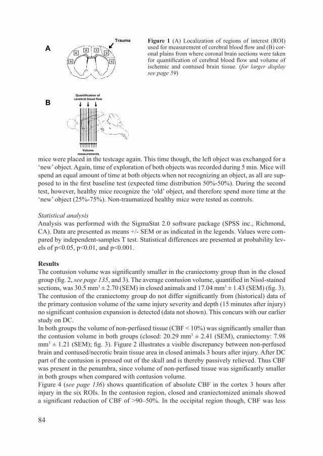
84
mice were placed in the testcage again. This time though, the left object was exchanged for a ‘new’ object. Again, time of exploration of both objects was recorded during 5 min. Mice will spend an equal amount of time at both objects when not recognizing an object, as all are sup-posedtointhefirstbaselinetest(expectedtimedistribution50%-50%).Duringthesecondtest, however, healthy mice recognize the ‘old’ object, and therefore spend more time at the ‘new’ object (25%-75%). Non-traumatized healthy mice were tested as controls.
StatisticalanalysisAnalysis was performed with the SigmaStat 2.0 software package (SPSS inc., Richmond, CA). Data are presented as means +/- SEM or as indicated in the legends. Values were com-pared by independent-samples T test. Statistical differences are presented at probability lev-els of p<0.05, p<0.01, and p<0.001.
ResultsThecontusionvolumewassignificantlysmallerinthecraniectomygroupthanintheclosedgroup(fig.2,seepage135,and3).Theaveragecontusionvolume,quantifiedinNissl-stainedsections, was 30.5 mm3 ± 2.70 (SEM) in closed animals and 17.04 mm3±1.43(SEM)(fig.3).Thecontusionofthecraniectomygroupdonotdiffersignificantlyfrom(historical)dataofthe primary contusion volume of the same injury severity and depth (15 minutes after injury) nosignificantcontusionexpansionisdetected(datanotshown).Thisconcurswithourearlierstudy on DC.Inbothgroupsthevolumeofnon-perfusedtissue(CBF<10%)wassignificantlysmallerthanthe contusion volume in both groups (closed: 20.29 mm3 ± 2.41 (SEM), craniectomy: 7.98 mm3±1.21(SEM);fig.3).Figure2illustratesavisiblediscrepancybetweennon-perfusedbrain and contused/necrotic brain tissue area in closed animals 3 hours after injury. After DC part of the contusion is pressed out of the skull and is thereby passively relieved. Thus CBF waspresentinthepenumbra,sincevolumeofnon-perfusedtissuewassignificantlysmallerin both groups when compared with contusion volume. Figure 4 (seepage136)showsquantificationofabsoluteCBFin thecortex3hoursafterinjury in the six ROIs. In the contusion region, closed and craniectomized animals showed asignificantreductionofCBFof>90–50%.Intheoccipitalregionthough,CBFwasless
Figure 1 (A) Localization of regions of interest (ROI) usedformeasurementofcerebralbloodflowand(B)cor-onal plains from where coronal brain sections were taken forquantificationofcerebralbloodflowandvolumeofischemic and contused brain tissue. (for larger displayseepage59)
Figure 1
Quantification of cerebral blood flow
Volume measurements
6
54
3
1A
B
Trauma
2

85
Figure 3. Contused versus non-per-fused parenchyma.Volume of contused (black) versus vol-ume of brain parenchyma with CBF < 10% of baseline (grey) 3 hr after TBI in closed (left) and craniectomized (right) animals (n= 5-10).
Figure 5. Quantification of absolute CBF in the hippocampus. CBF in contralateral (left) and ipsilat-eral (right) to injury in closed (black) and craniectomized (grey) animals. (*/@ p < 0.05, n=6-7).
Figure 6. Object Recognition Test. Percentage of time spend on the right object in control (black), closed (light grey) and craniectomized (dark grey) animals in respect of exploration of 2 similar objects (left) and 2 different (1 old,1new)objects(right).(#/€/*p<0.05, @p < 0.001, n=6-7)
Figure 3
closed craniectomy
Con
tusi
on (m
m3 )
0
10
20
30
40
50Nissl-staining"CBF < 10%"
@
@*
*#
#
€
all significant p < 0.01
Contusion volume vs "non-perfused volume"
€
Figure 3. Contused versus non-perfused parenchyma. Volume of contused (black) versus volume of brain parenchyma with CBF < 10% of baseline (grey) 3 hrs after TBI in closed (left) and craniectomized (right) animals (n= 5-10).
Figure 5
CBF in hippocampus
3 hours after injurycontralateral ipsilateral
% o
f con
trol
0
50
100
150
200closed craniectomy
*
* p < 0.05 vs control
@
@
@ p < 0.05, contra vs ipsilateral
Figure 5. Quantification of absolute CBF in the hippocampus. CBF in contralateral (left) andipsilateral (right) to injury in closed (black) and craniectomized (grey) animals. (*/@ p < 0.05, n=6-7)
Figure 6
Object Recognition Test
sessionsimilar objects different objects
% o
f tim
e sp
end
on ri
ght o
bjec
t
0
20
40
60
80
100controlclosedcraniectomy *
*@
@
€
€#
#
# € * p < 0.5 @ p < 0.001
Figure 6. Object Recognition Test. Percentage of time spend on the right object in control (black),closed (light grey) and craniectomized (dark grey) animals in respect of exploration of 2 similar objects (left) and 2 different (1 old, 1 new) objects (right). (#/€/* p < 0.05, @p < 0.001, n=6-7)
8�
reducedinthecraniectomizedgroup(1.2%vs21.0%).(fig.4D).Intheipsilateralcortex,inthemedialfrontalpart(ROI4),closedanimalsshowedasignificantreductionwhencra-niectomizeddidnot(fig4B).Thiswouldconcurwiththecontusionexpansionsuggestedinfigure2.Lateraltothecontusion(ROI6)bothgroupsshowedatrendtowardsareducedCBFfrontal,parietalandoccipital,thoughfewsignificant.Theclosedgroupshowedasignificantreductionparietal.Thecraniectomizedgroup,ontheotherhand,showedasignificantreduc-tionintheoccipitalROI(fig.4F).Inthecontralateralcortexnosignificantdifferencewasmeasuredbetweentheclosedandcraniectomizedgroup(fig.4A,C,E),norincomparisontocontrol values (data not shown).CBF in the hippocampus on the ipsilateral, injured, hemisphere was in both groups sig-nificantlydecreased. In thecraniectomygroupCBF in thehippocampusof the ipsilateralhemispherewassignificantlyhigher than in theclosedgroup(80.60%±15.81(SEM)vs.30.41%±20.07(SEM),resp.;fig.5).Contralateral,theclosedgroupshowedatrendtowardsreduction of CBF (86.16% ± 11.81 (SEM)).Hippocampalfunctionwasanalyzedbytheobjectrecognitiontest.NobeneficialeffectsofDCwerefound(fig.6).Theclosedgrouprecognizedtheoldobjectsignificantlybetterthanthe craniectomy group. However, this implies that the test was impure. Usually injured ani-mals are not capable of recognizing the old object. Reasons for this discrepancy are unclear. Unexpected mortality was 1 of 14 mice in both experiments (3 hours and 7 days).
DiscussionTwo recent studies from our laboratory using CCI injury, demonstrated that DC reduces con-tusionexpansionandbrainedemaformation,andimprovesfunctionaloutcomesignificantlywhen performed up to three hours after TBI (Zweckberger 2003, 2006). The way DC affects pathophysiological mechanisms besides reducing ICP itself is scarcely known. A vicious circle of known pathophysiological pathways leading to ischemia, apoptosis and necrosis is discontinued when ICP is reduced. Therefore, CBF was likely to be altered and improvement on neurological functioning expected. CBF was measured using 14C-iodoantipyrine autoradiography (Engel et al., 2007 (chapter 3b); Maeda et al., 2000). This method is from a practical point complicated, but is possible to perform in mice. Autoradiography still is the golden standard for CBF measurements. This methodwasthereforefirstchoiceforthisstudy.ItwaspreviouslyperformedinmiceafterCCI by Lundblad et al. (2004 and 2007). These investigators had used the same injury device and the same sort of mice, but different injury parameters. They used mild injury that didn’t cause a real primary contusion. Therefore our present data aren’t comparable with their pre-vious ones. Neither did they use whole body snap freezing, but decapitated mice simultane-ously with the last blood sample. The authors opinion is that whole body snap freezing is the best, and most simultaneous, way of harvesting heart blood samples and brains of the same timepoint, with loosing least radioactivity. CBF and contusion volume are measured only at 3 hours after injury/DC. Reason for this is that past this timepoint edema will develop in closed animals (Zweckberger et al., 2006), which distorts a pure comparison of CBF between closed and craniectomized animals. Be-cause DC is a permanent open system ICP will not raise after that time in mice (Zweckberger et al., 2006).Contusions in craniectomized animals 3 hours after injury didn’t expand during previous ex-periments (Zweckberger 2003, 2006). Nor do our current data show expansion of this volume

8�
at this timepoint in craniectomized animals. Besides contusion volume we have measured CBF after CCI injury previously which showed no expansion of non-perfused brain volume over time, between 15 min and 24 hours (Engel et al., 2007). In the present study 3 hours afterinjurynon-perfusedbrainvolumewassignificantlysmallerincraniectomizedanimalsthaninclosedones.Thismeansthatperfusionaroundthecontusionwassignificantlybetterafter early DC.In our previous study CBF was reduced 15 minutes after injury throughout the whole brain (Engel et al 2007). A reduction of approximately 50% was shown contralateral. Twenty-four hours after injury CBF recovered to control values. Our current data show that the time win-dow for recovery is smaller, already within 3 hours after injury. Inthecontusion,thatwassignificantlysmallerintheDCgroup,bothgroupsshowedareduc-tion of CBF of >90 – 50%. In the occipital region though, CBF was reduced less in the DC group. This can be explained by the fact that in closed animals contusions expand into the occipital cortex, and emphasizes thereby that contusions do not expand after early DC. In our animal model the contusion is evacuated spontaneously by leaving the DC open. Only skin closes on top of it. This automatically leaves space for the hippocampus and subcortical nuclei, which are otherwhise compressed by the expanding hemorrhagic contusion. We hy-pothesized that this leads to an improved function of these brain regions. As expected, CBF measured in the hippocampus on the injured side was reduced in the closed group, whereas incraniectomizedanimalsnosignificantreductionwasmeasured.Surprisingly,hippocampalfunction was not better in craniectomized animals, measured by ORT. The object recognition test has been proven an effecitve method in another injury mouse model (closed head injury) (Shein et al., 2005; Beni et al., 2006; Yatsiv et al., 2005). Unfortunately ORT has failed in our investigation, because control traumatized animals seemed to have either not enough hippo-campaldamage,orhaverecoveredquickerasexpected.Bynomeansabeneficialeffectonhippocampal function in combination with early DC was shown.Clinically, improved outcome expressed in Glasgow Outcome Scale (GOS) has been pub-lished previously (Aarabi et al., 2006, Taylor et al., 2001, Bayir et al., 2003, Yamakami and Yamaura, 1993). Jaeger (2005) and Stiefel (2004) have shown that brain tissue oxygen (PbrO2) improved immediately in the contusion area. Translation of preclinical data into the clinic is always complicated. Our present results differ from the few clinical data avaible on CBF and DC (Yamakami 1993). Yamakami et al. (1993) report on CBF measurements in craniectomized patients after TBI. CBF increased immediately underneath the area of de-compression. This differs from our rodent model, where an apparent focal contusion is made and CBF isn’t detectable. Partially this can be explained by the fact that contusions in human beings are more heterogeneous than the standardized contusion in mice, in which vasculature is damaged acutely in the primary contusion.In a pediatric population early DC has been accepted as a legitimate treatment for uncon-trollableraisedICP(Tayloretal.,2001).TaylorandcollaeguesperformedthefirstRCTtoinvestigate DC in a pediatric population. Although the sample size was small (n=27 in seven years) a better outcome was seen after DC. Subsequently two multicentre RCTs have been commenced in Australia (DECRA) and other countries (RESCUEicp). These RCTs will in-form us in a few months to years whether early DC indeed will grow up to be a legitimate therapy instead of a “second tier” therapy. Our preclinical data show that besides other patho-physiological parameters CBF is improved after TBI in mice, and the authors would like to advocate a more mature position of early DC in the treatment of uncontrollable raised ICP


89
CHAPTER 5. GENERAL DISCUSSION
The overall aim of this thesis was to investigate the importance of ischemia in traumatic brain injury from different perspectives, i.e. epidemiology, pathophysiology and therapeutic options, hypothesizing that the role of ischemia is essential in secondary brain damage after TBI. Although many preventive strategies such as motor cycle helmets and seat belts have reduced the incidence of severe TBI in the past, incidence has remained stable in an urban area between 1990-2000 (7.3 per 100.000) (see chapter 2A). The start of every medical query is epidemiology: incidence, outcome and potential prognostic factors of TBI, for many fac-torsmightinfluence(patho)physiologicalconditionsandoutcome.Aproblemneedstoex-ist before we can explore, investigate its processes and attempt to solve it. Outside the city boundaries incidence might have been higher because the majority of high-velocity injuries occur here. Young men are most affected by TBI. Despite the preventive strategies mentioned previouslyroadtrafficaccidents(RTA)havealwaysbeenandnowadaysstillarethemajorcause of injury. Our urban data though, give the impression this number has decreased. Other studies, not performed within city limits show higher rates of RTAs (Butcher et al., 2007). Conversely, the amount of falls will increase. The population is ageing; therefore more elder-lyTBIpatientscanbeexpectedinthefuture(Sundstrometal.,2007).Asignificantincreaseof elderly patients was found between 1990 and 2000 (chapter 2A). This ‘second age peak’ of age related incidence will develop into a serious impediment to society, leading to both care andfinancialdifficulties.Fallascauseofinjurywillincreaseaswellwiththistrend(Butcheret al., 2007). Overall mortality is higher in the present study: 45.8%. Lower numbers had been expected because optimal care was possible within 45 min after injury. However, other stud-iesdifferfromourfindingsbecausemorepre-hospitaldeathswereincludedbysystematicpre-hospital registration in chapter 2A. The higher mortality among elderly and the growing incidence of TBI in the present study will be responsible for a small part of higher mortality. Of the patients that survived more than half was discharged successfully, living a life outside health care facilities with none to minor disabilities. One third remained disabled and only 1.4% was discharged in a persistent vegetative state. These numbers hardly differ from previ-ously published studies (Maas et al., 1997, Marshall, 2000, Thornhill et al., 2001). Prognostic factors in chapter 2A were age, cause of injury, initial GCS and AIShead. These prognostic factors have been studied before in smaller study samples and showed to more or lesser extent the same effect (Hukkelhoven et al., 2003, Chesnut et al., 1995, Manley et al., 2001). In chapters 2B and 2C several factors were investigated in more detail. High age is associated with worse outcome (OR 2.14), with a change point at 30 years. African-American people had a higher risk for poorer outcome than Caucasians. Several reasons can be taken into consideration; lower social integration (Hart et al., 2005), lower educational level (Harri-son-Felix et al., 1998, Jorge et al., 1994), lower pre-morbid employment (Burnett et al., 2003, Wagner et al., 2000), higher drug abuse rates pre-injury (Wagner et al., 2000) and worse ac-cess to optimal acute care and rehabilitation (Bazarian et al., 2003, Burnett et al., 2003). None of these factors have been investigated thoroughly yet. A weak association exists between lower educational level and worse outcome, hypothesizing that less educated people come from societies with lower socio-economic status and thereby have less ability to access the correctcareandarepsychologicallylessabletofullybenefitrehabilitationtreatment(John-stone et al., 2003). After years of discussion gender is no prognostic factor in the present, larger, study. Even when corrected for age, and thereby eliminating the difference between

90
pre-andpostmenopausalwomen,nosignificantdifferencewasfound.Hypotension,hypoxiaand hypothermia are different prognostic factors. An insult of either factor is associated with poorer outcome (ORs 2.7, 2.1, 2.2 respectively). Several insults of one or a combination of hypotension and hypoxia lead to worse outcome than one insult alone (Chapter 2C, Manley et al., 2001). They merge into an evil triad, causing cumulative secondary brain damage. These factors differ from e.g. age in that these directly change physiological conditions in the brain into e.g. decreased CBF. Hypothetically hypoxia, hypotension and hypothermia can be prevented or treated. Unfortunately comorbidity overrules prevention or treatment chronically through i.e. cardiovascular disease. Acute comorbidity is caused by polytrauma, e.g. (excessive) extracranial bleeding, or drugs needed for intubation leading to hypotension. The current studies proved stronger as well as previous smaller studies the detrimental effects of these prognostic factors (Chapter 2A,B,C, Hukkelhoven et al., 2003, Chesnut et al., 1993, Manley et al., 2001, Wagner et al., 2000). These facts will assist physicians with decision-making in the future.
The pathophysiology of TBI has been investigated to a large extent during the past decades, but certainly not everything is clear. Secondary brain damage caused by ischemia was inves-tigated in chapter 3. Ischemia itself leads to cell death invitro also when superimposed by increased CO2 after mechanical injury (chapter 3A). Reactive oxygen species are generated after injury (Leker and Shohami, 2002, Samdani et al., 1997). Superoxide, hydroxyl radicals and nitric oxide, which is generated by nitric oxide synthase, can interact with each other and form one of the most damaging free radical species, peroxynitrite (Dawson and Dawson, 1996). The protective role of free radical scavengers was tested (chapter 3A). Only one of them,7-NINA,hadasmallsignificanteffectwhenadministeredpre-injury.Combiningtwofree radical scavengers, with a broader range of effects, neither had the expected effect. According to data shown in chapter 3B ischemia is actually not the main reason for second-ary contusion expansion in CCI injury in mice. The volume of a cortical contusion expands significantlyovertimeduringthefirst24hoursafterexperimentalTBI(Zweckbergeretal.,2003, Chapter 3B). Measurements of absolute CBF by 14C-iodoantipyrine autoradiography revealed that thevolumeof ischemic tissuedoesnotexpandsignificantlyafterTBI.Thisindicates that progressive penumbral ischemia may play only a minor role in delayed peri-contusional cell death and hence contusion expansion. The ischemic threshold (20 ml/mg/min) was only found within the primary contusion. Hypoperfusion was seen throughout the whole brain, thus in the penumbra as well. Nevertheless, contusions expand and a reduction of CBF is strongly related to an adverse outcome after TBI (Hlatky et al., 2004, Soustiel et al., 2005). Other factors must be more accountable. The vasculature is altered after injury. The blood brain barrier (BBB) becomes permeable to proteins early after injury (Kawamata et al., 2000, Maeda et al., 2003). This results in vasogenic oedema, and therefore causes secondary cell death by fuelling cytotoxic oedema. Osmolality in the penumbra increases, which ag-gravates further cell death by switching on apoptotic pathways and increasing excitotoxicity (Katayamaetal.,1998).Ourfindingsindicatethatischemiaisnotpresenteither15minnor24 hours after injury, and that mechanisms such as vasogenic and cytotoxic oedema lead-ing to increased intracranial pressure (ICP) fuel apoptosis and excitotoxicity, may be more relevant to contusion expansion. TBI has broad pathophysiological effects and a small and earlytreatmentwindow,whereasthefirsttwenty-fourtoforty-eighthoursareoftenthemostimportant. In conclusion, ischemia does kill cells invitro, but free radical scavengers alone

91
can hardly reduce this cell death. Hence, ischemia is disproved to be the most important fac-tor to SCE after CCI in mice in chapter 3B. With the understanding of several pathophysi-ological pathways, many therapeutic options have gone through trial and error. Decompres-sive craniectomy (DC) is one of these therapeutic options that is currently living its third life during modern history. Current research methods combined with old methods explored the pathophysiology and effects of DC in more depth. DC cracks the Monroe-Kellie doctrine of the rigid case in which pressure can only build up. DC releases pressure from a medical uncontrollable raised ICP (chapter 4A). Several parameters have been measured in chapter 4.ICPraiseandoedemaformationweresignificantlylessincraniectomisedmice,thoughtime of performing DC was of utmost importance. In mice, already 8 hours after trauma, DC showednobeneficialeffectsanymore.CBFmeasuredbyLaserDoppler(LD)andautoradi-ography differed on few parts in the brain only. The combination of LD and autoradiography is of great value, because LD measures CBF through time, but shallow, and autoradiography only at one time point, though with better resolution. In this thesis the non-perfused volume was significantly smaller than contusion volume inmice.The perfusion deficitwas evensmaller in DC animals; therefore ischemia cannot be the main reason for SCE in this type of injury. However, this does not mean that ischemia is not harmful. As discussed in chapter 3A, ischemia does kill cells, but in order to do so ischemia must be present. It is likely that ischemia is present in other types of injury, such as diffuse (vascular) injury. The presence of ischemia in the penumbra in human studies with multicontusional injury was actually shown (Bouma et al., 1991, Hlatky et al., 2004, Liu et al., 2002).
FutureThe expression “treatable” in the context of TBI should be interpreted broadly in the con-text of prevention, pre/in-hospital medical care and rehabilitation. Prevention is a hot topic in today’s medical world. Is it possible to prevent TBI in the future with further preventive strategies? Can cars become safer for driver, pedestrian and cyclist, although the amount of patients suffering from whiplash injury has increased since the use of seatbelts (Montfoort et al., 2006)? Lower social integration, lower educational level, lower pre-morbid employ-ment, higher drug abuse pre-injury rates, less access to optimal acute care/rehabilitation are all factors that are shown to have a negative effect on outcome. None of these factors have been investigated thoroughly enough yet. A strenuous effort should be made towards the investigation of these parameters. AlthoughnobeneficialeffectswereseenspecificallyonCBFafterearlyDC,earlyDCdoesshowbeneficial effectsonoutcomeofmice.More information isneededonmechanismsofDC,inordertoimprovetheidentificationofpatientswhopossiblybenefitfromDC.Inseveral chapters the BBB was discussed in the role of oedema formation, and thereby ICP increase. As we know now from CBF, oedema formation and ICP data is that oedema and raised ICP itself, and not a low CBF might be responsible for SCE. In our model oedema couldbecausedbyadisruptedBBBduringthefirstfewhoursafterinjury(Zweckbergeretal., 2003). Whether and how early DC affects the BBB is unknown. The outcome of TBI patients after DC will be revealed through the earlier mentioned tri-als DECRA and RESCUEicp. These studies are very important in future determination of guidelines for the treatment of severe TBI. A great challenge will be standardizing possible changes in favour of performing early DC. In a running study large differences are seen when data between cities and even countries are compared. For example, The Vrije Universiteit

92
Medical Centre in Amsterdam has performed many more DCs than University Medical Cen-tre Groningen in the past three years (approximately 20% and 4% respectively of all severe TBI patients). Average amount of DCs in the Netherlands (± 9%) in comparison to Germany and Austria (both ± 16%) differ as well. Data on outcome seems promising (data not shown, personalcommunication),butofcoursetherandomisedcontrolledtrialswillprovideafinalanswer. AnotherfieldofinterestwithinTBItreatmentis,asdiscussedinchapter3A,pharmacologicalagents. An excellent review was written by Schouten (2007), but with disappointing conclu-sions; no pharmacological agents have been associated with better outcome yet. A combina-tion of pharmacological agents might be useful, though in chapter 3A the use of several reac-tiveoxygenspecies-inhibitorsdidnotshowtheexpectedbeneficialeffect.Targetingmoreparts of pathophysiological pathways at once might be the future. Combination of therapeutic optionsisvaluableine.g.thefieldofoncology.However,pharmacologicalagentsusedintheacute phase can also aggravate damage by causing hypotension. This might be the case with the use of rapid sequence intubation (pre-)hospital.
This thesis shows that TBI is still a harmful disease that is not likely to be diminished eas-ily. Many factors play a role; though the hypothesis that ischemia plays a main role must be falsifiedforcontusionalinjury.Thisthesisthoughverifiesthatpathophysiologicalpathwaysstart very early and treatment must be initiated as early as possible. There is no time to sit and wait, and think about what can be done in a few hours, when ICP has reached medical uncon-trollable heights. DC must be considered early in the decisive therapeutic process i.e. within hours of rising ICP. This necessitates ongoing optimal cooperation among (neuro)surgeons, intensivists and anaesthesiologists.


95
ReferencesAarabi B, Hesdorffer DC, Ahn ES, Aresco C, Scalea TM, Eisenberg HM (2006) Outcome following de-compressive craniectomy for malignant swelling due to severe head injury. J Neurosurg 104:469-479.
Acosta JA, Yang JC, Winchell RJ, Simons RK, Fortlage DA, Hollingsworth-Fridlund P, Hoyt DB (1998) Lethal injuries and time to death in a level I trauma center. J Am Coll Surg 186:528-533.
Albanese J, Leone M, Alliez JR, Kaya JM, Antonini F, Alliez B, Martin C (2003) Decompressive craniectomy for severe traumatic brain injury: Evaluation of the effects at one year. Crit Care Med 31:2535-2538.
Alexander E, Ball MR, Laster DW (1987) Subtemporal decompression: radiological observations and current surgical experience. Br J Neurosurg 1:427-433.
AlexanderMJ,MartinNA,KhannaR, CaronM, BeckerDP (1994) Regional cerebral blood flowtrends in head injured patients with focal contusions and cerebral edema. Acta Neurochir Suppl (Wien) 60:479-481.
Andlin-Sobocki P, Jonsson B, Wittchen HU, Olesen J (2005) Cost of disorders of the brain in Europe. Eur J Neurol 12 Suppl 1:1-27.
Andrews PJ, Sleeman DH, Statham PF, McQuatt A, Corruble V, Jones PA, Howells TP, Macmillan CS (2002) Predicting recovery in patients suffering from traumatic brain injury by using admission variables and physiological data: a comparison between decision tree analysis and logistic regression. J Neurosurg 97:326-336.
Annegers JF, Grabow JD, Kurland LT, Laws ER, Jr. (1980) The incidence, causes, and secular trends of head trauma in Olmsted County, Minnesota, 1935-1974. Neurology 30:912-919.
Annoni JM, Beer S, Kesselring J (1991) [Sequelae of severe craniocerebral injuries. An epidemiologi-cal study in the Canton of St. Gallen]. Schweiz Med Wochenschr 121:207-213.
Asikainen I, Kaste M, Sarna S (1996) Patients with traumatic brain injury referred to a rehabilitation and re-employment programme: social and professional outcome for 508 Finnish patients 5 or more years after injury. Brain Inj 10:883-899.
Asikainen I, Kaste M, Sarna S (1998) Predicting late outcome for patients with traumatic brain injury referred to a rehabilitation programme: a study of 508 Finnish patients 5 years or more after injury. Brain Inj 12:95-107.
Asikainen I, Kaste M, Sarna S (1999) Early and late posttraumatic seizures in traumatic brain injury rehabilitationpatients:braininjuryfactorscausinglateseizuresandinfluenceofseizuresonlong-termoutcome. Epilepsia 40:584-589.
Asikainen I, Nybo T, Muller K, Sarna S, Kaste M (1999) Speed performance and long-term functional and vocational outcome in a group of young patients with moderate or severe traumatic brain injury. Eur J Neurol 6:179-185.
Baethmann A, Maier-Hauff K, Kempski O, Unterberg A, Wahl M, Schurer L (1988) Mediators of brain edema and secondary brain damage. Crit Care Med 16:972-978.
Bahloul M, Chelly H, Ben Hmida M, Ben Hamida C, Ksibi H, Kallel H, Chaari A, Kassis M, Rekik N, Bouaziz M (2004) Prognosis of traumatic head injury in South Tunisia: a multivariate analysis of 437 cases. J Trauma 57:255-261.

9�
BalestreriM,CzosnykaM,ChatfieldDA,SteinerLA,SchmidtEA,SmielewskiP,MattaB,PickardJD(2004) Predictive value of Glasgow Coma Scale after brain trauma: change in trend over the past ten years. J Neurol Neurosurg Psychiatry 75:161-162.
Bayir H, Clark RS, Kochanek PM (2003) Promising strategies to minimize secondary brain injury after head trauma. Crit Care Med 31:S112-117.
Bayir H, Marion DW, Puccio AM, Wisniewski SR, Janesko KL, Clark RS, Kochanek PM (2004) Marked gender effect on lipid peroxidation after severe traumatic brain injury in adult patients. J Neu-rotrauma 21:1-8.
Bazarian JJ, Pope C, McClung J, Cheng YT, Flesher W (2003) Ethnic and racial disparities in emer-gency department care for mild traumatic brain injury. Acad Emerg Med 10:1209-1217.
Bell RM, Krantz BE, Weigelt JA (1999) ATLS: a foundation for trauma training. Ann Emerg Med 34:233-237.
Beni SM, Tsenter J, Alexandrovich AG, Galron-Krool N, Barzilai A, Kohen R, Grigoriadis N, Sime-onidouC, ShohamiE (2006)CuZn-SODdeficiency, rather than overexpression, is associatedwithenhanced recovery and attenuated activation of NF-kappaB after brain trauma in mice. J Cereb Blood Flow Metab 26:478-490.
BetzAL,IannottiF,HoffJT(1989)Brainedema:aclassificationbasedonblood-brainbarrierintegrity.Cerebrovasc Brain Metab Rev 1:133-154.
Bouillon B, Troidl H, Neugebauer E, Tiling T, Schweins M, Vorweg M (1990) The Cologne prehospital trauma system: results and criteria for its evaluation. Theor Surg 5:36-43.
Bouillon B, Lefering R, Vorweg M, Tiling T, Neugebauer E, Troidl H (1997) Trauma score systems: Cologne Validation Study. J Trauma 42:652-658.
Bouillon B, Raum M, Fach H, Buchheister B, Lefering R, Menzel J, Klug N (1999) The incidence and outcomeofseverebraintrauma-Designandfirstresultsofanepidemiologicalstudyinanurbanarea.Restor Neurol Neurosci 14:85-92.
BoumaGJ,MuizelaarJP(1992)Cerebralbloodflow,cerebralbloodvolume,andcerebrovascularreac-tivity after severe head injury. J Neurotrauma 9 Suppl 1:S333-348.
Bouma GJ, Muizelaar JP, Choi SC, Newlon PG, Young HF (1991) Cerebral circulation and metabolism after severe traumatic brain injury: the elusive role of ischemia. J Neurosurg 75:685-693.
Braakman R, Gelpke GJ, Habbema JD, Maas AI, Minderhoud JM (1980) Systematic selection of prog-nostic features in patients with severe head injury. Neurosurgery 6:362-370.
Bradbury A, Robertson C (1993) Prospective audit of the pattern, severity and circumstances of injury sustainedbyvehicleoccupantsasaresultofroadtrafficaccidents.ArchEmergMed10:15-23.
Brain Trauma Foundation, American Association of Neurological Surgeons, Joint Section on Neu-rotrauma and Critical Care (1996) Guidelines for the management of severe head injury. J Neurotrauma 13:641-734.
Brain Trauma Foundation (BTF), American Association of Neurological Surgeons (AANS), Joint Sec-tion on Neurotrauma and Critical Care (2000) Guidelines for the management of severe head injury. J Neurotrauma 17:457-627.
Brain Trauma Foundation (BTF) (2000) Guidelines for the prehospital management of traumatic brain injury. In: Brain Trauma Foundation. New York.

9�
Bricolo A, Turazzi S, Alexandre A, Rizzuto N (1977) Decerebrate rigidity in acute head injury. J Neu-rosurg 47:680-689.
BrindleR(1992)LocalstreetspeedmanagementinAustralia--isit“trafficcalming”?AccidAnalPrev24:29-38.
Britt RH, Hamilton RD (1978) Large decompressive craniotomy in the treatment of acute subdural hematoma. Neurosurgery 2:195-200.
Bruns J, Jr., Hauser WA (2003) The epidemiology of traumatic brain injury: a review. Epilepsia 44 Suppl 10:2-10.
BryanRM,Jr.,CherianL,RobertsonC(1995)Regionalcerebralbloodflowaftercontrolledcorticalimpact injury in rats. Anesth Analg 80:687-695.
Bullock R, Maxwell WL, Graham DI, Teasdale GM, Adams JH (1991) Glial swelling following human cerebral contusion: an ultrastructural study. J Neurol Neurosurg Psychiatry 54:427-434.
Bullock R, Sakas D, Patterson J, Wyper D, Hadley D, Maxwell W, Teasdale GM (1992) Early post-traumaticcerebralbloodflowmapping:correlationwithstructuraldamageafterfocalinjury.ActaNeu-rochir Suppl (Wien) 55:14-17.
Burkert W, Plaumann H (1989) [The value of large pressure-relieving trepanation in treatment of refrac-tory brain edema. Animal experiment studies, initial clinical results]. Zentralbl Neurochir 50:106-108.
Burnett DM, Kolakowsky-Hayner SA, Slater D, Stringer A, Bushnik T, Zafonte R, Cifu DX (2003) Ethnographic analysis of traumatic brain injury patients in the national Model Systems database. Arch Phys Med Rehabil 84:263-267.
Butcher I, Maas AI, Lu J, Marmarou A, Murray GD, Mushkudiani NA, McHugh GS, Steyerberg EW (2007) Prognostic value of admission blood pressure in traumatic brain injury: results from the IM-PACT study. J Neurotrauma 24:294-302.
Butcher I, McHugh GS, Lu J, Steyerberg EW, Hernandez AV, Mushkudiani N, Maas AI, Marmarou A, Murray GD (2007) Prognostic value of cause of injury in traumatic brain injury: results from the IMPACT study. J Neurotrauma 24:281-286.
CaldwellMT,McGovernEM(1993)Fataltrauma:afiveyearreviewinaDublinhospital.IrJMedSci162:309-312.
Chamelian L, Feinstein A (2004) Outcome after mild to moderate traumatic brain injury: the role of dizziness. Arch Phys Med Rehabil 85:1662-1666.
Champion HR, Sacco WJ, Carnazzo AJ, Copes W, Fouty WJ (1981) Trauma score. Crit Care Med 9:672-676.
Champion HR, Sacco WJ, Copes WS, Gann DS, Gennarelli TA, Flanagan ME (1989) A revision of the Trauma Score. J Trauma 29:623-629.
Changaris DG, McGraw CP, Richardson JD, Garretson HD, Arpin EJ, Shields CB (1987) Correlation of cerebral perfusion pressure and Glasgow Coma Scale to outcome. J Trauma 27:1007-1013.
Chen Y, Constantini S, Trembovler V, Weinstock M, Shohami E (1996) An experimental model of closedhead injury inmice: pathophysiology, histopathology, and cognitive deficits. JNeurotrauma13:557-568.
98
Cherian L, Robertson CS, Contant CF, Jr., Bryan RM, Jr. (1994) Lateral cortical impact injury in rats: cerebrovascular effects of varying depth of cortical deformation and impact velocity. J Neurotrauma 11:573-585.
Chesnut RM, Marshall LF, Klauber MR, Blunt BA, Baldwin N, Eisenberg HM, Jane JA, Marmarou A, Foulkes MA (1993) The role of secondary brain injury in determining outcome from severe head injury. J Trauma 34:216-222.
Chiu WT, Huang SJ, Tsai SH, Lin JW, Tsai MD, Lin TJ, Huang WC (2007) The impact of time, legisla-tion, and geography on the epidemiology of traumatic brain injury. J Clin Neurosci 14:930-935.
Cho S, Park EM, Zhou P, Frys K, Ross ME, Iadecola C (2005) Obligatory role of inducible nitric oxide synthase in ischemic preconditioning. J Cereb Blood Flow Metab 25:493-501.
Choi DW (1995) Calcium: still center-stage in hypoxic-ischemic neuronal death. Trends Neurosci 18:58-60.
Choi SC, Muizelaar JP, Barnes TY, Marmarou A, Brooks DM, Young HF (1991) Prediction tree for severely head-injured patients. J Neurosurg 75:251-255.
Clark RS, Kochanek PM, Dixon CE, Chen M, Marion DW, Heineman S, DeKosky ST, Graham SH (1997) Early neuropathologic effects of mild or moderate hypoxemia after controlled cortical impact injury in rats. J Neurotrauma 14:179-189.
Clark RS, Kochanek PM, Chen M, Watkins SC, Marion DW, Chen J, Hamilton RL, Loeffert JE, Gra-ham SH (1999) Increases in Bcl-2 and cleavage of caspase-1 and caspase-3 in human brain after head injury. Faseb J 13:813-821.
Clifton GL, Miller ER, Choi SC, Levin HS, McCauley S, Smith KR, Jr., Muizelaar JP, Wagner FC, Jr., Marion DW, Luerssen TG, Chesnut RM, Schwartz M (2001) Lack of effect of induction of hypothermia after acute brain injury. N Engl J Med 344:556-563.
Colicos MA, Dixon CE, Dash PK (1996) Delayed, selective neuronal death following experimental corticalimpactinjuryinrats:possibleroleinmemorydeficits.BrainRes739:111-119.
Colohan AR, Alves WM, Gross CR, Torner JC, Mehta VS, Tandon PN, Jane JA (1989) Head injury mortality in two centers with different emergency medical services and intensive care. J Neurosurg 71:202-207.
Combes P, Fauvage B, Colonna M, Passagia JG, Chirossel JP, Jacquot C (1996) Severe head injuries: an outcome prediction and survival analysis. Intensive Care Med 22:1391-1395.
Cooper PR, Rovit RL, Ransohoff J (1976) Hemicraniectomy in the treatment of acute subdural hema-toma: a re-appraisal. Surg Neurol 5:25-28.
Cooper PR, Hagler H, Clark WK, Barnett P (1979) Enhancement of experimental cerebral edema after decompressive craniectomy: implications for the management of severe head injuries. Neurosurgery 4:296-300.
Culmsee C, Junker V, Wolz P, Semkova I, Krieglstein J (1998) Lubeluzole protects hippocampal neurons from excitotoxicity in vitro and reduces brain damage caused by ischemia. Eur J Pharmacol 342:193-201.
Dawson VL (1999) Potent neuroprotectants linked to bifunctional inhibition. Proc Natl Acad Sci U S A 96:10557-10558.
Dawson VL, Dawson TM (1996) Nitric oxide in neuronal degeneration. Proc Soc Exp Biol Med 211:33-40.

99
Della Corte F, Giordano A, Pennisi MA, Barelli A, Caricato A, Campioni P, Galli G (1997) Quantitative cerebralbloodflowandmetabolismdeterminationinthefirst48hoursaftersevereheadinjurywithanew dynamic SPECT device. Acta Neurochir (Wien) 139:636-641; discussion 641-632.
Demetriades D, Kuncir E, Murray J, Velmahos GC, Rhee P, Chan L (2004) Mortality prediction of head Abbreviated Injury Score and Glasgow Coma Scale: analysis of 7,764 head injuries. J Am Coll Surg 199:216-222.
Demetriades D, Kuncir E, Velmahos GC, Rhee P, Alo K, Chan LS (2004) Outcome and prognostic fac-tors in head injuries with an admission Glasgow Coma Scale score of 3. Arch Surg 139:1066-1068.
Demetriades D, Kuncir E, Murray J, Velmahos GC, Rhee P, Chan L (2004) Mortality prediction of head Abbreviated Injury Score and Glasgow Coma Scale: analysis of 7,764 head injuries. J Am Coll Surg 199:216-222.
Dereeper E, Ciardelli R, Vincent JL (1998) Fatal outcome after polytrauma: multiple organ failure or cerebral damage? Resuscitation 36:15-18.
DeWittDS,ProughDS,TaylorCL,WhitleyJM(1992)Reducedcerebralbloodflow,oxygendelivery,and electroencephalographic activity after traumatic brain injury and mild hemorrhage in cats. J Neu-rosurg 76:812-821.
DeWittDS,JenkinsLW,WeiEP,LutzH,BeckerDP,KontosHA(1986)Effectsoffluid-percussionbraininjuryonregionalcerebralbloodflowandpialarteriolardiameter.JNeurosurg64:787-794.
Diener HC, Cortens M, Ford G, Grotta J, Hacke W, Kaste M, Koudstaal PJ, Wessel T (2000) Lubeluzole in acute ischemic stroke treatment: A double-blind study with an 8-hour inclusion window comparing a 10-mg daily dose of lubeluzole with placebo. Stroke 31:2543-2551.
Dietrich WD, Alonso O, Busto R, Prado R, Zhao W, Dewanjee MK, Ginsberg MD (1998) Posttraumatic cerebralischemiaafterfluidpercussionbraininjury:anautoradiographicandhistopathologicalstudyinrats. Neurosurgery 43:585-593; discussion 593-584.
Edna TH (1983) Risk factors in traumatic head injury. Acta Neurochir (Wien) 69:15-21.
Eldadah BA, Faden AI (2000) Caspase pathways, neuronal apoptosis, and CNS injury. J Neurotrauma 17:811-829.
Ellenberg JH, Levin HS, Saydjari C (1996) Posttraumatic Amnesia as a predictor of outcome after severe closed head injury. Prospective assessment. Arch Neurol 53:782-791.
Ellis EF, McKinney JS, Willoughby KA, Liang S, Povlishock JT (1995) A new model for rapid stretch-induced injury of cells in culture: characterization of the model using astrocytes. J Neurotrauma 12:325-339.
Engberg A (1995) Severe traumatic brain injury--epidemiology, external causes, prevention, and reha-bilitation of mental and physical sequelae. Acta Neurol Scand Suppl 164:1-151.
Ennaceur A, Delacour J (1988) A new one-trial test for neurobiological studies of memory in rats. 1: Behavioral data. Behav Brain Res 31:47-59.
EvansL(1991)Trafficsafetyandthedriver.NewYork:VanNostrandReinhold.
Farace E, Alves WM (2000) Do women fare worse: a metaanalysis of gender differences in traumatic brain injury outcome. J Neurosurg 93:539-545.

100
Farin A, Deutsch R, Biegon A, Marshall LF (2003) Sex-related differences in patients with severe head injury: greater susceptibility to brain swelling in female patients 50 years of age and younger. J Neurosurg 98:32-36.
Farin A, Deutsch R, Biegon A, Marshall LF (2003) Sex-related differences in patients with severe head injury: greater susceptibility to brain swelling in female patients 50 years of age and younger. J Neurosurg 98:32-36.
Fearnside FR, Simpson AD (1997) Epidemiology. In: Head Injury (Reilly P, Bullock R, eds). London: Chapman and Hall.
FieschiC,BattistiniN,BeduschiA,BoselliL,RossandaM(1974)Regionalcerebralbloodflowandintraventricular pressure in acute head injuries. J Neurol Neurosurg Psychiatry 37:1378-1388.
Figaji AA, Fieggen AG, Peter JC (2003) Early decompressive craniotomy in children with severe trau-matic brain injury. Childs Nerv Syst 19:666-673.
Fiskum G (2000) Mitochondrial participation in ischemic and traumatic neural cell death. J Neurotrau-ma 17:843-855.
Force BTT (2000) Management and prognosis of severe traumatic brain injury. J Neurotrauma 17:451-553.
Foulkes MA, Eisenberg HM, Jane JA, Marmarou A, Marshall LF (1991) The Traumatic Coma Data Bank: design, methods, and baseline characteristics. J Neurosurg Suppl. 75:S8-S13.
Gaab MR, Rittierodt M, Lorenz M, Heissler HE (1990) Traumatic brain swelling and operative decom-pression: a prospective investigation. Acta Neurochir Suppl (Wien) 51:326-328.
GaabM,KnoblichOE,FuhrmeisterU,PflughauptKW,DietrichK(1979)Comparisonoftheeffectsof surgical decompression and resection of local edema in the therapy of experimental brain trauma. Investigation of ICP, EEG and cerebral metabolism in cats. Childs Brain 5:484-498.
Gan BK, Lim JH, Ng IH (2004) Outcome of moderate and severe traumatic brain injury amongst the elderly in Singapore. Ann Acad Med Singapore 33:63-67.
Garnett MR, Blamire AM, Corkill RG, Rajagopalan B, Young JD, Cadoux-Hudson TA, Styles P (2001) Abnormal cerebral blood volume in regions of contused and normal appearing brain following trau-matic brain injury using perfusion magnetic resonance imaging. J Neurotrauma 18:585-593.
Gennarelli TA, Champion HR, Copes WS, Sacco WJ (1994) Comparison of mortality, morbidity, and severity of 59,713 head injured patients with 114,447 patients with extracranial injuries. J Trauma 37:962-968.
GennarelliTA,SpielmanGM,LangfittTW,GildenbergPL,HarringtonT,JaneJA,MarshallLF,MillerJD,PittsLH(1982)Influenceofthetypeofintracraniallesiononoutcomefromsevereheadinjury.JNeurosurg 56:26-32.
Gentleman D (1992) Causes and effects of systemic complications among severely head injured pa-tients transferred to a neurosurgical unit. Int Surg 77:297-302.
Geraldina P, Mariarosaria L, Annarita A, Susanna G, Michela S, Alessandro D, Sandra S, Enrico C (2003) Neuropsychiatric sequelae in TBI: a comparison across different age groups. Brain Inj 17:835-846.Ghajar J (2000) Traumatic brain injury. Lancet 356:923-929.
Ghajar J, Hariri RJ, Narayan RK, Iacono LA, Firlik K, Patterson RH (1995) Survey of critical care management of comatose, head-injured patients in the United States. Crit Care Med 23:560-567.
101
Ginsberg MD, Zhao W, Alonso OF, Loor-Estades JY, Dietrich WD, Busto R (1997) Uncoupling of local cerebralglucosemetabolismandbloodflowafteracutefluid-percussioninjuryinrats.AmJPhysiol272:H2859-2868.
Glass TF, Reeves B, Sharp FR (2002) Modeling both the mechanical and hypoxic features of traumatic brain injury in vitro in rats. Neurosci Lett 328:133-136.
Gomez PA, Lobato RD, Boto GR, De la Lama A, Gonzalez PJ, de la Cruz J (2000) Age and outcome after severe head injury. Acta Neurochir (Wien) 142:373-380; discussion 380-371.
Gordon E, von Holst H, Rudehill A (1995) Outcome of head injury in 2298 patients treated in a single clinic during a 21-year period. J Neurosurg Anesthesiol 7:235-247.
Graham DI, Adams JH, Doyle D (1978) Ischaemic brain damage in fatal non-missile head injuries. J Neurol Sci 39:213-234.
Grande PO, Naredi S (2002) Clinical studies in severe traumatic brain injury: a controversial issue. Intensive Care Med 28:529-531.
Groger M, Lebesgue D, Pruneau D, Relton J, Kim SW, Nussberger J, Plesnila N (2005) Release of bra-dykinin and expression of kinin B2 receptors in the brain: role for cell death and brain edema formation after focal cerebral ischemia in mice. J Cereb Blood Flow Metab 25:978-989.
Groswasser Z, Cohen M, Keren O (1998) Female TBI patients recover better than males. Brain Inj 12:805-808.
Guerra WK, Gaab MR, Dietz H, Mueller JU, Piek J, Fritsch MJ (1999) Surgical decompression for traumatic brain swelling: indications and results. J Neurosurg 90:187-196.
Hall ED, Braughler JM (1989) Central nervous system trauma and stroke. II. Physiological and phar-macological evidence for involvement of oxygen radicals and lipid peroxidation. Free Radic Biol Med 6:303-313.
Hall ED, Detloff MR, Johnson K, Kupina NC (2004) Peroxynitrite-mediated protein nitration and lipid peroxidation in a mouse model of traumatic brain injury. J Neurotrauma 21:9-20.
Harris C, DiRusso S, Sullivan T, Benzil DL (2003) Mortality risk after head injury increases at 30 years. J Am Coll Surg 197:711-716.
Harrison-Felix C, Zafonte R, Mann N, Dijkers M, Englander J, Kreutzer J (1998) Brain injury as a resultofviolence:preliminaryfindingsfromthetraumaticbraininjurymodelsystems.ArchPhysMedRehabil 79:730-737.
Hart T, Whyte J, Polansky M, Kersey-Matusiak G, Fidler-Sheppard R (2005) Community outcomes following traumatic brain injury: impact of race and preinjury status. J Head Trauma Rehabil 20:158-172.
Hartwig E, Dirks B, Oldenkott P, Pfenninger E, Helm M, Kinzl L (1993) [Management of the patient with craniocerebral injuries at the accident site and clinic admission]. Unfallchirurg 96:564-568.
Heiskanen O, Sipponen P (1970) Prognosis of severe brain injury. Acta Neurol Scand 46:343-348.
Hejazi N, Witzmann A, Fae P (2002) Unilateral decompressive craniectomy for children with severe brain injury. Report of seven cases and review of the relevant literature. Eur J Pediatr 161:99-104.
Hendrich KS, Kochanek PM, Williams DS, Schiding JK, Marion DW, Ho C (1999) Early perfusion aftercontrolledcorticalimpactinrats:quantificationbyarterialspin-labeledMRIandtheinfluenceofspin-lattice relaxation time heterogeneity. Magn Reson Med 42:673-681.

102
HlatkyR,ContantCF,Diaz-MarchanP,ValadkaAB,RobertsonCS(2004)Significanceofareducedcerebralbloodflowduringthefirst12hoursaftertraumaticbraininjury.NeurocritCare1:69-83.
HodgsonNF,StewartTC,GirottiMJ(2000)Autopsiesanddeathcertificationindeathsduetoblunttrauma: what are we missing? Can J Surg 43:130-136.
Hoffman WE, Charbel FT, Edelman G, Hannigan K, Ausman JI (1996) Brain tissue oxygen pressure, carbon dioxide pressure and pH during ischemia. Neurol Res 18:54-56.
Hoffmann B, Duwecke C, von Wild KR (2002) Neurological and social long-term outcome after early rehabilitation following traumatic brain injury. 5-year report on 240 TBI patients. Acta Neurochir Suppl 79:33-35.
HolmstromA,AkesonJ(2005)Sevofluraneinduceslesscerebralvasodilationthanisofluraneat thesame A-line autoregressive index level. Acta Anaesthesiol Scand 49:16-22.
HoofienD,VakilE,GilboaA,DonovickPJ,BarakO(2002)Comparisonofthepredictivepowerofsocio-economic variables, severity of injury and age on long-term outcome of traumatic brain injury: sample-specificvariablesversusfactorsaspredictors.BrainInj16:9-27.
Huang HM, Shen CC, Ou HC, Yu JY, Chen HL, Kuo JS, Hsieh SJ (2002) Neuroprotective MK801 is associated with nitric oxide synthase during hypoxia/reoxygenation in rat cortical cell cultures. J Cell Biochem 84:367-376.
Hukkelhoven CW, Steyerberg EW, Farace E, Habbema JD, Marshall LF, Maas AI (2002) Regional differences in patient characteristics, case management, and outcomes in traumatic brain injury: experi-ence from the tirilazad trials. J Neurosurg 97:549-557.
Hukkelhoven CW, Steyerberg EW, Rampen AJ, Farace E, Habbema JD, Marshall LF, Murray GD, Maas AI (2003) Patient age and outcome following severe traumatic brain injury: an analysis of 5600 patients. J Neurosurg 99:666-673.
Ikeda J, Ma L, Morita I, Murota S (1994) Involvement of nitric oxide and free radical (O2-) in neuronal injury induced by deprivation of oxygen and glucose in vitro. Acta Neurochir Suppl (Wien) 60:94-97.
Ingebrigtsen T, Mortensen K, Romner B (1998) The epidemiology of hospital-referred head injury in northern Norway. Neuroepidemiology 17:139-146.
Ishige N, Pitts LH, Berry I, Carlson SG, Nishimura MC, Moseley ME, Weinstein PR (1987) The effect of hypoxia on traumatic head injury in rats: alterations in neurologic function, brain edema, and cere-bralbloodflow.JCerebBloodFlowMetab7:759-767.
Jaeger M, Soehle M, Meixensberger J (2005) Improvement of brain tissue oxygen and intracranial pres-sure during and after surgical decompression for diffuse brain oedema and space occupying infarction. Acta Neurochir Suppl 95:117-118.
Jagannathan J, Okonkwo DO, Dumont AS, Ahmed H, Bahari A, Prevedello DM, Jane JA, Sr., Jane JA, Jr. (2007) Outcome following decompressive craniectomy in children with severe traumatic brain injury: a 10-year single-center experience with long-term follow up. J Neurosurg 106:268-275.
Jager TE, Weiss HB, Coben JH, Pepe PE (2000) Traumatic brain injuries evaluated in U.S. emergency departments, 1992-1994. Acad Emerg Med 7:134-140.
JaggiJL,ObristWD,GennarelliTA,LangfittTW(1990)Relationshipofearlycerebralbloodflowandmetabolism to outcome in acute head injury. J Neurosurg 72:176-182.
JayTM,LucignaniG,CraneAM,JehleJ,SokoloffL(1988)Measurementoflocalcerebralbloodflowwith [14C]iodoantipyrine in the mouse. J Cereb Blood Flow Metab 8:121-129.
103
Jenkins LW, Moszynski K, Lyeth BG, Lewelt W, DeWitt DS, Allen A, Dixon CE, Povlishock JT, Ma-jewski TJ, Clifton GL, et al. (1989) Increased vulnerability of the mildly traumatized rat brain to cere-bral ischemia: the use of controlled secondary ischemia as a research tool to identify common or differ-ent mechanisms contributing to mechanical and ischemic brain injury. Brain Res 477:211-224.
Jennett B (1996) Epidemiology of head injury. J Neurol Neurosurg Psychiatry 60:362-369.
Jennett B (1998) Epidemiology of head injury. Arch Dis Child 78:403-406.
Jennett B, Bond M (1975) Assessment of outcome after severe brain damage. Lancet 1:480-484.
Jennett B, Snoek J, Bond MR, Brooks N (1981) Disability after severe head injury: observations on the use of the Glasgow Outcome Scale. J Neurol Neurosurg Psychiatry 44:285-293.
Jennett B, Teasdale G, Braakman R, Minderhoud J, Heiden J, Kurze T (1979) Prognosis of patients with severe head injury. Neurosurgery 4:283-289.
Jennett B, Teasdale G, Fry J, Braakman R, Minderhoud J, Heiden J, Kurze T (1980) Treatment for severe head injury. J Neurol Neurosurg Psychiatry 43:289-295.
Jeremitsky E, Omert L, Dunham CM, Protetch J, Rodriguez A (2003) Harbingers of poor outcome the day after severe brain injury: hypothermia, hypoxia, and hypoperfusion. J Trauma 54:312-319.
Jiang JY, Gao GY, Li WP, Yu MK, Zhu C (2002) Early indicators of prognosis in 846 cases of severe traumatic brain injury. J Neurotrauma 19:869-874.
Johnstone B, Mount D, Gaines T, Goldfader P, Bounds T, Pitts O, Jr. (2003) Race differences in a sample of vocational rehabilitation clients with traumatic brain injury. Brain Inj 17:95-104.
JorgeRE,RobinsonRG,StarksteinSE,ArndtSV(1994)Influenceofmajordepressionon1-yearout-come in patients with traumatic brain injury. J Neurosurg 81:726-733.
Kamii H, Mikawa S, Murakami K, Kinouchi H, Yoshimoto T, Reola L, Carlson E, Epstein CJ, Chan PH (1996) Effects of nitric oxide synthase inhibition on brain infarction in SOD-1-transgenic mice follow-ing transient focal cerebral ischemia. J Cereb Blood Flow Metab 16:1153-1157.
Kannus P, Palvanen M, Niemi S (2001) Time trends in severe head injuries among elderly Finns. Jama 286:673-674.
KatayamaY,KawamataT(2003)Edemafluidaccumulationwithinnecroticbraintissueasacauseofthe mass effect of cerebral contusion in head trauma patients. Acta Neurochir Suppl 86:323-327.
Katayama Y, Mori T, Maeda T, Kawamata T (1998) Pathogenesis of the mass effect of cerebral contu-sions: rapid increase in osmolality within the contusion necrosis. Acta Neurochir Suppl 71:289-292.
Kawamata T, Katayama Y, Aoyama N, Mori T (2000) Heterogeneous mechanisms of early edema for-mation in cerebral contusion: diffusion MRI and ADC mapping study. Acta Neurochir Suppl 76:9-12.
Kermer P, Klocker N, Bahr M (1999) Neuronal death after brain injury. Models, mechanisms, and therapeutic strategies in vivo. Cell Tissue Res 298:383-395.
Kirkness CJ, Burr RL, Mitchell PH, Newell DW (2004) Is there a sex difference in the course following traumatic brain injury? Biol Res Nurs 5:299-310.
Kjellberg RN, Prieto A, Jr. (1971) Bifrontal decompressive craniotomy for massive cerebral edema. J Neurosurg 34:488-493.
104
Klauber MR, Marshall LF, Barrett-Connor E, Bowers SA (1981) Prospective study of patients hospital-ized with head injury in San Diego County, 1978. Neurosurgery 9:236-241.
Klemen P, Grmec S (2006) Effect of pre-hospital advanced life support with rapid sequence intubation on outcome of severe traumatic brain injury. Acta Anaesthesiol Scand 50:1250-1254.
Kochanek PM, Marion DW, Zhang W, Schiding JK, White M, Palmer AM, Clark RS, O’Malley ME, Styren SD, Ho C, et al. (1995) Severe controlled cortical impact in rats: assessment of cerebral edema, bloodflow,andcontusionvolume.JNeurotrauma12:1015-1025.
Kocher T (1901) Die Therapie des Hirndruckes. In: Hirnerschuetterung, Hirndruck und chirurgische Eingriffe bei Hirnkrankheiten. (Hoelder A, ed), pp 262-266. Vienna: Hoelder, A.
KontopoulosV,ForoglouN,PatsalasJ,MagrasJ,ForoglouG,Yiannakou-PephtoulidouM,SofianosE, Anastassiou H, Tsaoussi G (2002) Decompressive craniectomy for the management of patients with refractory hypertension: should it be reconsidered? Acta Neurochir (Wien) 144:791-796.
Kontos HA (1985) George E. Brown memorial lecture. Oxygen radicals in cerebral vascular injury. Circ Res 57:508-516.
Kopjar B, Wickizer TM (2000) Age gradient in the cost-effectiveness of bicycle helmets. Prev Med 30:401-406.Kraus JF (1992) Epidemiology of Head Injury. In: Head Injury 3rd revision (Cooper P, ed), pp 1-25. Baltimore: Williams & Wilkins.
Kraus JF, Nourjah P (1988) The epidemiology of mild, uncomplicated brain injury. J Trauma 28:1637-1643.
Kraus JF, McArthur DL (1996) Epidemiologic aspects of brain injury. Neurol Clin 14:435-450.
Kraus JF, Peek C, McArthur DL, Williams A (1994) The effect of the 1992 California motorcycle hel-met use law on motorcycle crash fatalities and injuries. Jama 272:1506-1511.
Kraus JF, Anderson CL, Arzemanian S, Salatka M, Hemyari P, Sun G (1993) Epidemiological aspects of fatal and severe injury urban freeway crashes. Accid Anal Prev 25:229-239.
Kraus JF, Black MA, Hessol N, Ley P, Rokaw W, Sullivan C, Bowers S, Knowlton S, Marshall L (1984) Theincidenceofacutebraininjuryandseriousimpairmentinadefinedpopulation.AmJEpidemiol119:186-201.
Kroppenstedt SN, Stroop R, Kern M, Thomale UW, Schneider GH, Unterberg AW (1999) Lubeluzole following traumatic brain injury in the rat. J Neurotrauma 16:629-637.
Lafuente JV, Cervos-Navarro J (1999) Craniocerebral trauma induces hemorheological disturbances. J Neurotrauma 16:425-430.
Lannoo E, Van Rietvelde F, Colardyn F, Lemmerling M, Vandekerckhove T, Jannes C, De Soete G (2000) Early predictors of mortality and morbidity after severe closed head injury. J Neurotrauma 17:403-414.
Langlois JA, Rutland-Brown W, Wald MM (2006) The epidemiology and impact of traumatic brain injury: a brief overview. J Head Trauma Rehabil 21:375-378.
Lehr D, Baethmann A, Reulen HJ, Steiger HJ, Lackner C, Stummer W, Wirth A, Holzel D, Stolpe E, Assal J, Belg A, Schrodel M, Muller N, Ueblacker P, Chlistalla A, Schneider G, Schweiberer L, Dietz HG, Trappe A, Gobel WE, Jaksche H, Messner V, Grumme T, Wenger P, Weess T, et al. (1997) Man-agement of patients with severe head injury in the preclinical phase: a prospective analysis. J Trauma 42:S71-75.

105
Leitgeb J, Erb K, Mauritz W, Janciak I, Wilbacher I, Rusnak M (2007) Severe traumatic brain injury in AustriaV:CTfindingsandsurgicalmanagement.WienKlinWochenschr119:56-63.
Leker RR, Shohami E (2002) Cerebral ischemia and trauma-different etiologies yet similar mecha-nisms: neuroprotective opportunities. Brain Res Brain Res Rev 39:55-73.
Levin HS, Saydjari C, Eisenberg HM, Foulkes M, Marshall LF, Ruff RM, Jane JA, Marmarou A (1991) Vegetative state after closed-head injury. A Traumatic Coma Data Bank Report. Arch Neurol 48:580-585.
Levin HS, Zhang L, Dennis M, Ewing-Cobbs L, Schachar R, Max J, Landis JA, Roberson G, Scheibel RS, Miller DL, Hunter JV (2004) Psychosocial outcome of TBI in children with unilateral frontal le-sions. J Int Neuropsychol Soc 10:305-316.
LeweltW,JenkinsLW,MillerJD(1980)Autoregulationofcerebralbloodflowafterexperimentalfluidpercussion injury of the brain. J Neurosurg 53:500-511.
Lewen A, Matz P, Chan PH (2000) Free radical pathways in CNS injury. J Neurotrauma 17:871-890.
Lieberman JD, Pasquale MD, Garcia R, Cipolle MD, Mark Li P, Wasser TE (2003) Use of admission Glasgow Coma Score, pupil size, and pupil reactivity to determine outcome for trauma patients. J Trauma 55:437-442; discussion 442-433.
Lighthall JW (1988) Controlled cortical impact: a new experimental brain injury model. J Neurotrauma 5:1-15.
Liu H, Goodman JC, Robertson CS (2002) The effects of L-arginine on cerebral hemodynamics after controlled cortical impact injury in the mouse. J Neurotrauma 19:327-334.
Ljung C (1975) A model for brain deformation due to rotation of the skull. J Biomech 8:263-274.
Lundblad C, Grande PO, Bentzer P (2004) A mouse model for evaluation of capillary perfusion, micro-vascularpermeability,corticalbloodflow,andcorticaledemainthetraumatizedbrain.JNeurotrauma21:741-753.
Luukinen H, Herala M, Koski K, Kivela SL, Honkanen R (1999) Rapid increase of fall-related severe head injuries with age among older people: a population-based study. J Am Geriatr Soc 47:1451-1452.
Maas AI, Dearden M, Servadei F, Stocchetti N, Unterberg A (2000) Current recommendations for neu-rotrauma. Curr Opin Crit Care 6:281-292.
Maas AI, Murray G, Henney H, 3rd, Kassem N, Legrand V, Mangelus M, Muizelaar JP, Stocchetti N, KnollerN(2006)Efficacyandsafetyofdexanabinolinseveretraumaticbraininjury:resultsofaphaseIII randomised, placebo-controlled, clinical trial. Lancet Neurol 5:38-45.
Maas AI, Steyerberg EW, Butcher I, Dammers R, Lu J, Marmarou A, Mushkudiani NA, McHugh GS, Murray GD (2007) Prognostic value of computerized tomography scan characteristics in traumatic brain injury: results from the IMPACT study. J Neurotrauma 24:303-314.
Maas AI, Dearden M, Teasdale GM, Braakman R, Cohadon F, Iannotti F, Karimi A, Lapierre F, Murray G, Ohman J, Persson L, Servadei F, Stocchetti N, Unterberg A (1997) EBIC-guidelines for management of severe head injury in adults. European Brain Injury Consortium. Acta Neurochir (Wien) 139:286-294.
Macciocchi SN, Reid DB, Barth JT (1993) Disability following head injury. Curr Opin Neurol 6:773-777.
10�
MacKenzie EJ, Edelstein SL, Flynn JP (1989) Hospitalized head-injured patients in Maryland: inci-dence and severity of injuries. Md Med J 38:725-732.
MadsenFF(1990)Regionalcerebralbloodflowafteralocalizedcerebralcontusioninpigs.ActaNeu-rochir (Wien) 105:150-157.
Maeda K, Mies G, Olah L, Hossmann KA (2000) Quantitative measurement of local cerebral blood flowin theanesthetizedmouseusing intraperitoneal [14C]iodoantipyrine injectionandfinalarterialheart blood sampling. J Cereb Blood Flow Metab 20:10-14.
Maeda T, Katayama Y, Kawamata T, Aoyama N, Mori T (1997) Hemodynamic depression and micro-thrombosis in the peripheral areas of cortical contusion in the rat: role of platelet activating factor. Acta Neurochir Suppl 70:102-105.
Maeda T, Katayama Y, Kawamata T, Koyama S, Sasaki J (2003) Ultra-early study of edema formation in cerebral contusion using diffusion MRI and ADC mapping. Acta Neurochir Suppl 86:329-331.
Maiese K, TenBroeke M, Kue I (1997) Neuroprotection of lubeluzole is mediated through the signal transduction pathways of nitric oxide. J Neurochem 68:710-714.
Mamelak AN, Pitts LH, Damron S (1996) Predicting survival from head trauma 24 hours after injury: a practical method with therapeutic implications. J Trauma 41:91-99.
Manley G, Knudson MM, Morabito D, Damron S, Erickson V, Pitts L (2001) Hypotension, hypoxia, and head injury: frequency, duration, and consequences. Arch Surg 136:1118-1123.
Marmarou A, Foda MA, van den Brink W, Campbell J, Kita H, Demetriadou K (1994) A new model of diffuse brain injury in rats. Part I: Pathophysiology and biomechanics. J Neurosurg 80:291-300.
Marmarou A, Lu J, Butcher I, McHugh GS, Mushkudiani NA, Murray GD, Steyerberg EW, Maas AI (2007) IMPACT database of traumatic brain injury: design and description. J Neurotrauma 24:239-250.
Marmarou A, Lu J, Butcher I, McHugh GS, Murray GD, Steyerberg EW, Mushkudiani NA, Choi S, Maas AI (2007) Prognostic value of the Glasgow Coma Scale and pupil reactivity in traumatic brain injury assessed pre-hospital and on enrollment: an IMPACT analysis. J Neurotrauma 24:270-280.
Marshall LF (2000) Head injury: recent past, present, and future. Neurosurgery 47:546-561.
Marshall LF, Marshall SB, Klauber MR, Van Berkum Clark M, Eisenberg H, Jane JA, Luerssen TG, MarmarouA,FoulkesMA(1992)Thediagnosisofheadinjuryrequiresaclassificationbasedoncom-puted axial tomography. J Neurotrauma 9 Suppl 1:S287-292.
MartinsAN,DoyleTF(1977)Bloodflowandoxygenconsumptionofthefocallytraumatizedmonkeybrain. J Neurosurg 47:346-352.
MartinsAN,DoyleTF(1978)Cerebralbloodflowinthemonkeyafterfocalcryogenicinjury.Stroke9:509-513.
Mazaux JM, Masson F, Levin HS, Alaoui P, Maurette P, Barat M (1997) Long-term neuropsychological outcome and loss of social autonomy after traumatic brain injury. Arch Phys Med Rehabil 78:1316-1320.
Mazzini L, Campini R, Angelino E, Rognone F, Pastore I, Oliveri G (2003) Posttraumatic hydrocepha-lus: a clinical, neuroradiologic, and neuropsychologic assessment of long-term outcome. Arch Phys Med Rehabil 84:1637-1641.
10�
McAllister TW (1992) Neuropsychiatric sequelae of head injuries. Psychiatr Clin North Am 15:395-413.
McCleary C, Satz P, Forney D, Light R, Zaucha K, Asarnow R, Namerow N (1998) Depression after traumatic brain injury as a function of Glasgow Outcome Score. J Clin Exp Neuropsychol 20:270-279.
McGraw J, Hiebert GW, Steeves JD (2001) Modulating astrogliosis after neurotrauma. J Neurosci Res 63:109-115.
McHugh GS, Butcher I, Steyerberg EW, Lu J, Mushkudiani N, Marmarou A, Maas AI, Murray GD (2007) Statistical approaches to the univariate prognostic analysis of the IMPACT database on trau-matic brain injury. J Neurotrauma 24:251-258.
McIntosh TK, Hayes RL, DeWitt DS, Agura V, Faden AI (1987) Endogenous opioids may mediate secondary damage after experimental brain injury. Am J Physiol 253:E565-574.
McKinney JS, Willoughby KA, Liang S, Ellis EF (1996) Stretch-induced injury of cultured neuro-nal, glial, and endothelial cells. Effect of polyethylene glycol-conjugated superoxide dismutase. Stroke 27:934-940.
McSwain NE, Jr., Belles A (1990) Motorcycle helmets--medical costs and the law. J Trauma 30:1189-1197; discussion 1197-1189.
Meier U, Grawe A (2003) The importance of decompressive craniectomy for the management of severe head injuries. Acta Neurochir Suppl 86:367-371.
MeixensbergerJ,RoosenK(1998)Clinicalandpathophysiologicalsignificanceofsevereneurotraumain polytraumatized patients. Langenbecks Arch Surg 383:214-219.
Messing-Junger AM, Marzog J, Wobker G, Sabel M, Bock WJ (2003) Decompressive craniectomy in severe brain injury. Zentralbl Neurochir 64:171-177.
Miller JD, Sweet RC, Narayan R, Becker DP (1978) Early insults to the injured brain. Jama 240:439-442.
Miller JD, Butterworth JF, Gudeman SK, Faulkner JE, Choi SC, Selhorst JB, Harbison JW, Lutz HA, Young HF, Becker DP (1981) Further experience in the management of severe head injury. J Neurosurg 54:289-299.
Moecke H, Schaeper J, Herden HN, Doerges V, Friedrich HJ (1994) Das Bundeseinheitliche Rettungs-dienstprotokoll. Empfelung der DIVI. Intensivmed 31:96-99.
Moody RA, Ruamsuke S, Mullan SF (1968) An evaluation of decompression in experimental head injury. J Neurosurg 29:586-590.
Mosenthal AC, Lavery RF, Addis M, Kaul S, Ross S, Marburger R, Deitch EA, Livingston DH (2002) Isolated traumatic brain injury: age is an independent predictor of mortality and early outcome. J Trau-ma 52:907-911.
Mueller RN, Deyo DJ, Brantley DR, Disterhoft JF, Zornow MH (2003) Lubeluzole and conditioned learning after cerebral ischemia. Exp Brain Res 152:329-334.
MuirJK,BoerschelM,EllisEF(1992)Continuousmonitoringofposttraumaticcerebralbloodflowusinglaser-Dopplerflowmetry.JNeurotrauma9:355-362.
108
MuizelaarJP(1989)Cerebralbloodflow,cerebralbloodvolume,andcerebralmetabolismafterseverehead injury. In: Textbook of head injury (Becker DP, S.K. G, eds), pp 221-240. Philadephia, PA: W.B. Saunders.
Mullins RJ, Veum-Stone J, Hedges JR, Zimmer-Gembeck MJ, Mann NC, Southard PA, Helfand M, GainesJA,TrunkeyDD(1996)Influenceofastatewidetraumasystemonlocationofhospitalizationand outcome of injured patients. J Trauma 40:536-545; discussion 545-536.
Munro PT, Smith RD, Parke TR (2002) Effect of patients’ age on management of acute intracranial haematoma: prospective national study. Bmj 325:1001.
Murray CJ, Lopez AD (1997) Global mortality, disability, and the contribution of risk factors: Global Burden of Disease Study. Lancet 349:1436-1442.
Murray GD, Butcher I, McHugh GS, Lu J, Mushkudiani NA, Maas AI, Marmarou A, Steyerberg EW (2007) Multivariable prognostic analysis in traumatic brain injury: results from the IMPACT study. J Neurotrauma 24:329-337.
Murray GD, Teasdale GM, Braakman R, Cohadon F, Dearden M, Iannotti F, Karimi A, Lapierre F, Maas A, Ohman J, Persson L, Servadei F, Stocchetti N, Trojanowski T, Unterberg A (1999) The Euro-pean Brain Injury Consortium survey of head injuries. Acta Neurochir (Wien) 141:223-236.
Narayan RK, Greenberg RP, Miller JD, Enas GG, Choi SC, Kishore PR, Selhorst JB, Lutz HA, 3rd, BeckerDP(1981)Improvedconfidenceofoutcomepredictioninsevereheadinjury.Acomparativeanalysis of the clinical examination, multimodality evoked potentials, CT scanning, and intracranial pressure. J Neurosurg 54:751-762.
Naredi S, Eden E, Zall S, Stephensen H, Rydenhag B (1998) A standardized neurosurgical neuroin-tensive therapy directed toward vasogenic edema after severe traumatic brain injury: clinical results. Intensive Care Med 24:446-451.
Naredi S, Olivecrona M, Lindgren C, Ostlund AL, Grande PO, Koskinen LO (2001) An outcome study of severe traumatic head injury using the “Lund therapy” with low-dose prostacyclin. Acta Anaesthe-siol Scand 45:402-406.
NewcombJK,ZhaoX,PikeBR,HayesRL(1999)Temporalprofileofapoptotic-likechangesinneu-rons and astrocytes following controlled cortical impact injury in the rat. Exp Neurol 158:76-88.
NilssonB,NordstromCH(1977)Experimentalheadinjuryintherat.Part3:Cerebralbloodflowandoxygen consumption after concussive impact acceleration. J Neurosurg 47:262-273.
Nilsson B, Ponten U, Voigt G (1977) Experimental head injury in the rat. Part 1: Mechanics, patho-physiology, and morphology in an impact acceleration trauma model. J Neurosurg 47:241-251.
Nilsson P, Hillered L, Ponten U, Ungerstedt U (1990) Changes in cortical extracellular levels of en-ergy-related metabolites and amino acids following concussive brain injury in rats. J Cereb Blood Flow Metab 10:631-637.
Nilsson P, Gazelius B, Carlson H, Hillered L (1996) Continuous measurement of changes in regional cerebralbloodflowfollowingcorticalcompressioncontusiontraumaintherat.JNeurotrauma13:201-207.
O’Dell DM, Muir JK, Zhang C, Bareyre FM, Saatman KE, Raghupathi R, Welsh F, McIntosh TK (2000) Lubeluzole treatment does not attenuate neurobehavioral dysfunction or CA3 hippocampal neu-ronal loss following traumatic brain injury in rats. Restor Neurol Neurosci 16:127-134.
Ogawa M, Minami T, Katsurada K, Sugimoto T (1974) Evaluation of external cranial decompression for traumatic acute brain swelling. Med J Osaka Univ 25:73-78.

109
Ogungbo B, Kumar V, Gregson B, Mendelow AD (2004) Mortality risk after head injury. J Am Coll Surg 198:852-853; author reply 853.
Ommaya AK, Rockoff SD, Baldwin M (1964) Experimental Concussion; a First Report. J Neurosurg 21:249-265.
Ono J, Yamaura A, Kubota M, Okimura Y, Isobe K (2001) Outcome prediction in severe head injury: analyses of clinical prognostic factors. J Clin Neurosci 8:120-123.
Orsay EM, Dunne M, Turnbull TL, Barrett JA, Langenberg P, Orsay CP (1990) Prospective study of the effect of safety belts in motor vehicle crashes. Ann Emerg Med 19:258-261.
OslerT,BakerSP,LongW(1997)Amodificationoftheinjuryseverityscorethatbothimprovesac-curacyandsimplifiesscoring.JTrauma43:922-925;discussion925-926.
Paden M, McGee K, Krug E (2002) Injury: a leading cause of the global burden of disease. Geneva: WHO.
Pfenninger J, Santi A (2002) Severe traumatic brain injury in children--are the results improving? Swiss Med Wkly 132:116-120.
Pentland B, Jones PA, Roy CW, Miller JD (1986) Head injury in the elderly. Age Ageing 15:193-202.Pfenninger J, Santi A (2002) Severe traumatic brain injury in children--are the results improving? Swiss Med Wkly 132:116-120.
Pike BR, Zhao X, Newcomb JK, Glenn CC, Anderson DK, Hayes RL (2000) Stretch injury causes cal-pain and caspase-3 activation and necrotic and apoptotic cell death in septo-hippocampal cell cultures. J Neurotrauma 17:283-298.
Plesnila N, Zhu C, Culmsee C, Groger M, Moskowitz MA, Blomgren K (2004) Nuclear translocation of apoptosis-inducing factor after focal cerebral ischemia. J Cereb Blood Flow Metab 24:458-466.
Plesnila N, von Baumgarten L, Retiounskaia M, Engel D, Ardeshiri A, Zimmermann R, Hoffmann F, Landshamer S, Wagner E, Culmsee C (2007) Delayed neuronal death after brain trauma involves p53-dependent inhibition of NF-kappaB transcriptional activity. Cell Death Differ 14:1529-1541.
Polin RS, Ayad M, Jane JA (2003) Decompressive craniectomy in pediatric patients. Crit Care 7:409-410.
Polin RS, Shaffrey ME, Bogaev CA, Tisdale N, Germanson T, Bocchicchio B, Jane JA (1997) De-compressive bifrontal craniectomy in the treatment of severe refractory posttraumatic cerebral edema. Neurosurgery 41:84-92; discussion 92-84.
Pollay M, Stevens FA (1980) Blood-brain barrier restoration following cold injury. Neurol Res 1:239-245.
Prough DS, Lang J (1997) Therapy of patients with head injuries: key parameters for management. J Trauma 42:S10-18.
QuigleyMR,VidovichD,CantellaD,WilbergerJE,MaroonJC,DiamondD(1997)Definingthelimitsof survivorship after very severe head injury. J Trauma 42:7-10.
Raghupathi R, McIntosh TK (1998) Pharmacotherapy for traumatic brain injury: a review. Proc West Pharmacol Soc 41:241-246.
Raghupathi R, Graham DI, McIntosh TK (2000) Apoptosis after traumatic brain injury. J Neurotrauma 17:927-938.

110
Ransohoff J, Benjamin MV, Gage EL, Jr., Epstein F (1971) Hemicraniectomy in the management of acute subdural hematoma. J Neurosurg 34:70-76.
Ratanalert S, Chompikul J, Hirunpat S, Pheunpathom N (2002) Prognosis of severe head injury: an experience in Thailand. Br J Neurosurg 16:487-493.
Reilly PL, Bullock MR, eds (2005) Head Injury. Pathophysiology and Management., 2nd Edition. London: Hodder Arnold.
Ribas GC, Jane JA (1992) Traumatic contusions and intracerebral hematomas. J Neurotrauma 9 Suppl 1:S265-278.
Rimel RW, Giordani B, Barth JT, Boll TJ, Jane JA (1981) Disability caused by minor head injury. Neurosurgery 9:221-228.
Rinaldi A, Mangiola A, Anile C, Maira G, Amante P, Ferraresi A (1990) Hemodynamic effects of de-compressive craniectomy in cold induced brain oedema. Acta Neurochir Suppl (Wien) 51:394-396.
Rivara JB, Jaffe KM, Polissar NL, Fay GC, Martin KM, Shurtleff HA, Liao S (1994) Family function-ing and children’s academic performance and behavior problems in the year following traumatic brain injury. Arch Phys Med Rehabil 75:369-379.
Rosenfeld JV (2006) A neurosurgeon in Iraq: a personal perspective. J Clin Neurosci 13:986-990.
Rosomoff HL, Kochanek PM, Clark R, DeKosky ST, Ebmeyer U, Grenvik AN, Marion DW, Obrist W, Palmer AM, Safer P, White RJ (1996) Resuscitation from severe brain trauma. Crit Care Med 24:S48-56.
Ross DT, Graham DI, Adams JH (1993) Selective loss of neurons from the thalamic reticular nucleus following severe human head injury. J Neurotrauma 10:151-165.
Roy PD (1987) The value of trauma centres: a methodologic review. Can J Surg 30:17-22.Rudehill A, Bellander BM, Weitzberg E, Bredbacka S, Backheden M, Gordon E (2002) Outcome of traumatic brain injuries in 1,508 patients: impact of prehospital care. J Neurotrauma 19:855-868.
Rudehill A, Bellander BM, Weitzberg E, Bredbacka S, Backheden M, Gordon E (2002) Outcome of traumatic brain injuries in 1,508 patients: impact of prehospital care. J Neurotrauma 19:855-868.
Rutigliano D, Egnor MR, Priebe CJ, McCormack JE, Strong N, Scriven RJ, Lee TK (2006) Decompres-sive craniectomy in pediatric patients with traumatic brain injury with intractable elevated intracranial pressure. J Pediatr Surg 41:83-87; discussion 83-87.
Saito K, Packianathan S, Longo LD (1997) Free radical-induced elevation of ornithine decarboxylase activity in developing rat brain slices. Brain Res 763:232-238.
Samdani AF, Dawson TM, Dawson VL (1997) Nitric oxide synthase in models of focal ischemia. Stroke 28:1283-1288.
Sampalis JS, Lavoie A, Boukas S, Tamim H, Nikolis A, Frechette P, Brown R, Fleiszer D, Denis R, Bergeron E, et al. (1995) Trauma center designation: initial impact on trauma-related mortality. J Trau-ma 39:232-237; discussion 237-239.
Satz P, Forney DL, Zaucha K, Asarnow RR, Light R, McCleary C, Levin H, Kelly D, Bergsneider M, Hovda D, Martin N, Namerow N, Becker D (1998) Depression, cognition, and functional correlates of recovery outcome after traumatic brain injury. Brain Inj 12:537-553.

111
Satz P, Zaucha K, Forney DL, McCleary C, Asarnow RF, Light R, Levin H, Kelly D, Bergsneider M, Hovda D, Martin N, Caron MJ, Namerow N, Becker D (1998) Neuropsychological, psychosocial and vocational correlates of the Glasgow Outcome Scale at 6 months post-injury: a study of moderate to severe traumatic brain injury patients. Brain Inj 12:555-567.
Scaling CoI (1990) Abbreciated Injury Scale 1990 Revision. In. Des Plaines, Illinois: Association for Advancement of Automovement Medicine.
Schneider GH, Bardt T, Lanksch WR, Unterberg A (2002) Decompressive craniectomy following trau-matic brain injury: ICP, CPP and neurological outcome. Acta Neurochir Suppl 81:77-79.
Schouten JW (2007) Neuroprotection in traumatic brain injury: a complex struggle against the biology of nature. Curr Opin Crit Care 13:134-142.
Schreiber D, Gennarelli TA, Meany DF (1995) Proceedings of the 1995 International Research Confer-ence on Biomechanics of Impact (Brunen, Switserland). In: International Research Council on Bioki-netics of Impact (A C, ed), pp 233-244. Lyon, France.
Schreiber MA, Aoki N, Scott BG, Beck JR (2002) Determinants of mortality in patients with severe blunt head injury. Arch Surg 137:285-290.
Schroder ML, Muizelaar JP, Bullock MR, Salvant JB, Povlishock JT (1995) Focal ischemia due to traumatic contusions documented by stable xenon-CT and ultrastructural studies. J Neurosurg 82:966-971.
Schwarzmaier S, Kim SW, Trabold R, Plesnila N (2007) Microvasculatory alterations following experi-mental TBI. J Neurotrauma 23.
Servadei F, Antonelli V, Betti L, Chieregato A, Fainardi E, Gardini E, Giuliani G, Salizzato L, Kraus JF (2002) Regional brain injury epidemiology as the basis for planning brain injury treatment. The Romagna (Italy) experience. J Neurosurg Sci 46:111-119.
ShafiS,GilbertJC,LoghmaneeF,AllenJE,CatyMG,GlickPL,CardenS,AzizkhanRG(1998)Im-pact of bicycle helmet safety legislation on children admitted to a regional pediatric trauma center. J Pediatr Surg 33:317-321.
Shanker G, Aschner M (2003) Methylmercury-induced reactive oxygen species formation in neonatal cerebral astrocytic cultures is attenuated by antioxidants. Brain Res Mol Brain Res 110:85-91.
Shein NA, Horowitz M, Alexandrovich AG, Tsenter J, Shohami E (2005) Heat acclimation increases hypoxia-inducible factor 1alpha and erythropoietin receptor expression: implication for neuroprotec-tion after closed head injury in mice. J Cereb Blood Flow Metab 25:1456-1465.
Siesjo BK (1992) Pathophysiology and treatment of focal cerebral ischemia. Part I: Pathophysiology. J Neurosurg 77:169-184.
Siesjo BK (1992) Pathophysiology and treatment of focal cerebral ischemia. Part II: Mechanisms of damage and treatment. J Neurosurg 77:337-354.
Signorini DF, Andrews PJ, Jones PA, Wardlaw JM, Miller JD (1999) Predicting survival using simple clinical variables: a case study in traumatic brain injury. J Neurol Neurosurg Psychiatry 66:20-25.
Simma B, Tscharre A, Hejazi N, Krasznai L, Fae P (2002) Neurologic outcome after decompressive craniectomy in children. Intensive Care Med 28:1000.
Skoglund TS, Eriksson-Ritzen C, Jensen C, Rydenhag B (2006) Aspects on decompressive craniectomy in patients with traumatic head injuries. J Neurotrauma 23:1502-1509.
112
Slemmer JE, Weber JT, De Zeeuw CI (2004) Cell death, glial protein alterations and elevated S-100 beta release in cerebellar cell cultures following mechanically induced trauma. Neurobiol Dis 15:563-572.
Slemmer JE, Matser EJ, De Zeeuw CI, Weber JT (2002) Repeated mild injury causes cumulative dam-age to hippocampal cells. Brain 125:2699-2709.
Smith FM, Raghupathi R, MacKinnon MA, McIntosh TK, Saatman KE, Meaney DF, Graham DI (2000) TUNEL-positive staining of surface contusions after fatal head injury in man. Acta Neuropathol (Berl) 100:537-545.
Sosin DM, Sacks JJ, Smith SM (1989) Head injury-associated deaths in the United States from 1979 to 1986. Jama 262:2251-2255.
Sosin DM, Sacks JJ, Holmgreen P (1990) Head injury--associated deaths from motorcycle crashes. Relationship to helmet-use laws. Jama 264:2395-2399.
Sosin DM, Sniezek JE, Thurman DJ (1996) Incidence of mild and moderate brain injury in the United States, 1991. Brain Inj 10:47-54.
Soustiel JF, Glenn TC, Shik V, Boscardin J, Mahamid E, Zaaroor M (2005) Monitoring of cerebral bloodflowandmetabolismintraumaticbraininjury.JNeurotrauma22:955-965.
Stein DG (2005) The case for progesterone. Ann N Y Acad Sci 1052:152-169.Steinemann S, Shackford SR, Davis JW (1990) Implications of admission hypothermia in trauma pa-tients. J Trauma 30:200-202.
Steinemann S, Shackford SR, Davis JW (1990) Implications of admission hypothermia in trauma pa-tients. J Trauma 30:200-202.
Steiner J, Rafols D, Park HK, Katar MS, Rafols JA, Petrov T (2004) Attenuation of iNOS mRNA exac-erbates hypoperfusion and upregulates endothelin-1 expression in hippocampus and cortex after brain trauma. Nitric Oxide 10:162-169.
SteinerLA,ColesJP,JohnstonAJ,CzosnykaM,FryerTD,SmielewskiP,ChatfieldDA,SalvadorR,Aigbirhio FI, Clark JC, Menon DK, Pickard JD (2003) Responses of posttraumatic pericontusional ce-rebralbloodflowandbloodvolumetoanincreaseincerebralperfusionpressure.JCerebBloodFlowMetab 23:1371-1377.
Stewart TC, Girotti MJ, Nikore V, Williamson J (2003) Effect of airbag deployment on head injuries in severe passenger motor vehicle crashes in Ontario, Canada. J Trauma 54:266-272.
Stiefel MF, Heuer GG, Smith MJ, Bloom S, Maloney-Wilensky E, Gracias VH, Grady MS, LeRoux PD (2004) Cerebral oxygenation following decompressive hemicraniectomy for the treatment of refractory intracranial hypertension. J Neurosurg 101:241-247.
Strachan RD, Whittle IR, Miller JD (1989) Hypothermia and severe head injury. Brain Inj 3:51-55.
SullivanHG,MartinezJ,BeckerDP,MillerJD,GriffithR,WistAO(1976)Fluid-percussionmodelofmechanical brain injury in the cat. J Neurosurg 45:521-534.
Sundstrom T, Sollid S, Wentzel-Larsen T, Wester K (2007) Head injury mortality in the Nordic coun-tries. J Neurotrauma 24:147-153.
Sutton RL, Hovda DA, Adelson PD, Benzel EC, Becker DP (1994) Metabolic changes following cortical contusion: relationships to edema and morphological changes. Acta Neurochir Suppl (Wien) 60:446-448.

113
Tagliaferri F, Compagnone C, Korsic M, Servadei F, Kraus J (2006) A systematic review of brain injury epidemiology in Europe. Acta Neurochir (Wien) 148:255-268; discussion 268.
Tanaka H, Katayama Y, Kawamata T, Tsubokawa T (1994) Excitatory amino acid release from contused brain tissue into surrounding brain areas. Acta Neurochir Suppl (Wien) 60:524-527.
Tate RL, McDonald S, Lulham JM (1998) Incidence of hospital-treated traumatic brain injury in an Australian community. Aust N Z J Public Health 22:419-423.
TaylorA,ButtW,RosenfeldJ,ShannF,DitchfieldM,LewisE,KlugG,WallaceD,HenningR,Tib-balls J (2001) A randomized trial of very early decompressive craniectomy in children with traumatic brain injury and sustained intracranial hypertension. Childs Nerv Syst 17:154-162.
Teasdale G, Jennett B (1974) Assessment of coma and impaired consciousness. A practical scale. Lan-cet 2:81-84.
Teasdale G, Skene A, Parker L, Jennett B (1979) Age and outcome of severe head injury. Acta Neuro-chir Suppl (Wien) 28:140-143.
Tenjin H, Ueda S, Mizukawa N, Imahori Y, Hino A, Yamaki T, Kuboyama T, Ebisu T, Hirakawa K, Yamashita M, et al. (1990) Positron emission tomographic studies on cerebral hemodynamics in pa-tients with cerebral contusion. Neurosurgery 26:971-979.
Terao H (1963) [the Experimental Study on Head Injury.]. Rocz Akad Med Im Juliana Marchlewskiego Bialymst 15:1063-1072.
Thal SC, Plesnila N (2007) Non-invasive intraoperative monitoring of blood pressure and arterial pCO2 during surgical anesthesia in mice. J Neurosci Methods 159:261-267.
Thompson DC, Rivara FP, Thompson R (2000) Helmets for preventing head and facial injuries in bicy-clists. Cochrane Database Syst Rev:CD001855.
Thornhill S, Teasdale GM, Murray GD, McEwen J, Roy CW, Penny KI (2000) Disability in young people and adults one year after head injury: prospective cohort study. Bmj 320:1631-1635.
Tiesman H, Zwerling C, Peek-Asa C, Sprince N, Cavanaugh JE (2007) Non-fatal injuries among urban and rural residents: the National Health Interview Survey, 1997-2001. Inj Prev 13:115-119.
Tiret L, Hausherr E, Thicoipe M, Garros B, Maurette P, Castel JP, Hatton F (1990) The epidemiology of head trauma in Aquitaine (France), 1986: a community-based study of hospital admissions and deaths. Int J Epidemiol 19:133-140.
ToescuEC(1998)Apoptosisandcelldeathinneuronalcells:wheredoesCa2+fitin?CellCalcium24:387-403.
Tolias CM, Bullock MR (2004) Critical appraisal of neuroprotection trials in head injury: what have we learned? NeuroRx 1:71-79.
Tornheim PA, McDermott F, Shiguma M (1990) Effect of experimental blunt head injury on acute regionalcerebralbloodflowandedema.AdvNeurol52:377-384.
Trabold R, Schueler OG, Eriskat J, Plesnila N, Baethmann AJ, Back T (2006) Arterial hypotension triggers perifocal depolarizations and aggravates secondary damage in focal brain injury. Brain Res 1071:237-244.
Unterberg AW, Stover J, Kress B, Kiening KL (2004) Edema and brain trauma. Neuroscience 129:1021-1029.

114
Unterharnscheidt F (1963) [Closed Brain Injury. Experimental Studies with Single, Repeated and Fre-quent Blunt Skull Trauma.]. Monogr Gesamtgeb Neurol Psychiatr 103:1-117.
Vahedi K, Hofmeijer J, Juettler E, Vicaut E, George B, Algra A, Amelink GJ, Schmiedeck P, Schwab S, Rothwell PM, Bousser MG, van der Worp HB, Hacke W (2007) Early decompressive surgery in malignant infarction of the middle cerebral artery: a pooled analysis of three randomised controlled trials. Lancet Neurol 6:215-222.
Vakili A, Kataoka H, Plesnila N (2005) Role of arginine vasopressin V1 and V2 receptors for brain damage after transient focal cerebral ischemia. J Cereb Blood Flow Metab 25:1012-1019.
van den Brink WA, van Santbrink H, Steyerberg EW, Avezaat CJ, Suazo JA, Hogesteeger C, Jansen WJ, Kloos LM, Vermeulen J, Maas AI (2000) Brain oxygen tension in severe head injury. Neurosurgery 46:868-876; discussion 876-868.
van der Naalt J, van Zomeren AH, Sluiter WJ, Minderhoud JM (1999) One year outcome in mild to moderate head injury: the predictive value of acute injury characteristics related to complaints and return to work. J Neurol Neurosurg Psychiatry 66:207-213.
van Santbrink H, Schouten JW, Steyerberg EW, Avezaat CJ, Maas AI (2002) Serial transcranial Dop-pler measurements in traumatic brain injury with special focus on the early posttraumatic period. Acta Neurochir (Wien) 144:1141-1149.
van Santbrink H, van den Brink WA, Steyerberg EW, Carmona Suazo JA, Avezaat CJ, Maas AI (2003) Brain tissue oxygen response in severe traumatic brain injury. Acta Neurochir (Wien) 145:429-438; discussion 438.
Venes JL, Collins WF (1975) Bifrontal decompressive craniectomy in the management of head trauma. J Neurosurg 42:429-433.
Verweij BH, Amelink GJ, Muizelaar JP (2007) Current concepts of cerebral oxygen transport and en-ergy metabolism after severe traumatic brain injury. Prog Brain Res 161:111-124.
Vestrup JA, Stormorken A, Wood V (1988) Impact of advanced trauma life support training on early trauma management. Am J Surg 155:704-707.
Vitaz TW, Jenks J, Raque GH, Shields CB (2003) Outcome following moderate traumatic brain injury. Surg Neurol 60:285-291; discussion 291.
Vollmer DG, Dacey RG, Jr. (1991) The management of mild and moderate head injuries. Neurosurg Clin N Am 2:437-455.
vonOettingenG,BergholtB,GyldenstedC,AstrupJ(2002)Bloodflowandischemiawithintraumaticcerebral contusions. Neurosurgery 50:781-788; discussion 788-790.
Wada K, Chatzipanteli K, Busto R, Dietrich WD (1998) Role of nitric oxide in traumatic brain injury in the rat. J Neurosurg 89:807-818.
Wada K, Chatzipanteli K, Busto R, Dietrich WD (1999) Effects of L-NAME and 7-NI on NOS catalytic activity and behavioral outcome after traumatic brain injury in the rat. J Neurotrauma 16:203-212.
Wagner AK, Sasser HC, Hammond FM, Wiercisiewski D, Alexander J (2000) Intentional traumatic brain injury: epidemiology, risk factors, and associations with injury severity and mortality. J Trauma 49:404-410.
Wald SL, Shackford SR, Fenwick J (1993) The effect of secondary insults on mortality and long-term disability after severe head injury in a rural region without a trauma system. J Trauma 34:377-381; discussion 381-372.

115
Walder AD, Yeoman PM, Turnbull A (1995) The abbreviated injury scale as a predictor of outcome of severe head injury. Intensive Care Med 21:606-609.
Walia S, Sutcliffe AJ (2002) The relationship between blood glucose, mean arterial pressure and out-come after severe head injury: an observational study. Injury 33:339-344.
WattsDD,HanflingD,WallerMA,GilmoreC,FakhrySM,TraskAL(2004)Anevaluationoftheuseof guidelines in prehospital management of brain injury. Prehosp Emerg Care 8:254-261.
Weber JT (2004) Calcium homeostasis following traumatic neuronal injury. Curr Neurovasc Res 1:151-171.
Weber JT, Rzigalinski BA, Willoughby KA, Moore SF, Ellis EF (1999) Alterations in calcium-mediated signal transduction after traumatic injury of cortical neurons. Cell Calcium 26:289-299.
Wei EP, Dietrich WD, Povlishock JT, Navari RM, Kontos HA (1980) Functional, morphological, and metabolic abnormalities of the cerebral microcirculation after concussive brain injury in cats. Circ Res 46:37-47.
Wellons JC, 3rd, Sheng H, Laskowitz DT, Burkhard Mackensen G, Pearlstein RD, Warner DS (2000) A comparison of strain-related susceptibility in two murine recovery models of global cerebral ischemia. Brain Res 868:14-21.
Werner C, Engelhard K (2007) Pathophysiology of traumatic brain injury. Br J Anaesth 99:4-9.
Wesson D, Spence L, Hu X, Parkin P (2000) Trends in bicycling-related head injuries in children after implementation of a community-based bike helmet campaign. J Pediatr Surg 35:688-689.
WhitfieldPC,PatelH,HutchinsonPJ,CzosnykaM,ParryD,MenonD,PickardJD,KirkpatrickPJ(2001) Bifrontal decompressive craniectomy in the management of posttraumatic intracranial hyperten-sion. Br J Neurosurg 15:500-507.
Wilberger J, Chen DA (1991) Management of head injury. The skull and meninges. Neurosurg Clin N Am 2:341-350.
WinchellRJ,HoytDB(1997)Endotrachealintubationinthefieldimprovessurvivalinpatientswithse-vere head injury. Trauma Research and Education Foundation of San Diego. Arch Surg 132:592-597.
Winter CD, Adamides A, Rosenfeld JV (2005) The role of decompressive craniectomy in the manage-ment of traumatic brain injury: a critical review. J Clin Neurosci 12:619-623.
WyllieAH,KerrJF,CurrieAR(1980)Celldeath:thesignificanceofapoptosis.IntRevCytol68:251-306.
YamakamiI,McIntoshTK(1989)Effectsoftraumaticbraininjuryonregionalcerebralbloodflowinrats as measured with radiolabeled microspheres. J Cereb Blood Flow Metab 9:117-124.
YamakamiI,YamauraA(1993)Effectsofdecompressivecraniectomyonregionalcerebralbloodflowin severe head trauma patients. Neurol Med Chir (Tokyo) 33:616-620.
Yamakami I, Yamaura A, Isobe K (1993) Types of traumatic brain injury and regional cerebral blood flowassessedby99mTc-HMPAOSPECT.NeurolMedChir(Tokyo)33:7-12.
Yatsiv I, Grigoriadis N, Simeonidou C, Stahel PF, Schmidt OI, Alexandrovitch AG, Tsenter J, Shohami E (2005) Erythropoietin is neuroprotective, improves functional recovery, and reduces neuronal apop-tosisandinflammationinarodentmodelofexperimentalclosedheadinjury.FasebJ19:1701-1703.
11�
Yeates KO, Taylor HG, Woodrome SE, Wade SL, Stancin T, Drotar D (2002) Race as a moderator of parent and family outcomes following pediatric traumatic brain injury. J Pediatr Psychol 27:393-403.
Yoo DS, Kim DS, Cho KS, Huh PW, Park CK, Kang JK (1999) Ventricular pressure monitoring during bilateral decompression with dural expansion. J Neurosurg 91:953-959.
Young B, Runge JW, Waxman KS, Harrington T, Wilberger J, Muizelaar JP, Boddy A, Kupiec JW (1996) Effects of pegorgotein on neurologic outcome of patients with severe head injury. A multicenter, randomized controlled trial. Jama 276:538-543.
Yuan XQ, Prough DS, Smith TL, Dewitt DS (1988) The effects of traumatic brain injury on regional cerebralbloodflowinrats.JNeurotrauma5:289-301.
Zador PL, Ciccone MA (1991) Driver fatalities in frontal impacts: comparisons between cars with air bags and manual belts. In, p 12. Arlington, VA: Insurance Institute for Highway Safety.
Zauner A, Doppenberg E, Soukup J, Menzel M, Young HF, Bullock R (1998) Extended neuromonitor-ing: new therapeutic opportunities? Neurol Res 20 Suppl 1:S85-90.
Zhao X, Newcomb JK, Pike BR, Wang KK, d’Avella D, Hayes RL (2000) Novel characteristics of glu-tamate-induced cell death in primary septohippocampal cultures: relationship to calpain and caspase-3 protease activation. J Cereb Blood Flow Metab 20:550-562.
Zhuang J, Schmoker JD, Shackford SR, Pietropaoli JA (1992) Focal brain injury results in severe cere-bral ischemia despite maintenance of cerebral perfusion pressure. J Trauma 33:83-88.
Zipfel GJ, Babcock DJ, Lee JM, Choi DW (2000) Neuronal apoptosis after CNS injury: the roles of glutamate and calcium. J Neurotrauma 17:857-869.
Zweckberger K, Stoffel M, Baethmann A, Plesnila N (2003) Effect of decompression craniotomy on increase of contusion volume and functional outcome after controlled cortical impact in mice. J Neu-rotrauma 20:1307-1314.
Zweckberger K, Eros C, Zimmermann R, Kim SW, Engel D, Plesnila N (2006) Effect of early and de-layed decompressive craniectomy on secondary brain damage after controlled cortical impact in mice. J Neurotrauma 23:1083-1093.


119
Samenvatting:Secundaire Schade na Traumatisch Schedelhersenletsel:
Epidemiologie, Pathofysiologie en Behandeling
Traumatisch schedelhersenletsel is een van de meest voorkomende doodsoorzaken van jonge mannen in de westerse samenleving, maar mensen van alle leeftijden hebben hiermee te maken. De oorzaak van traumatisch schedelhersenletsel is vaak een verkeersongeluk of een val van bepaalde hoogte, maar ook veroorzaakt tijdens sport of vuurwapengevecht en an-derszins geweld. Eenieder kan er dus mee in aanraking komen. De incidentie op Europees niveau is ongeveer 235 per 100 000 inwoners. Zowel mild, middelmatig en ernstig trauma-tisch schedelhersenletsel worden hiertoe gerekend. Vijftien per 100 000 inwoners overlijdt per jaar hieraan. De overige patiënten zijn echter niet gevrijwaard van late gevolgen. Zo’n 70 van de 235 patiënten blijven gehandicapt en hebben de rest van hun leven hulp nodig bij de algemene dagelijkse levensbehoeften. Ongeveer een zelfde hoeveelheid patiënten zal een li-chte handicap overhouden en niet meer op het niveau van voor het letsel kunnen functioneren. Dit brengt niet alleen veel teweeg bij de patiënten zelf, maar evenzo bij familie en vrienden. Ook de kans op de ziekte van Alzheimer is groter na een traumatisch schedelhersenletsel. De epidemiologische kenmerken zijn in dit proefschrift nogmaals bevestigd in het 2e hoofdstuk. Daarbij kwam als nieuwe informatie naar voren dat naarmate de bevolking verouderd ook het aantal traumatisch schedelhersenletsel patiënten van hogere leeftijd aanzienlijk stijgt, met als zeer waarschijnlijk gevolg een verhoging van de daarbij behorende (zorg)kosten. Verdere maatregelen ter preventie zijn hier geïndiceerd. Enkele omstreden prognostische fac-torenwerdengeverifieerd.Uitdemeta-analyseisgeblekendathetgeslachtvandepatiëntweliswaar op epidemiologisch niveau een verschil maakt in de hoeveelheid mannen versus vrouwen, maar hun afzonderlijke uitkomst na trauma wordt er niet door bepaald. Afro-ameri-kaanse afkomst en een lager opleidingsniveau toonde wel een slechtere prognose. Gezien het feit dat deze factoren vaak samen voorkomen moeten deze factoren nog in meer detail uitgezocht worden. Het voorkomen van hypotensie, hypoxie en hypothermie tijdens de eerste 24 uur na trauma heeft ook een slechtere prognose. De prognose verslechtert nog meer als deze hypo’s samen voorkomen.Het pathofysiologische deel van dit proefschrift is in een in vitro en een in vivo model uit-gevoerd. De hoofdgedachte was dat in focaal trauma een lage doorbloeding van de hersenen tot ischemie leidt, en daarmee meer hersenweefsel verloren gaat. Uitkomsten van het in vitro onderzoek toonden dat ischemie een cumulatief schadelijk effect heeft in combinatie met mechanisch letsel. Uit andere onderzoeken is reeds bekend dat vrije radicalen een grote rol spelen bij ischemische schade. In het in vitro model is vervolgens met enkele farmaceutische middelen (Lubeluzole, 7-NINA, SOD-dismutase) geprobeerd de schade tegen te gaan, dan wel te verminderen. Het effect van de ischemische schade tezamen met de mechanische is echter zo overweldigend dat slechts een klein effect werd gezien. In het vivo model is gekek-en naar de doorbloeding (perfusie) in het brein na trauma op verschillende tijdsmomenten. Er werd wel een hypoperfusie gezien, maar geen duidelijke ischemie, terwijl in het gebied om het trauma heen (de penumbra) het weefselverval wel toenam. Ook hieruit blijkt dat er andere factoren zijn, zoals een verhoogde intracraniële druk en verschillende moleculaire processen (apoptose pathways), die belangrijker zijn. De reeds beschreven hypoperfusie en de ander-szins onbehandelbare verhoogde intracraniële druk zijn vervolgens getracht te verbeteren

120
in hetzelfde in vivo model door middel van een craniëctomie. Een craniëctomie betekent het weghalen van een deel van de schedel, waardoor de gezwollen hersenen meer ruimte krijgen. De hypoperfusie, weefselverval en de intracraniële druk verbeterden inderdaad. De craniëctomie is reeds in 1901 beschreven door Kocher en is daardoor zeker geen nieuwe therapie, maar, door nieuwe meettechnieken krijgt deze oude therapie een nieuwe kans. Door dit onderzoek, en het onderzoek van enkele andere groepen over de wereld, wordt steeds duidelijker wanneer het zinvol is een craniëctomie uit te voeren en welke patiënt hier baat bij kunnen hebben. Voor de keuze van patiënten wordt ook gebruik gemaakt van prognostische factoren, waarover hierboven reeds is geschreven. Het belangrijkste is dat de craniëctomie vroeg na het trauma, of na het begin van het bestaan van een verhoogde intracraniële druk, die niet reageert op andere (non-invasieve) technieken, wordt uitgevoerd. Dit is een groot discussiepunt in de kliniek tussen neurologen, neurochirurgen en intensivisten. Dit proefschrift draagt bij aan de actuele kennis van de epidemiologie van traumatisch sche-delhersenletsel: de samenstelling van de bevolking verandert, de incidentie van dit ziekte-beeld echter ook. Het zal hoogstwaarschijnlijk leiden tot zorgproblemen op zowel praktisch alsfinancieelniveau.Hetsignalerenervanbiedtdemogelijkheidvroegteanticiperen,danwelverdere preventie te ontwikkelen. Door de resultaten over de doorbloeding van de hersenen na unifocaal letsel is duidelijk geworden dat de doorbloeding weliswaar belangrijk is voor se-cundaire schade, maar niet het belangrijkste. Het belangrijkste lijkt vooralsnog de verhoogde intracraniële druk, die door middel van een wel overwogen, doch spoedige, craniëctomie in de toekomst meer patiënten beter behandeld kunnen worden.


123
List of Publications
Engel DC, Slemmer JE, Vlug AS, Maas AI, Weber JT (2005) Combined effects of mechani-cal and ischemic injury to cortical cells: secondary ischemia increases damage and decreases effects of neuroprotective agents. Neuropharmacology 49:985-995.
Maegele M, Engel D, Bouillon B, Lefering R, Fach H, Raum M, Buchheister B, Schaefer U, Klug N, Neugebauer E (2007) Incidence and outcome of traumatic brain injury in an urban area in Western Europe over 10 years. Eur Surg Res 39:372-379.
McHugh GS, Engel DC, Butcher I, Steyerberg EW, Lu J, Mushkudiani N, Hernandez AV, Marmarou A, Maas AI, Murray GD (2007) Prognostic value of secondary insults in traumatic brain injury: results from the IMPACT study. J Neurotrauma 24:287-293.
Mushkudiani NA, Engel DC, Steyerberg EW, Butcher I, Lu J, Marmarou A, Slieker F, McHugh GS, Murray GD, Maas AI (2007) Prognostic value of demographic characteristics in traumatic brain injury: results from the IMPACT study. J Neurotrauma 24:259-269.
Plesnila N, von Baumgarten L, Retiounskaia M, Engel D, Ardeshiri A, Zimmermann R, Hoffmann F, Landshamer S, Wagner E, Culmsee C (2007) Delayed neuronal death after brain trauma involves p53-dependent inhibition of NF-kappaB transcriptional activity. Cell Death Differ 14:1529-1541.
Slemmer JE, Haasdijk ED, Engel DC, Plesnila N, Weber JT (2007) Aldolase C-positive cer-ebellar Purkinje cells are resistant to delayed death after cerebral trauma and AMPA-medi-ated excitotoxicity. Eur J Neurosci 26:649-656.
Zweckberger K, Eros C, Zimmermann R, Kim SW, Engel D, Plesnila N (2006) Effect of early and delayed decompressive craniectomy on secondary brain damage after controlled cortical impact in mice. J Neurotrauma 23:1083-1093.
Engel DC, Mies G, Terpolili NA, Loch A, De Zeeuw CI, Weber JT, Maas AIR, Plesnila N. Changesofcerebralbloodflowduringthesecondaryexpansionofacorticalcontusionas-sessed by non-invasive 14C-iodoantipyrene autoradiography in mice. Submitted.
EngelDC,MiesG,LochA,WeberJT,MaasAIR,PlesnilaN.Theroleofcerebralbloodflowin decompressive craniectomy after controlled cortical impact in mice. Inpreparation.

DANK U WEL
DANK JE WEL
THANK YOU VERY MUCH
DANKE SCHÖN
MERCI
OBRIGADO
GRAZIE
GRACIAS
TODA RABA

125
Acknowledgements/Dankwoord/Dankeswort
for personal support (in alphabetical order):
Alexander Baethmann Suzanne Cörvers Carsten Culmsee
Alexander Cupido Eddie Dalm Bjorn Dortland
Georg Enders Aagje Engel Cees Engel
Jan Engel Willem Engel Miranda Geelhoed
Uwe Grützner Elize Haasdijk Oliver Kempski
Sara Khosrovani Helga Kleylein Edith Klink
Sandro Krieg Lucy Andrew Maas
Marc Maegele Uta Mamrak Anthony Marmarou
Gillian McHugh Heidi Mehne James Meiring
Günter Mies Gordon Murray Nino Mushkudiani
Loes Nijs Saskia Peerdeman Angelique Pijpers
Nikolaus Plesnila Mieke Pleumeekers Neeltje Rauwerda
Mia Rauwerda Alke Schropp Susanne Schwarzmaier
Esther Shohami Jenn Slemmer Sebastian Sonanini
Ewout Steyerberg Anwar Tarabishi Nicole Terpolilli
Eva Teuling Tierstall / EDC Elisabeth Török
Raimund Trabold Renee van Batenburg Marja van Gemerden
Angela Vlug Louisa von Baumgarten John Weber
Rosa Wiedemann Seoong Wong Kim Mei Ping Wu
Phebe Wulf Chris de Zeeuw Klaus Zweckberger
for financial support: Gerrit Jan Mulderstichting Erasmus MCBreedtestrategie Subsidie Erasmus MC
Vereniging Trustfonds Erasmus Universiteit RotterdamNederlandse Organisatie voor Wetenschappelijk Onderzoek (NWO)
Hersenstichting NederlandNational Institute of Health (NIH)
Deutsche Forschungsgemeinschaft (DFG)


12�
Curriculum Vitae
I, Doortje Caroline Engel, was born the 27th of September 1983 in Rot-terdam. I went to the ‘van Oldenbarnevelt’ primary school, and thereaf-ter to the ‘Marnix Gymnasium’. I started medical school at the Erasmus University Rotterdam in 2001. During medical school I did an orienta-tion project at the department of Neurosurgery with Sanjay Harhangi, who was one of the senior residents. From then on my interest in neuro-science started to grow. I joined the Master of Neuroscience Programme of the Erasmus MC in 2002 and started research in Neurotrauma in the
group of dr. John Weber, in cooperation with dr. Andrew Maas of the department of Neu-rosurgery. To learn about patients, patient care & the clinical part of neuroscience as well I worked as a nurse-assistant at the department of Neurosurgery for two years.After obtaining the ‘Doctoraal’ of medical school and the Master of Science’s degree in February 2005 I left Rotterdam to live and work in Munich for over a year. I worked in ‘the neuroteam’ of prof. Nikolaus PlesnilaintheInstituteforSurgicalResearch.DuringthattimeIflewto Jerusalem to learn about different experiments from Esther Shoha-miandIwenttoColognetofinalizeotherexperimentswithprofessorGunter Mies at the Max Plank Institute for Neurological Research. I also worked for the IMPACT study of dr. Andrew Maas. During the past few years I was given the opportunity to travel around the world (Switzerland, France, USA, Australia, Germany, Italy & Israel); of course it was entirely my pleasure to take these opportunities. In June 2006 I returned to Rotterdam to start my internships (‘Co-schappen’), write articles and this thesis. I missed being abroad so I left for my surgical internships and a bit of research toCologne(Germany)andMelbourne(Australia).IwillfinishmedicalschoolonJune27thof this year, and what the future will hold isn’t clear when this thesis was being printed. I do knowIwouldliketocontinuecombininghospitalandsciencewithinthesurgicalfield.Thequestion is, however, where? And next to all this study & work there was also some play: hockey, cello and repairing house/cars/bikes etc. Some of these hobbies hope to get a bit more of my time after June 27th 2008…

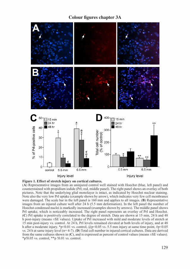
129
Figure 1. Effect of stretch injury on cortical cultures. (A) Representative images from an uninjured control well stained with Hoechst (blue, left panel) and counterstained with propidium iodide (PrI; red, middle panel). The right panel shows an overlay of both pictures. Note that the underlying glial monolayer is intact, as indicated by Hoechst nuclear staining. Note also the very low PrI uptake (example shown by arrow), which indicates very few cell membranes were damaged. The scale bar in the left panel is 160 mm and applies to all images. (B) Representative images from an injured culture well after 24 h (5.5 mm deformation). In the left panel the number of Hoechst condensed nuclei is markedly increased (examples shown by arrows). The middle panel shows PrI uptake, which is noticeably increased. The right panel represents an overlay of PrI and Hoechst. (C) PrI uptake is positively correlated to the degree of stretch. Data are shown at 15 min, 24 h and 48 h post-injury (means ±SE values). Uptake of PrI increased with mild and moderate levels of stretch at 15 min post-injury vs. control. At 24 h, PrI levels remained elevated at both levels of injury, and at 48 h after a moderate injury. *p<0.01 vs. control, @p<0.05 vs. 5.5 mm injury at same time point, #p<0.05 vs. 24 h at same injury level (n= 4-7). (D) Total cell number in injured cortical cultures. Data arederived from the same cultures shown in (C), and is expressed as percent of control values (means ±SE values). *p!0.05 vs. control, **p !0.01 vs. control.
Colour figures chapter 3A

130
Figure 2. Stretch injury causes a reduction in MAP2-positive neurons. (A) Representative im-age from an uninjured control well stained against MAP2 (red) and counterstained with DAPI (blue). Uninjured neurons have an intact soma and smooth neurites. Note that the underlying glial monolayer is intact, as indicated by DAPI nuclear staining. The scale bar is 160 mm and applies to both im-ages. (B) Representative image from an injured culture well (6.5 mm deformation). Injury caused beaded neurites (indicated by the arrow) as well as a disruption of the glial monolayer, indicated by DAPIstained nuclei. (C) Summary of the number of MAP2-positive neurons at 15 min, 24 h and 48 h after injury. Data are expressed as percent of con-trol values (meansGSE values; n=6-8). *p < 0.01 vs. control, @p < 0.05 vs. 5.5 mm injury at same time point.

131
Figure 2. (A) Representative 14C-IAP autoradiograms of non-injured control animals. (B-D) Quanti-ficationofcerebralbloodflow(ml/100gtissue/min)intheregionsofinterest(ROI)definedinfigure1. Data are arranged according to the anatomical location of the ROIs, i.e. from the left lateral to right lateral cortex (n=5). Panel B shows the data from the most rostrally, panel D the data from the most occipitally located brain section.
Colour figures chapter 3B
Figure 2
rostral
100 ml/100g/min
400 ml/100g/min
200 ml/100g/min
50 ml/100g/min
occipital
Cer
ebra
l blo
od fl
ow(m
l/100
g/m
in)
0
20
40
60
80
100
120
medialleft right
ROI1
ROI3
ROI2
ROI6
ROI5
ROI4
Cer
ebra
l blo
od fl
ow(m
l/100
g/m
in)
0
20
40
60
80
100
120
medialleft right
ROI1
ROI3
ROI2
ROI6
ROI5
ROI4
Cer
ebra
l blo
od fl
ow(m
l/100
g/m
in)
0
20
40
60
80
100
120
medialleft right
ROI1
ROI3
ROI2
ROI6
ROI5
ROI4
rostraloccipital
A
B
C
D

132
Figure 3. (A) Representative 14C-IAP autoradiograms of mice subjected to experimental traumatic braininjurybycontrolledcorticalimpacttotherighthemisphere(arrows)andscarified15minutesor24hoursafterTBI.Ascomparedtonon-injuredcontrolscerebralbloodflowdecreasedinthewholebrainincludingthehemispherecontralateraltotheimpact15minafterTBI.Nobloodflowwasdetect-ableatthelocationofthecontusion.Twenty-fourhoursafterTBIcerebralbloodflowrecoveredinthecontralateralhemispherewhilestillnobloodflowcouldbedetectedatthecontusionsite.(B) Temporal changesoflocalcerebralbloodflowmeasuredbylaserDopplerfluxmetryoverthecontralateralhemi-sphere after controlled cortical impact in mice.
frontal medial occipital
control
15 min
24 hrs
100 ml/100g/min
400 ml/100g/min
200 ml/100g/min
50 ml/100g/min
Figure 3
A
B
Time after trauma (min)0 20 40 60 80 100 120
Lase
r Dop
pler
flux
(% b
asel
ine)
040
50
60
70
80
90
100
110
120 Mean +/- SEM, n=8
p<0.001

133
Figure 6. Two pairs of adjacent coronal brain sections taken at equal distances throughout the contused brain were used for the determination of ischemic and necrotic brain tissue volume by 14C-IAP autora-diography and Nissl staining, respectively (see Fig. 1B). A-D. Representative autoradiograms (A & C) and histological sections (B & D) obtained 15 min and 24 hours after TBI showing the areas of ischemic and contused brain tissue, respectively. E. The volume of contused (white bars) as compared to the volume of ischemic/not perfused brain parenchyma (CBF < 10% of baseline; grey bars) 15 min and 24 h after TBI. While the volume of contused brain tissue expands significantly over time (+43%; *p<0.01), the volume of non-perfused tissue shows only amoderate,notsignificantchangeovertime(n=5-7).
DB
C
15 min 24 hrA
Figure 6
15 min 24 hrs
Tiss
ue v
olum
e (m
m3 )
0
10
20
30
40
50NecrosisCBF < 10%
Time after trauma
*Mean +/- SDp<0.01
E

134
Colour figures chapter 4A
Figure 2. Nissl-stained coronal brain section showing a cortical contusion 15 min (A) and 24 h (B) aftercontrolledcorticalinjury.Highermagnifications(200)fromtheareasindicatedinA(C,D) and B (E).Autoradiographyshowingbloodflowvaluesinthebrainofamousesacrificed15minaftertrauma(F).Nobloodflow(black)isdetectablewithintheprimarycontusion.
Figure 2
15‘ 24 hA B
F
DC
E
100 µm
15‘
24 h
Control
15‘

135
Figure 2. Representative adjacent brain sections.Autoradiogram (A) and Nissl-stained (B). Note the difference between the contusion/non-perfused area.
Colour figures chapter 4B
Figure 2
A
B
3 hr
Figure 2. Representative adjacent brain sections: autoradiogram (top) and Nissl-stained (bottom).
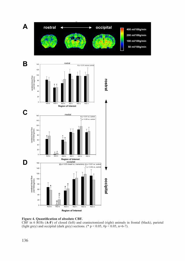
13�
Figure 4. Quantification of absolute CBF. CBF in 6 ROIs (A-F) of closed (left) and craniectomized (right) animals in frontal (black), parietal (light grey) and occipital (dark grey) sections. (* p < 0.05, #p < 0.05, n=6-7).
occipital
cere
bral
blo
od fl
ow(m
l/100
mg/
min
)
0
20
40
60
80
100
120
140
160
Region of Interest
ROI 6 ROI 5 ROI 4 ROI 1 ROI 2 ROI 3
#
#
***
# p < 0.01 vs. control* p < 0.05 vs. control
@
@
@ p< 0.05 closed vs. craniectomy
Figure 4
A
Figure 2
rostral
100 ml/100g/min
400 ml/100g/min
200 ml/100g/min
50 ml/100g/min
occipitalA
B
C
D
rostraloccipital
medial
cere
bral
blo
od fl
ow(m
l/100
mg/
min
)
0
20
40
60
80
100
120
140
160
Region of InterestROI 6 ROI 5 ROI 4 ROI 1 ROI 2 ROI 3
#
*
# #
# p < 0.01 vs. control* p < 0.05 vs. control
*
rostral
cere
bral
blo
od fl
ow (m
l/100
mg/
min
)
0
20
40
60
80
100
120
140
Region of InterestROI 6 ROI 5 ROI 4 ROI 1 ROI 2 ROI 3
# #
# p < 0.01 versus control
Cerebral blood flow expressed as % of CBF in normal brain (dotted line)15minutes after experimental TBI inmice (n=5-6) in rostral (A), striatal(B), and occipital (C) coronal brain
sections. CBF was quantified in six regions of interest (ROI) arranged according to their anatomical location, i.e. from the left lateral to theright lateral cortex. 15 min after trauma CBF decreased significantlyin all regions of the brain (A-C; *p<0.01), most dramatically at
the site of the impact (#p<0.001).