sea urchin goosecoid regulatory roles - anger's lab · l. m. angerer and others. sea urchin...
TRANSCRIPT

INTRODUCTION
Patterning of cell fates along the animal-vegetal (AV) and oral-aboral (OA) axes of the sea urchin embryo requires a signalingprocess that is initiated in the vegetal organizing center.Recent studies have established that the inductive processesthat pattern mesoderm, endoderm and ectoderm fates alongthe AV axis are initiated by cell-autonomous activation ofdownstream components of the canonical Wnt pathway, i.e.β-catenin and TCF-Lef (Emily-Fenouil et al., 1998; Huanget al., 2000; Logan et al., 1999; Vonica et al., 2000;Wikramanayake et al., 1998). β-catenin/TCF-Lef activates, inat least one case directly (Howard et al., 2001), production ofdownstream transcription factors (SpKrl) and signalingligands, including SpWnt8 (A. Wikramanayake and W. H.Klein, personal communication) and probably Delta (H. Sweetand C. Ettensohn, personal communication), which functionin specifying vegetal fates.
The OA axis is established after fertilization by amechanism that may involve polarized differences in redox
potential (Coffman and Davidson, 2001). The point at whichthis axis is conditionally specified varies from the two-cellstage in Strongylocentrotus purpuratus(Cameron et al., 1989),the organism used in the studies reported here, to as late asthe eight-cell stage in some other species (Kominami, 1988).OA polarity is displayed primarily in the ectoderm, by thedifferentiation of distinct oral and aboral epithelial cell typesthat are separated by a thin band of ciliated cuboidal cells.Although OA polarity is established after fertilization by amechanism different from that which fixes the AV axis duringoogenesis, patterning of different cell types arrayed along theOA axis nevertheless requires signaling from the vegetal pole.Animal halves of eight-cell embryos or intact embryos inwhich β-catenin function is blocked by injection of cadherinmRNA fail to differentiate aboral ectoderm or ciliary bands(Wikramanayake and Klein, 1997; Wikramanayake et al.,1998). Differentiation of these tissues can be rescued inanimal half embryos by treating them with LiCl, whichinhibits the β-catenin-destabilizing kinase, GSK3β, or byinjection at the one-cell stage of mRNA encoding a stable
4393Development 128, 4393-4404 (2001)Printed in Great Britain © The Company of Biologists Limited 2001DEV5925
We have identified a single homolog of goosecoid, SpGsc,that regulates cell fates along both the animal-vegetal andoral-aboral axes of sea urchin embryos. SpGsc mRNA isexpressed briefly in presumptive mesenchyme cells of the~200-cell blastula and, beginning at about the same time,accumulates in the presumptive oral ectoderm throughpluteus stage. Loss-of-function assays with morpholine-substituted antisense oligonucleotides show that SpGsc isrequired for endoderm and pigment cell differentiation andfor gastrulation. These experiments and gain-of-functiontests by mRNA injection show that SpGsc is a repressorthat antagonizes aboral ectoderm fate specification andpromotes oral ectoderm differentiation. We show thatSpGsc competes for binding to specific cis elements withSpOtx, a ubiquitous transcription activator that promotesaboral ectoderm differentiation. Moreover, SpGscrepresses transcription in vivo from an artificial promoterdriven by SpOtx. As SpOtx appears long before SpGsc
transcription is activated, we propose that SpGsc divertsectoderm towards oral fate by repressing SpOtx targetgenes. Based on the SpGsc-SpOtx example and otheravailable data, we propose that ectoderm is first specifiedas aboral by broadly expressed activators, including SpOtx,and that the oral region is subsequently respecified bythe action of negative regulators, including SpGsc.Accumulation of SpGsc in oral ectoderm depends on cell-cell interactions initiated by nuclear β-catenin function,which is known to be required for specification of vegetaltissues, because transcripts are undetectable in dissociatedor in cadherin mRNA-injected embryos. This is the firstidentified molecular mechanism underlying the knowndependence of oral-aboral ectoderm polarity onintercellular signaling.
Key words: Morpholino, Transcriptional repressor, Embryonic axes,Otx, β-catenin, Sea urchin
SUMMARY
Sea urchin goosecoid function links fate specification along the animal-
vegetal and oral-aboral embryonic axes
Lynne M. Angerer 1,*, David W. Oleksyn 1, Amy M. Levine 1, Xiaotao Li 2, William H. Klein 2 andRobert C. Angerer 1
1Department of Biology, University of Rochester, Rochester, NY 14627, USA2Department of Biochemistry and Molecular Biology, The University of Texas M.D. Anderson Cancer Center, and GraduateProgram in Genes and Development, Houston, TX 77030, USA*Author for correspondence (e-mail: [email protected])
Accepted 13 August 2001
4394
form of β-catenin (Wikramanayake et al., 1998). The fact thatanimal halves and cadherin-expressing embryos express theEctoV epitope, which also accumulates late in differentiatedoral ectoderm (both facial epithelium and ciliary band), hasled to the proposal that β-catenin-dependent signals arerequired specifically for aboral ectoderm differentiation(Wikramanayake et al., 1998).
In vertebrate embryos, activation of the canonical Wntpathway leads to the establishment of a gastrulation organizingcenter that expresses the goosecoid transcription factor(Lemaire and Kodjabachian, 1996). The fact that ectopicexpression of goosecoid can induce a second dorsal axis inXenopus embryos suggests that it is a critical upstreamregulator of dorsal fate in this embryo (Cho et al., 1991).Echinoderms are a sister clade to the chordates, whoseembryos have been shown to use a number of molecularpathways employed in vertebrate embryos. In addition to themolecules noted above, BMP2/4, whose function isantagonized by goosecoid in Xenopusembryos, plays animportant role in ectoderm specification in sea urchin embryos(Angerer et al., 2000). These considerations suggest thatgoosecoid might also play an important role in early sea urchindevelopment. We present our characterization of an S.purpuratus goosecoidhomolog, SpGsc, and our investigationsof its role in sea urchin embryogenesis. We have determinedits time and sites of expression, characterized phenotypesproduced in loss-of-function and misexpression experimentsand determined whether its expression depends on thecanonical Wnt pathway. Our data show that SpGsc is requiredfor gastrulation and subsequent differentiation of endodermand pigment cells, one of the derivatives of secondarymesenchyme. SpGsc also plays a central role in patterning cellfates along the OA axis, in that its restricted expression in oralectoderm is required to repress gene(s) that promote aboralectoderm differentiation. The data presented here support amodel in which SpGsc function links patterning of cell fatesalong the AV and OA axes of sea urchin embryos.
MATERIALS AND METHODS
Embryo and single-cell culturesAdult sea urchins (S. purpuratus) were obtained from Charles M.Hollahan (Santa Barbara, CA). Embryos were cultured (Angerer andAngerer, 1981) and cell separation experiments were carried out(Reynolds et al., 1992) essentially as described previously.Blastomeres were resuspended in Ca2+-free seawater and cultured inspinner flasks as described (Hurley et al., 1989).
Cloning and construct preparationDegenerate primers representing sequences conserved in thehomeodomains of Xenopusand Drosophilagoosecoid proteins wereused with a very early blastula random-primed cDNA library templateto amplify the homologous cDNA. Full-length cDNA containing a320 amino acid residue open reading frame (GenBank AccessionNumber, AF315231) was generated by 5′ and 3′ RACE (LifeTechnologies). SpGsccDNA was inserted into Tclone, derived fromthe plasmid vector, pSp64T (Angerer et al., 2000), and syntheticmRNAs were transcribed with Sp6 RNA polymerase (Sp6 mESSAGEmACHINE, Ambion) from templates truncated with XbaI. TheSpGsc-VP16 fusion construct was prepared as follows. The SpGscDNA-binding domain (DBD) was obtained by PCR from the full-length cDNA clone described above using forward and reverse
primers containing SacI and NotI sites (underlined below),respectively:
forward, 5′CCC CGA GCT CTT GGT GAT GGA ATT CAA GAGAAA GAG GCG ACA;
reverse, 5′ CCC CGC GGC CGC TTA CCG TTT CTG CTT CCTC 3′.
This fragment was inserted into Tclone between the correspondingrestriction sites and the VP16 transcriptional activation domain fromclone pCS2-VP16∆βXtcf-3 (corresponding to amino acid residues411 to 490) (Vonica et al., 2000), which was contained on an EcoRIfragment, was inserted upstream of the SpGsc DBD at the EcoRI sitein the forward primer (italics).
Hybridization assaysBlots of genomic DNA digested with either EcoRI or RsaI wereprepared as described previously (Yang et al., 1989a) and probed withrandom-primed cDNA sequence encoding the homeodomain and 22additional 5′ amino acid residues in a solution containing 5×SSPE(0.75 M NaCl, 20 mM sodium phosphate, pH 7.4, 5 mM trisodiumEDTA), 0.1% sodium pyrophosphate, 5×Denhardt’s (0.5%polyvinylpyrrolidone, 0.5% bovine serum albumin and 0.5% Ficoll),0.5% sodium dodecyl sulfate at 60°C and washed with 0.15×SSPE,0.1% SDS at 45°C, which corresponds to a low stringency (Tm–45°C).RNA blotting was carried out as described previously (Howard et al.,2001) with 2 µg of mesenchyme blastula polyA+RNA and the blotwas washed at moderate stringency (Tm–35°C; 0.1×SSPE, 55°C).For RNase protection assays, total RNA (10 µg) from normal ordissociated embryos was purified with TRIzol reagent (Gibco BRL,Bethesda, MD) and hybridized to probes for SpGsc(1×10⁸ cpm/µg),Spec2a(1×108 cpm/µg) (Hardin et al., 1988) or SpHE(1×10⁷ cpm/µg)(Wei et al., 1999). Hybridization and analysis of RNase-resistantfragments were as described previously (Yang et al., 1989a). We useda radioactive method for in situ hybridization assays because itprovides greater sensitivity and reproducibility for early embryonicstages than we have been able to achieve with whole-mount methods.Sections (5 µm) of embryos at selected developmental stages werehybridized with 33P-labeled RNA probes for SpGsc (2.5×10⁵ dpm/ng)as described previously (Angerer et al., 1987).
Generation of a glutathione-S-transferase (GST)-SpGscfusion proteinA synthetic SpGsc protein was used to determine DNA-bindingproperties. To generate the pGST-GSC construct, pGEX-KG vector(Amersham Pharmacia Biotech, NJ) was digested with BamHI andNotI. The SpGsccDNA coding sequence was amplified by PCR usingprimers containing BamHI and NotI sites:
Gsc-Bam forward, TGG GAT CCT GGA CTA TTA TCT CCCCGA CGT C;
Gsc-Not reverse, CGA TGC GGC CGCGGC GAG GAG ACCCCG ATG GTG AG.
The digested PCR fragment was ligated into pGEX-KG to producethe pGST-GSC construct. The fusion protein was expressed in E. coliBL21 cells. The GST-SpGsc fusion protein was purified using theGST Purification Module from Amersham. Briefly, an overnightculture derived from a single colony was diluted 100-fold with 500ml NZY medium and incubated for 3 hours at 37°C. IPTG was addedto 0.2 mM and the culture was incubated for an additional 2 hours at37°C. Cells were harvested, resuspended in phosphate-buffered saline(PBS) with 1% N-laurylsarcosine (sarkosyl) and sonicated on ice for1 minute (power level 4, 50% duty cycle). The lysate was clarifiedand Triton X-100 was added to the supernatant to a final concentrationof 2%. Washed glutathione-agarose bead suspension (0.5 ml; 50% v/vin PBS) was added, and the lysate was incubated at room temperatureon a shaker for 30 minutes. The beads were transferred to a suppliedcolumn and washed six times with ice-cold PBS. The fusion proteinwas eluted from the beads with 10 mM reduced glutathione andsubsequently dialyzed into storage buffer (50 mM Hepes, pH 7.4, 100
L. M. Angerer and others

4395Sea urchin goosecoid regulatory roles
mM KCL, 1 mM DTT, 1 mM PMSF, 10%(v/v) glycerol). The GSTmoiety on the SpGsc-GST fusion protein was removed by proteolyticdigestion with Factor Xa for 10 hours at room temperature.
Electromobility shift assay (EMSA)EMSA was performed as described previously (Yuh et al., 2001). Thereactions contained 20-50 ng of the GST-SpGsc fusion protein orSpGsc protein with the GST moiety removed, 1×10⁴ cpm of 32P-end-labeled oligonucleotide probe, 100 ng of the indicated competitor, and0.5 µg of poly(dI-dC) in a final volume of 15 µl 1×EMSA buffer (12%glycerol, 20 mM Hepes (pH 7.9), 5 mM MgCl2, 100 mM KCl, 1 mMDTT) for 20 minutes at 4°C. DNA-protein complexes were resolvedin a 5% polyacrylamide gel in 0.5×TBE and signals from the driedgels were recorded by a phosphorimager (Molecular Dynamics:Image Quant).
Injection of mRNAs and morpholino oligonucleotides. Constructs for in vitro transcription of mRNAs were verified bysequencing. Synthetic mRNAs were suspended in 30% glycerol,quantitated by spectrophotometry and by gel electrophoresis, andmicroinjected as described previously (Angerer et al., 2000). Either2×105 or 6×10⁵ RNA molecules were injected into each egg.Morpholine-substituted oligonucleotides complementary tonucleotides –30 to –6 with respect to the translation start site of SpGscmRNA and a control morpholino were obtained from Gene Tools(Corvallis, Oregon) and dissolved in diethylpyrocarbonate-treatedwater at a concentration of 8 mM. This stock solution was dilutedto either 200 or 400 µM and 2 pl were injected to give a finalconcentration in the egg of 2-4 µM.
Promoter assayA promoter containing multimerized SpOtx ciselements linked to theCAT (chloramphenicol acetyl transferase) reporter gene (5C) (Mao etal., 1996) and synthetic SpGscmRNA were microinjected into one-cell sea urchin zygotes. CAT assays were carried out as describedpreviously (Wei et al., 1995).
Immunostaining and microscopyEmbryos were fixed in artificial sea watercontaining 4% paraformaldehyde and stainedwith a polyclonal antibody against SpSoxB1,with a monoclonal antibody against a PMC-specific epitope (6e10, kindly supplied byDr Chuck Ettensohn, Carnegie MellonUniversity) as described previously (Kennyet al., 1999), with Sp1 monoclonal antibodyobtained from the Developmental BiologyHybridoma Bank (Dieter Soll, University ofIowa), and with polyclonal antibodies againstSpec1 (Carpenter et al., 1984) and EctoV(Coffman and McClay, 1990) as describedpreviously (Angerer et al., 2000). Fluorescentsignals were captured by sequential scanningusing a LeicaTS confocal microscope.
RT-PCRRNA from 24-hour embryos injected withglycerol (control), synthetic mRNAs ormorpholinos was purified with TRIzolreagent (Gibco BRL, Bethesda, MD), DNaseI digestion, organic extraction and ethanolprecipitation. One-step RT-PCR (AdvancedBiotechnologies, Surrey, UK) using theprimers listed below was employed toamplify SpGsc, Endo16, BMP2/4, or 12Smitochondrial rRNA sequences (Mito) as aload control for embryo RNA.
SpGsc forward primer: CCT GAG TAT CAC TTA GCT GCCSpGsc reverse primer: TCG TTC CTC TTT GAG GTC GAEndo16 forward primer: AAC AAG GTG CGT GCG GCT GCEndo 16 reverse primer: GCT GGG GCG AGC ACA TTA TTMito forward primer: ACT CTC TCC TCG GAG CTA TAMito reverse primer: GTA TAA TTT TTG CGT ATT CGG CSpBMP2/4 forward primer: 5 CAG GCC TAC TAT TGT CGCSpBMP2/4 reverse primer: GGT ACT AGT GCT GGG TTGSignals were compared within the linear phase of amplification, as
verified by analyzing samples collected at different cycles. Afterelectrophoresis of the samples through 6% polyacrylamide gels inTBE (0.1 M Tris, 0.1 M sodium borate, 2 mM EDTA, pH 8.3), signalswere quantitated by phosphorimagery.
RESULTS
Isolation and characterization of the SpGsc cDNAsequenceFull-length SpGsc cDNA was isolated as described inMaterials and Methods. It encodes a predicted peptide thatcontains two conserved regions corresponding to thehomeodomain (Fig. 1A) and the goosecoid/engrailedhomology (GEH) domain (Fig. 1B). Alignment of thehomeodomain sequence with homologous sequences fromother organisms indicates clearly that the closest relative toSpGsc is Drosophila Gsc (Accession Number, S70617),followed by homologs in the polychaete annelid (Platynereisdumerilii; Accession Number, CAC19336) and amphioxus(Branchiostoma floridae; Accession Number, AAF97935)(Neidert et al., 2000). Interestingly, most of the sequencedivergence between these homeodomains and those ofvertebrate Gsc proteins (Fig. 1A, boxed sequences) maps to the
Fig. 1. (A) Goosecoid homeodomain sequence alignment. Red and blue residues indicatenonconservative and conservative amino acid substitutions, respectively. The asterisk indicatesK50 a residue found in all bicoid-class homeodomain-containing proteins that is essential forbinding to cognate ciselements. Vertebrate sequences are contained within the black rectangle.(B) Representative goosecoid proteins are aligned with respect to their homeodomain (HD)sequences. The relative positions of the conserved goosecoid-engrailed homology (GEH)domain that mediates repression are indicated by red boxes. An additional sequence that issimilar between SpGsc and DrosophilaGsc is shown in gray. (C) Blot of sperm DNA from twoindividuals that has been digested with either EcoRI (a) or RsaI (b) and hybridized at reducedstringency with a 32P-labeled probe representing the homeodomain sequence (see Materialsand Methods for details).

4396
central region; both N- and C-terminal regions are extremelywell conserved among all the members of this family. Becausesea urchins are evolutionarily much more closely related tovertebrates than to Drosophila, and because multiple Gscgenes have been identified in both chickens and mice, weinvestigated the possibility that a second sea urchin Gscgeneexists that is more closely related to those of vertebrates.However, no evidence for a second gene could be detected inblots of DNA from individual sperm samples that were probedwith the SpGschomeodomain sequence at low stringency (Fig.1C). In addition, reduced stringency hybridization screens ofan arrayed 20 hour blastula cDNA library did not identify otherGsc-like sequences, although they did recover SpGscagain, aswell as other cDNAs encoding homeodomain-containingproteins distinct from the bicoid-like class of which SpGscisa member. The strong conservation of the Gsc homeodomainsamong an echinoderm, an arthropod, a polychaete annelid anda cephalochordate suggests that this is the ancestral gene. Weconclude that after the divergence of cephalochordates andvertebrates, this gene was duplicated several times in thevertebrate line, after which the ancestral gene was lost.Nevertheless, despite some sequence differences betweenthe central regions of the ancestral and vertebrate Gschomeodomains, these proteins probably remain functionallysimilar. For example, DrosophilaGsc can elicit secondary axeswhen ectopically expressed in Xenopusembryos (Goriely etal., 1996).
The GEH peptide sequence in DrosophilaGsc is required tobind a co-repressor that mediates the repressor activity of Gsc(Mailhos et al., 1998). As shown in Fig. 1B, this sequence iswell conserved in SpGsc, in which it probably functionssimilarly because, as shown below, SpGsc also has repressoractivity in sea urchin embryos.
SpGsc expression pattern during embryogenesisSpGsctranscripts are not detectable in the maternal RNApopulation or during cleavage; they accumulate between thevery early blastula and mesenchyme blastula stages and persistat similar levels in the pluteus larva (Fig. 2). Blots ofpolyadenylated RNA detect a single 3.6 kb mRNA species(data not shown). In situ hybridization verifies that SpGsctranscripts are absent from eggs (Fig. 3A,B), cleaving embryosand very early blastulae (data not shown), but are detectable inhatched blastulae (Fig. 3C-H). At the latter stage the majorregion of expression encompasses about one half of the
presumptive ectoderm. The hybridization patterns of gastrulae(Fig. 3I,J) and plutei (Fig. 3K,L) identify this region aspresumptive oral ectoderm. Lower signals are observed over afew cells in the vegetal plate region of early mesenchymeblastulae (Fig. 3D,F,H), which can be identified by thepresence of a few adjacent, unlabeled, ingressed primarymesenchyme cells (PMCs). Comparison of adjacent sections(compare Fig. 3E,F with 3G,H) demonstrates thereproducibility of these vegetal signals, while hybridizationpatterns on fortuitously oriented sections (e.g. Fig. 3C,D)suggest that the positive cells are arranged in a torus around afew unlabeled cells at the vegetal pole. The size and positionof the unlabeled region are characteristic of small micromeredaughters, suggesting that the adjacent labeled cells are PMCprecursors that have not yet ingressed into the blastocoel.SpGscmessage accumulation in mesenchyme cells is transientand is downregulated as they enter the blastocoel. Based ontheir number and position, we favor the view that these cells
L. M. Angerer and others
Fig. 2.RNase protection assays show that SpGsctranscriptsaccumulate at mesenchyme blastula (MB), gastrula (G) and pluteus(P) stages. A negative control is provided by hybridization to yeasttRNA (ytRNA). Unhybridized probe (P), egg (E), 16-cell stage(16-c), VEB (very early blastula, ~150 cells). The arrow indicates theband produced by the hybridized probe; trace amounts of undigestedprobe persist in some of the samples.
Fig. 3. In situ hybridization with 33P-labeled antisense SpGsc probeshows that SpGsc mRNA accumulates in oral ectoderm throughoutthe mesenchyme blastula-pluteus period and transiently duringblastula stages in presumptive mesenchyme cells in the vegetal plate.(A,C,E,G,I,K) Bright field images; (B,D,F,H,J,L) corresponding darkfield images. (A,B) Egg; (C-H) early mesenchyme blastula;(I,J) gastrula; (K,L) pluteus. The arrowheads in C-H indicate recentlyingressed primary mesenchyme cells. The black line in H indicatesthe approximate plane of section that would produce thehybridization pattern shown in C,D. The locations of oral and aboralectoderm in I-L are marked with the labels oe and aoe, respectively.The distribution of SpGsc mRNAs is indicated in black in M(blastula) and N (pluteus). cm, coelomic mesenchyme; endo,endoderm; pmc, primary mesenchyme cells; smc, secondarymesenchyme cells. Embryos shown in C-L are oriented with thevegetal pole down. Scale bar: 20 µm.
4397Sea urchin goosecoid regulatory roles
are PMC precursors, but we cannot exclude the possibilitythat some presumptive secondary mesenchyme cells alsotransiently express SpGsc. The early and late expressionpatterns are indicated by black shading in the diagrams inFig. 3M,N.
SpGsc is required both for gastrulation and forestablishing oral-aboral polarityWe examined the effects of loss of SpGsc function bymorpholino-mediated inhibition of translation. We verified thatthe SpGsc morpholino inhibits translation in sea urchinembryos using the method described previously (Howard et al.,2001): SpGscsequence complementary to the morpholino orthe morpholino sequence itself (non-complementary) wasinserted in the 5′UTR of the GFP cDNA template. Embryossimultaneously injected at the one-cell stage with the SpGscmorpholino and synthetic mRNA containing thecomplementary sequence did not produce detectable GFP,whereas embryos co-injected with mRNA containing the non-complementary sequence were brightly fluorescent (data notshown).
Loss of SpGsc function produced a striking phenotype. Mostembryos failed to gastrulate or to differentiate endoderm, whilea few produced only small gut rudiments (Fig. 4A, Table 1).As shown by RTPCR, 24-hour embryos from this populationhad greatly reduced levels of Endo16RNA (Fig. 4B), whoseexpression in the vegetal plate (presumptive endoderm andsecondary mesenchyme) normally begins about the same timeas does that of SpGsc. These embryos also lacked any vegetalderivatives labeled with monoclonal antibodies against EctoV,which labels foregut cells (Fig. 4A). The differentiation ofpigment cells, a subset of secondary mesenchyme, wasstrongly suppressed: the embryo shown in Fig. 4C illustratesthe maximum level of immunostaining with the Sp1 antibody,which recognizes an epitope expressed early in thedifferentiation of these cells, (Gibson and Burke, 1985);however, at this dose of SpGsc morpholino, most embryoslacked detectable Sp1 staining.
In addition to these defects in differentiation of vegetal celltypes, embryos lacking SpGsc did not establish OA polarity,but instead assumed a radialized morphology that lackeddefinable oral and aboral territories separated by a ciliary band.Immunostaining reveals that all cells in the ectoderm expressthe aboral Spec1 marker. Little or no signal was detectable afterimmunostaining for the EctoV epitope, which is expressed athigh levels and confined to the differentiated oral ectoderm ofnormal plutei (Fig. 4A). These observations demonstrate thatSpGsc function is required for differentiation of endoderm, atleast some, if not all, secondary mesenchyme, facial epitheliumand ciliary band. By contrast, early differentiation of PMCs, asindicated by the presence of ingressed cells that stainspecifically with the 6e10 antibody, is not affected (Fig. 4A,right; Fig. 4C).
SpGsc functions as a repressor in the oral ectodermThe role of SpGsc within presumptive oral ectoderm could beto activate genes required for oral ectoderm fate, to repressthose required for aboral fate, or both. The fact that the domainof expression of Spec1, a late aboral ectoderm marker, expandsin the absence of SpGsc function and the presence of a GEHdomain suggest a repressor function. One potential mechanism
would be for SpGsc to antagonize the function of activators,such as SpOtx, another bicoid-class factor that is an essentialearly transcriptional activator in aboral ectoderm (Li et al.,1999; Mao et al., 1996). When SpOtx is converted to an activerepressor by linking its DNA-binding domain to the engrailedrepression domain and this protein is expressed in sea urchinembryos, accumulation of aboral ectoderm-specific markers isstrongly suppressed (Li et al., 1999). Because Otx and Gschave been shown to bind to the same cis-acting elements inother systems (Mailhos et al., 1998), it is likely that SpGsc cancompete for binding at SpOtx target genes in oral ectoderm.
If this hypothesis is correct, then misexpression of SpGscshould divert aboral ectoderm toward oral fate, while anactivator consisting of the SpGsc DNA-binding domain linkedto the VP16 activation domain should have the opposite effect.That this is, indeed, the case is shown in Fig. 5. The ectodermof embryos injected with SpGscmRNA expresses EctoV(oral), but little or no Spec1 (aboral). Conversely, embryosinjected with SpGsc-VP16mRNA express predominantlySpec1. Thus, SpGsc and SpOtx-Eng behave similarly (Fig. 5,middle) (Li et al., 1999), as do SpGsc-VP16 and SpOtx (Fig.5, bottom) (Mao et al., 1996).
The severity of the ectodermal defects caused bymisexpression of SpGsc-VP16, as measured by the relativeintensities of Spec1 and EctoV staining, appears to be greaterthan that produced by blocking SpGsc translation. This is notunexpected as SpGsc-VP16, when present at high levelsthroughout the embryo, is likely to have more transcriptionalactivation activity than that which is elicited by normalendogenous levels of SpOtx in the absence of SpGsc.
Both SpGsc loss-of-function experiments and the effects ofSpGsc and SpGsc-VP16 misexpression support the idea thatSpGsc can bind at SpOtx target sites and act as a competingrepressor. To test this model directly, we performed EMSA tocharacterize SpGsc DNA binding properties in vitro, andtransactivation assays to test the ability of SpGsc to directlycompete with SpOtx function in vivo. EMSA showed that abacterially produced GST-SpGsc fusion protein bindsspecifically to a DNA element contained on a 39 bp fragment(CII) previously shown to bind SpOtx and to mediate SpOtxactivation of the aboral ectoderm-specific Spec2agene (Maoet al., 1994). The CII probe also contains another cis element(OER) recently shown to mediate the binding of an oralectoderm repressor unrelated to SpGsc (Yuh et al., 2001). Asexpected, the complex formed with GST-Gsc can be shiftedwith an anti-GST antibody (compare lanes 2 and 4 in Fig. 6A).This protein binds specifically to the Otx cis element, ascompetition with either the wild-type CII sequence or anoligonucleotide with mutations in the OER site effectivelyinhibited SpGsc-DNA complex formation (Fig. 6A, lanes 3, 7).
Table 1. Gut formation is suppressed in embryos injectedwith SpGsc-morpholino (Gsc-M)
% embryos
Glycerol Gsc-M (n=49) (n=79)
No gut 10 79Small gut 23 17Large gut 52 2Dead embryos 15 9
4398
By contrast, sequences with mutated Otx sites incapable ofbinding to SpOtx were ineffective as competitors (Fig. 6A,lanes 5, 6). In particular, a mutation changing TAATCC toTAATTG, which is known to specifically affect binding ofclass K50 homeobox proteins, including Otx and Gsc (Hanesand Brent, 1991), did not compete for SpGsc binding at theOtx site (lane 5). To ensure that SpGsc was responsible forcomplex formation, we removed the GST moiety byproteolytic digestion with Factor Xa and repeated the EMSA.As expected, an SpGsc-DNA complex formed that waseffectively competed by the CII wild-type oligonucleotide (Fig.6A, lane 9) but not by the oligonucleotides with mutations inthe Otx site (lanes 10, 11).
These experiments suggest that SpGsc binds to the Otx sitewith an affinity roughly comparable with that of SpOtx. Thiswas confirmed by gel shift assays, which showed that therelative binding constant of SpGsc is within fourfold of that ofSpOtx in vitro (3.1×10³ versus 1.3×10⁴). Direct competitionbetween SpGsc and SpOtx for binding at the Otx site isdemonstrated in Fig. 6B. In order to better distinguish betweenSpGsc- and SpOtx-DNA complexes by EMSA, we comparedSpGsc with the GST moiety removed and the entire SpOtx-GST fusion protein. SpGsc alone formed a complex that wasunaffected by the addition of an SpOtx antibody (Fig. 6B, lane3). SpOtx-GST formed two lower mobility complexes thatwere both effectively supershifted with the SpOtx antibody
(Fig. 6B, compare lanes 4 and 5). The faster migrating of thetwo SpOtx-GST complexes is likely to be a degradationproduct of the intact SpOtx-GST. When we fixed the amountof SpOtx-GST at 5.6 pmol and added increasing amounts ofSpGsc from 2.8 to 19.9 pmol, we observed a decrease in theamount of SpOtx-GST complexes with a correspondingappearance of the SpGsc complex (Fig. 6B, lanes 6-8). Aboutequal amounts of SpGsc and SpOtx-GST complexes wereobserved at an input ratio of 2 to 3 (lanes 7 and 8). In additionto the SpGsc complex, we observed a slightly slower migratingcomplex that behaved in a manner generally similar to theSpGsc complex and was dependent on added SpOtx-GST (Fig.6B, lanes 6-8). Although the origin of this complex is unclear,it suggests an interaction between SpGsc and SpOtx-GST.
Conversely, when we kept SpGsc levels constant at 2.8 pmoland added increasing amounts of SpOtx-GST (1.4 to 11.2pmol), we observed a decrease in the SpGsc complex and acorresponding increase in the SpOtx-GST complexes (Fig. 6B,lanes 9-13). Again, equimolar inputs of SpGsc and SpOtx-GST(lane 11) yield about three times more of the latter complex.Taken together, the EMSA results demonstrate that SpOtx andSpGsc are able to compete with one another at an Otx site.
To test the functional significance of the interaction betweenSpGsc and the Otx cis element in vivo, we used a transgeneconstruct driven by a promoter whose activity is dependent onSpOtx cis elements (Mao et al., 1994). When increasing
L. M. Angerer and others
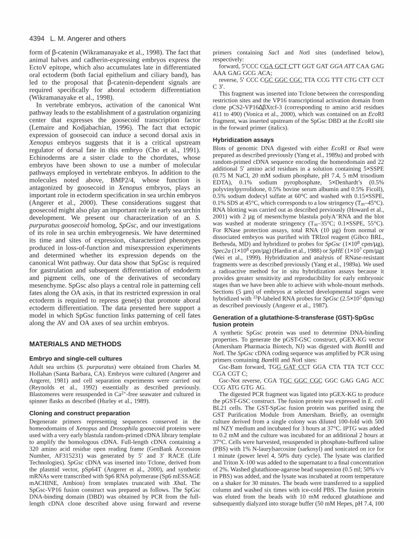
Fig. 4.Morpholino knockdown of SpGsc blocks oral endoderm and vegetal differentiation. (A) Confocal images of 3-day-old embryos injectedeither with 30% glycerol (top panels) or SpGsc morpholino in 30% glycerol (bottom panels). Embryos were stained either with antibodiesagainst EctoV (red), which stains late oral ectoderm and foregut, and Spec1 (green), which stains aboral ectoderm at this stage (left side), orwith antibody 6e10 that recognizes ingressed primary mesenchyme cells. (B) RT-PCR analysis of Endo16 mRNA levels in 24-hour embryosinjected either with glycerol, or morpholinos against SpGsc (SpGsc-M) or SpKrl (SpKrl-M); RT, reverse transcriptase. Samples were analyzedat cycles 22 and 25 to verify that signals were compared during the linear phase of PCR amplification. Endo16 signals were normalized withrespect to mitochondrial 12S rRNA values and these were set to a value of 1 for the positive control, which was RNA from glycerol-injectedembryos. As SpKrl is required for Endo16 expression, SpKrl-M (morpholino) provides a negative control (Howard et al., 2001). (C) 2-dayembryos that had been injected with glycerol (top) or the SpGsc morpholino (bottom) were stained with antibodies specific for the Sp1 epitopethat is expressed on pigment cells (green) or for 6e10 (PMCs; red). Weakly Sp1-positive cells in SpGsc morpholino-injected embryos areindicated by arrowheads. Bars: 20 µm in A,C.
4399Sea urchin goosecoid regulatory roles
amounts of SpGscmRNA were co-injected with this constructat the one-cell stage, its promoter activity was progressivelyand strongly reduced (Fig. 6C). The straightforwardinterpretation of these results is that SpGsc downregulates theactivity of this promoter by competition for binding at theSpOtx cis elements. An alternative mechanism, i.e. that SpGscsequesters SpOtx in inactive heterodimers, is unlikely becausein other systems formation of such heterodimers dependson the binding of both proteins to a palindromic pairedhomeodomain binding site (Mailhos et al., 1998), which islacking in the target promoter used here.
Although SpOtx was identified as an activator of aboralectoderm-specific genes, it accumulates in the nuclei of allectoderm cells (Li et al., 1997). By contrast, SpGsc isexpressed only in the oral facial epithelial region of theectoderm where it can act to repress SpOtx target genes, as thedata presented above demonstrate. This suggests that the ratioof SpGsc and SpOtx levels is an important factor in regulatingoral versus aboral fates, and spatial regulation of SpGsctranscription is therefore an essential feature of oral ectodermspecification.
SpGsc transcription depends on cell-cellinteractions that include β-catenin-dependent signalsDifferentiation of ectoderm requires signaling from vegetal
cells. Thus, animal halves of embryos, or embryoids derivedfrom egg animal hemispheres, remain as dauerblastulaeand avariety of experimental manipulations that interfere withsignaling by vegetal blastomeres also secondarily lead toradialization of the ectoderm (Angerer and Angerer, 2000).Such embryoids or embryos have been interpreted todifferentiate as oral ectoderm because they express the late oralectoderm marker, EctoV throughout. SpGsc is expressed inoral ectoderm beginning at hatching blastula stage, muchearlier the EctoV synthesis can be detected, and it is requiredfor establishing OA polarity. Therefore, it was of interest to testwhether accumulation of SpGsc mRNA is activated cellautonomously in oral ectoderm and, if not, then whether thesesignals are dependent on β-catenin, which is a majorcomponent of the vegetal signaling mechanism.
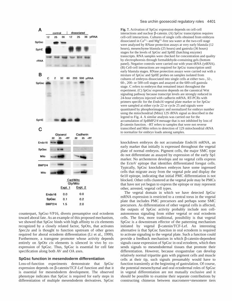
To test whether SpGsc expression is dependent on signalsfrom other cells, transcript levels were measured by RNaseprotection in RNA from cells of embryos continuouslydissociated beginning at the two-cell stage (Fig. 7A). No SpGscmRNA was detectable in dissociated embryos by this sensitiveassay, while control embryos showed the expected temporalpattern of accumulation. These results indicate that SpGscexpression in oral ectoderm, the major site of expression, is notactivated cell autonomously, but they do not reveal whether thelow level of transient SpGsc transcription in presumptivemesenchyme is sensitive to dissociation. In contrast to thesensitivity of SpGsc transcription to embryo dissociation,SpHEtranscripts accumulate cell autonomously as previouslyreported (Ghiglione et al., 1993; Reynolds et al., 1992). Levelsof 26S and 18S ribosomal RNAs in the stained gel demonstratethat the quality and recovery of RNA are equivalent in thesesamples.
To determine how late cell-cell interactions are required forSpGsctranscription, we dissociated embryos at progressivelylater cleavage and blastula stages and assayed SpGscRNAlevels when controls reached the gastrula stage (Fig. 7B).SpGsc message levels were strongly reduced even whenembryos were dissociated as late as the ~200-cell early blastulastage, but not when they were left intact until the mesenchymeblastula stage (~500 cells), by which time SpGsctranscriptionhas already begun in normal embryos. In this experiment,SpHE provides a contrasting control pattern of expression:SpHEtranscripts are not present in 500-cell normal gastrulae(Reynolds et al., 1992), but they do persist to this stage indissociated cells (Fig. 7B, dissociation at 2-, 32-, 64- and 200-cell stages), because interactions among cells are required forSpHEmRNA turnover (Ghiglione et al., 1993; Reynolds et al.,1992). These results indicate that the signaling required toinitiate SpGsctranscription must continue until just a few hoursbefore the gene is normally activated.
To determine whether SpGsctranscription in oral ectodermrequires nuclear β-catenin-dependent signals, SpGsc RNAlevels were compared by RT-PCR in RNA of embryos injectedwith either glycerol or cadherin mRNA. As describedpreviously (Howard et al., 2001; Logan et al., 1999;Wikramanayake et al., 1998), cadherin mRNA-injectedembryos developed into epithelial spheres that lackedendoderm and mesenchyme (data not shown). The results oftwo experiments using different egg batches were in excellentagreement and are quantitated at the bottom of Fig. 7C, whichalso shows the primary data for experiment 2. In both cases,
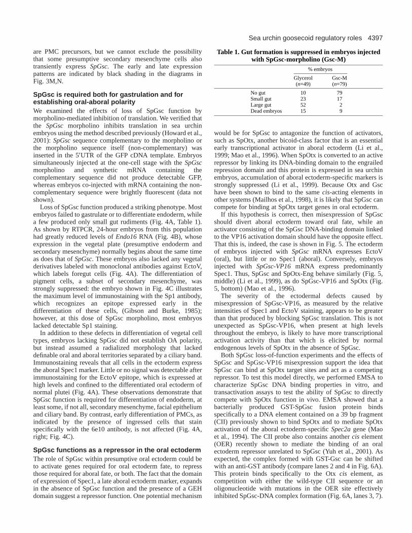
Fig. 5.Misexpression of SpOtx and SpGsc drive ectoderm towardaboral and oral fate, respectively. Confocal images of 3-day-oldembryos double-stained with anti-EctoV and anti-Spec1 that (toppanels) identify oral (red) and aboral (green) ectodermal territories innormal embryos at this stage. Sibling embryos were injected withmRNAs encoding either SpGsc (middle panels) or a fusion proteinconsisting of the SpGsc DNA-binding domain linked to the VP16transcriptional activation domain (bottom panels). Separate Spec1(left panels) and EctoV (center panels) signals are merged in theright panels. Misexpression of SpGsc promotes expression of the oralectoderm marker in all ectodermal cells while misexpression of theVP16 fusion protein has the reciprocal effect, driving these cells toexpress predominantly the aboral ectoderm marker. All theseconfocal images were obtained at the same photomultipliersensitivity. Scale bar: 10 µm.

4400
controls in which reverse transcriptase was omitted werenegative and signal intensities were consistent with samplingduring the linear phase of amplification. These measurements
show that SpGscRNA concentration is reduced between five-and tenfold in cadherin mRNA-injected 24-hour embryos. Asexpected, mRNA encoding the early vegetal plate marker,Endo16, is undetectable (Li et al., 1999; Wikramanayake et al.,1998).
To control for the formal possibility that lack of SpGscexpression results from a general arrest of ectodermdifferentiation in cadherin-expressing embryos, we analyzedthese embryos for accumulation of BMP2/4mRNA. In normalembryos, this message begins to be transcribed about the sametime as SpGscmRNA, and it accumulates throughout ectodermwith higher levels in the oral region (Angerer et al., 2000). Asshown in Fig. 7C, BMP2/4 mRNA accumulates to similarlevels in cadherin-expressing and control embryos (compareBMP2/4 signals with mitochondrial rRNA load controls). Weconclude that blocking β-catenin function does not generallyinhibit activation of at least some relatively late ectoderm-specific genes.
DISCUSSION
The experiments presented here support the following majorconclusions: (1) SpGsc acts downstream of nuclear β-cateninin sea urchin embryos, as demonstrated by its marked downregulation in embryos injected with cadherin mRNA; (2) asshown by morpholino antisense translational inhibition, SpGscis required for gastrulation, for expression of the Endo16vegetal marker, and for differentiation of pigment cells; and (3)SpGsc is required for differentiation of oral ectoderm andestablishment of OA polarity, as also demonstrated by theseloss-of-function experiments. Our results strongly suggest thatSpGsc promotes oral fate by antagonizing aboral-specific genefunctions. Thus, loss of SpGsc function in presumptive oralectoderm causes it to express the Spec1 aboral ectodermmarker and misexpression of a transcription-activating
L. M. Angerer and others
Fig. 6.SpGsc competes for binding at SpOtx ciselements.(A) SpGsc binds with specificity to an SpOtx site from the Spec2apromoter (contained on the CII fragment). Bacterially producedGST-GSC fusion protein purified by glutathione affinitychromatography formed a complex that was competable with thesequences containing an intact SpOtx ciselement (lane 2 versuslanes 3 and 7), but not with those in which this element was alteredby point mutation (lane 5) or by sequence replacement as defined byYuh et al. (Yuh et al., 2001) (lane 6). Addition of GST antibody (lane4) supershifted a significant fraction of this complex. GSC proteinderived from GSC-GST also formed specific complexes (lane 8) asshown by competition reactions with the probe sequence (lane 9),and those containing mutated Otx elements (lanes 10,11). (B) SpGscand SpOtx compete for binding to CII. GSC/CII complexes areshown in lanes 2 and 3. GST-Otx/CII complexes (lane 4) aresupershifted with Otx antibody (lane 5). Reactions containingmixtures of GSC and GST-Otx were carried out under limiting probeconcentrations. Lanes 6-8, constant amounts of GST-Otx were mixedwith increasing quantities of GSC; Lanes 10-13, constant amounts ofGSC were mixed with increasing amounts of GST-Otx. (C) SpGscdown regulates the activity of a promoter driven by SpOtx. Embryos(100) were injected with a promoter/CAT transgene construct andeither no (–) or 2×10⁶ or 6×10⁶ molecules of SpGsc mRNA. St refersto a positive control reaction containing chloramphenicol acetyltransferase.
4401Sea urchin goosecoid regulatory roles
counterpart, SpGsc-VP16, diverts presumptive oral ectodermtoward aboral fate. As an example of this proposed mechanism,we showed that SpGsc binds with high affinity to cis elementsrecognized by a closely related factor, SpOtx, that activatesSpec2aand is thought to function upstream of other genesrequired for aboral ectoderm differentiation (Li et al., 1999).Furthermore, a transgene promoter whose activity dependsentirely on SpOtx ciselements is silenced in vivo by co-expression of SpGsc. Thus, SpGsc is essential for cell fatespecification along both AV and OA axes.
SpGsc function in mesendoderm differentiationLoss-of-function experiments demonstrate that SpGscexpression depends on β-catenin/TCF-Lef function and that itis essential for mesendoderm development. The observedphenotype indicates that SpGsc is required for early steps indifferentiation of multiple mesendoderm derivatives. SpGsc
knockdown embryos do not accumulate Endo16 mRNA, anearly marker that initially is expressed throughout the vegetalplate of normal embryos. Pigment cells, the major SMC typedo not differentiate as assayed by expression of the early Sp1marker. No archenteron develops and no vegetal cells expressthe EctoV epitope that identifies differentiated foregut cells.Typically, SpGsc knockdown embryos have some ingressedcells that migrate away from the vegetal pole and display the6e10 epitope, indicating that initial PMC differentiation is notblocked. Other cells clustered at the vegetal pole may be PMCsthat have not yet begun to express the epitope or may representother, arrested, vegetal cell types.
The vegetal domain in which we have detected SpGscmRNA expression is restricted to a central torus in the vegetalplate that includes PMC precursors and perhaps some SMCprecursors. As differentiation of other vegetal cells is affected,the outputs of SpGsc activity probably include non cell-autonomous signaling from either vegetal or oral ectodermcells. The first, more traditional, possibility is that vegetalSpGsc is a downstream effector of the patterning mechanisminitiated by vegetal β-catenin/TCF-Lef. An interestingalternative is that SpGsc function in oral ectoderm is requiredto activate signaling to the vegetal plate. Such a function couldprovide a feedback mechanism in which β-catenin-dependentsignals cause expression of SpGsc in oral ectoderm, which thensends signals to mesendodermal tissues that promote theirdifferentiation. However, because exogastrulae can developrelatively normal tripartite guts with pigment cells and musclecells at their tip, such signals presumably would have tofunction transiently at the beginning of gastrulation. Of course,the potential mesenchymal and oral ectodermal roles of SpGscin vegetal differentiation are not mutually exclusive and itshould be possible to examine their separate contributions byconstructing chimeras between macromere+mesomere tiers
Fig. 7.Activation of SpGsc expression depends on cell-cellinteractions and nuclear β-catenin. (A) SpGsctranscription requirescell-cell interactions. Cultures of single cells obtained from embryosdissociated in Ca2+- and Mg2+-free sea water at the two-cell stagewere analyzed by RNase protection assays at very early blastula (12hours), mesenchyme blastula (25 hours) and gastrula (36 hours)stages for the levels of SpGscand SpHE(hatching enzyme)transcripts. RNA samples were checked for concentration and qualityby electrophoresis through formaldehyde-containing gels (bottompanel). Negative controls were carried out with yeast tRNA (ytRNA).(B) Cell-cell interactions are required for SpGsctranscription untilearly blastula stages. RNase protection assays were carried out with amixture of SpGscand SpHEprobes on samples isolated fromcultures of embryos dissociated into single cells at either two-, 32-,60-, 200- or 500-cell stages and assayed at the 600-cell gastrulastage. C refers to embryos that remained intact throughout theexperiment. (C) SpGscexpression depends on the canonical Wntsignaling pathway because transcript levels are strongly reduced in24-hour embryos injected with cadherin mRNA. RT-PCRs withprimers specific for the Endo16vegetal plate marker or for SpGscwere sampled at either cycle 22 or cycle 25 and signals werequantitated by phosphorimagery and normalized for embryo numberusing the mitochondrial (Mito) 12S rRNA signal as described in thelegend to Fig. 4. A similar analysis was carried out for theaccumulation of SpBMP2/4message that is not inhibited by loss ofβ-catenin function. –RT refers to samples that were not reversetranscribed and Mito refers to detection of 12S mitochondrial rRNAto normalize for embryo loads among samples.
4402
and micromeres in which SpGsc translation is blocked in onecomponent by a morpholino knockdown.
SpGsc function in oral ectoderm developmentActivation of SpGsc in oral ectoderm clearly depends onnuclear β-catenin function. As differentiation of ectoderm isknown to require vegetal signals, the most likely hypothesis isthat expression in oral ectoderm is a consequence of theactivity of the canonical, vegetal Wnt signaling pathway. Whilethe possibility that β-catenin also functions in ectoderm nucleicannot be excluded, it has never been detected there, even whenthese cells are induced to form endoderm. We found itsurprising that SpGsc expression was blocked whennuclearization ofβ-catenin was inhibited by cadherin, becausecadherin-treated embryos and embryos derived from animalhalves express EctoV strongly throughout the ectoderm, whichhas consequently been interpreted to be oral ectoderm (Li etal., 1999). Thus, we anticipated that SpGsc would be upregulated in cadherin-treated embryos. The fact that it wasdownregulated leads us to suspect that EctoV accumulation isnot always an indicator of oral ectoderm fate. Although EctoVaccumulates specifically in differentiated oral ectoderm (bothfacial epithelium and ciliary band) at late stages, its initialpattern of zygotic synthesis, which begins at least several hoursafter SpGsc expression at late mesenchyme blastula stage(Coffman and McClay, 1990), is not known. While furtherstudies are required to resolve this question, the relatively highand uniform concentration of EctoV in cadherin-expressing oranimal-half embryos may alternatively reflect its abnormallyprolonged, uniform synthesis in embryos in which aboralectoderm specification is inhibited. We suggest it is not thecase, however, that these embryos are arrested at a stage beforeSpGsc transcription begins, because they do transcribeBMP2/4, which is activated at the same time as SpGscinnormal embryos (Angerer et al., 2000).
If the nuclear β-catenin-dependent signals that activateSpGsc transcription in oral ectoderm come from vegetalblastomeres, then this mechanism would help explain whyelaboration of oral-aboral patterning depends on vegetalsignals. SpGsc message begins to accumulate in thepresumptive oral territory of hatched blastulae only after majorregions of the vegetal plate are thought to be conditionallyspecified but before the endoderm-ectoderm border isestablished (Angerer and Angerer, 2000). Our cell dissociationexperiments show that the signaling required for SpGsctranscription must occur (or persist until) shortly beforeactivation occurs. Therefore, activation of SpGsc in oralectoderm most likely depends on signals originating from theadjacent conditionally specified endoderm. As oral ectodermdifferentiation continues throughout gastrulation and theendoderm-ectoderm border remains subject to respecificationduring this time (McClay and Logan, 1996), continuedsignaling may be required relatively late in development. Thepersistence of high levels of SpGsc expression in oral ectodermthrough the end of embryogenesis suggests that it acts withinthis territory to help maintain oral ectoderm fate.
Three steps in ectoderm patterning Our observations on SpGsc function lead to a three-step modelfor ectoderm patterning. First, an autonomously acting set ofanimalizing transcription factors (ATFs) can drive initial
specification of a pre-ectoderm state (Angerer and Angerer,2000). Embryonic ectoderm passes only transiently throughthis state during cleavage in normal embryos. If embryos areartificially locked in this pattern of gene expression, as areanimal-half embryos or embryos deprived of β-catenin/TCF-Lef function, they develop as classic dauerblastulae, whichhave a thickened wall at the animal pole and eventually expressEctoV over most or all of their surface (depending on thespecies). Second, β-catenin-dependent signals emitteduniformly from the vegetal hemisphere then up regulatetranscriptional regulators required for ectoderm to progress toconditional specification as aboral ectoderm. One of theseregulators is SpOtx, which is present at similar concentrationin all ectoderm nuclei (Li et al., 1997). This factor is presentmaternally and is likely to act upstream in the aboral ectodermspecification pathway. In addition to its accumulating in nucleiduring cleavage stages, exogenously supplied SpOtx canrescue uniform expression of aboral ectoderm-specific genesin embryos injected with cadherin mRNA, including actinCyIIIa, which is thought not to be activated directly by SpOtx.In the third step, patterning of ectoderm to form separate oraland aboral territories requires activation of repressors of aboralectoderm genes in the oral territory. This proposed sequenceof events is consistent with previous observations that manygenes ultimately expressed in only the aboral ectoderm areinitially activated at the end of cleavage throughout most orall of the ectoderm (Kingsley et al., 1993; Yang et al., 1989a;Yang et al., 1989b). Only subsequently are these genesdownregulated in oral ectoderm, beginning aroundmesenchyme blastula stage, which is the time when SpGscbegins to accumulate. Interestingly, the other spatial regulatorsof aboral ectoderm-specific genes identified to date (p3A2)(Kirchhamer and Davidson, 1996) (OER) (Yuh et al., 2001)also function as repressors in oral ectoderm rather than asspatially restricted activators in the aboral territory.
We have provided one mechanism for SpGsc function in oralectoderm differentiation by demonstrating that it can competewith SpOtx to repress genes promoting aboral ectoderm fate.Thus, establishment of discrete ectoderm territories does notoccur until competitive levels of repressor activities, such asthat of SpGsc, are reached just after hatching blastula stage.When SpGsc is misexpressed precociously and ectopically bymRNA injection, it presumably displaces SpOtx from its targetgenes throughout the presumptive ectoderm, leading tosuppression of aboral ectoderm differentiation. Conversely,overexpression of SpOtx effectively competes SpGsc functionin oral ectoderm, thereby allowing continued transcription ofaboral-specific genes (Mao et al., 1996). This remarkableability of individual factors like SpGsc and SpOtx to drivedifferentiation towards a specific cell type may suggest thatthese factors function far upstream in the pathways that specifyectoderm tissues. We think a more likely alternative is that eachof these factors is a member of a network of crossregulatingactivators and repressors, and that these networks can bemanipulated by altering the levels of individual members.Thus, it will be of interest to identify other regulatory factorsoperating in these tissues.
A critical component of this model that remains to beidentified is the activity that restricts SpGscexpression to theoral region. The specific activation of SpGscin presumptive oralectoderm demonstrates that a unique transcriptional territory
L. M. Angerer and others
4403Sea urchin goosecoid regulatory roles
has been established there by about the 250-cell stage. Thisactivity cannot depend on vegetal nuclear β-catenin, whichexhibits no OA polarity. This polarizing component mostprobably arises from the poorly understood mechanism thatinitially specifies the OA axis. It might originate in the earlyredox gradient that predicts the alignment of this axis and hasbeen shown to affect the activity of at least one transcriptionalregulator (Coffman and Davidson, 2001). Although vegetalsignals clearly are required for establishing OA polarity, it isnot yet known whether these signals are instructive (i.e. thepolarity is imposed from the vegetal cells) or permissive (i.e.uniform vegetal signals are required to implement an inherentOA polarity in the ectoderm). Evidence that a relatively early,vegetal OA polarity exists is that the Notch receptor is enrichedon the apical surfaces of presumptive endoderm cells on theaboral side around the blastula stage (Sherwood and McClay,1997). If the model we have presented is correct, thenidentification of the spatial regulatory elements of the SpGscpromoter should define the additional activities that specify oralectoderm, bringing us much closer to understanding how theoral-aboral axis is established.
This work was supported by grants from NIGMS (EM25553)(R. C. A.), NICHD (HD22619 to W. H. K.) and the Robert A. WelchFoundation (G-0010 to W. H. K.). We would like to thank AthulaWikramanayake and Ruthie Zearfoss for generating the originalSpGscPCR product, and Drs Alan Vonica and Judy Venuti for aplasmid containing the VP16 domain.
REFERENCES
Angerer, L. M. and Angerer, R. C. (1981). Detection of poly A+ RNA insea urchin eggs and embryos by quantitative in situ hybridization. NucleicAcids Res.9, 2819-2840.
Angerer, L. M. and Angerer, R. C. (2000). Animal-vegetal axis patterningmechanisms in the early sea urchin embryo. Dev. Biol.218, 1-12.
Angerer, L. M., Cox, K. H. and Angerer, R. C.(1987). Demonstration oftissue-specific gene expression by in situ hybridization. Methods Enzymol.152, 649-661.
Angerer, L. M., Oleksyn, D. W., Logan, C. Y., McClay, D. R., Dale, L. andAngerer, R. C.(2000). A BMP pathway regulates cell fate allocation alongthe sea urchin animal-vegetal embryonic axis. Development127, 1105-1114.
Cameron, R. A., Fraser, S. E., Britten, R. J. and Davidson, E. H.(1989).The oral-aboral axis of a sea urchin embryo is specified by first cleavage.Development106, 641-647.
Carpenter, C. D., Bruskin, A. M., Hardin, P. E., Keast, M. J., Anstrom, J.,Tyner, A. L., Brandhorst, B. P. and Klein, W. H. (1984). Novel proteinsbelonging to the troponin C superfamily are encoded by a set of mRNAs insea urchin embryos. Cell 36, 663-671.
Cho, K. W., Blumberg, B., Steinbeisser, H. and De Robertis, E. M.(1991).Molecular nature of Spemann’s organizer: the role of the Xenopushomeobox gene goosecoid. Cell 67, 1111-1120.
Coffman, J. A. and McClay, D. R. (1990). A hyaline layer protein thatbecomes localized to the oral ectoderm and foregut of sea urchin embryos.Dev. Biol.140, 93-104.
Coffman, J. A. and Davidson, E. H.(2001). Oral-Aboral axis specificationin the sea urchin embryo. Dev. Biol.230, 18-28.
Emily-Fenouil, F., Ghiglione, C., Lhomond, G., Lepage, T. and Gache, C.(1998). GSK3beta/shaggy mediates patterning along the animal-vegetal axisof the sea urchin embryo. Development125, 2489-2498.
Ghiglione, C., Lhomond, G., Lepage, T. and Gache, C.(1993). Cell-autonomous expression and position-dependent repression by Li+ of twozygotic genes during sea urchin early development. EMBO J.12, 87-96.
Gibson, A. W. and Burke, R. D. (1985). The origin of pigment cells inembryos of the sea urchin Strongylocentrotus purpuratus. Dev. Biol.107,414-419.
Goriely, A., Stella, M., Coffinier, C., Kessler, D., Mailhos, C., Dessain, S.and Desplan, C. (1996). A functional homologue of goosecoid inDrosophila. Development122, 1641-1650.
Hanes, S. D. and Brent, R.(1991). A genetic model for interaction of thehomeodomain recognition helix with DNA. Science251, 426-430.
Hardin, P. E., Angerer, L. M., Hardin, S. H., Angerer, R. C. and Klein, W.H. (1988). Spec2 genes of Strongylocentrotus purpuratus. Structure anddifferential expression in embryonic aboral ectoderm cells. J. Mol. Biol.202,417-431.
Howard, E. W., Newman, L. A., Oleksyn, D. W., Angerer, R. C. andAngerer, L. M. (2001). SpKrl: a direct target of β-catenin regulationrequired for endoderm differentiation in sea urchin embryos. Development128, 365-375.
Huang, L., Li, X., El-Hodiri, H. M., Dayal, S., Wikramanayake, A. H.and Klein, W. H. (2000). Involvement of Tcf/Lef in establishing cell typesalong the animal- vegetal axis of sea urchins. Dev. Genes Evol.210, 73-81.
Hurley, D. L., Angerer, L. M. and Angerer, R. C.(1989). Altered expressionof spatially regulated embryonic genes in the progeny of separated seaurchin blastomeres. Development106, 567-579.
Kenny, A. P., Kozlowski, D., Oleksyn, D. W., Angerer, L. M. and Angerer,R. C. (1999). SpSoxB1, a maternally encoded transcription factorasymmetrically distributed among early sea urchin blastomeres.Development126, 5473-5483.
Kingsley, P. D., Angerer, L. M. and Angerer, R. C.(1993). Major temporaland spatial patterns of gene expression during differentiation of the seaurchin embryo. Dev. Biol.155, 216-234.
Kirchhamer, C. V. and Davidson, E. H. (1996). Spatial and temporalinformation processing in the sea urchin embryo: modular and intramodularorganization of the CyIIIa gene cis-regulatory system. Development122,333-348.
Kominami, T. (1988). Determination of dorso-ventral axis in early embryosof the sea urchin, Hemicentrotus pulcherrimus. Dev. Biol.127, 187-196.
Lemaire, P. and Kodjabachian, L.(1996). The vertebrate organizer: structureand molecules. Trends Genet.12, 525-531.
Li, X., Chuang, C. K., Mao, C. A., Angerer, L. M. and Klein, W. H.(1997).Two Otx proteins generated from multiple transcripts of a single gene inStrongylocentrotus purpuratus. Dev. Biol.187, 253-266.
Li, X., Wikramanayake, A. H. and Klein, W. H. (1999). Requirement ofSpOtx in cell fate decisions in the sea urchin embryo and possible role as amediator of beta-catenin signaling. Dev. Biol.212, 425-439.
Logan, C. Y., Miller, J. R., Ferkowicz, M. J. and McClay, D. R.(1999).Nuclear beta-catenin is required to specify vegetal cell fates in the sea urchinembryo. Development126, 345-357.
Mailhos, C., Andre, S., Mollereau, B., Goriely, A., Hemmati-Brivanlou, A.and Desplan, C. (1998). Drosophila Goosecoid requires a conservedheptapeptide for repression of paired-class homeoprotein activators.Development125, 937-947.
Mao, C. A., Gan, L. and Klein, W. H. (1994). Multiple Otx binding sitesrequired for expression of the Strongylocentrotus purpuratus Spec2agene.Dev. Biol.165, 229-242.
Mao, C. A., Wikramanayake, A. H., Gan, L., Chuang, C. K., Summers,R. G. and Klein, W. H. (1996). Altering cell fates in sea urchin embryosby overexpressing SpOtx, an orthodenticle-related protein. Development122, 1489-1498.
McClay, D. R. and Logan, C. Y. (1996). Regulative capacity of thearchenteron during gastrulation in the sea urchin. Development122, 607-616.
Neidert, A., Panopoulou, G. and Langeland, J.(2000). Amphioxusgoosecoid and the evolution of the head organizer and prechordal plate.Evol. Dev.2, 303-310.
Reynolds, S. D., Angerer, L. M., Palis, J., Nasir, A. and Angerer, R. C.(1992). Early mRNAs, spatially restricted along the animal-vegetal axis ofsea urchin embryos, include one encoding a protein related to tolloid andBMP-1. Development114, 769-786.
Sherwood, D. R. and McClay, D. R.(1997). Identification and localizationof a sea urchin Notch homologue: insights into vegetal plate regionalizationand Notch receptor regulation. Development124, 3363-3374.
Vonica, A., Weng, W., Gumbiner, B. M. and Venuti, J. M.(2000). TCF isthe nuclear effector of the beta-catenin signal that patterns the sea urchinanimal-vegetal axis. Dev. Biol.217, 230-243.
Wei, Z., Angerer, L. M., Gagnon, M. L. and Angerer, R. C. (1995).Characterization of the SpHEpromoter that is spatially regulated alongthe animal-vegetal axis of the sea urchin embryo. Dev. Biol.171, 195-211.

4404
Wei, Z., Angerer, L. M. and Angerer, R. C. (1999). Spatially regulatedSpEts4 transcription factor activity along the sea urchin embryo animal-vegetal axis. Development126, 1729-1737.
Wikramanayake, A. H. and Klein, W. H. (1997). Multiple signaling eventsspecify ectoderm and pattern the oral-aboral axis in the sea urchin embryo.Development124, 13-20.
Wikramanayake, A. H., Huang, L. and Klein, W. H. (1998). beta-cateninis essential for patterning the maternally specified animal-vegetal axis in thesea urchin embryo. Proc. Natl. Acad. Sci. USA95, 9343-9348.
Yang, Q., Angerer, L. M. and Angerer, R. C.(1989a). Structure and tissue-specific developmental expression of a sea urchin arylsulfatase gene. Dev.Biol. 135, 53-65.
Yang, Q., Angerer, L. M. and Angerer, R. C.(1989b). Unusual pattern ofaccumulation of mRNA encoding EGF-related protein in sea urchinembryos. Science246, 806-808.
Yuh, C. H., Li, X., Davidson, E. H. and Klein, W. H. (2001). Correctexpression of Spec2ain the sea urchin embryo requires both Otx and othercis-regulatory elements. Dev. Biol.232, 424-438.
L. M. Angerer and others