scutellarin suppresses platelet aggregation and stalls ... fileoriginal article scutellarin...
TRANSCRIPT
Original Article
Scutellarin Suppresses Platelet Aggregationand Stalls Lesional Progression in MouseWith Induced Endometriosis
Ding Ding, MD, PhD1,3, Xianjun Cai, MD2, Hanxi Zheng, MD1,Sun-Wei Guo, PhD1, and Xishi Liu, MD, PhD1,3
AbstractPlatelets play an important role in the development of endometriosis. Scutellarin is a flavonoid isolated from a medicinal herbtraditionally used as a potent antiplatelet agent. In this study, we sought to evaluate its potential therapeutic effect, if any, in micewith induced endometriosis. Endometriosis was induced in 27 female Balb/c mice by intraperitoneal injection of uterine fragments.Two weeks after the induction, the 27 mice were randomly divided in equal sizes into 3 groups: untreated, which received onlyvehicle, and low-dose and high-dose groups, which received low- and high dose of scutellarin treatment. Hotplate test wasadministrated to all mice before endometriosis induction, and before and after the scutellarin treatment. Two weeks after thetreatment, a blood sample was drawn before sacrifice and all lesions were harvested. The peripheral platelet activation rate andtotal lesion weight were assessed, and immunohistochemistry and histochemistry analyses were performed to evaluate the extentof proliferation, angiogenesis, fibroblast-to-myofibroblast transdifferentiation (FMT), and fibrosis in lesions. Compared withuntreated mice, mice in both low-dose and high-dose groups had significantly reduced lesion weight and improved hyperalgesia.Scutellarin also reduced the peripheral-activated platelets rate and resulted in significantly reduced platelet aggregation, cellularproliferation, angiogenesis, the extent of FMT, and the extent of fibrosis in lesions. Thus, we conclude that scutellarin is efficaciousin treating endometriosis in vivo by suppressing platelet aggregation, inhibiting proliferation, angiogenesis, and fibrogenesis,resulting in reduced lesion size and improved pain behavior. As such, scutellarin may be a potentially promising therapeutics forthe treatment of endometriosis.
Keywordsendometriosis, fibrogenesis, hyperalgesia, mouse, platelet, scutellarin
Introduction
Endometriosis, defined as the deposition of endometrial-like
tissues outside the uterine cavity, is a common gynecologic
disease affecting 6% to 10% of reproductive-aged women.1 It
is the major contributing cause of dysmenorrhea, chronic pel-
vic pain, and infertility, negatively impacting on the quality of
life of affected patients. Due mainly to the lack of understand-
ing of its pathogenesis and pathophysiology, its clinical man-
agement is challenging.1 While surgery is proven efficacious in
relieving endometriosis-associated pain,2 the high recurrence
risk3,4 and the increased risk of premature ovarian failure due to
repeated surgery render medical treatment a viable option.5-7
The current medical treatment relies on inhibition of ovulation
and reduction of estradiol levels through hormonal manipula-
tion, but their efficacy is limited by short duration.8 The devel-
opment of nonhormonal drugs has been painfully slow.9,10
One defining feature of ectopic endometrium is cyclic
bleeding.11 Accumulating data have shown, indeed, that endo-
metriotic lesions are essentially wounds that undergo repeated
tissue injury and repair.12-14 As such, platelets are found to be
aggregated in endometriotic lesions, which play a critical
role in the development and progression of endometriosis.13
Activated platelets upregulate vascular endothelial growth
factor (VEGF) and matrix metallopeptidase 9 and induce
angiogenesis.13 Through the release of transforming growth
factor b1 (TGF-b1) and the induction of TGF-b/Smad
signaling pathway, activated platelets also drive epithelial–
mesenchymal transition (EMT), fibroblast-to-myofibroblast
transdifferentiation (FMT), and smooth muscle metaplasia
1 Shanghai OB/GYN Hospital, Fudan University, Shanghai, China2 Ningbo No. 7 Hospital, Ningbo, Zhejiang, China3 Shanghai Key Laboratory of Female Reproductive Endocrine-Related
Diseases, Fudan University, Shanghai, China
Corresponding Authors:
Sun-Wei Guo and Xishi Liu, Shanghai OB/GYN Hospital, Fudan University, 128
Shenyang Road, Shanghai 200090, China.
Emails: [email protected]; [email protected]
Reproductive Sciences1-12ª The Author(s) 2018Article reuse guidelines:sagepub.com/journals-permissionsDOI: 10.1177/1933719118817661journals.sagepub.com/home/rsx
(SMM) in endometriotic lesions, resulting ultimately in fibro-
sis.15,16 Activated platelets are also shown to be responsible for
increased estrogen receptor b expression17 and reduced natural
killer cell cytotoxicity in endometriosis.18 Consistent with the
important roles of platelets in lesional development, women
with endometriosis have been shown to be in a hypercoagulable
state.19,20 Consequently, platelet depletion and antiplatelet
treatment effectively suppress lesion growth in mouse with
induced endometriosis.13,21,22
The drug research and development (R&D) has always been
a winding, arduous, tortuous, costly, and often precarious
endeavor. From discovery to the successful regulatory approval
for marketing, there is an astoundingly high attrition rate: Over
99% of compounds do not make to the final end of the R&D
pipeline.23 Many compounds failed simply because of unac-
ceptable safety profiles or inferior efficacy or both. Given the
difficulty in drug R&D, one seemingly shortcut would be the
screening of compounds derived from herbal medicine, which
are often known to have a good safety profile.
Scutellarin (40,5,6-trihydroxyflavone-7-O-glucuronide) is a
flavonoid isolated from the plant Erigeron breviscapus (Vant.)
Hand.-Mazz., a Chinese herbal medicine used over a thousand
years.24 It is the major active component of breviscapine, which
is the total flavonoid extract of E breviscapus containing �90%scutellarin and �10% apigenin-7-O-glucronide in content. Bre-
viscapine is a prescription drug in China for the treatment of
cardiovascular and cerebrovascular diseases with an excellent
safety profile.25 Scutellarin has been demonstrated to have mul-
tiple pharmacological effects, having antioxidant, antiplatelet,
and anti-inflammatory properties.26-28 Scutellarin has also been
shown to exhibit anticancer activities by suppressing prolifera-
tion, invasion, metastasis, and angiogenesis on various types of
cancer.29-32 It was also reported that scutellarin could enhance
cisplatin-induced apoptosis and autophagy to overcome cisplatin
resistance in non-small cell lung cancer via extracellular
regulated protein kinases (ERK)/p53 and c-met/AKT signaling
pathways.33 Moreover, scutellarin has been shown to be
antifibrotic.34 Since coagulation, ERK/p53, c-met/AKT, and
fibrogenesis have all been reported to be involved in endome-
triosis,14,35-37 and since scutellarin has an excellent safety pro-
file, naturally one may wonder whether scutellarin could have
any therapeutic potential. Unfortunately, to our best knowledge,
there has been no report on its use in endometriosis.
In this study, we sought to investigate the therapeutic poten-
tial, if any, of scutellarin in mouse with induced endometriosis
by examining the lesion growth, peripheral platelet activation,
and the expression of molecules involved in the development
of endometriosis.
Materials and Methods
Mice
Forty-one virgin female Balb/c mice, 7 weeks old and about 18
to 20 g in weight, were purchased from Shanghai BiKai
Laboratory Animal Center (Shanghai, China) and used for this
study. They were housed individually in cages, maintained
under controlled conditions with a light/dark cycle of
12/12-hour, and had access to chow and water ad libitum. All
experiments in this study were performed under the guidelines
of the National Research Council’s Guide for the Care and Use
of Laboratory Animals38 and approved by the institutional
experimental animals review board of Shanghai OB/GYN Hos-
pital, Fudan University (on file). Among the 41 mice, 14 were
randomly selected as donors, while the rest were designated as
recipient mice.
Induction of Endometriosis and the Experiment Protocol
We used an established mouse model of endometriosis by intra-
peritoneal (ip) injection of endometrial fragments39 as used in
our previous studies.13,40 Briefly, donor mice were initially
injected intramuscular with estradiol benzoate (3 mg/mouse,
Xinyi Chemistry, Shanghai, China). Seven days later, mice
were sacrificed and their uteri were harvested, seeded in a
Petri dish containing sterile saline, and split longitudinally
with a pair of scissors. To minimize any potential bias, 3
uterine horns, one from one mouse and the other pair from
another mouse, were identically processed, minced together,
mixed well, then divide them into 3 roughly equal parts, with
each part injected into one each recipient mouse from the 3
groups: UT (for untreated), LS (for low-dose scutellarin), and
HS (for high-dose scutellarin). The baseline body weight was
measured and hotplate test was administrated before induc-
tion to all mice.
Two weeks after the induction of endometriosis, hotplate test
and body weight measurement were again administrated to all
mice. Before induction, the 27 recipient mice were randomly
divided into 3 equal-sized groups: Mice in group HS received
ip injections of scutellarin (Sigma, Sigma-Aldrich Co, St. Louis,
Missouri) 15 mg/kg/mouse in 300 mL sterile saline, group LS
received ip injections of scutellarin (7.5 mg/kg/mouse in 300 mL
sterile saline), and group UT received ip injections of just 300
mL sterile saline, the solvent for scutellarin solvent. All ip injec-
tions were repeated every 2 days for 2 weeks.
The choice of scutellarin doses was based on the conversion
of breviscapine (Shineway Pharmaceutical Company, Shijiaz-
huang, Hebei, China) for human usage to mouse, assuming that
90% of breviscapine is scutellarin. Given the usual dosage of
20 mg/60 kg/d or 0.333 mg/kg/d of breviscapine or 0.300 mg/
kg/d of scutellarin for intravenous (iv) injection (Shineway
Pharmaceutical Company), the body surface area–based con-
version from human to mouse41 would be 0.300 � 12.3 ¼3.68 mg/kg/d scutellarin by iv injection or about 3.68 �1.1 ¼ 4.05 to 3.68 � 1.25 ¼ 4.6 mg/kg/d for ip injection. As
we used injection every 2 days, the dosage was doubled to
about 8.10 to 9.2 mg/kg for one ip injection. Of course, this
was a very crude estimation. Given that the dosage used orally
for mouse ranged from 20 mg/kg42 to as high as 60 mg/kg43
which amounts to approximately 5 to 20 mg/kg by iv injection,
we decided to have the low- and high-dose set to be 7.5 and
15 mg/kg, respectively.
2 Reproductive Sciences XX(X)
Two weeks after the treatment started, the final hotplate test
and body weight measurement were administrated to all mice,
and before sacrifice by cervical dislocation a blood sample
(about 0.8-1 mL) was drawn through right orbit and mixed with
3.2% citric acid for anticoagulation purpose. The abdominal
cavity was immediately opened up and examined very care-
fully, and all visible lesions were excised and processed for
disease assessment or immunohistochemistry evaluation. The
extent of endometriosis was evaluated by assessing the total
weight of all excised lesions from each mouse.
Hotplate Test
The hotplate test was employed to assess the extent of
endometriosis-associated hyperalgesia.44 The hotplate latency
evaluated with a commercially available Hot Plate Analgesia
Meter (Model BME-480, Institute of Biomedical Engineering,
Chinese Academy of Medical Sciences, Tianjin, China) con-
sisting of a metal plate of 25 cm by 25 cm in size, which can be
heated to a constant temperature of 55.0�C + 0.18�C, on which
a plastic cylinder (20 cm in diameter, 18 cm in height) was
placed. Mice were brought to the testing room and allowed to
acclimatize for 10 minutes before the test began. The latency to
respond to thermal stimulus, defined as the time (in second)
elapsed from the moment when the mouse was inserted inside
the cylinder to the time when it licked or flicked its hind paws,
or jolted or jumped off the hot plate. Each animal was tested
only once in one session. The latency was calculated as the
mean of 2 readings recorded at intervals of 24 hours.
Assessment of Platelet Activation Rate by Flow Cytometry
The platelet activation rate was evaluated by flow cytometry
as previously described.19 Briefly, the blood samples were
centrifuged at room temperature immediately after collection
to avoid activation of platelets as much as possible. After
platelets were isolated, they were incubated at room tempera-
ture with allophycocyanin-conjugated anti-mouse CD61 anti-
body (eBioscience, San Diego, California) labeling total
mouse platelets and fluorescein isothiocyanate-conjugated
anti-mouse CD62p (P-selectin) antibody (eBioscience) label-
ing activated mouse platelets and kept from light for 30 min-
utes. Platelets were washed by phosphate-buffered saline and
analyzed by flow-cytometry cell sorting (BD FACS Calibur,
San Jose, California).
Histologic Analysis and Masson Trichrome Staining
All lesion samples were fixed for 24 hours at room temperature
with 10% formalin neutral buffer solution. After fixation, the
tissues were placed in 70% ethanol overnight at 4�C, then
embedded in paraffin and sectioned in 4-mm thickness. Each
tissue sample was evaluated by hematoxylin and eosin (H&E)
staining using an H&E staining kit (SunBiotec, Shanghai,
China) according to the manufacturer’s instructions. To stain
cell nuclei, sections were dipped into hematoxylin for
10 minutes, rinsed by tap water, then dipped into 0.1% HCl
thrice, washed by tap water thrice, dipped into 0.1% NH4OH
thrice, and washed by tap water thrice. To stain cytoplasm,
sections were dipped into eosin for 3 minutes. Slides were
dehydrated, mounted, and examined under an Olympus micro-
scope (Olympus, Tokyo, Japan) at �200 magnification to con-
firm the establishment of the endometriotic lesion by typical
epithelial and stromal components.
Masson trichrome staining was used to detect the collagen
fibers in tissue samples.16 Tissue sections were deparaffinized
in xylene and rehydrated in a graded alcohol series and then were
immersed in Bouin solution at 37�C for 2 hours. Bouin solution
was made with 75 mL of saturated picric acid, 25 mL of 10%formalin (w/v) solution, and 5 mL of acetic acid. The tissue sec-
tions were stained using Masson trichrome staining kit (Baso,
Wuhan, China) following the manufacturer’s instructions. Slides
were then mounted and evaluated under an Olympus microscope
(Olympus) at �200 magnification, capturing 4 to 5 different
fields of sections. The areas of the collagen fiber layer stained
in blue in proportion to the entire field of the ectopic implants
were calculated by the Image Pro-Plus 6.0 (Media Cybernetics,
Inc). Masson staining parameters assessed in the area detected
included (1) integrated optical density (IOD), (2) total stained
area (S), and (3) mean optical density (MOD), which was defined
as MOD ¼ IOD/S and used as the extent of lesional fibrosis.16
Immunohistochemistry
Serial 4-mm sections from the mouse experimental ectopic
lesions (see below) were obtained from each paraffin-
embedded tissue block, with the first resultant slide being
stained for H&E to confirm pathologic diagnosis, and the sub-
sequent slides stained for CD41 (a marker for platelets), pro-
liferating cell nuclear antigen (PCNA, a marker for cellular
proliferation), VEGF, CD31 (for counting microvessel density
or MVD), collagen I, a-smooth muscle actin (a-SMA), and
lysyl oxidase (LOX). Routine deparaffinization and rehydra-
tion procedures were performed following published protocols.
For antigen retrieval, the slides were heated at 98�C in the
ethylenediaminetetraacetic acid buffer (pH 8.0) or the citric
acid solution (pH 6.0; according to the primary antibody) for
a total of 30 minutes and cooled naturally at room temperature.
The rabbit anti-mouse CD41 (1:200, Abcam, Cambridge, Mas-
sachusetts), rabbit polyclonal antibodies against PCNA (1:100,
Thermo Littleton, Waltham, Massachusetts), VEGF (1:50,
Santa Cruz, Dallas, Texas), rabbit anti-mouse CD31 (1:50,
Abcam), rabbit anti-mouse a-SMA (1:100, Abcam), rabbit
anti-mouse collagen I (1:100, Abcam), and rabbit anti-mouse
LOX (1:100, LifeSpan BioScience, Seattle, Washington) were
used as primary antibodies.
The slides were incubated with the primary antibodies over-
night at 4�C. After the slides were rinsed, the horseradish
peroxidase-labeled secondary anti-rabbit or anti-mouse anti-
body detection reagent (Shanghai SunBiotec Company) was
incubated at room temperature for 30 minutes. The bound anti-
body complexes were stained for 1 minute or until appropriate
Ding et al 3
for microscopic examination with diaminobenzidine and then
counterstained with hematoxylin (30 seconds) and mounted.
Human invasive breast cancer or mice spleen tissue samples
were used as positive controls. For negative controls, the endo-
metriotic tissue samples from mouse were incubated with rab-
bit serum instead of the primary antibody (Supplementary
Information).
Immunostaining results for CD41, PCNA, VEGF, CD31,
collagen I, a-SMA, and LOX were evaluated using a semiquan-
titative scoring system. Briefly, the number and intensity of
positive cells were counted by Image Pro-Plus 6.0 (Media
Cybernetics) without prior knowledge of any information
regarding which group the mouse was in. A series of 5 random
images on several sections were taken for each immunostained
parameter to obtain a mean value. Staining was defined via
color intensity, and a color mask was made. The mask was
then applied equally to all images, and measurement readings
were obtained. Immunohistochemical parameters assessed in
the area detected included (1) integrated optical density (IOD),
(2) total stained area (S), and (3) mean optical density (MOD),
which was defined as MOD ¼ IOD/S, equivalent to the inten-
sity of stain in all positive cells. The numbers of CD31-labeled
MVD were counted under �400 microscopic magnification.
Statistical Analysis
The comparison of distributions of continuous variables
between or among 2 or more groups was made using the Wil-
coxon and Kruskal tests, respectively, and the paired Wilcoxon
test was used when the before-after comparison was made for
the same group of participants. A Pearson or Spearman rank
correlation coefficient was used when evaluating correlations
between 2 variables when both variables were continuous or
when at least one variable was ordinal. Multiple linear regres-
sion analysis was used to identify which factor(s) were associ-
ated with the lesion weight or immunoreactivity measure (all
squared-root or log-transformed to improve normality, where
appropriate).
The P values of less than .05 were considered statistically
significant. All computations were made with R 3.5.145 (www.
r-project.org).
Results
No mouse died during the entire experimental period. Scutel-
larin appeared to be well tolerated in treated mice. As previ-
ously reported, endometriosis was successfully induced in all
mice and the induced endometriotic lesions were histologically
confirmed.
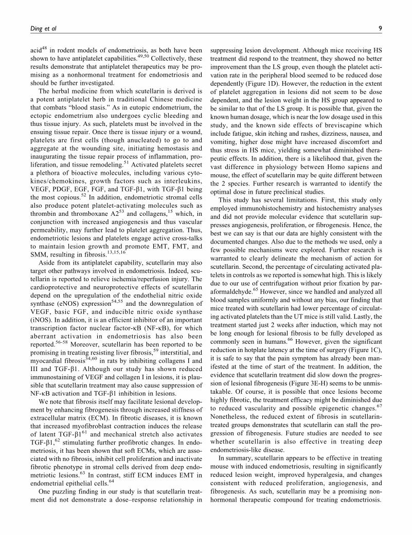
Scutellarin Treatment Reduces Lesion Weightand Improves Hyperalgesia
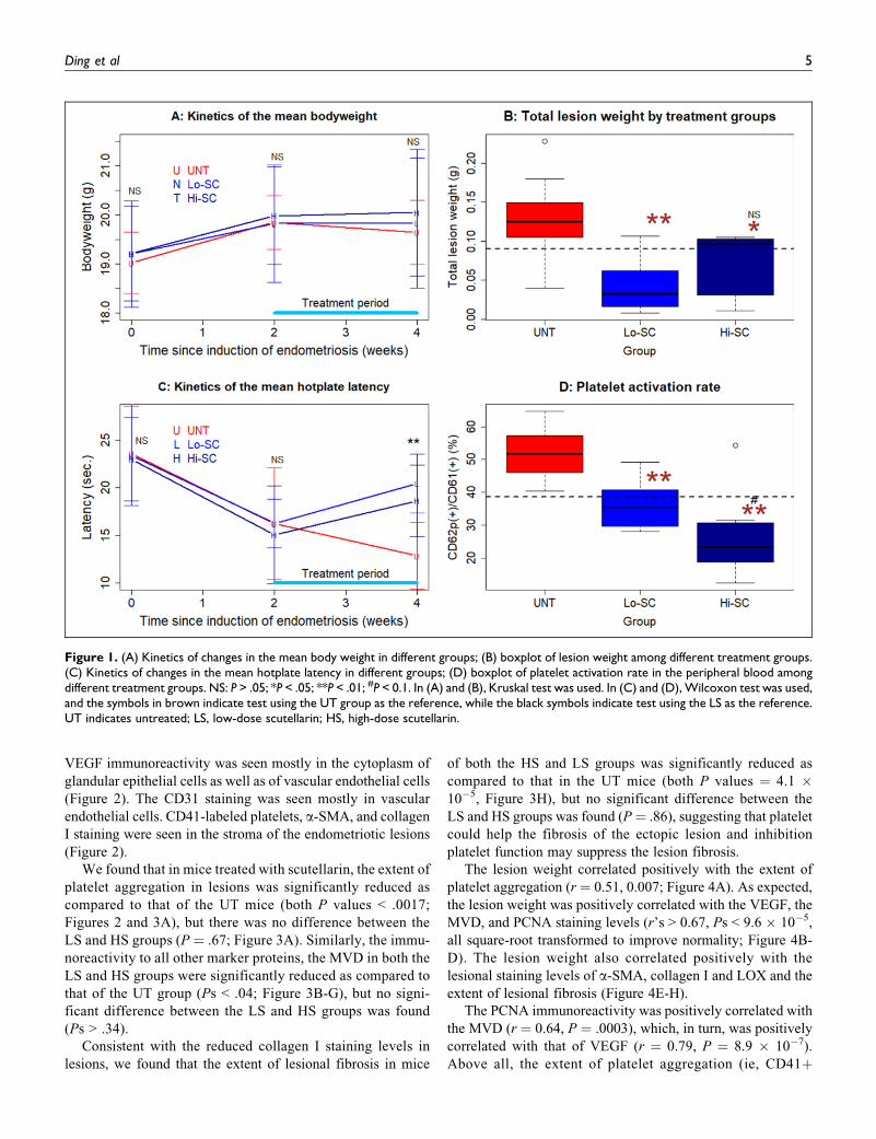
The body weight of all groups of mice increased gradually after
induction of endometriosis induction (P ¼ .0002; Figure 1A),
but no significant difference in body weight among the 3
groups was found before and 2 weeks after the induction, and
at the end of the experiment (Ps > .72; Figure 1A).
Mice that received LS and HS treatment had significantly
lower lesion weight than the UT mice (P¼ .0019 and P¼ .017,
respectively; Figure 1B). The average weight in LS and HS
groups of mice was reduced by 67.0% and 42.2%, respectively,
as compared to that of the UT mice (Figure 1B), suggesting that
the scutellarin treatment suppresses lesion growth. While LS
appeared to resulted in more suppressive effect than that the
HS, the difference in lesion weight between the 2 groups was
not statistically significant (P ¼ .14; Figure 1B).
As expected, there was no difference in hotplate latency
prior to the induction of endometriosis as well as prior to the
scutellarin treatment (both P values > .75; Figure 1C). How-
ever, there was a significant reduction in latency 2 weeks after
the endometriosis induction (P ¼ 5.9 � 10�6; Figure 1C),
consistent with what we reported previously.22,46 Two weeks
after treatment, the difference in hotplate latency among the 3
groups was highly significant (P ¼ .0026; Figure 1C). In fact,
mice treated with either LS or HS had significant improvement
in hotplate latency (P¼ .004 and P¼ .027, respectively; Figure
1C). In contrast, UT mice had worsened latency (P ¼ .008;
Figure 1C). No significant difference in latency between the LS
and HS groups (P ¼ .23; Figure 1C). A multiple linear regres-
sion analysis incorporating the latency before treatment, dose
and body weight indicated that before-treatment latency and
whether or not treated with scutellarin were significantly and
positively associated with the latency after treatment (P ¼ 9.0
� 10�6 and P ¼ 7.4 � 10�8, respectively; R2 ¼ 0.79).
Scutellarin Suppresses Platelet Activation Ratein the Peripheral Blood
We also evaluated the platelet activation rate in the peripheral
blood from all groups of mice. We found that the platelet
activation rate in both the LS and HS groups was significantly
lower than that of the UT group (both P values < .0037; Figure
1D). The rate in the HS group was marginally significantly
lower than that of the LS group (P ¼ .072; Figure 1D). A
multiple linear regression analysis incorporating the lesion
weight and dose indicated that the dose was significantly and
negatively associated with the activation rate (P ¼ 3.9 � 10�5;
R2 ¼ 0.56). These data indicate that scutellarin treatment sup-
presses platelet activation dose dependently.
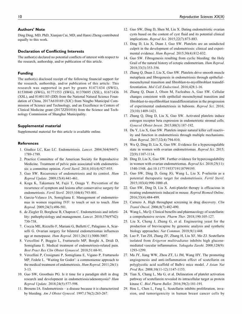
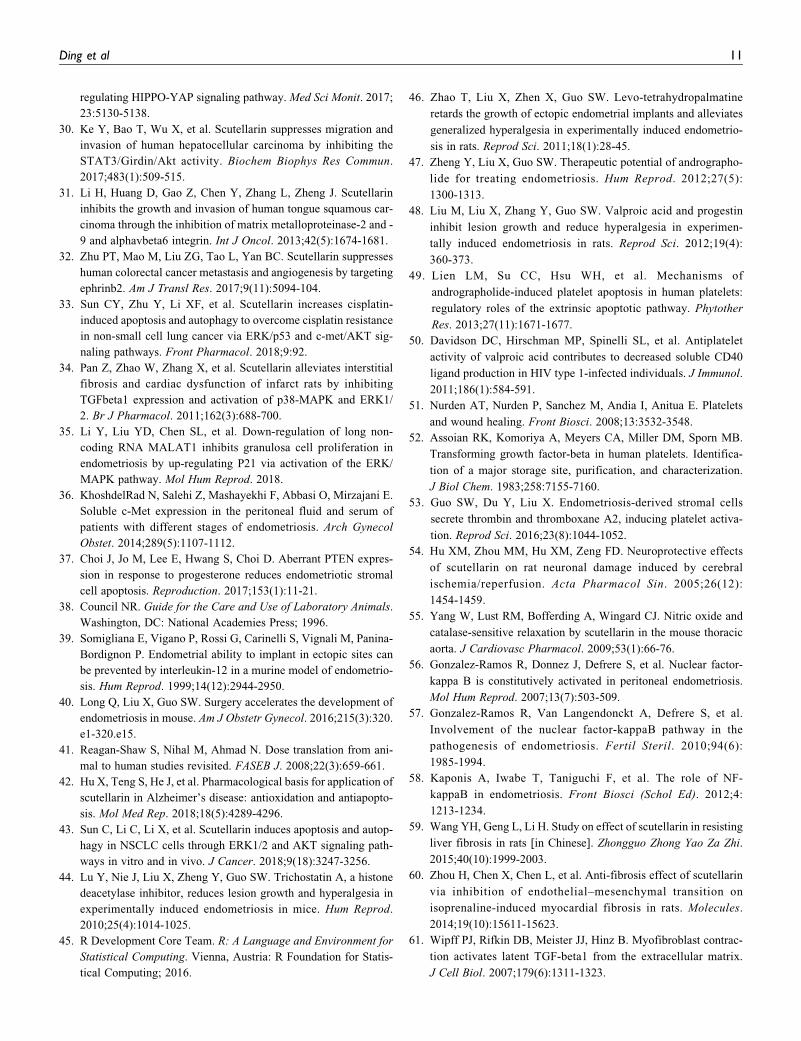
Scutellarin Suppresses Platelet Aggregation,Angiogenesis, Proliferation, and Fibrogenesis in Lesions
To gain insight into the possible mechanisms underlying the
scutellarin suppressive effect, we also performed an immuno-
histochemistry analysis of CD41 (for platelet aggregation),
VEGF, CD31 (for MVD), PNCA, a-SMA, collagen I, and LOX
in ectopic endometrium. In addition, we evaluated the extent of
lesional fibrosis by Masson trichrome staining. We found that
PCNA and LOX staining was seen in cellular nuclei in both the
stromal and epithelial cells of the ectopic endometrium, while
4 Reproductive Sciences XX(X)
VEGF immunoreactivity was seen mostly in the cytoplasm of
glandular epithelial cells as well as of vascular endothelial cells
(Figure 2). The CD31 staining was seen mostly in vascular
endothelial cells. CD41-labeled platelets, a-SMA, and collagen
I staining were seen in the stroma of the endometriotic lesions
(Figure 2).
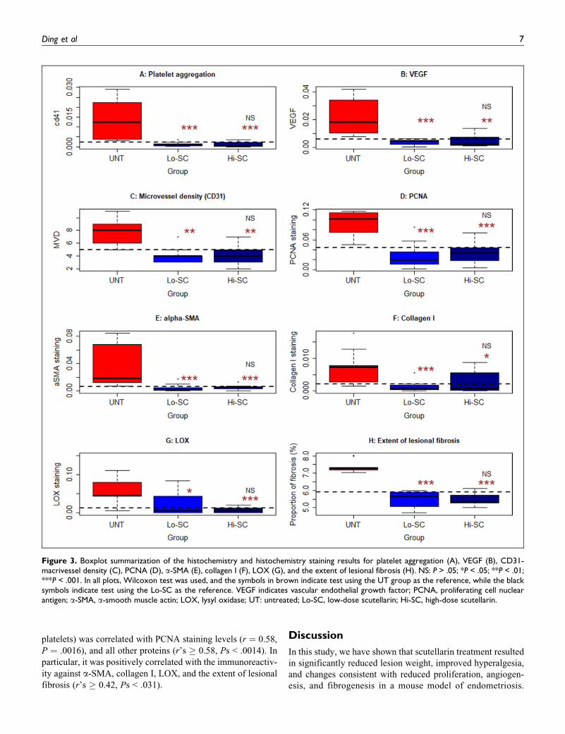
We found that in mice treated with scutellarin, the extent of
platelet aggregation in lesions was significantly reduced as
compared to that of the UT mice (both P values < .0017;
Figures 2 and 3A), but there was no difference between the
LS and HS groups (P ¼ .67; Figure 3A). Similarly, the immu-
noreactivity to all other marker proteins, the MVD in both the
LS and HS groups were significantly reduced as compared to
that of the UT group (Ps < .04; Figure 3B-G), but no signi-
ficant difference between the LS and HS groups was found
(Ps > .34).
Consistent with the reduced collagen I staining levels in
lesions, we found that the extent of lesional fibrosis in mice
of both the HS and LS groups was significantly reduced as
compared to that in the UT mice (both P values ¼ 4.1 �10�5, Figure 3H), but no significant difference between the
LS and HS groups was found (P¼ .86), suggesting that platelet
could help the fibrosis of the ectopic lesion and inhibition
platelet function may suppress the lesion fibrosis.
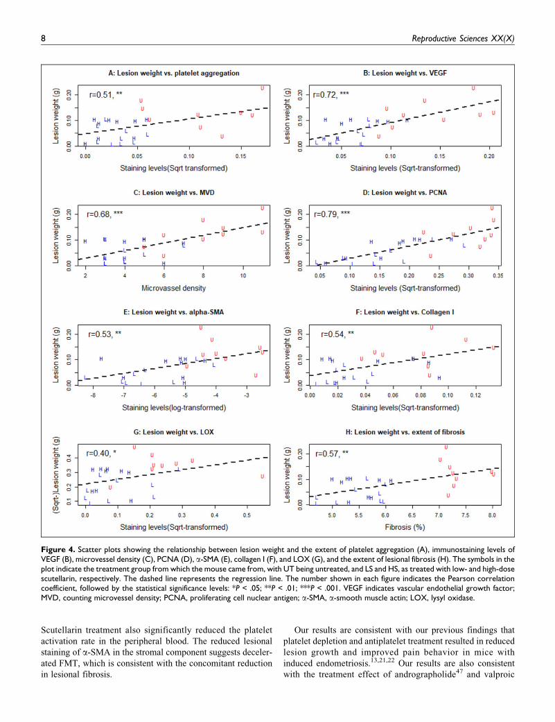
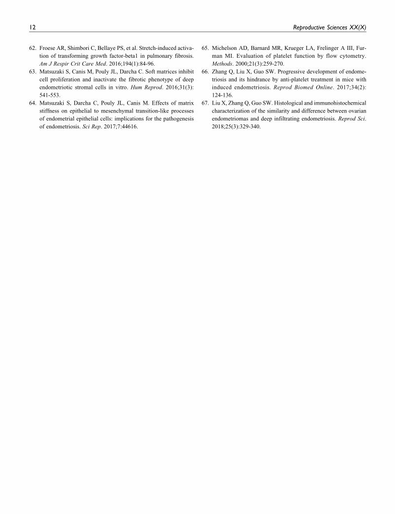
The lesion weight correlated positively with the extent of
platelet aggregation (r ¼ 0.51, 0.007; Figure 4A). As expected,
the lesion weight was positively correlated with the VEGF, the
MVD, and PCNA staining levels (r’s > 0.67, Ps < 9.6 � 10�5,
all square-root transformed to improve normality; Figure 4B-
D). The lesion weight also correlated positively with the
lesional staining levels of a-SMA, collagen I and LOX and the
extent of lesional fibrosis (Figure 4E-H).
The PCNA immunoreactivity was positively correlated with
the MVD (r ¼ 0.64, P ¼ .0003), which, in turn, was positively
correlated with that of VEGF (r ¼ 0.79, P ¼ 8.9 � 10�7).
Above all, the extent of platelet aggregation (ie, CD41þ
Figure 1. (A) Kinetics of changes in the mean body weight in different groups; (B) boxplot of lesion weight among different treatment groups.(C) Kinetics of changes in the mean hotplate latency in different groups; (D) boxplot of platelet activation rate in the peripheral blood amongdifferent treatment groups. NS: P > .05; *P < .05; **P < .01; #P < 0.1. In (A) and (B), Kruskal test was used. In (C) and (D), Wilcoxon test was used,and the symbols in brown indicate test using the UT group as the reference, while the black symbols indicate test using the LS as the reference.UT indicates untreated; LS, low-dose scutellarin; HS, high-dose scutellarin.
Ding et al 5
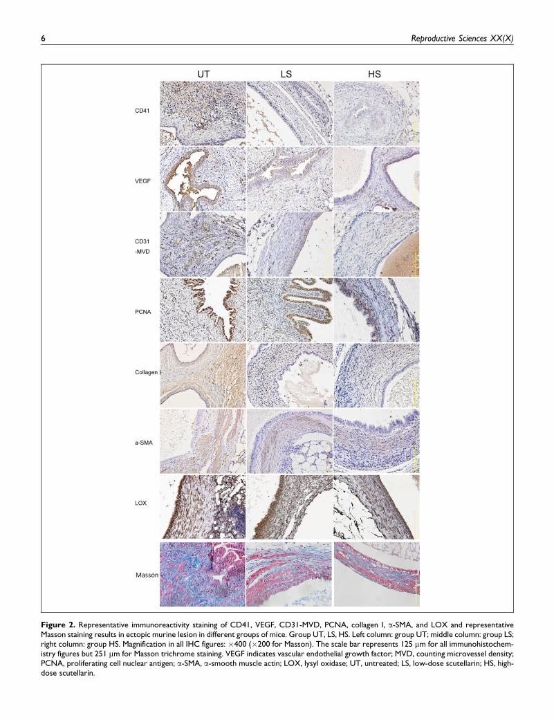
Figure 2. Representative immunoreactivity staining of CD41, VEGF, CD31-MVD, PCNA, collagen I, a-SMA, and LOX and representativeMasson staining results in ectopic murine lesion in different groups of mice. Group UT, LS, HS. Left column: group UT; middle column: group LS;right column: group HS. Magnification in all IHC figures: �400 (�200 for Masson). The scale bar represents 125 mm for all immunohistochem-istry figures but 251 mm for Masson trichrome staining. VEGF indicates vascular endothelial growth factor; MVD, counting microvessel density;PCNA, proliferating cell nuclear antigen; a-SMA, a-smooth muscle actin; LOX, lysyl oxidase; UT, untreated; LS, low-dose scutellarin; HS, high-dose scutellarin.
6 Reproductive Sciences XX(X)
platelets) was correlated with PCNA staining levels (r ¼ 0.58,
P ¼ .0016), and all other proteins (r’s � 0.58, Ps < .0014). In
particular, it was positively correlated with the immunoreactiv-
ity against a-SMA, collagen I, LOX, and the extent of lesional
fibrosis (r’s � 0.42, Ps < .031).
Discussion
In this study, we have shown that scutellarin treatment resulted
in significantly reduced lesion weight, improved hyperalgesia,
and changes consistent with reduced proliferation, angiogen-
esis, and fibrogenesis in a mouse model of endometriosis.
Figure 3. Boxplot summarization of the histochemistry and histochemistry staining results for platelet aggregation (A), VEGF (B), CD31-macrivessel density (C), PCNA (D), a-SMA (E), collagen I (F), LOX (G), and the extent of lesional fibrosis (H). NS: P > .05; *P < .05; **P < .01;***P < .001. In all plots, Wilcoxon test was used, and the symbols in brown indicate test using the UT group as the reference, while the blacksymbols indicate test using the Lo-SC as the reference. VEGF indicates vascular endothelial growth factor; PCNA, proliferating cell nuclearantigen; a-SMA, a-smooth muscle actin; LOX, lysyl oxidase; UT: untreated; Lo-SC, low-dose scutellarin; Hi-SC, high-dose scutellarin.
Ding et al 7
Scutellarin treatment also significantly reduced the platelet
activation rate in the peripheral blood. The reduced lesional
staining of a-SMA in the stromal component suggests deceler-
ated FMT, which is consistent with the concomitant reduction
in lesional fibrosis.
Our results are consistent with our previous findings that
platelet depletion and antiplatelet treatment resulted in reduced
lesion growth and improved pain behavior in mice with
induced endometriosis.13,21,22 Our results are also consistent
with the treatment effect of andrographolide47 and valproic
Figure 4. Scatter plots showing the relationship between lesion weight and the extent of platelet aggregation (A), immunostaining levels ofVEGF (B), microvessel density (C), PCNA (D), a-SMA (E), collagen I (F), and LOX (G), and the extent of lesional fibrosis (H). The symbols in theplot indicate the treatment group from which the mouse came from, with UT being untreated, and LS and HS, as treated with low- and high-dosescutellarin, respectively. The dashed line represents the regression line. The number shown in each figure indicates the Pearson correlationcoefficient, followed by the statistical significance levels: *P < .05; **P < .01; ***P < .001. VEGF indicates vascular endothelial growth factor;MVD, counting microvessel density; PCNA, proliferating cell nuclear antigen; a-SMA, a-smooth muscle actin; LOX, lysyl oxidase.
8 Reproductive Sciences XX(X)
acid48 in rodent models of endometriosis, as both have been
shown to have antiplatelet capabilities.49,50 Collectively, these
results demonstrate that antiplatelet therapeutics may be pro-
mising as a nonhormonal treatment for endometriosis and
should be further investigated.
The herbal medicine from which scutellarin is derived is
a potent antiplatelet herb in traditional Chinese medicine
that combats “blood stasis.” As in eutopic endometrium, the
ectopic endometrium also undergoes cyclic bleeding and
thus tissue injury. As such, platelets must be involved in the
ensuing tissue repair. Once there is tissue injury or a wound,
platelets are first cells (though anucleated) to go to and
aggregate at the wounding site, initiating hemostasis and
inaugurating the tissue repair process of inflammation, pro-
liferation, and tissue remodeling.51 Activated platelets secret
a plethora of bioactive molecules, including various cyto-
kines/chemokines, growth factors such as interleukins,
VEGF, PDGF, EGF, FGF, and TGF-b1, with TGF-b1 being
the most copious.52 In addition, endometriotic stromal cells
also produce potent platelet-activating molecules such as
thrombin and thromboxane A253 and collagens,15 which, in
conjunction with increased angiogenesis and thus vascular
permeability, may further lead to platelet aggregation. Thus,
endometriotic lesions and platelets engage active cross-talks
to maintain lesion growth and promote EMT, FMT, and
SMM, resulting in fibrosis.13,15,16
Aside from its antiplatelet capability, scutellarin may also
target other pathways involved in endometriosis. Indeed, scu-
tellarin is reported to relieve ischemia/reperfusion injury. The
cardioprotective and neuroprotective effects of scutellarin
depend on the upregulation of the endothelial nitric oxide
synthase (eNOS) expression54,55 and the downregulation of
VEGF, basic FGF, and inducible nitric oxide synthase
(iNOS). In addition, it is an efficient inhibitor of an important
transcription factor nuclear factor-kB (NF-kB), for which
aberrant activation in endometriosis has also been
reported.56-58 Moreover, scutellarin has been reported to be
promising in treating resisting liver fibrosis,59 interstitial, and
myocardial fibrosis34,60 in rats by inhibiting collagens I and
III and TGF-b1. Although our study has shown reduced
immunostaining of VEGF and collagen I in lesions, it is plau-
sible that scutellarin treatment may also cause suppression of
NF-kB activation and TGF-b1 inhibition in lesions.
We note that fibrosis itself may facilitate lesional develop-
ment by enhancing fibrogenesis through increased stiffness of
extracellular matrix (ECM). In fibrotic diseases, it is known
that increased myofibroblast contraction induces the release
of latent TGF-b161 and mechanical stretch also activates
TGF-b1,62 stimulating further profibrotic changes. In endo-
metriosis, it has been shown that soft ECMs, which are asso-
ciated with no fibrosis, inhibit cell proliferation and inactivate
fibrotic phenotype in stromal cells derived from deep endo-
metriotic lesions.63 In contrast, stiff ECM induces EMT in
endometrial epithelial cells.64
One puzzling finding in our study is that scutellarin treat-
ment did not demonstrate a dose–response relationship in
suppressing lesion development. Although mice receiving HS
treatment did respond to the treatment, they showed no better
improvement than the LS group, even though the platelet acti-
vation rate in the peripheral blood seemed to be reduced dose
dependently (Figure 1D). However, the reduction in the extent
of platelet aggregation in lesions did not seem to be dose
dependent, and the lesion weight in the HS group appeared to
be similar to that of the LS group. It is possible that, given the
known human dosage, which is near the low dosage used in this
study, and the known side effects of breviscapine which
include fatigue, skin itching and rashes, dizziness, nausea, and
vomiting, higher dose might have increased discomfort and
thus stress in HS mice, yielding somewhat diminished thera-
peutic effects. In addition, there is a likelihood that, given the
vast difference in physiology between Homo sapiens and
mouse, the effect of scutellarin may be quite different between
the 2 species. Further research is warranted to identify the
optimal dose in future preclinical studies.
This study has several limitations. First, this study only
employed immunohistochemistry and histochemistry analyses
and did not provide molecular evidence that scutellarin sup-
presses angiogenesis, proliferation, or fibrogenesis. Hence, the
best we can say is that our data are highly consistent with the
documented changes. Also due to the methods we used, only a
few possible mechanisms were explored. Further research is
warranted to clearly delineate the mechanism of action for
scutellarin. Second, the percentage of circulating activated pla-
telets in controls as we reported is somewhat high. This is likely
due to our use of centrifugation without prior fixation by par-
aformaldehyde.65 However, since we handled and analyzed all
blood samples uniformly and without any bias, our finding that
mice treated with scutellarin had lower percentage of circulat-
ing activated platelets than the UT mice is still valid. Lastly, the
treatment started just 2 weeks after induction, which may not
be long enough for lesional fibrosis to be fully developed as
commonly seen in humans.66 However, given the significant
reduction in hotplate latency at the time of surgery (Figure 1C),
it is safe to say that the pain symptom has already been man-
ifested at the time of start of the treatment. In addition, the
evidence that scutellarin treatment did slow down the progres-
sion of lesional fibrogenesis (Figure 3E-H) seems to be unmis-
takable. Of course, it is possible that once lesions become
highly fibrotic, the treatment efficacy might be diminished due
to reduced vascularity and possible epigenetic changes.67
Nonetheless, the reduced extent of fibrosis in scutellarin-
treated groups demonstrates that scutellarin can stall the pro-
gression of fibrogenesis. Future studies are needed to see
whether scutellarin is also effective in treating deep
endometriosis-like disease.
In summary, scutellarin appears to be effective in treating
mouse with induced endometriosis, resulting in significantly
reduced lesion weight, improved hyperalgesia, and changes
consistent with reduced proliferation, angiogenesis, and
fibrogenesis. As such, scutellarin may be a promising non-
hormonal therapeutic compound for treating endometriosis.
Ding et al 9
Authors’ Note
Ding Ding, MD, PhD, Xianjun Cai, MD, and Hanxi Zheng contributed
equally to this work.
Declaration of Conflicting Interests
The author(s) declared no potential conflicts of interest with respect to
the research, authorship, and/or publication of this article.
Funding
The author(s) disclosed receipt of the following financial support for
the research, authorship, and/or publication of this article: This
research was supported in part by grants 81471434 (SWG),
81530040 (SWG), 81771553 (SWG), 81370695 (XSL), 81671436
(XSL), and 81401183 (DD) from the National Natural Science Foun-
dation of China, 2017A610169 (XJC) from Ningbo Municipal Com-
mission of Science and Technology, and an Excellence in Centers of
Clinical Medicine grant (2017ZZ01016) from the Science and Tech-
nology Commission of Shanghai Municipality.
Supplemental material
Supplemental material for this article is available online.
References
1. Giudice LC, Kao LC. Endometriosis. Lancet. 2004;364(9447):
1789-1799.
2. Practice Committee of the American Society for Reproductive
Medicine. Treatment of pelvic pain associated with endometrio-
sis: a committee opinion. Fertil Steril. 2014;101(4):927-935.
3. Guo SW. Recurrence of endometriosis and its control. Hum
Reprod Update. 2009;15(4):441-461.
4. Koga K, Takamura M, Fujii T, Osuga Y. Prevention of the
recurrence of symptom and lesions after conservative surgery for
endometriosis. Fertil Steril. 2015;104(4):793-801.
5. Garcia-Velasco JA, Somigliana E. Management of endometrio-
mas in women requiring IVF: to touch or not to touch. Hum
Reprod. 2009;24(3):496-501.
6. de Ziegler D, Borghese B, Chapron C. Endometriosis and inferti-
lity: pathophysiology and management. Lancet. 2010;376(9742):
730-738.
7. Coccia ME, Rizzello F, Mariani G, Bulletti C, Palagiano A, Scar-
selli G. Ovarian surgery for bilateral endometriomas influences
age at menopause. Hum Reprod. 2011;26(11):3000-3007.
8. Vercellini P, Buggio L, Frattaruolo MP, Borghi A, Dridi D,
Somigliana E. Medical treatment of endometriosis-related pain.
Best Pract Res Clin Obstet Gynaecol. 2018;51:68-91.
9. Vercellini P, Crosignani P, Somigliana E, Vigano P, Frattaruolo
MP, Fedele L. ‘Waiting for Godot’: a commonsense approach to
the medical treatment of endometriosis. Hum Reprod. 2011;26(1):
3-13.
10. Guo SW, Groothuis PG. Is it time for a paradigm shift in drug
research and development in endometriosis/adenomyosis? Hum
Reprod Update. 2018;24(5):577-598.
11. Brosens IA. Endometriosis – a disease because it is characterized
by bleeding. Am J Obstet Gynecol. 1997;176(2):263-267.
12. Guo SW, Ding D, Shen M, Liu X. Dating endometriotic ovarian
cysts based on the content of cyst fluid and its potential clinical
implications. Reprod Sci. 2015;22(7):873-883.
13. Ding D, Liu X, Duan J, Guo SW. Platelets are an unindicted
culprit in the development of endometriosis: clinical and experi-
mental evidence. Hum Reprod. 2015;30(4):812-832.
14. Guo SW. Fibrogenesis resulting from cyclic bleeding: the Holy
Grail of the natural history of ectopic endometrium. Hum Reprod.
2018;33(3):353-356.
15. Zhang Q, Duan J, Liu X, Guo SW. Platelets drive smooth muscle
metaplasia and fibrogenesis in endometriosis through epithelial–
mesenchymal transition and fibroblast-to-myofibroblast transdif-
ferentiation. Mol Cell Endocrinol. 2016;428:1-16.
16. Zhang Q, Duan J, Olson M, Fazleabas A, Guo SW. Cellular
changes consistent with epithelial–mesenchymal transition and
fibroblast-to-myofibroblast transdifferentiation in the progression
of experimental endometriosis in baboons. Reprod Sci. 2016;
23(10):1409-1421.
17. Zhang Q, Ding D, Liu X, Guo SW. Activated platelets induce
estrogen receptor beta expression in endometriotic stromal cells.
Gynecol Obstet Invest. 2015;80(3):187-192.
18. Du Y, Liu X, Guo SW. Platelets impair natural killer cell reactiv-
ity and function in endometriosis through multiple mechanisms.
Hum Reprod. 2017;32(4):794-810.
19. Wu Q, Ding D, Liu X, Guo SW. Evidence for a hypercoagulable
state in women with ovarian endometriomas. Reprod Sci. 2015;
22(9):1107-1114.
20. Ding D, Liu X, Guo SW. Further evidence for hypercoagulability
in women with ovarian endometriomas. Reprod Sci. 2018;25(11):
1540-1548. doi:10.1177/1933719118799195.
21. Guo SW, Ding D, Geng JG, Wang L, Liu X. P-selectin as a
potential therapeutic target for endometriosis. Fertil Steril.
2015;103(4):990-1000 e8.
22. Guo SW, Ding D, Liu X. Anti-platelet therapy is efficacious in
treating endometriosis induced in mouse. Reprod Biomed Online.
2016;33(4):484-499.
23. Carnero A. High throughput screening in drug discovery. Clin
Transl Oncol. 2006;8(7):482-490.
24. Wang L, Ma Q. Clinical benefits and pharmacology of scutellarin:
a comprehensive review. Pharm Ther. 2018;190:105-127.
25. Liu X, Cheng J, Zhang G, et al. Engineering yeast for the
production of breviscapine by genomic analysis and synthetic
biology approaches. Nat Commun. 2018;9(1):448.
26. Luo P, Tan ZH, Zhang ZF, Zhang H, Liu XF, Mo ZJ. Scutellarin
isolated from Erigeron multiradiatus inhibits high glucose-
mediated vascular inflammation. Yakugaku Zasshi. 2008;128(9):
1293-1299.
27. Ma JY, Jiang WW, Zhou ZT, Li JM, Wang HY. The promoting
angiogenesis and anti-inflammation effect of scutellarin on
polyglycolic acid scaffold of Balb/c mice model. J Asian Nat
Prod Res. 2008;10(11-12):1147-1153.
28. Tian X, Chang L, Ma G, et al. Delineation of platelet activation
pathway of scutellarein revealed its intracellular target as protein
kinase C. Biol Pharm Bullet. 2016;39(2):181-191.
29. Hou L, Chen L, Fang L. Scutellarin inhibits proliferation, inva-
sion, and tumorigenicity in human breast cancer cells by
10 Reproductive Sciences XX(X)
regulating HIPPO-YAP signaling pathway. Med Sci Monit. 2017;
23:5130-5138.
30. Ke Y, Bao T, Wu X, et al. Scutellarin suppresses migration and
invasion of human hepatocellular carcinoma by inhibiting the
STAT3/Girdin/Akt activity. Biochem Biophys Res Commun.
2017;483(1):509-515.
31. Li H, Huang D, Gao Z, Chen Y, Zhang L, Zheng J. Scutellarin
inhibits the growth and invasion of human tongue squamous car-
cinoma through the inhibition of matrix metalloproteinase-2 and -
9 and alphavbeta6 integrin. Int J Oncol. 2013;42(5):1674-1681.
32. Zhu PT, Mao M, Liu ZG, Tao L, Yan BC. Scutellarin suppresses
human colorectal cancer metastasis and angiogenesis by targeting
ephrinb2. Am J Transl Res. 2017;9(11):5094-104.
33. Sun CY, Zhu Y, Li XF, et al. Scutellarin increases cisplatin-
induced apoptosis and autophagy to overcome cisplatin resistance
in non-small cell lung cancer via ERK/p53 and c-met/AKT sig-
naling pathways. Front Pharmacol. 2018;9:92.
34. Pan Z, Zhao W, Zhang X, et al. Scutellarin alleviates interstitial
fibrosis and cardiac dysfunction of infarct rats by inhibiting
TGFbeta1 expression and activation of p38-MAPK and ERK1/
2. Br J Pharmacol. 2011;162(3):688-700.
35. Li Y, Liu YD, Chen SL, et al. Down-regulation of long non-
coding RNA MALAT1 inhibits granulosa cell proliferation in
endometriosis by up-regulating P21 via activation of the ERK/
MAPK pathway. Mol Hum Reprod. 2018.
36. KhoshdelRad N, Salehi Z, Mashayekhi F, Abbasi O, Mirzajani E.
Soluble c-Met expression in the peritoneal fluid and serum of
patients with different stages of endometriosis. Arch Gynecol
Obstet. 2014;289(5):1107-1112.
37. Choi J, Jo M, Lee E, Hwang S, Choi D. Aberrant PTEN expres-
sion in response to progesterone reduces endometriotic stromal
cell apoptosis. Reproduction. 2017;153(1):11-21.
38. Council NR. Guide for the Care and Use of Laboratory Animals.
Washington, DC: National Academies Press; 1996.
39. Somigliana E, Vigano P, Rossi G, Carinelli S, Vignali M, Panina-
Bordignon P. Endometrial ability to implant in ectopic sites can
be prevented by interleukin-12 in a murine model of endometrio-
sis. Hum Reprod. 1999;14(12):2944-2950.
40. Long Q, Liu X, Guo SW. Surgery accelerates the development of
endometriosis in mouse. Am J Obstetr Gynecol. 2016;215(3):320.
e1-320.e15.
41. Reagan-Shaw S, Nihal M, Ahmad N. Dose translation from ani-
mal to human studies revisited. FASEB J. 2008;22(3):659-661.
42. Hu X, Teng S, He J, et al. Pharmacological basis for application of
scutellarin in Alzheimer’s disease: antioxidation and antiapopto-
sis. Mol Med Rep. 2018;18(5):4289-4296.
43. Sun C, Li C, Li X, et al. Scutellarin induces apoptosis and autop-
hagy in NSCLC cells through ERK1/2 and AKT signaling path-
ways in vitro and in vivo. J Cancer. 2018;9(18):3247-3256.
44. Lu Y, Nie J, Liu X, Zheng Y, Guo SW. Trichostatin A, a histone
deacetylase inhibitor, reduces lesion growth and hyperalgesia in
experimentally induced endometriosis in mice. Hum Reprod.
2010;25(4):1014-1025.
45. R Development Core Team. R: A Language and Environment for
Statistical Computing. Vienna, Austria: R Foundation for Statis-
tical Computing; 2016.
46. Zhao T, Liu X, Zhen X, Guo SW. Levo-tetrahydropalmatine
retards the growth of ectopic endometrial implants and alleviates
generalized hyperalgesia in experimentally induced endometrio-
sis in rats. Reprod Sci. 2011;18(1):28-45.
47. Zheng Y, Liu X, Guo SW. Therapeutic potential of andrographo-
lide for treating endometriosis. Hum Reprod. 2012;27(5):
1300-1313.
48. Liu M, Liu X, Zhang Y, Guo SW. Valproic acid and progestin
inhibit lesion growth and reduce hyperalgesia in experimen-
tally induced endometriosis in rats. Reprod Sci. 2012;19(4):
360-373.
49. Lien LM, Su CC, Hsu WH, et al. Mechanisms of
andrographolide-induced platelet apoptosis in human platelets:
regulatory roles of the extrinsic apoptotic pathway. Phytother
Res. 2013;27(11):1671-1677.
50. Davidson DC, Hirschman MP, Spinelli SL, et al. Antiplatelet
activity of valproic acid contributes to decreased soluble CD40
ligand production in HIV type 1-infected individuals. J Immunol.
2011;186(1):584-591.
51. Nurden AT, Nurden P, Sanchez M, Andia I, Anitua E. Platelets
and wound healing. Front Biosci. 2008;13:3532-3548.
52. Assoian RK, Komoriya A, Meyers CA, Miller DM, Sporn MB.
Transforming growth factor-beta in human platelets. Identifica-
tion of a major storage site, purification, and characterization.
J Biol Chem. 1983;258:7155-7160.
53. Guo SW, Du Y, Liu X. Endometriosis-derived stromal cells
secrete thrombin and thromboxane A2, inducing platelet activa-
tion. Reprod Sci. 2016;23(8):1044-1052.
54. Hu XM, Zhou MM, Hu XM, Zeng FD. Neuroprotective effects
of scutellarin on rat neuronal damage induced by cerebral
ischemia/reperfusion. Acta Pharmacol Sin. 2005;26(12):
1454-1459.
55. Yang W, Lust RM, Bofferding A, Wingard CJ. Nitric oxide and
catalase-sensitive relaxation by scutellarin in the mouse thoracic
aorta. J Cardiovasc Pharmacol. 2009;53(1):66-76.
56. Gonzalez-Ramos R, Donnez J, Defrere S, et al. Nuclear factor-
kappa B is constitutively activated in peritoneal endometriosis.
Mol Hum Reprod. 2007;13(7):503-509.
57. Gonzalez-Ramos R, Van Langendonckt A, Defrere S, et al.
Involvement of the nuclear factor-kappaB pathway in the
pathogenesis of endometriosis. Fertil Steril. 2010;94(6):
1985-1994.
58. Kaponis A, Iwabe T, Taniguchi F, et al. The role of NF-
kappaB in endometriosis. Front Biosci (Schol Ed). 2012;4:
1213-1234.
59. Wang YH, Geng L, Li H. Study on effect of scutellarin in resisting
liver fibrosis in rats [in Chinese]. Zhongguo Zhong Yao Za Zhi.
2015;40(10):1999-2003.
60. Zhou H, Chen X, Chen L, et al. Anti-fibrosis effect of scutellarin
via inhibition of endothelial–mesenchymal transition on
isoprenaline-induced myocardial fibrosis in rats. Molecules.
2014;19(10):15611-15623.
61. Wipff PJ, Rifkin DB, Meister JJ, Hinz B. Myofibroblast contrac-
tion activates latent TGF-beta1 from the extracellular matrix.
J Cell Biol. 2007;179(6):1311-1323.
Ding et al 11
62. Froese AR, Shimbori C, Bellaye PS, et al. Stretch-induced activa-
tion of transforming growth factor-beta1 in pulmonary fibrosis.
Am J Respir Crit Care Med. 2016;194(1):84-96.
63. Matsuzaki S, Canis M, Pouly JL, Darcha C. Soft matrices inhibit
cell proliferation and inactivate the fibrotic phenotype of deep
endometriotic stromal cells in vitro. Hum Reprod. 2016;31(3):
541-553.
64. Matsuzaki S, Darcha C, Pouly JL, Canis M. Effects of matrix
stiffness on epithelial to mesenchymal transition-like processes
of endometrial epithelial cells: implications for the pathogenesis
of endometriosis. Sci Rep. 2017;7:44616.
65. Michelson AD, Barnard MR, Krueger LA, Frelinger A III, Fur-
man MI. Evaluation of platelet function by flow cytometry.
Methods. 2000;21(3):259-270.
66. Zhang Q, Liu X, Guo SW. Progressive development of endome-
triosis and its hindrance by anti-platelet treatment in mice with
induced endometriosis. Reprod Biomed Online. 2017;34(2):
124-136.
67. Liu X, Zhang Q, Guo SW. Histological and immunohistochemical
characterization of the similarity and difference between ovarian
endometriomas and deep infiltrating endometriosis. Reprod Sci.
2018;25(3):329-340.
12 Reproductive Sciences XX(X)