salinity-induced changes in branchia atpase...
TRANSCRIPT

J. exp. Bwl. 151, 279-296 (1990) 2 7 9Printed in Great Britain © The Company of Biologists Limited 1990
SALINITY-INDUCED CHANGES IN BRANCHIAL Na+/K+-ATPase ACTIVITY AND TRANSEPITHELIAL POTENTIALDIFFERENCE IN THE BRINE SHRIMP ARTEMIA SAUNA
BY CHARLES W. HOLLIDAY*
Department of Biology, Lafayette College, Easton, PA 18042, USA
DAVID B. ROYE AND ROBERT D. ROER
Center for Marine Science Research, University of North Carolina,Wilmington, NC, USA
Accepted 2 March 1990
Summary
Silver staining of the adult brine shrimp, Artemia salina, revealed that only themetepipodites of the phyllopodia were significantly permeable to chloride and/orsilver ions. The metepipodites stained in a reticulated pattern, possibly indicatingareas in the cuticle over cells specialized for chloride secretion. Crude homogen-ates of metepipodites had very high Na+/K+-ATPase enzyme specific activity(ESA) which increased in proportion to the salinity of the external medium and,thus, in proportion to the need for outward salt transport in these strongly hypo-osmoregulating animals. Metepipodite ESA as a percentage of whole-body ESAincreased from 7.6% in 50% sea water (SW) to 25.0% in 400% SW. Gut andmaxillary gland also had high Na+/K+-ATPase ESAs, implicating these organs inosmoregulatory processes as well. The time courses of increases in phyllopodialand gut ESAs in brine shrimps transferred from 100 % SW to 400 % SW areconsistent with the induction of new Na+/K+-ATPase; 4-7 days was required forsignificant increases to occur. Haemolymph ion analyses and transepithelialpotential differences, measured in brine shrimp acclimated in all the SW media,indicate that chloride is actively transported out of the brine shrimp while sodiumis very close to electrochemical equilibrium across the body wall. Thus, themetepipodites of the brine shrimp appear to possess cells with many functionalsimilarities to the teleost branchial chloride cells.
Introduction
Brine shrimps, Artemia salina, live in some of the harshest aquatic environmentsknown. Their ability to survive and reproduce in hypersaline waters inhabited only
*To whom reprint requests should be addressed.
Key words: osmoregulation, sodium chloride transport, Crustacea, gill.
280 C. W. HOLLIDAY, D. B. ROYE AND R. D . ROER
by algae, bacteria and a few species of insects has aroused much interest in themechanisms used by these creatures to survive the ionic and osmotic stressesimposed by their environment. Conte's group has elegantly outlined the mechan-isms of osmotic and ionic regulation in the nauplii of A. salina (reviewed in Conte,1984), which use a special salt-secreting gland, the neck organ, to rid themselves ofsalts that enter by diffusion from the hypersaline medium. Further, Russler andMangos (1978) have shown that the neck organ of A. salina nauplii is the majorroute for sodium efflux.
The mechanisms of osmotic and ionic regulation in adult A. salina suggested bythe studies of Copeland (1967), Croghan (1958a,b,c,d,e), Smith (1969a,b) andThuet et al. (1968) are very similar to those proposed for marine teleosts. Brineshrimps are strong hypo-osmotic regulators in all media more concentrated thanapproximately 30 % sea water. In these concentrated media they drink to replacewater lost by osmosis to the hyperosmotic external medium and they use their gillsto secrete salts which enter with the ingested medium and by diffusion (Croghan,1958b,c,d). Sodium and chloride are actively transported out of the gut into thehaemolymph and, presumably, water follows passively; the ions are then secretedinto the medium at the gills. The metepipodites of the phyllopodia appear to bethe sites of outward ion transport (Croghan, 1958c) and a special cell type found inthese structures (the 'dark cell', Copeland, 1967), rich in mitochondria, is thoughtto be responsible for this ion transport. Augenfeld (1969) found that whole-bodyNa+/K+-ATPase activity in immature and adult A. salina increased with thesalinity of the external medium. Further, based on measurements of transepi-thelial potential difference in brine shrimps in 100 % sea water (Smith, 1969a), itappears that chloride is the ion that is actively transported out of the brine shrimpby the metepipodites, while sodium is in or very close to electrochemicalequilibrium across the body wall. Thus, it appears that brine shrimps, too, possess'chloride cells' with transport characteristics similar to those in the gills of marineteleosts. A second brine shrimp, Parartemia zletzlana, has been studied by Geddes(1975a,b,c) and its osmotic and ionic regulation appear to be very similar to thoseof A. salina.
The present study was undertaken to investigate further the apparent similaritybetween teleost and brine shrimp osmoregulatory mechanisms. Silver staining wasused to show that the metepipodite cuticle has cell-sized areas of very highpermeability to chloride and/or silver ions. The role of the cellular sodium pump,the Na+/K+-ATPase, in ion transport by the brine shrimp was investigated byassaying gills and other body parts from shrimps acclimated in media of varyingsalinity to determine whether enzyme specific activity increases with the salinity ofthe external medium, as does branchial Na+/K+-ATPase activity in many teleosts.Transepithelial potential differences were also measured in 50 %, 100 %, 200 %and 400% sea water to determine whether chloride and/or sodium ions areactively transported out of the animal in these media. We report here results whichconfirm the functional similarities between the marine teleost gill and themetepipodites of the brine shrimp. Our data on A. salina metepipodites are
Brine shrimp branchial Na+/K+-ATPase and TEP 281
consistent with the transport model proposed for teleost branchial chloride cells byZadunaisky (1984).
Materials and methodsAnimals and acclimation media
Anemia salina used in this study were purchased from several commercialsuppliers as San Francisco Bay Brand (Newark, California) cysts or live adults.The cysts were obtained by the vendors from both the Great Salt Lake, Utah, andthe San Francisco Bay, California (Leslie Salt Co. ponds), populations of A.salina; live adults were from the San Francisco Bay population only.
The normal culture medium for brine shrimps was 100 % sea water (SW) whichwas made from a commercial synthetic sea salt mixture (Lobster Tank Salt, DaynoManufacturing Co., Lynn, Massachusetts; lOOOmosmolkg"1). Other media(50 %, 200 % and 400 % SW) were also made using this salt mixture and had jinalosmotic pressures of 500, 2000 and 4000mosmolkg~1, respectively. Culturemedium osmotic pressure was measured at intervals of 2-3 days and adjusted byadding distilled water to replace that lost by evaporation. Cysts were hatched in100 % SW, transferred to 51 of clean, gently aerated medium and fed powderedtropical fish food (Staple Food, Tetra Werke, Melle, West Germany) daily. Onlyadult, female brine shrimps were used in this study and all animals were acclimatedin the various media for at least 14 days before being used in experiments.
Silver staining
The so-called 'silver stain' was used to identify areas of the brine shrimp whichhave a high permeability to chloride ions and in which silver chloride precipitateswhen the animals are treated with a dilute solution of silver nitrate. Brine shrimpswere removed from the culture medium using a wide-mouthed Pasteur pipette andrinsed for 30 s in each of three changes of deionized water to remove adherentchloride ions. The animals were then transferred to 0.5% AgNO3 for 30 s andrinsed in deionized water as before. Finally, shrimps were transferred to KodakD-19 photographic developer (Eastman Kodak, Inc., Rochester, New York) for30 s and rinsed in deionized water three times.
Haemolymph sampling
To avoid the difficulties of sampling haemolymph with micropipettes, a newmethod for rapidly sampling haemolymph from adult A. salina was developed forthis study. Animals were removed individually from the culture medium using awide-mouthed Pasteur pipette and gently expelled onto a dry paper towel. Whenall adherent culture medium had been drawn off by the paper towel, the brineshrimp was grasped firmly just behind the head from the dorsal side with jeweller'sforceps, crushing the nerve cord. After a brief period of struggle the brine shrimprelaxed and straightened out; it was then held over a small plastic Petri dish filledwith water-equilibrated mineral oil so as to immerse the last few millimeters of the
282 C. W. HOLLIDAY, D. B. ROYE AND R. D . ROER
abdomen in the oil. Using fine scissors, the tip of the abdomen was then cut offbeneath the surface of the oil as the brine shrimp was lifted up and away from theoil. This procedure caused the brine shrimp's body muscles to contract reflexly andto expel approximately 1-3 JJ\ of haemolymph as a droplet into the oil as the bodywas lifted away from it. If the brine shrimp was not lifted away from the oil as thetip of the abdomen was severed, the haemolymph frequently ran up along theabdomen by capillary action and wetted the phyllopodia, making it very difficult toobtain a sample. Sampling was repeated with 5-10 individuals until 10-20 jA ofhaemolymph had been collected as a pooled sample for analyses.
It could be argued that haemolymph obtained by the new method would becontaminated with gut fluid, but this appears not to be the case for two reasons.First, few samples were visibly contaminated with faeces and these were notincluded in the pooled samples used for analyses. Second, the gut fluid of A. salinahas been reported to be much more concentrated than the haemolymph. Forexample, the data reported by Croghan (19586) show that gut fluid averaged 2-3times the osmotic pressure of haemolymph in all media (e.g. shrimp in approxi-mately 100% SW had gut fluid averaging about 720 mosmol kg"1 and haemolymphaveraging about 350 mosmol kg"1). Haemolymph osmolality in brine shrimpacclimated in 100 % SW in the present study averaged 362 mosmol kg~1, indicatingthat no significant contamination by the much more concentrated gut fluid hadoccurred.
Measurement of osmotic pressure and ion concentrations
Osmotic pressure of media and haernolymph samples was measured using aWescor 5100 C vapour pressure osmometer. Chloride concentrations in media andhaemolymph samples were measured using a coulometric titrator (Buchler-Cot-love chloridometer). Sodium concentrations in media and haemolymph sampleswere measured using an Orion Research Ross sodium-ion-specific electrodeconnected to a Beckman 0 12 pH/lSE meter; all samples were diluted andadjusted for ionic strength, and sodium concentrations were measured at pH9.5.
Measurement of transepithelial potential difference
Transepithelial potential differences (TEPs) were measured in A. salina usingglass microelectrodes and a plastic chamber similar to that described by Smith(1969a). Brine shrimps were individually removed from the culture medium usinga wide-mouthed Pasteur pipette and gently expelled onto a paper towel to drythem. Pieces of human hair were tied with an overhand knot so as to form a3-4 mm loop in the middle of each hair. One loop was then placed over the headand pulled tight just behind it; a second loop was put over the end of the abdomenand pulled tight. Thus 'lassoed', the brine shrimp was secured in the chamber bythe four strands of hair protruding from it. Brine shrimps prepared in this way didnot struggle and continued to make steady, vigorous swimming movements in theTEP chamber medium. Animals survived for at least 6h in preliminary prep-arations; TEP measurements took less than 20min. The swimming movements of
Brine shrimp branchial Na+/ K+-ATPase and TEP 283
the brine shrimp were judged sufficient to minimize the effects of any unstirredlayers which might develop during the measurement of TEP. Media were changedby siphoning approximately five times the chamber volume of new medium intothe TEP chamber while the overflow was removed by suction.
TEP was measured by impaling the brine shrimp with glass microelectrodes heldin a micromanipulator and viewed with a dissecting microscope. The microelec-trodes were made with a Kopf model 700C electrode puller and were filled with3 mol I"1 KC1; they had tip impedances of 1-5 MQ. The microelectrode and holderwere plugged into the probe end of a WPIM701 electrometer. Grounding wasaccomplished with a Ag/AgCl reference electrode. TEPs were displayed on aTektronix model 5103N oscilloscope and read to the nearest millivolt from theoscilloscope screen.
Although TEPs were stable and varied little when measured at a variety oflocations over the body surface, the best site for impalement without breaking themicroelectrode tips was at the dorsal bases of the phyllopodia. Preliminaryexperiments showed that serial microelectrode impalement at the bases ofsuccessive phyllopodia did not cause the TEP to change. Impalements and TEPmeasurements were performed in quadruplicate for each medium to which thebrine shrimp was exposed. TEPs were first measured in brine shrimps bathed intheir medium of acclimation, then in the other three SW media, allowing 3-4 minfor the TEP to stabilize after each change of medium.
Na+/K+-ATPase assay
The activity of the cellular sodium pump in various tissues of A. salina wasmeasured as the enzyme specific activity (ESA) of Na+/K+-ATPase in crudehomogenates of tissues dissected and pooled for assay. Groups of 5-10 brineshrimps were rinsed in homogenizing medium (HM; 0.25 mol I"1 sucrose,6mmoll~1 EDTA) and dissected into the following parts, which were pooled forhomogenization and enzyme assay: heads, metepipodites, phyllopodia withoutmetepipodites, body wall and gut. After dissection, the gut was stripped free offaeces by grasping it at one end and gently pulling it between the tips of a secondpair of forceps. In some assays whole phyllopodia were used. Maxillary glandswere dissected by cutting free a small patch of the body wall containing them; pairsof glands from 25 brine shrimps were pooled and homogenized for each assay. In afinal assay, the ESA of the eleventh pair of metepipodites was measured; unlikemetepipodites 1-10, these appendages do not stain with silver and it was ofinterest to determine if they had lower ESAs than the other metepipodites. Theeleventh metepipodites from 40 brine shrimps acclimated in 100% SW weredissected, homogenized and assayed for ESA; metepipodites 1-10 were dissectedfrom the same animals and served as controls.
Body parts were homogenized on ice in 0.4-1.0 ml of HM in a ground glasshomogenizer and kept on ice for up to an hour until assayed. The assay media andmethods were identical to those used in previous studies (Holliday, 1985). Briefly,phosphate liberated from ATP by each homogenate was measured in two reaction
284 C. W. HOLLIDAY, D . B. ROYE AND R. D . ROER
media. One medium had optimum concentrations of all ions (lOOmmolF1 Na+,SOmmoir1 K+, SmmolP1 ATP, lOmmolT1 Mg2"1", 20mmoir 1 imidazole,pH7.2) whereas the other lacked potassium and contained ouabain (130mmoll~x
Na+, Smmoir 1 ATP, lOmmolP1 Mg2"1", ^mmoU" 1 imidazole, lmmoll"1
ouabain, pH7.2). After incubation at 30°C for 15min, the reaction was stoppedand phosphate concentrations in the reaction mixtures were measured colorimetri-cally as the reduced phosphomolybdate complex. Enzyme specific activity wascalculated as the difference in phosphate liberated by each homogenate in the twomedia and is expressed as /xmol phosphate liberated mg"1 protein h"1. Proteinconcentrations in the homogenates were measured colorimetrically using the Bio-Rad protein assay (Bio-Rad Laboratories, Richmond, California) with bovineserum albumin as a standard. All chemicals used in the chemical and enzymeassays were reagent grade or better.
Statistics
Unless otherwise stated, results are expressed as mean values±s.E. Student'sunpaired Mest was used to evaluate the significance of differences between meanvalues. A probability (P) value =S0.05 was considered to be significant. For thosepoints in figures without error bars, S.E. was smaller than the size of the symbol onthe figure.
Results
Silver staining
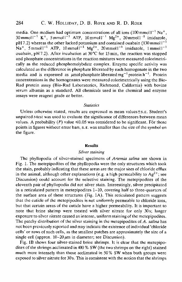
The phyllopodia of silver-stained specimens of Anemia salina are shown inFig. 1. The metepipodites of the phyllopodia were the only structures which tookthe stain, probably indicating that these areas are the major sites of chloride effluxin the animal, although other explanations (e.g. a high permeability to Ag2+; seeDiscussion) could account for the selective staining. The metepipodites of theeleventh pair of phyllopodia did not silver stain. Interestingly, silver precipitatedin a reticulated pattern in metepipodites 1-10, covering half to three-quarters ofthe surface area of these structures (Fig. 1A). This reticulated pattern suggeststhat the cuticle of the metepipodites is not uniformly permeable to chloride ions,but that certain areas of the cuticle have a higher permeability. It is important tonote that brine shrimp were treated with silver nitrate for only 30s; longerexposure to silver nitrate caused an intense, uniform staining of the metepipodites.The patchy distribution of the silver staining in the metepipodites of A salina hasnot been previously reported and may indicate the existence of individual 'chloridecells' or rows of such cells, as the smallest patches are approximately the size of asingle cell (approx. 10-20/HTI in diameter; see Discussion).
Fig. IB shows four silver-stained brine shrimps. It is clear that the metepipo-dites of the shrimps acclimated in 400 % SW (the two shrimps on the right) stainedmuch more intensely than those acclimated in 50 % SW when both groups wereexposed to silver nitrate for 30 s. This is consistent with the notion that the shrimps
Brine shrimp branchial Na+/K+-ATPase and TEP 285
Fig. 1. Silver-stained Artemia salina. (A) Lateral view of phyllopodia 6-10. Note thereticulated pattern of the stain; the smallest of the dark patches is approximately10-20/an in diameter; (B) dorsal view of four silver-stained A. salina; the two animalson the left were acclimated in 50 % SW, the two on the right were acclimated in 400 %SW. Note the darker staining of the metepipodites in the two brine shrimps on theright.
286 C. W. HOLLIDAY, D . B. ROYE AND R. D . ROER
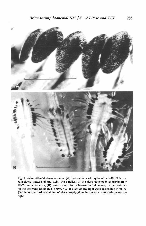
Table 1. Osmotic pressure and sodium and chloride ion concentrations in the haemo-lymph and external media-of Artemia salina
% Seawater
50
100
200
400
Mean
Osmoticpressure
(mosmolkg"1
4%(493, 499)
1034(1031, 1036)
1898(1890, 1907)
4012(4001, 4024)
values±s.E.(jV)
External medium
Sodium) (mmoir 1 )
243(239, 247)
498(497, 499)
900(898, 902)
1946(1940, 1952)
or mean (value!,
Chloride(mmoir 1 )
273(270, 276)
602(608, 596)
1075(1059, 1135)
2216(2223, 2210)
value2) where
Osmoticpressure
(mosmol kg"
353±4(6)
362±5(6)
411 ±3(6)
498±4(6)
TV is 2.
Haemolymph
Sodium') (mmoir1)
135±5(5)
155±5(5)
161±3(5)
185±5(5)
Chloride(mmoir1)
147(136, 158)
146(144, 148)
156(156, 155)
210(203, 217)
in 400 % SW were secreting chloride at a higher rate than those in 50 % SW, aswould be expected.
Osmotic and ionic regulation
Table 1 shows the osmoregulatory and ionoregulatory performance of brineshrimps, acclimated in four seawater media. As noted by other investigators, A.salina is a weak hyporegulator in 50 % SW and an increasingly strong hyporegula-tor in 100 %, 200 % and 400 % SW. Its haemolymph osmotic pressure and sodiumand chloride concentrations increased approximately 40 % in the face of eightfoldincreases in the osmotic pressure and sodium and chloride concentrations in theexternal medium.
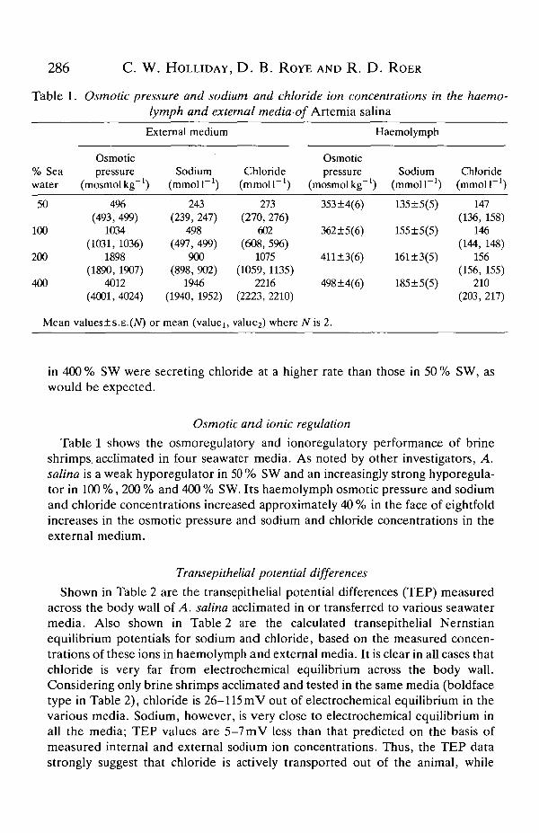
Transepithelial potential differences
Shown in Table 2 are the transepithelial potential differences (TEP) measuredacross the body wall of A. salina acclimated in or transferred to various seawatermedia. Also shown in Table 2 are the calculated transepithelial Nemstianequilibrium potentials for sodium and chloride, based on the measured concen-trations of these ions in haemolymph and external media. It is clear in all cases thatchloride is very far from electrochemical equilibrium across the body wall.Considering only brine shrimps acclimated and tested in the same media (boldfacetype in Table 2), chloride is 26-115 raV out of electrochemical equilibrium in thevarious media. Sodium, however, is very close to electrochemical equilibrium inall the media; TEP values are 5-7 mV less than that predicted on the basis ofmeasured internal and external sodium ion concentrations. Thus, the TEP datastrongly suggest that chloride is actively transported out of the animal, while
Brine shrimp branchial Na+/K+-ATPase and TEP 287
Table 2. Transepithelial potential differences measured in Artemia salina ac-climated in or acutely transferred to various seawater media and calculatedNernstian transepithelial equilibrium potentials for sodium and chloride ions in
acclimated animals
Transepithelial potential difference (mV, polarity inside)and medium in which transepithelial potential was
measured
Acclimationmedium
50%sea water
100%sea water
200%sea water
400%sea water
Equilibriumpotential (mV,polarity inside)
Sodium Chloride
50% SW100% SW200% SW400% SW
+10±l(4)
+ 10±l(4)+ 10±l(4)
+ 11±3(4)+23+1(7)+24±2(5)+25±2(4)
+28±1(4)+29±1(4)+37±2(5)+36±1(4)
+37±2(4)+40±2(3)+52±2(5)+55 ±1(5)
+ 15+30+44+60
-16-36-50-60
*Mean values±s.E. (N).For each brine shrimp, transepithelial potential difference was measured first in the medium
of acclimation, then in the other three media.Equilibrium potentials were calculated using the appropriate haemolymph and medium ion
concentrations shown in Table 1.Results for shrimps acclimated and tested in the same media are shown in bold type.
sodium is very close to being passively distributed across the body wall in all mediatested.
The TEPs measured after acute transfer of brine shrimps to media other thanthe acclimation medium (values other than boldface type in Table 2) showinteresting trends. First, when TEP was measured in media more dilute than theacclimation medium, the TEP was nearly identical to that measured in brineshrimps acclimated and measured in the same dilute medium (e.g. brine shrimpsacclimated in 400 % SW and measured in 50 % SW had the same TEP as shrimpsacclimated in 50% SW and measured in 50% SW). If it is assumed that themeasured TEP is due to active ion transport, the brine shrimp seems to be capableof rapidly reducing such transport when placed in media more dilute than theacclimation medium. However, the reverse is not true. In all cases the TEP inbrine shrimps measured in media more concentrated than the acclimation mediumwas lower than that measured in shrimps acclimated and measured in the samemedia (e.g. shrimps acclimated in 50 % SW and measured in 400 % SW showed amuch lower TEP, 37 mV, than that of shrimps acclimated in and measured in400% SW, 55 mV; Table 2). This may indicate that brine shrimps are unable to'turn on' active ion transport as rapidly as they can turn it off when the saltconcentration of the external medium changes rapidly. These differences haveinteresting implications for the relative roles of increased activity of existingmetepipodite Na+/K+-ATPase (enzyme activation) and synthesis of newNa+/K+-ATPase molecules (enzyme induction) in situations that require changesin ion transport (see Discussion).
288 C. W. HOLLIDAY, D. B. ROYE AND R. D. ROER
Na+1K+-ATPase assay optimisation
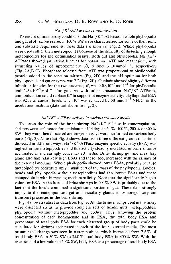
To ensure optimal assay conditions, the Na+/K+-ATPases in whole phyllopodiaand gut of A. salina reared in 100 % SW were characterized for some of their ionicand substrate requirements; these data are shown in Fig. 2. Whole phyllopodiawere used rather than metepipodites because of the difficulty of dissecting enoughmetepipodites for the optimisation assays. Both gut and phyllopodial Na+/K+-ATPases showed saturation kinetics for potassium, ATP and magnesium, withsaturating values of approximately 30, 5 and 3-lOmmolT1, respectively(Fig. 2A,B,C). Phosphate released from ATP was proportional to phyllopodialprotein added to the reaction mixture (Fig. 2D) and the pH optimum for bothphyllopodial and gut enzymes was 7.2 (Fig. 2E). Ouabain showed slightly differentinhibition kinetics for the two enzymes; K\ was 9.0xlO~6moll~1 for phyllopodiaand 1.3xl0~5moir1 for gut. As with other crustacean Na+/K+-ATPases,ammonium ion could replace K+ in support of enzyme activity; phyllopodial ESAwas 92% of control levels when K+ was replaced by SOmmoll"1 NH4C1 in theincubation medium (data not shown in Fig. 2).
Na+1K+-ATPase activity in various seawater media
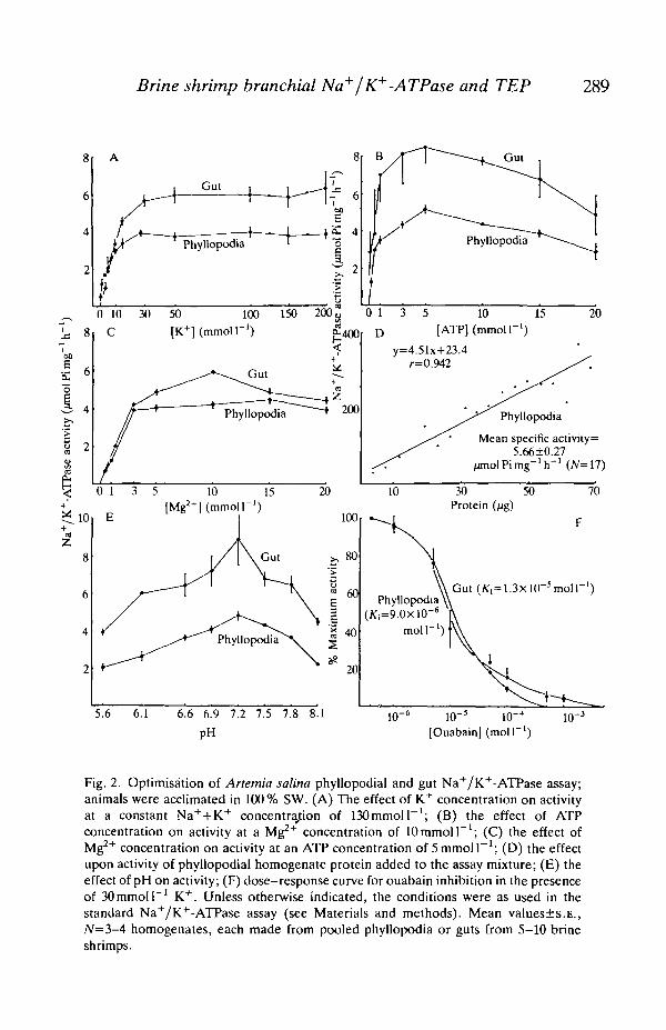
To assess the role of the brine shrimp Na+/K+-ATPase in osmoregulation,shrimps were acclimated for a minimum of 14 days in 50 %, 100 %, 200 % or 400 %SW; they were then dissected and enzyme assays were performed on various bodyparts (Fig. 3). Note that Fig. 3 shows data from three different groups of shrimpsdissected in different ways. Na+/K+-ATPase enzyme specific activity (ESA) washighest in the metepipodites and this activity steadily increased in brine shrimpsacclimated in increasingly concentrated media. Brine shrimp gut and maxillarygland also had relatively high ESAs and these, too, increased with the salinity ofthe external medium. Whole phyllopodia showed lower ESAs, probably becausemetepipodites constitute only a small part of the mass of the phyllopodia. Bodies,heads and phyllopodia without metepipodites had the lowest ESAs and thesechanged little with increasing medium salinity. Note that the significantly highervalue for ESA in the heads of brine shrimps in 400 % SW is probably due to thefact that the heads contained a significant portion of gut. These data stronglyimplicate the metepipodites, gut and maxillary glands in osmoregulatory iontransport processes in the brine shrimp.
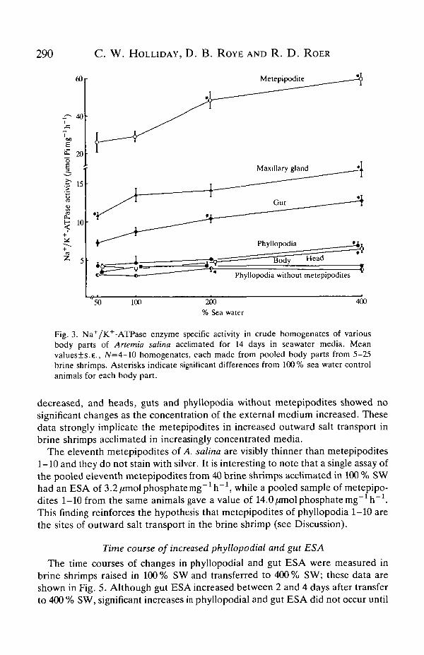
Fig. 4 shows a subset of data from Fig. 3. All the brine shrimps used in this assaywere dissected so as to provide complete sets of heads, guts, metepipodites,phyllopodia without metepipodites and bodies. Thus, knowing the proteinconcentration of each homogenate and its ESA, the total body ESA andpercentage of total body ESA for each dissected group of body parts could becalculated for shrimps acclimated in each of the four external media. The mostpronounced change was seen in metepipodites, which increased from 7.6% oftotal body ESA in 50% SW to 25.0% total body ESA in 400% SW. With theexception of a low value in 50 % SW, body ESA as a percentage of total body ESA
Brine shrimp branchial Na+/K+-ATPase and TEP 289
Gut7 6
Phyllopodia4—+5 4
1 o
Gut
0 10 30 50 100 150 200j,C [K+] (mmoir1)
0 1 3 5 10 15[ATP] (mmol I"1)
20
Mean specific activity=5.66±0.27
5.6 6.1 6.6 6.9 7.2 7.5 7.8 8.1
PH10" KT5 KT4 10"3
[Ouabain] (moir1)
Fig. 2. Optimisation of Artemia salina phyllopodial and gut Na+/K+-ATPase assay;animals were acclimated in 100% SW. (A) The effect of K+ concentration on activityat a constant Na++K+ concentration of 130 mmol T1; (B) the effect of ATPconcentration on activity at a Mg2+ concentration of 10 mmol F 1 ; (C) the effect ofMg2+ concentration on activity at an ATP concentration of 5 mmol I"1; (D) the effectupon activity of phyllopodial homogenate protein added to the assay mixture; (E) theeffect of pH on activity; (F) dose-response curve for ouabain inhibition in the presenceof 30 mmol I"1 K+. Unless otherwise indicated, the conditions were as used in thestandard Na+/K+-ATPase assay (see Materials and methods). Mean values±s.E.,N=3-4 homogenates, each made from pooled phyllopodia or guts from 5-10 brineshrimps.
290 C. W. HOLLIDAY, D. B. ROYE AND R. D . ROER
60
. i - 40
00
£• 20o
.f 15
Metepipodite
Phyllopodia without metepipodites
50 100 200% Sea water
400
Fig. 3. Na+/K+-ATPase enzyme specific activity in crude homogenates of variousbody parts of Anemia salina acclimated for 14 days in seawater media. Meanvalues±s.E., /V=4-10 homogenates, each made from pooled body parts from 5-25brine shrimps. Asterisks indicate significant differences from 100% sea water controlanimals for each body part.
decreased, and heads, guts and phyllopodia without metepipodites showed nosignificant changes as the concentration of the external medium increased. Thesedata strongly implicate the metepipodites in increased outward salt transport inbrine shrimps acclimated in increasingly concentrated media.
The eleventh metepipodites of A. salina axe. visibly thinner than metepipodites1-10 and they do not stain with silver. It is interesting to note that a single assay ofthe pooled eleventh metepipodites from 40 brine shrimps acclimated in 100 % SWhad an ESA of 3.2 /miol phosphate mg~1h~1, while a pooled sample of metepipo-dites 1-10 from the same animals gave a value of 14.0/zmolphosphatemg"^"1.This finding reinforces the hypothesis that metepipodites of phyllopodia 1-10 arethe sites of outward salt transport in the brine shrimp (see Discussion).
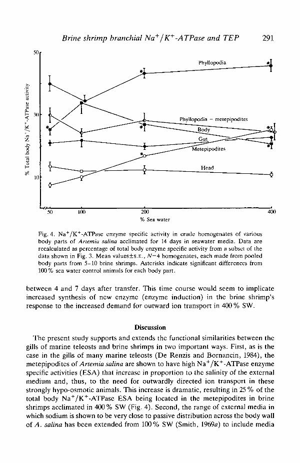
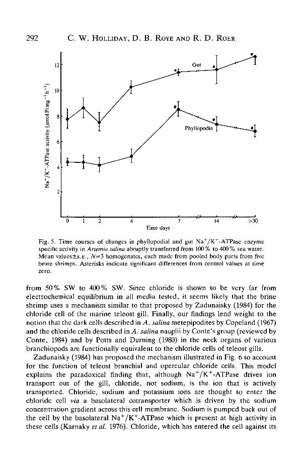
Time course of increased phyllopodial and gut ESA
The time courses of changes in phyllopodial and gut ESA were measured inbrine shrimps raised in 100% SW and transferred to 400% SW; these data areshown in Fig. 5. Although gut ESA increased between 2 and 4 days after transferto 400 % SW, significant increases in phyllopodial and gut ESA did not occur until
Brine shrimp branchial Na+/K+-ATPase and TEP 291
50
30
(2
10
Phyllopodia
Phyllopodia — metepipodites
50 100 200
% Sea water
400
Fig. 4. Na+/K+-ATPase enzyme specific activity in crude homogenates of variousbody parts of Anemia salina acclimated for 14 days in seawater media. Data arerecalculated as percentage of total body enzyme specific activity from a subset of thedata shown in Fig. 3. Mean values±s.E., N=4 homogenates, each made from pooledbody parts from 5-10 brine shrimps. Asterisks indicate significant differences from100 % sea water control animals for each body part.
between 4 and 7 days after transfer. This time course would seem to implicateincreased synthesis of new enzyme (enzyme induction) in the brine shrimp'sresponse to the increased demand for outward ion transport in 400 % SW.
Discussion
The present study supports and extends the functional similarities between thegills of marine teleosts and brine shrimps in two important ways. First, as is thecase in the gills of many marine teleosts (De Renzis and Bornancin, 1984), themetepipodites of Anemia salina are shown to have high Na+/K+-ATPase enzymespecific activities (ESA) that increase in proportion to the salinity of the externalmedium and, thus, to the need for outwardly directed ion transport in thesestrongly hypo-osmotic animals. This increase is dramatic, resulting in 25 % of thetotal body Na+/K+-ATPase ESA being located in the metepipodites in brineshrimps acclimated in 400% SW (Fig. 4). Second, the range of external media inwhich sodium is shown to be very close to passive distribution across the body wallof A. salina has been extended from 100% SW (Smith, 1969a) to include media
292 C. W. HOLLIDAY, D . B. ROYE AND R. D . ROER
12
'j. 10
soE
CU
•o 8i
vity
Q
i 68
CO
2
Gut #
1 ^^1 / #T
^ 1 y / Phyllopodia
/T y '
j T T ^ ^ ^ ^
• ^ 1 — y ^ ^ " ^
v~~~~~*-—Jit
14 >30Time days
Fig. 5. Time courses of changes in phyllopodial and gut Na+/K+-ATPase enzymespecific activity in Artemia salina abruptly transferred from 100 % to 400 % sea water.Mean values±s.E., 7V=3 homogenates, each made from pooled body parts from fivebrine shrimps. Asterisks indicate significant differences from control values at timezero.
from 50% SW to 400% SW. Since chloride is shown to be very far fromelectrochemical equilibrium in all media tested, it seems likely that the brineshrimp uses a mechanism similar to that proposed by Zadunaisky (1984) for thechloride cell of the marine teleost gill. Finally, our findings lend weight to thenotion that the dark cells described in A. salina metepipodites by Copeland (1967)and the chloride cells described in A. salina nauplii by Conte's group (reviewed byConte, 1984) and by Potts and Durning (1980) in the neck organs of variousbranchiopods are functionally equivalent to the chloride cells of teleost gills.
Zadunaisky (1984) has proposed the mechanism illustrated in Fig. 6 to accountfor the function of teleost branchial and opercular chloride cells. This modelexplains the paradoxical finding that, although Na+/K+-ATPase drives iontransport out of the gill, chloride, not sodium, is the ion that is activelytransported. Chloride, sodium and potassium ions are thought to enter thechloride cell via a basolateral cotransporter which is driven by the sodiumconcentration gradient across this cell membrane. Sodium is pumped back out ofthe cell by the basolateral Na+/K+-ATPase which is present at high activity inthese cells (Karnaky etal. 1976). Chloride, which has entered the cell against its
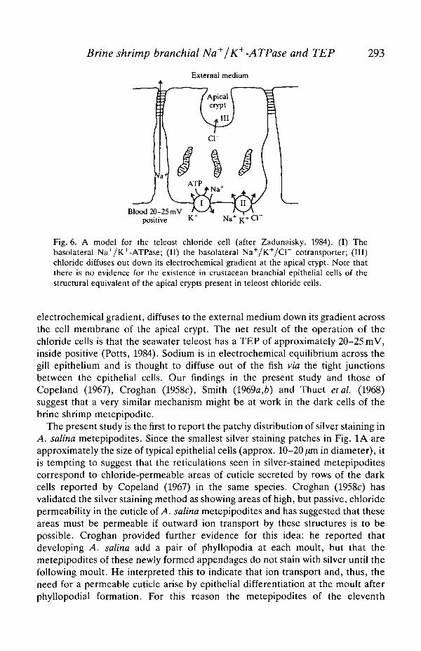
Brine shrimp branchial Na+/K+ -ATPase and TEP
External medium
293
Blood 20-25 mVpositive
Fig. 6. A model for the teleost chloride cell (after Zadunaisky, 1984). (I) Thebasolateral Na+/K+-ATPase; (II) the basolateral Na+/K+/Cl" cotransporter; (in)chloride diffuses out down its electrochemical gradient at the apical crypt. Note thatthere is no evidence for the existence in crustacean branchial epithelial cells of thestructural equivalent of the apical crypts present in teleost chloride cells.
electrochemical gradient, diffuses to the external medium down its gradient acrossthe cell membrane of the apical crypt. The net result of the operation of thechloride cells is that the seawater teleost has a TEP of approximately 20-25 mV,inside positive (Potts, 1984). Sodium is in electrochemical equilibrium across thegill epithelium and is thought to diffuse out of the fish via the tight junctionsbetween the epithelial cells. Our findings in the present study and those ofCopeland (1967), Croghan (1958c), Smith (1969a,6) and Thuet et al. (1968)suggest that a very similar mechanism might be at work in the dark cells of thebrine shrimp metepipodite.
The present study is the first to report the patchy distribution of silver staining inA. salina metepipodites. Since the smallest silver staining patches in Fig. 1A areapproximately the size of typical epithelial cells (approx. 10-20 pern in diameter), itis tempting to suggest that the reticulations seen in silver-stained metepipoditescorrespond to chloride-permeable areas of cuticle secreted by rows of the darkcells reported by Copeland (1967) in the same species. Croghan (1958c) hasvalidated the silver staining method as showing areas of high, but passive, chloridepermeability in the cuticle of A. salina metepipodites and has suggested that theseareas must be permeable if outward ion transport by these structures is to bepossible. Croghan provided further evidence for this idea: he reported thatdeveloping A. salina add a pair of phyllopodia at each moult, but that themetepipodites of these newly formed appendages do not stain with silver until thefollowing moult. He interpreted this to indicate that ion transport and, thus, theneed for a permeable cuticle arise by epithelial differentiation at the moult afterphyllopodial formation. For this reason the metepipodites of the eleventh
294 C. W. HOLLIDAY, D. B. ROYE AND R. D. ROER
phyllopodia do not stain with silver; they appear at the last moult and never have achance to develop an ion transport epithelium with a chloride-permeable cuticle.Croghan's hypothesis is supported by our finding that the eleventh metepipoditeshave a Na+/K+-ATPase ESA less than one-quarter that of metepipodites 1-10,indicating that the eleventh metepipodites do not develop the capacity to pumpsalts out of the haemolymph.
Although it seems likely that the silver staining technique identifies areas of highchloride permeability in A. salina, other mechanisms could also account for thedifferential staining. It is possible that the stain reveals areas of the cuticle that aregenerally very permeable to cations such as Ag2"1", which enters from the silvernitrate solution and then precipitates with chloride ion in the cuticle. If this is thecase, then silver staining may identify areas of the metepipodites that are verycation-permeable and that may be the sites of passive exit of sodium ions, as in theZadunaisky chloride cell model (Fig. 6). In this regard it is of interest that Barraetal. (1983), using X-ray microanalysis, have found that the precipitates whichform in the branchial cuticle of silver-lactate-treated crabs, Eriocheir sinensis, docontain large amounts of silver and chloride. Thus, although interpretation ofwhat the silver stain is actually showing may be problematic, it is clear that itidentifies areas of relatively high ion transport activity in the cuticle.
When A. salina is transferred from 100 % SW to 400 % SW, the time course ofchanges in metepipodite Na+/K+-ATPase ESA is consistent with the synthesis ofnew enzyme in response to the increased demands for outward ion transport (i.e.several days are required; Fig. 5). Although short-term changes in the rate of iontransport probably occur, synthesis of new enzyme also appears to be necessary forincreased osmoregulatory ion transport. The TEP data shown in Table 2 also lendindirect support to this idea. When transferred to media less concentrated than theacclimation medium, the TEPs decrease within' 3-4min to values very close tothose in shrimps acclimated in the same dilute media. Thus, if the TEP is assumedto be due to active ion transport, such ion transport can be rapidly decreased indilute media, suggesting rapid modulation (i.e. activation/deactivation of existingenzyme) of the Na+/K+-ATPase, which is assumed in the model above to drivechloride and sodium exit from the gills. However, when brine shrimps aretransferred to media more concentrated than the acclimation medium, the TEPdoes not rapidly increase to values close to those in brine shrimps acclimated in thesame concentrated media, but remains well below that of acclimated animals. Thisindicates that electrogenic ion transport and, presumably, the Na+/K+-ATPaseESA cannot be increased as rapidly as they are decreased and is consistent with theidea that Na+/K+-ATPase synthesis is necessary for the full response.
Morphological evidence also stresses the role of increased synthesis of Na+/K+-ATPase in acclimation of A. salina in concentrated media. When viewed edge-onin the dissection microscope, metepipodites dissected from shrimp acclimated in400 % SW were visibly thicker than metepipodites from shrimp acclimated in 50 %SW, probably indicating hypertrophy of ion transport tissue within the metepipo-dites in the more concentrated medium. Copeland (1967) and Croghan (1958c)
Brine shrimp branchial Na+/K+-ATPase and TEP 295
have also reported thickened metepipodite epithelium in brine shrimps acclimatedin concentrated media. Since brine shrimps are strong hypo-osmotic regulators in400% SW and, thus, tend to lose water to the external medium by osmosis, theincreased thickness of the metepipodites was probably not due to osmoticswelling. Thus, it appears that the population of dark cells in the metepipoditeepithelium may increase in thickness and/or multiply in response to transfer of thebrine shrimp to media of high salinity. If this is so, it seems likely that theseprocesses of hypertrophy in the metepipodite epithelium would require severaldays for completion.
Croghan (1959J), Smith (196%) and Thuet et al. (1968) have shown that A.salina, like marine teleosts, drinks hypertonic medium to replace water lost to themedium by osmosis. Drinking rates were quite high (48-72% body mass day"1),but the influx of sodium and chloride via the gut was calculated in the last tworeports above to be much smaller than influx across the body surface. Given thatthe concentrations of sodium and chloride in gut fluid are much lower than those inthe haemolymph, Croghan (1959J) and Smith (19696) suggested that sodium andchloride must be actively transported out of the gut fluid and that this transportmight drive the reabsorption of water from the gut. The correlation between gutNa+/K+-ATPase ESA and medium salinity seen in the present study supports thishypothesis in that gut ESA rises with the need to increase fluid uptake from thegut. The process is presumably driven by Na+/K+-ATPase, but no directmeasurements have been made of ion or fluid transport by the brine shrimp gut.Preliminary measurements of the TEP between the gut and haemolymph in thepresent study indicate that the gut is approximately 8mV lumen-positive withrespect to the haemolymph in shrimps from 50% and 100 % SW. Thus, transportof sodium and chloride out of the gut fluid is not a strongly electrogenic process inthis epithelium.
We note in closing that the brine shrimp maxillary gland has high a Na+/K+-ATPase ESA which increases in proportion to the salinity of the external medium,indicating that these glands may participate in osmotic or ionic regulation. Tyson(1969) has shown that the efferent duct of the maxillary gland of A. salina showsultrastructural features typical of transporting epithelia. However, since thesecretion of the gland has not been sampled, its role in osmotic and/or ionicbalance remains unknown.
This study was supported by a grant from Research Corporation and byLafayette College.
ReferencesAUGENFELD, J. M. (1969). The role of Na+-K+-activated, ouabain-sensitive ATPase in the
response of Anemia salina L. to salinity changes. Life Sci. 8, 973-978.BARRA, J.-A., PEQUEUX, A. AND HUMBERT, W. (1983). A morphological study of the gills of a
crab acclimated to fresh water. Tissue and Cell 15, 583-596.CONTE, F. P. (1984). Structure and function of the crustacean larval salt gland. Int. Rev. Cytol
91, 45-106.
296 C. W. HOLLIDAY, D. B. ROYE AND R. D. ROER
COPELAND, D. E. (1967). A study of the salt secreting cells in the brine shrimp (Anemia salina).In Protoplasma (ed. K. R. Porter), pp. 363-384. Vienna: Springer-Verlag.
CROGHAN, P. C. (1958a). The survival of Anemia salina (L.) in various media. J. exp. Biol. 35,213-218.
CROGHAN, P. C. (19586). The osmotic and ionic regulation of Artemia salina (L). J. exp. Biol.35, 219-233.
CROGHAN, P. C. (1958c). The mechanism of osmotic regulation in Artemia salina (L): thephysiology of the branchiae. J. exp. Biol. 35, 234-242.
CROGHAN, P. C. (1958d). The mechanism of osmotic regulation in Artemia salina (L): thephysiology of the gut. J. exp. Biol. 35,243-249.
CROGHAN, P. C. (1958e). Ionic fluxes in Artemia salina (L). J. exp. Biol. 35, 425-436.DE RENZIS, G. AND BORNANCIN, M. (1984). Ion transport and gill ATPases. In Fish Physiology,
vol. X (ed. W. S. Hoar and D. J. Randall), pp. 65-104. New York: Academic Press.GEDDES, M. C. (1975a). Studies on an Australian brine shrimp, Parartemia zeitziana Sayce
(Crustacea: Anostraca). I. Salinity tolerance. Comp. Biochem. Physiol. 51A, 553-559.GEDDES, M. C. (1975i>). Studies on an Australian brine shrimp, Parartemia zeitziana Sayce
(Crustacea: Anostraca). II. Osmotic and ionic regulation. Comp. Biochem. Physiol. 51A,561-571.
GEDDES, M. C. (1975C). Studies on an Australian brine shrimp, Parartemia zeitziana Sayce(Crustacea: Anostraca). III. The mechanisms of osmotic and ionic regulation. Comp.Biochem. Physiol. 51A, 573-578.
HOLLIDAY, C. W. (1985). Salinity-induced changes in gill Na, K-ATPase activity in the mudfiddler crab, Uca pugnax. J, exp. Zool. 233,199-208.
KARNAKY, K. J., JR, KINTER, L. B., KJNTER, W. B. AND STIRLING, C. E. (1976). Teleost chloridecell. II. Autoradiographic localization of gill Na, K-ATPase in killifish (Fundulus heteroclitus)adapted to low and high salinity environments. /. Cell Biol. 70, 157-177.
POTTS, W. T. W. (1984). Transepithelial potentials in fish gills. In Fish Physiology, vol. X (ed.W. S. Hoar and D. J. Randall), pp. 105-128. New York: Academic Press.
POTTS, W. T. W. AND DURNING, C. T. (1980). Physiological evolution in the branchiopods.Comp. Biochem Physiol. 67B, 475-484.
RUSSLER, D. AND MANGOS, J. (1978). Micropuncture studies of the osmoregulation in thenauplius of Artemia salina. Am. J. Physiol. 234, R216-R-222.
SMITH, P. G. (1969a). The ionic relations of Artemia salina (L.). I. Measurements of electricalpotential difference and resistance. J. exp. Biol. 51, 727-738.
SMITH, P. G. (19696). The ionic relations of Artemia salina (L.). II. Fluxes of sodium, chlorideand water. /. exp. Biol. 51, 739-757.
THUET, P., MOTAIS, R. ANDMAETZ, J. (1968). Les mechanismes de l'euryhalinite chez le crustacedes salines Artemia salina L. Comp. Biochem. Physiol. 26, 793-818.
TYSON, G. E. (1969). The fine structure of the maxillary gland of the brine shrimp, Artemiasalina: the efferent duct. Z. Zellforsch. mikrosk. Anat. 93, 151-163.
ZADUNAISKY, J. A. (1984). The chloride cell: the active transport of chloride and the paracellularpathways. In Fish Physiology, vol. X (ed. W. S. Hoar and D. J. Randall), pp. 129-176. NewYork: Academic Press.