ruben kuzniecky, m.d. nyu comprehensive epilepsy center new york university new york...
TRANSCRIPT
Ruben Kuzniecky, M.D.NYU Comprehensive Epilepsy Center
New York University
New York
Neuroimaging of Epilepsy
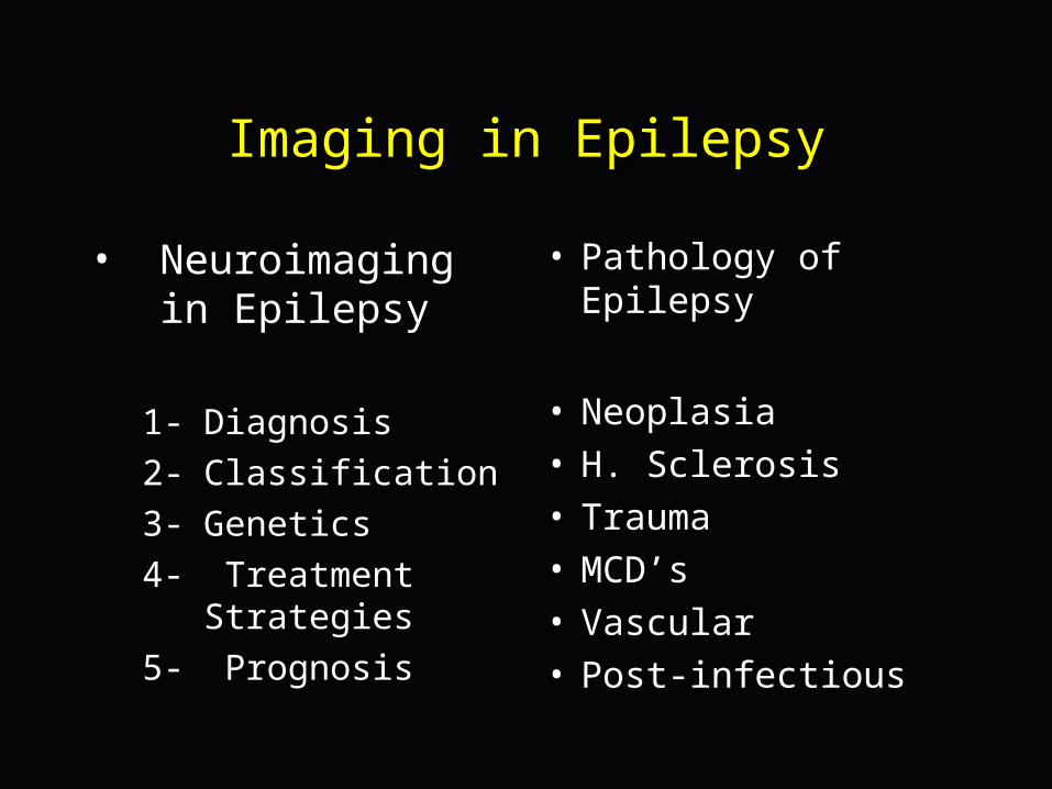
Imaging in EpilepsyImaging in Epilepsy
• Neuroimaging in Epilepsy
1- Diagnosis
2- Classification
3- Genetics
4- Treatment Strategies
5- Prognosis
• Pathology of Epilepsy
• Neoplasia• H. Sclerosis• Trauma• MCD’s• Vascular• Post-infectious
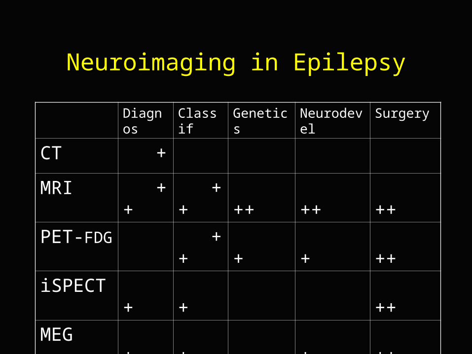
Neuroimaging in EpilepsyNeuroimaging in Epilepsy
Diagnos Classif Genetics Neurodevel Surgery
CT +
MRI ++ ++ ++ ++ ++
PET-FDG ++ + + ++
iSPECT + + ++
MEG + + + ++
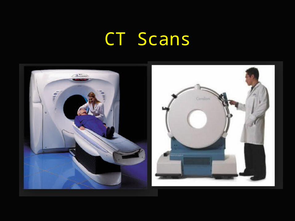
CT Scans
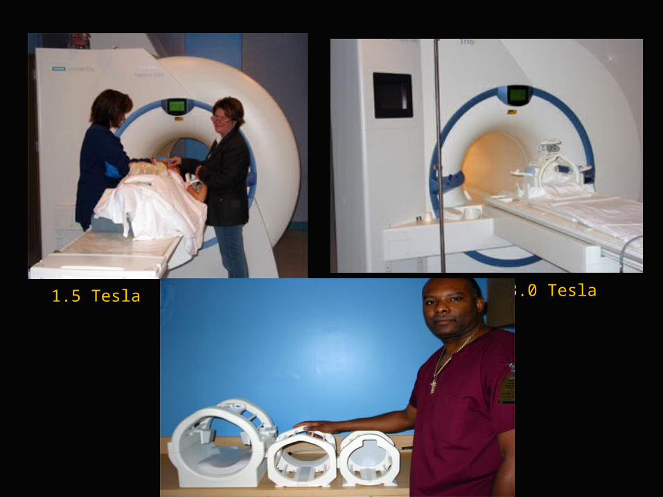
3.0 Tesla1.5 Tesla
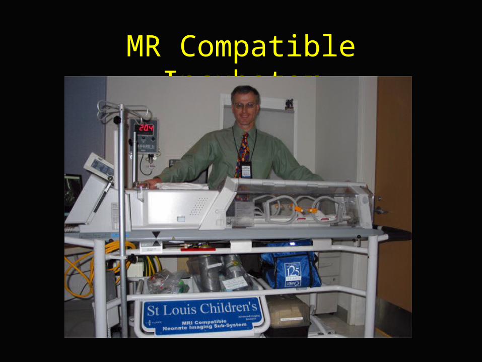
MR Compatible Incubator
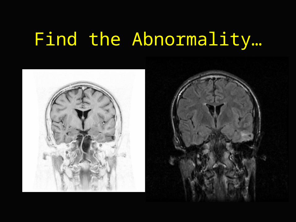
Find the Abnormality…
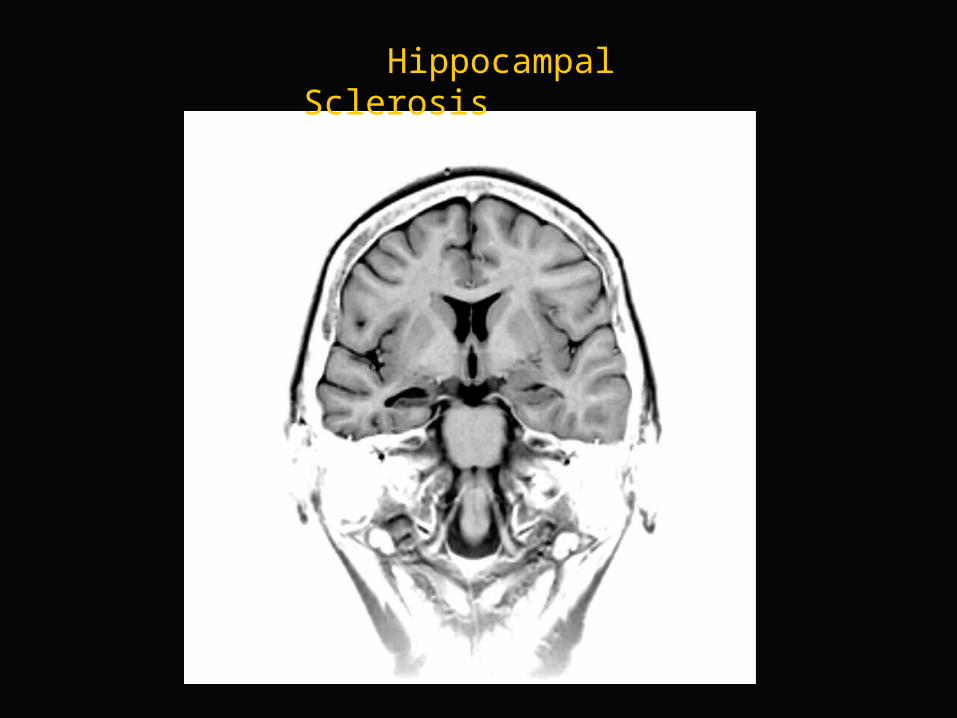
Hippocampal Sclerosis
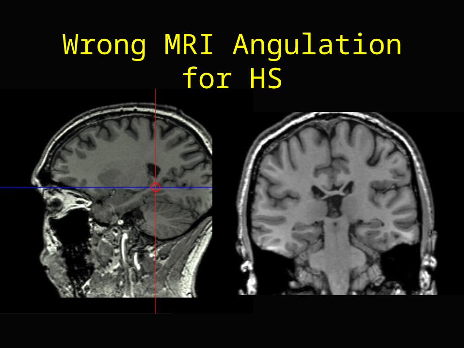
Wrong MRI Angulation for HS
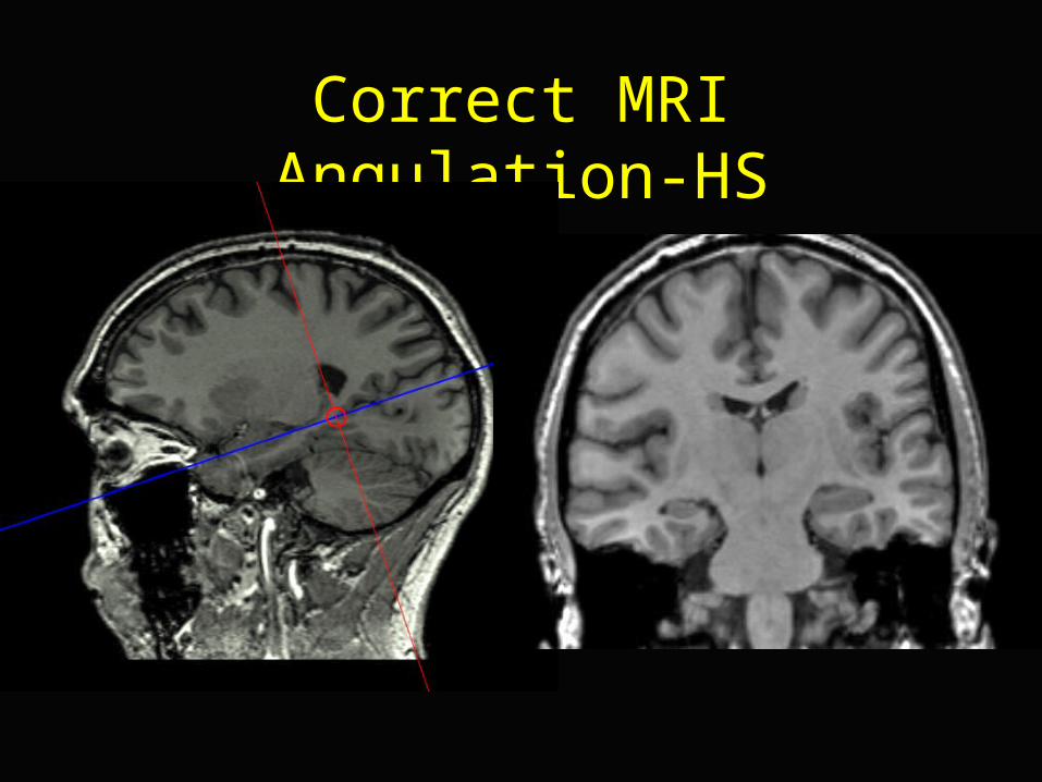
Correct MRI Angulation-HS
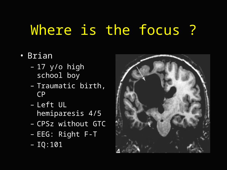
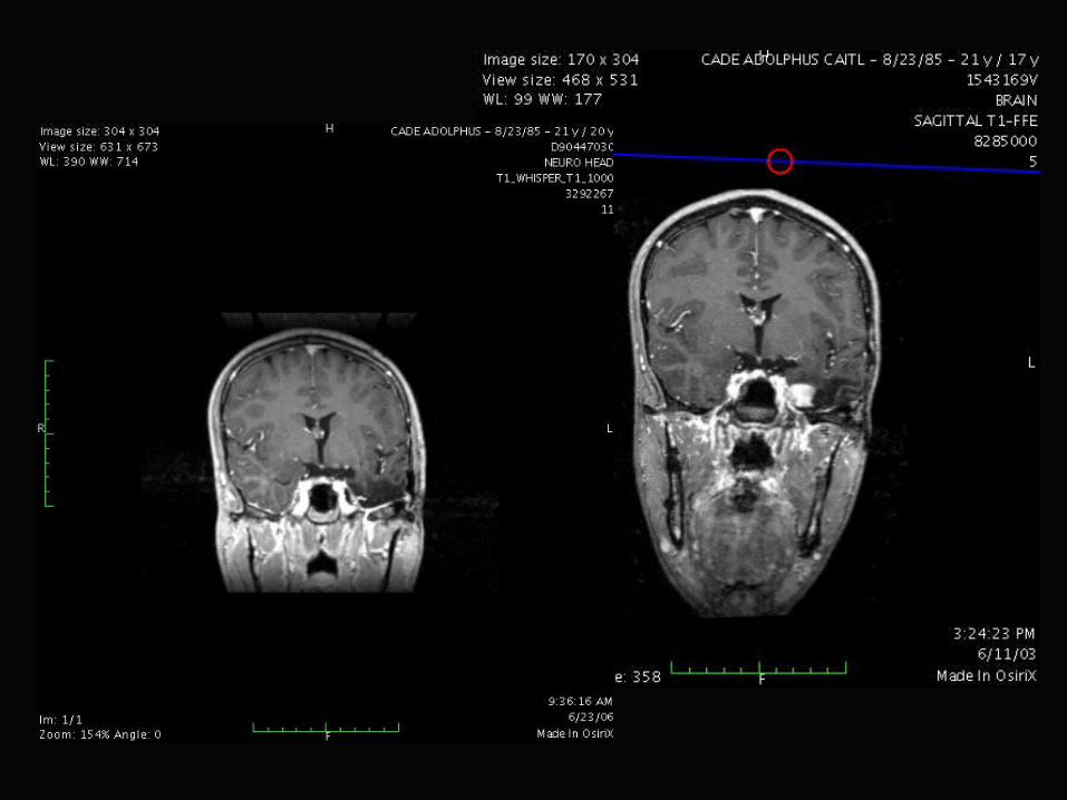
Where is the focus ?
• Brian– 17 y/o high school boy
– Traumatic birth, CP
– Left UL hemiparesis 4/5
– CPSz without GTC
– EEG: Right F-T
– IQ:101
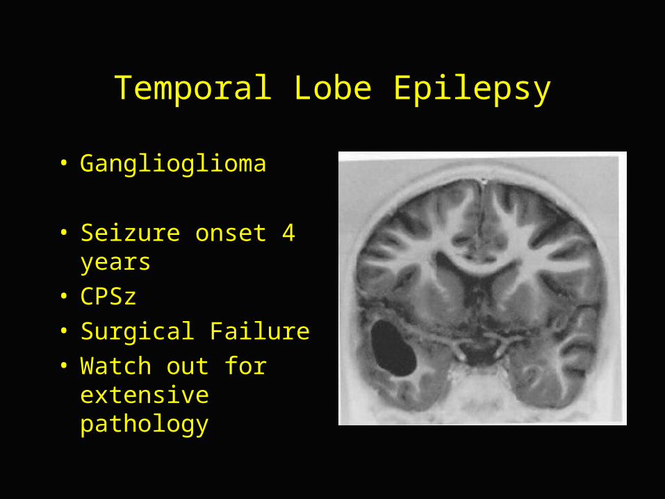
Temporal Lobe Epilepsy
• Ganglioglioma
• Seizure onset 4 years• CPSz• Surgical Failure• Watch out for
extensive pathology
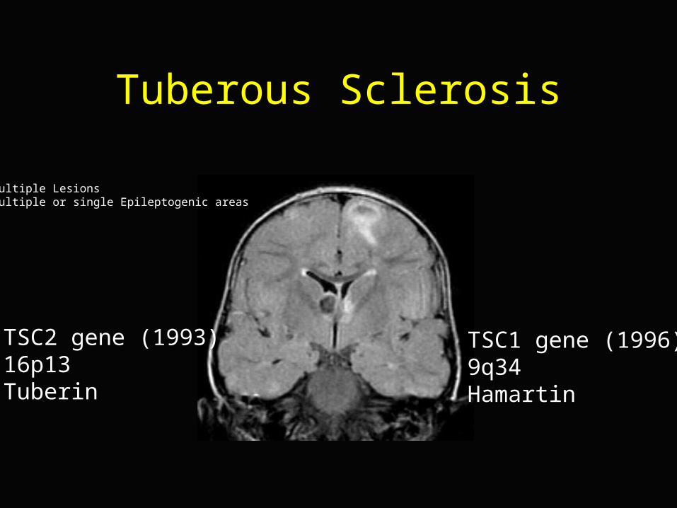
Tuberous Sclerosis
Multiple LesionsMultiple or single Epileptogenic areas
TSC2 gene (1993)16p13 Tuberin
TSC1 gene (1996)9q34Hamartin
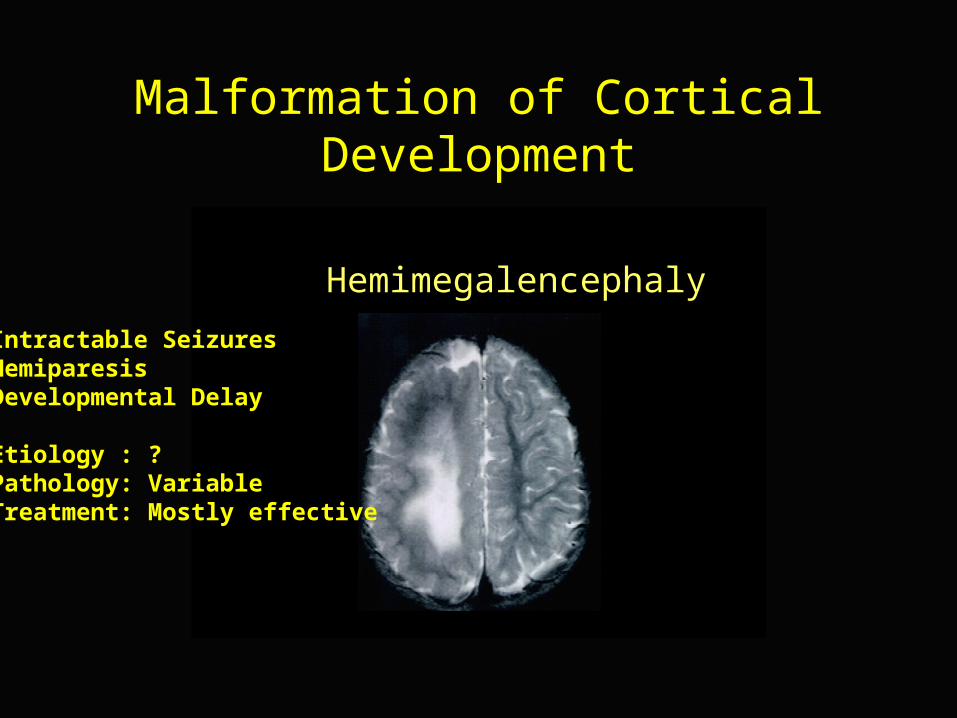
Malformation of Cortical Development
Hemimegalencephaly
Intractable SeizuresHemiparesisDevelopmental Delay
Etiology : ?Pathology: VariableTreatment: Mostly effective
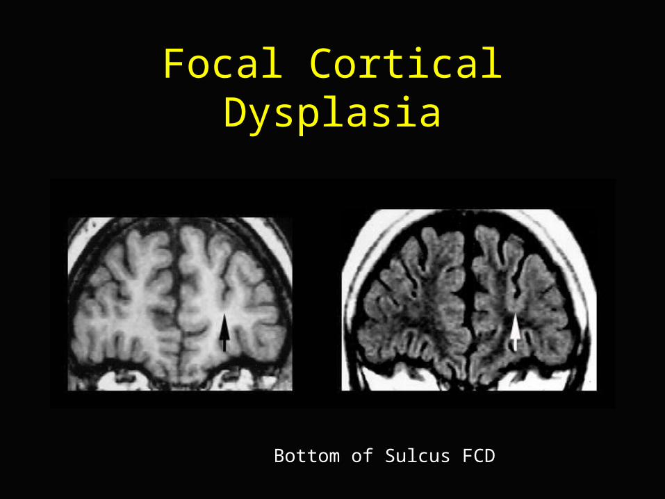
Focal Cortical Dysplasia
Bottom of Sulcus FCD
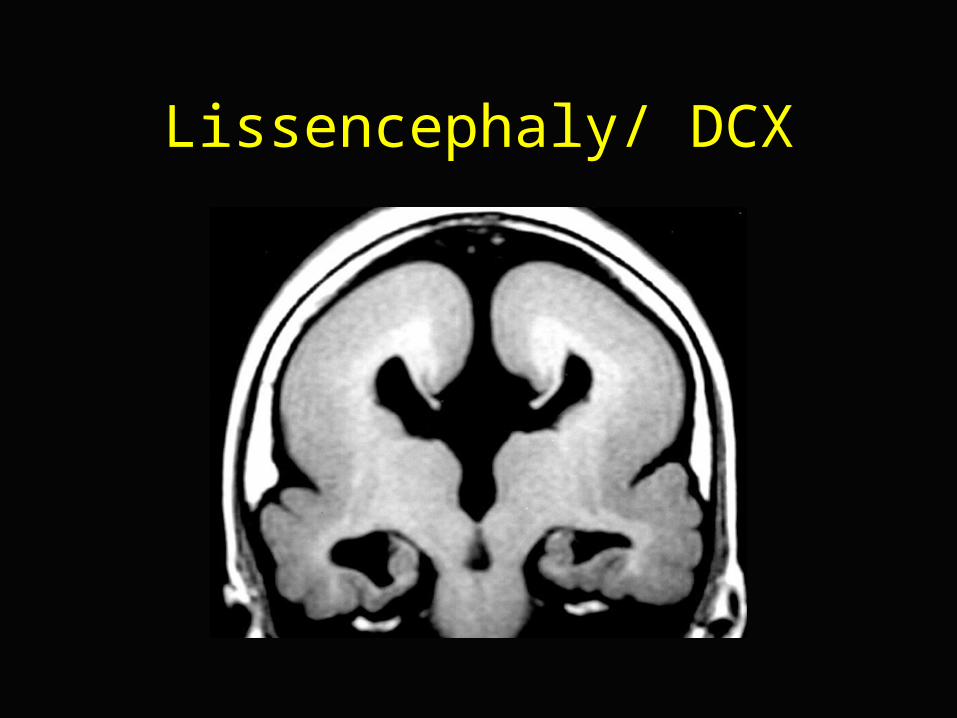
Lissencephaly/ DCX
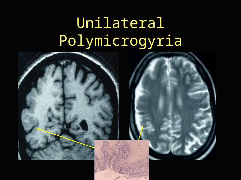
Unilateral Polymicrogyria
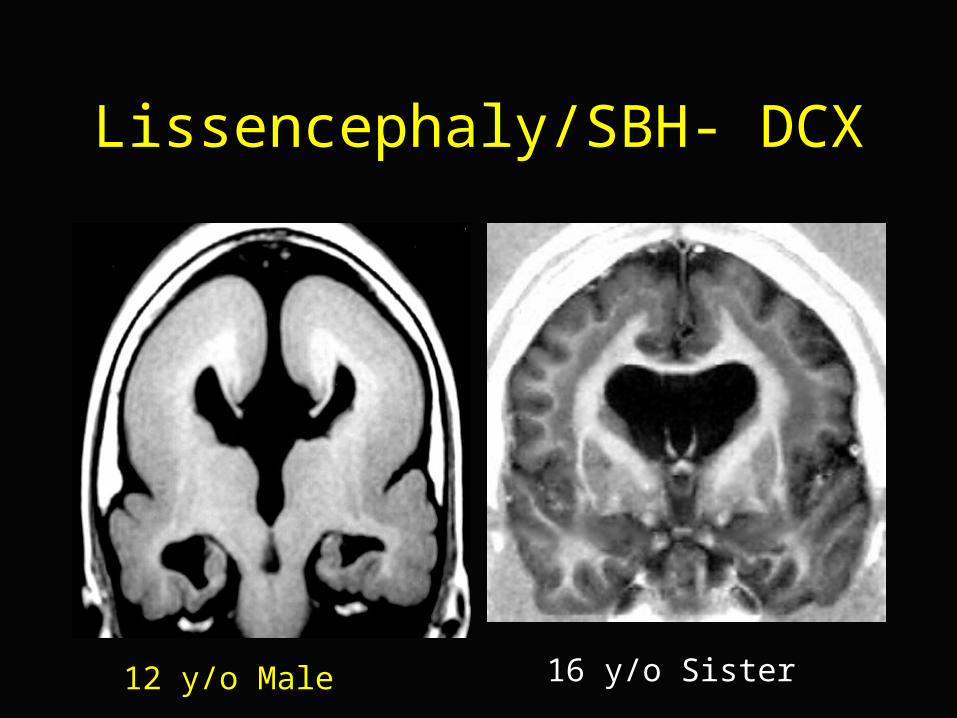
Lissencephaly/SBH- DCX
12 y/o Male 16 y/o Sister
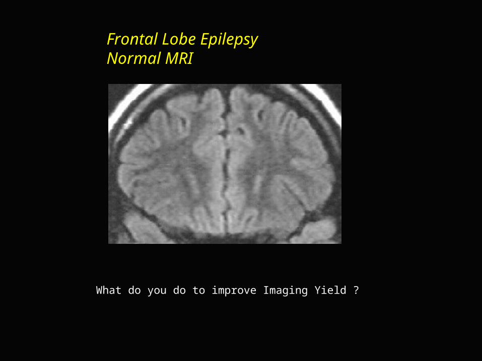
Frontal Lobe EpilepsyNormal MRI
What do you do to improve Imaging Yield ?
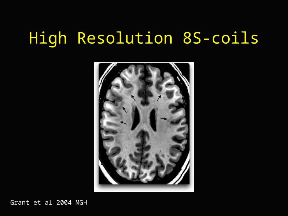
High Resolution 8S-coils
Grant et al 2004 MGH
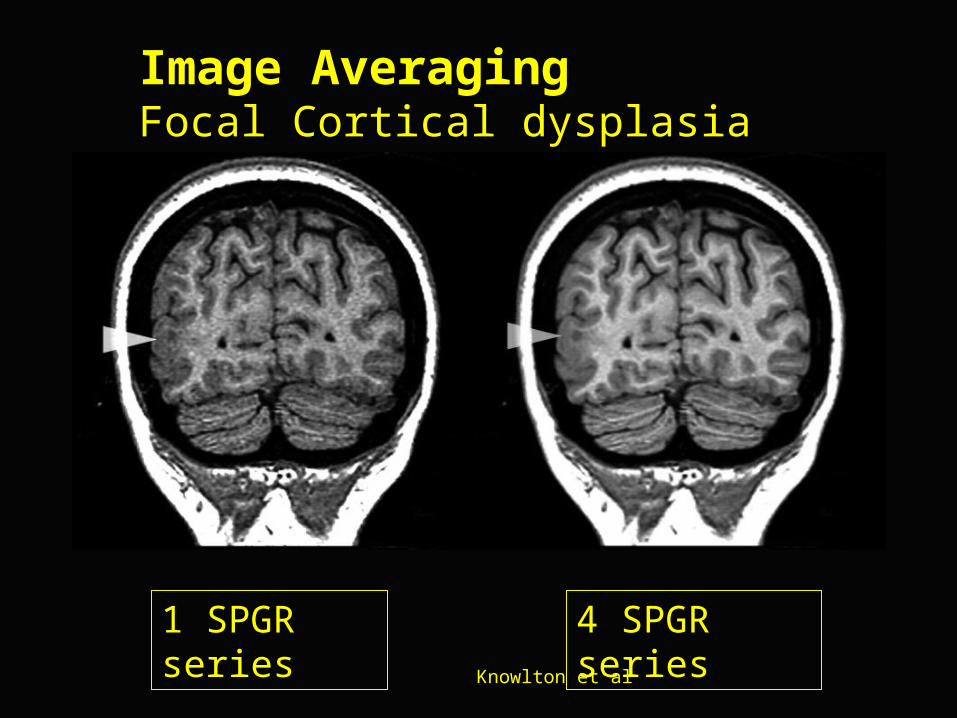
Image AveragingFocal Cortical dysplasiaFocal Cortical dysplasia
Knowlton et al
4 SPGR series1 SPGR series
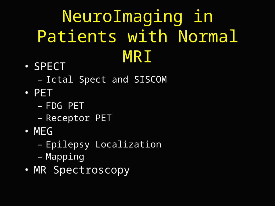
NeuroImaging in Patients with Normal MRI
• SPECT– Ictal Spect and SISCOM
• PET– FDG PET– Receptor PET
• MEG– Epilepsy Localization– Mapping
• MR Spectroscopy
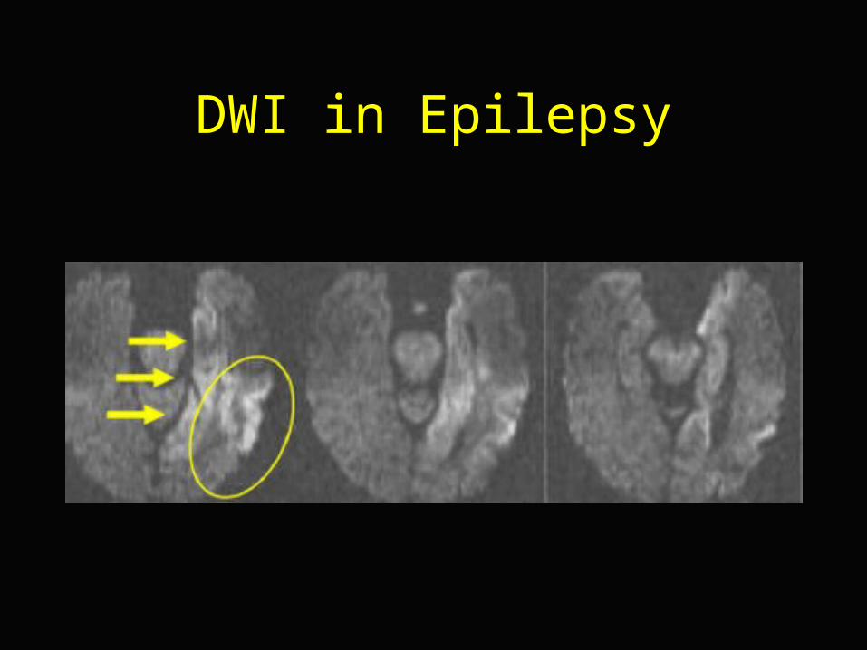
DWI in Epilepsy
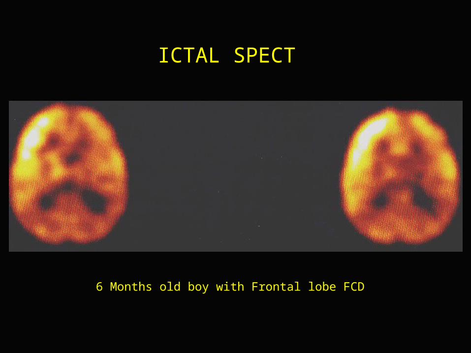
6 Months old boy with Frontal lobe FCD
ICTAL SPECT
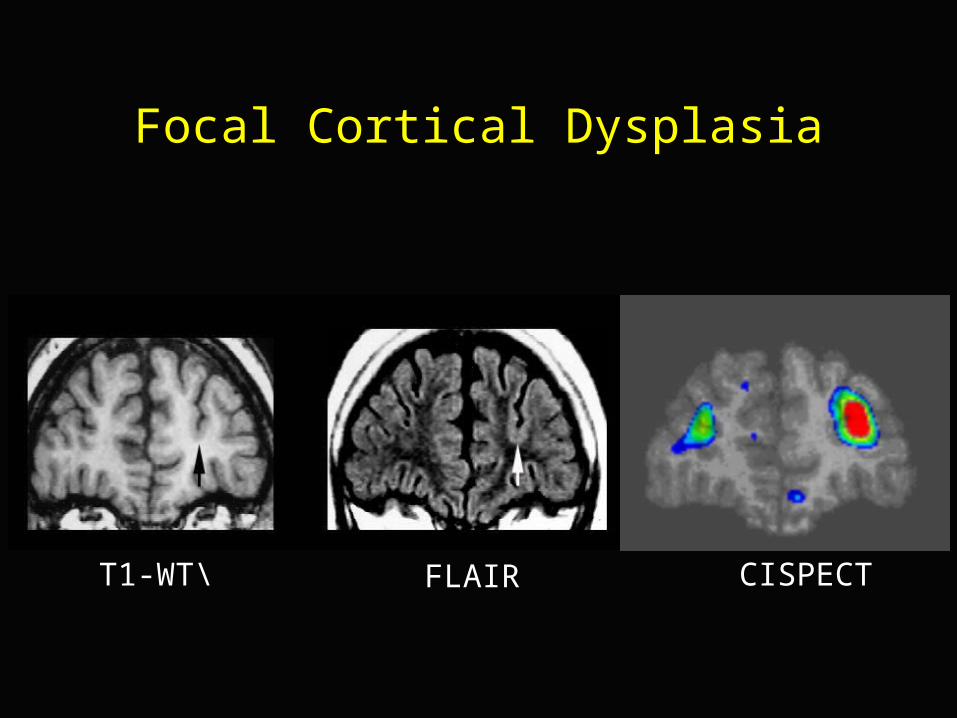
Focal Cortical Dysplasia
T1-WT\ FLAIR CISPECT
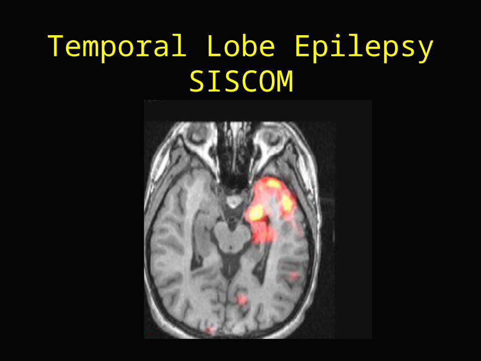
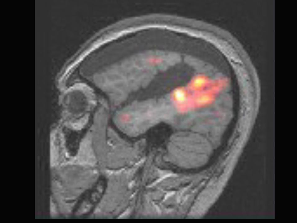
Temporal Lobe EpilepsySISCOM
PET
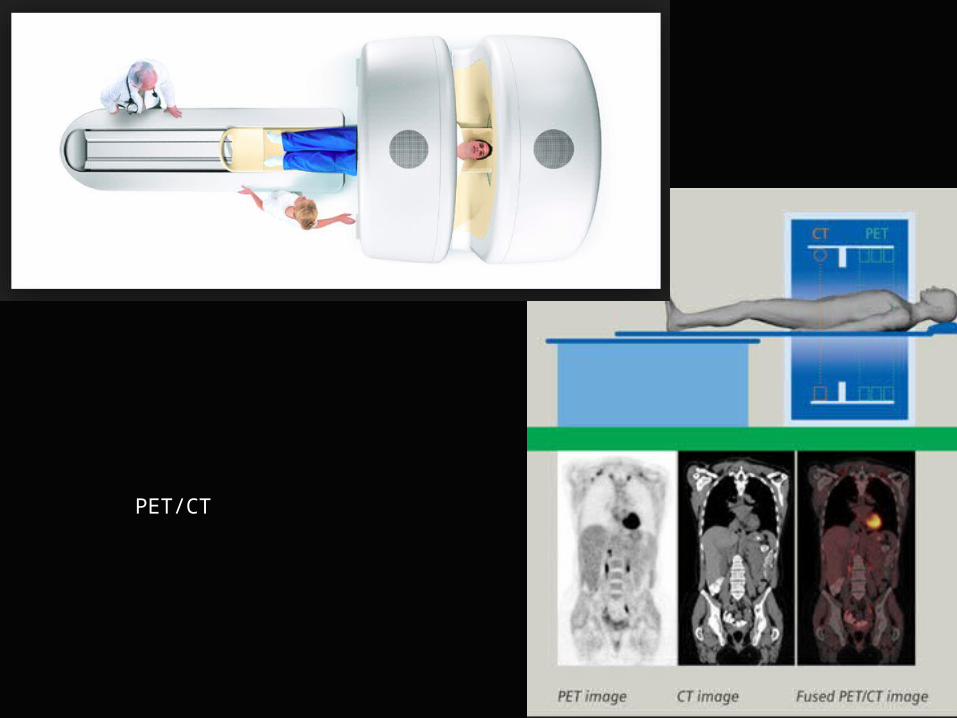
PET/CT
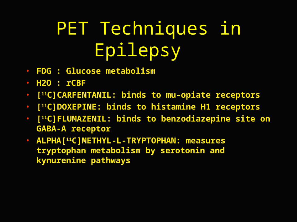
PET Techniques in Epilepsy
• FDG : Glucose metabolism• H2O : rCBF• [11C]CARFENTANIL: binds to mu-opiate receptors• [11C]DOXEPINE: binds to histamine H1 receptors• [11C]FLUMAZENIL: binds to benzodiazepine site on GABA-A
receptor• ALPHA[11C]METHYL-L-TRYPTOPHAN: measures
tryptophan metabolism by serotonin and kynurenine pathways
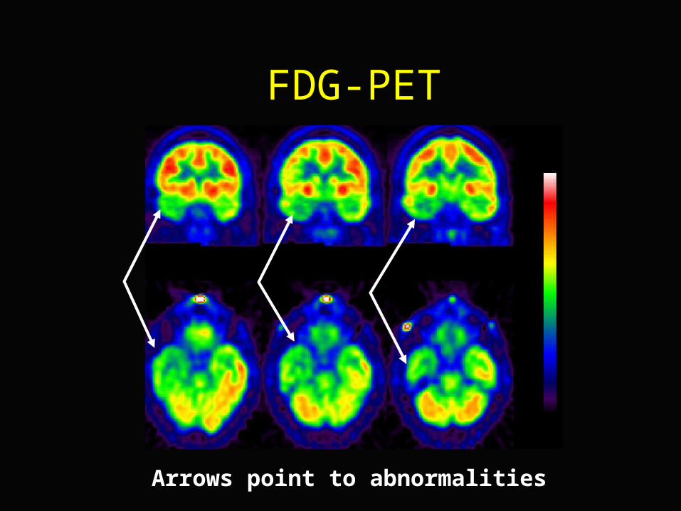
FDG-PET
Arrows point to abnormalities
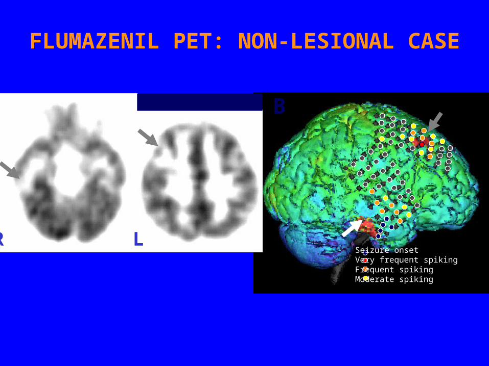
R L
A B
Seizure onsetVery frequent spikingFrequent spikingModerate spiking
FLUMAZENIL PET: NON-LESIONAL CASE
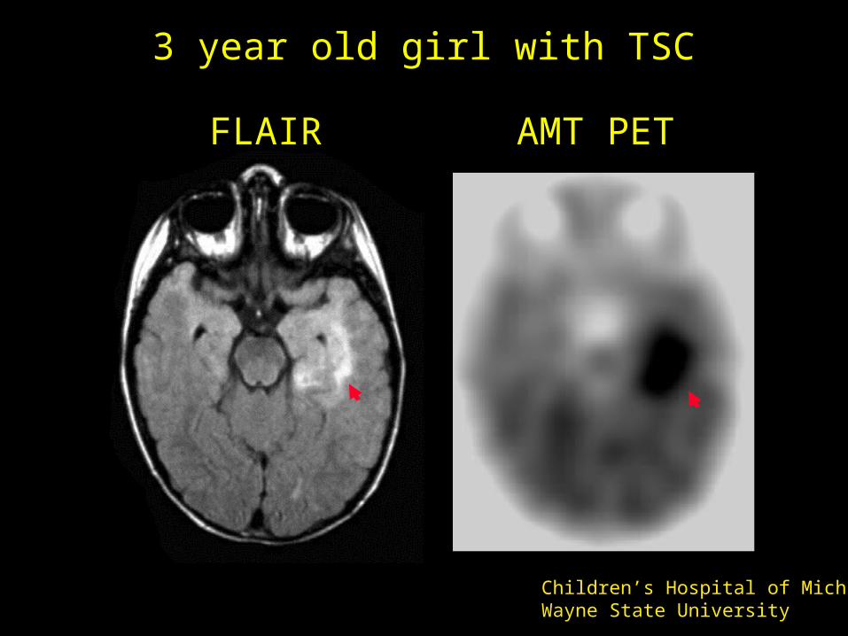
3 year old girl with TSC
FLAIR AMT PET
Children’s Hospital of MichiganWayne State University
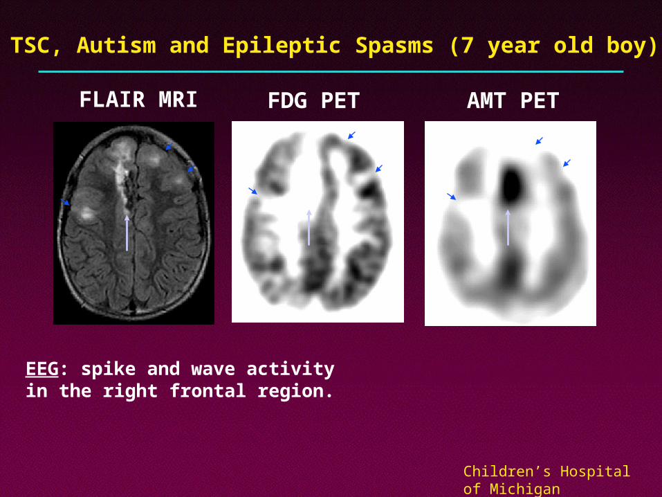
TSC, Autism and Epileptic Spasms (7 year old boy)
AMT PETFDG PETFLAIR MRI
EEG: spike and wave activity in the right frontal region.
Children’s Hospital of MichiganWayne State University
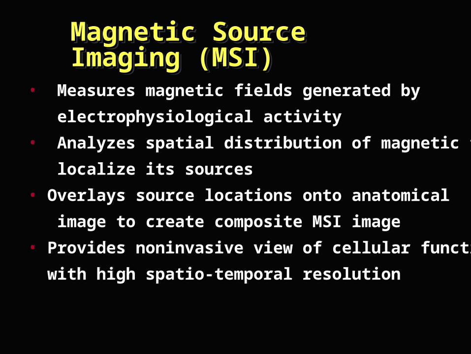
• Measures magnetic fields generated by Measures magnetic fields generated by
electrophysiological activityelectrophysiological activity
• Analyzes spatial distribution of magnetic field to Analyzes spatial distribution of magnetic field to
localize its sourceslocalize its sources
• Overlays source locations onto anatomical Overlays source locations onto anatomical
image to create composite MSI imageimage to create composite MSI image
• Provides noninvasive view of cellular function Provides noninvasive view of cellular function
with high spatio-temporal resolutionwith high spatio-temporal resolution
Magnetic Source Imaging Magnetic Source Imaging (MSI)(MSI)Magnetic Source Imaging Magnetic Source Imaging (MSI)(MSI)

MEG System 248 Channels
Superior Temporal Resolution
MEG-NYU Epilepsy Center
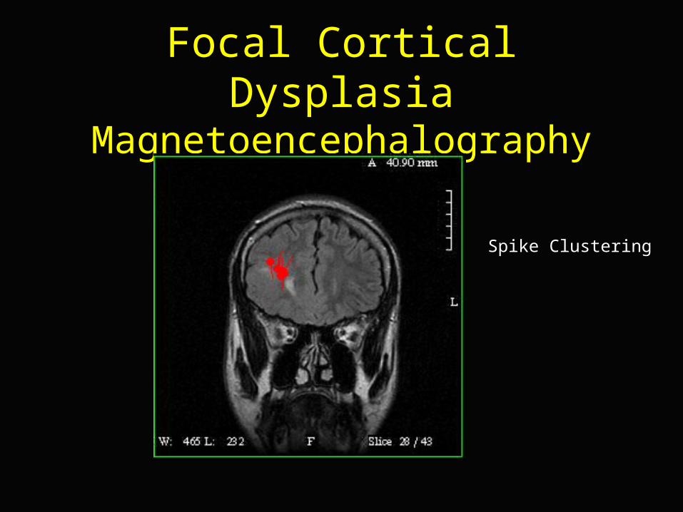
Focal Cortical DysplasiaMagnetoencephalography
Spike Clustering
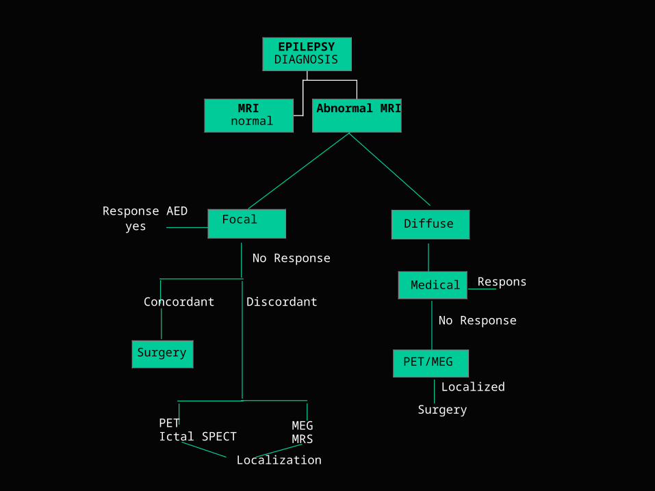
Focal Diffuse
Medical Tx Response
No Response
PET/MEG
Response AEDyes
No Response
Concordant
Surgery
Discordant
Surgery
Localized
PETIctal SPECT
MEGMRS
Localization
MRInormal
Abnormal MRI
EPILEPSYDIAGNOSIS
0
25
50
75
100
N=26Post-stroke
N=57Vascular
malformation
N=50Tumor
N=268Normal
MRI
N=50Closed Head trauma
N=81Cortical
dysgenesis
N=224Isolated
HS
N=38Dual
pathology
54%50%
46%42%
30%24%
11%3%
% o
f se
izur
e-fr
ee p
atie
nts
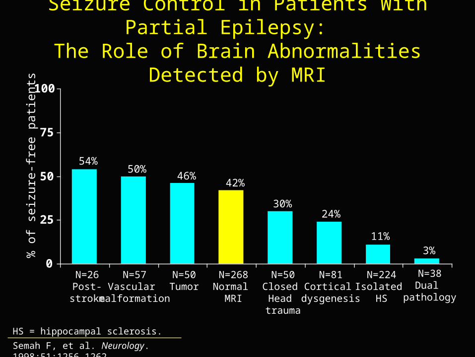
Seizure Control in Patients With Partial Epilepsy: The Role of Brain Abnormalities Detected by MRI
Semah F, et al. Neurology. 1998;51:1256-1262.
HS = hippocampal sclerosis.