royal entomological society · royal entomological society of london vol. ix. part i . handbooks...
TRANSCRIPT
Royal Entomological Society
HANDBOOKS FOR
THE IDENTIFICATION
OF BRITISH INSECTS
To purchase current handbooks and to download out-of-print parts visit: http://www.royensoc.co.uk/publications/index.htm
This work is licensed under a Creative Commons Attribution-NonCommercial-ShareAlike 2.0 UK: England & Wales License.
Copyright © Royal Entomological Society 2012
ROYAL ENTOMOLOGICAL SOCIETY OF LONDON Vol. IX. Part I .
HANDBOOKS FOR THE IDENTIFICATION OF BRITISH INSECTS
DIPTERA 1. Introduction and Key to Families
By
H. OLDROYD
LONDON Published by the Society and Sold at its Rooms
.p, Queen's Gate, S.W. 7
Revised and Reprinted, April, 1954- Price Seven Shillings and Sixpence
HANDBOOKS FOR THE IDENTIFICATION OF BRITISH INSECTS
The aim of this series of publications is to provide illustrated keys to the whole of the British Insects (in so far as this is possible), in ten volumes, as follows :
I. Part 1. General Introduction. , 2. Thysanura. , 3. Protura. , 4. Collem bola. , 5. Dermaptera and
Orthoptera. , 6. Plecoptera. , 7. Psocoptera. , 8. Anoplura.
II. Hemiptera. Ill. Lepidoptera. IV. and V. Coleoptera.
Part 9. Ephemeroptera. , 10. Odonata. , ll. Thysa.noptera. , 12. Neuroptera.. , 13. ~Ieooptera. , 14. Trichoptern. , 15. Strepsiptera.. , 16. Siphonaptera..
VI. Hymenoptera : Symphyta and Aculeata. VII. Hymenoptera : Ichneumonoidea.
VIII. Hymenoptera : Cynipoidea, Chalcidoidea, and Serphoidea.. IX. Diptera: Nematocera and Brachycera. X. Diptera : Cyclorrhapha. Volumes II to X will be divided into parts of convenient size, but it is
not possible to specify in advance the taxonomic content of each part.
Conciseness and cheapness are main objectives in this new series, and each part will be the work of a specialist, or of a group of specialists. Although much of the work will be based on existing published keys, suitably adapted, it is expected that it will also include much new and original matter.
Parts will be issued, separately paged and priced, as they become available.
Orders for the Series or for separate parts may be placed with the Registrar at the Society's rooms now, but prices can only be quoted for those parts already in the press.
The Society is indebted to the Royal Society for a grant towards the cost of initiating this series of Handbooks.
A list of parts now available appears on the back cover.
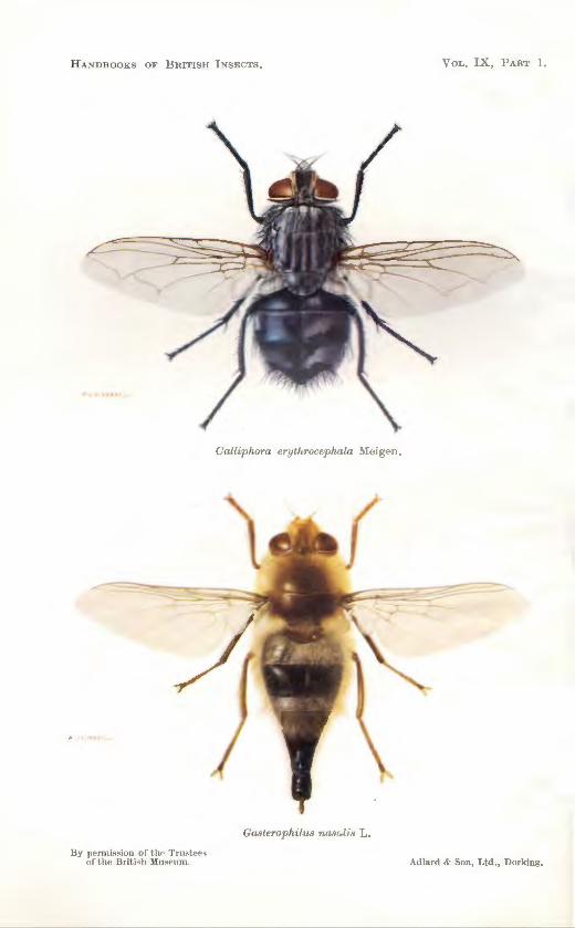
HAN DBOOKS OF BRITIS H 1 :-<SECT S .
B.v pC'rmis~ ion of t h" Tru~tcc~ of t he Brit.i ;.;h ~'lu;;;.rurn .
Calliphom e,·yth?-ocephala :VIc igen .
GasteTophilus na.so:lis L .
VoL. I X , J>ART I.
Ad lnrd & Son, Ltd., Dorking.

DIPTERA 1. INTRODUCTION AND KEY TO FAMILIES.
BY H. 0LDROYD.
HITHERTO the only key to the families of British Diptera has been that given in Wingate's Durham Diptera (1906), which is still widely used. If a more modern work is wanted one has to consult Lindner's Die Fliegen der Palaearktischen Region (1924- ), the various volumes of the Faune de France (1923- ), Lundbeck's Diptera Danica (1907-27), or Curran's North American Diptera (1934). Of these Lundbeck is the favourite, because it is not only work of first quality, but it is written in English. These works are expensive to buy, and not very accessible in libraries, and all suffer from the disadvantage that they cover a much wider fauna than the British, to which they give only slight attention. Other works, such as Verrall's British Flies, and many papers by Verrall, Collin, Edwards, Wainwright, Richards and others, deal exhaustively with smaller or larger groups, but offer little help to the beginner
To meet precisely this difficulty Grimshaw (1934) published an introductory account of the Order, and an English translation of Lindner's Key to Families, with reproductions of many of his figures. This paper, too, is relatively inaccessible, and the key is a literal translation of the German, without any attempt to correct even typographical errors.
The present key is widely based on previously published works, especially those listed above, but wherever possible I have checked it with named specimens, and freely modified it where necessary. Mr. J. E. Collin and Mr. L. Parmenter have very kindly tested parts of the Key to Families, and drawn attention to many errors and obscurities; neither should be blamed for any shortcomings that still remain.
I have tried to explain and illustrate as many technical terms as possible, and at the same time to indicate very generally the main lines of classification in the Diptera, and the variation of structure throughout the Order. Structural details used to define units smaller than a family will be discussed in later sections.
Besides Mr. Collin and Mr. Parmenter, I have had very welcome assistance from Dr. F. van Emden, Mr. Paul Freeman, Mr. R . L. Coe and Dr. John Smart, to whom I express my sincere thanks. The Trustees of the British Museum (Natural History) have very kindly consented to the use of a number of original drawings, first published in Dr. Smart's Insects of M edical Importance (1943) ; these drawings are by Mr. Arthur Smith. Mr. C. 0. Hammond has drawn figs . 92-97, and Mr. Paul Freeman figs. 39-48. The others are mine.
I

2 IX (1). DIPTERA
The Order Diptera, the two-winged, or true flies, comprises between 50,000 and 60,000 known species, of which the latest estimate (Kloet and Hincks, 1945) records 5199 as found in the British Isles. This last number is very far from being the final total of British Diptera. When the specific identity of some of the largest Diptera-Tabanus bovinus and its allies-is still by no means certain, it is not surprising that many of the smallest species are not easily named, and that much that is new about them is continually being recorded. The late Dr. F. W. Edwards estimated that in 25 years he had recorded as new to the British fauna some 500 Diptera, many of them new to science. The family AsiLIDAE is represented in the British Isles by only 26 species, yet three of these have been added to the List in the last 15 years, and one, new to science, was discovered in the Isle of Man in 1945. In 1947 CH:rnoso:mn.H new to science were still being described from British specimens, and periodical revisions of one or other of the numerous families of the Acalypterae, chiefly by l\ir. J. E. Collin, generally result in the recognition of ne-w species.
It -will be realized, therefore , t hat the Diptera, e>en in Britain, are Yery incompletely kno-wn. They are, in fact, at that most interesting stage of exploration -when there is enough known to provide a firm foundation, yet almost unlimited opportw1ity for detailed study of either their biology or their systematics.
The Diptera are one of the most specialized Orders of insects ; that is to say, they have travelled a long way from the primitive type of winged insect, whose nearest living relatives are the cockroaches and their allies. The latter are characterized by their biting mouthparts, associated with an omnivorous diet, their well developed legs, associated with a cursorial habit, and two pairs of leathery wings, with rather indifferent powers of flight. Diptera have lost the second pair of wings, which are modified into halteres, a pair of stalked bodies whose function is discussed later (p. 18). The loss of one pair of wings does not mean an impaired power of flight. On the contrary the Diptera are among the most accomplished of insect fliers both in speed and manoeuvrability.
With the use of the fore wings only in flight is associated a development of the mesothorax or second thoracic segment at the expense of the other two. Almost the whole of the thorax visible from above is the tergite of the mesothorax, even in those Diptera which have lost the power of flight and become secondarily wingless. The ventral parts of the thoracic segments are associated with the three pairs of legs, and are much more equally developed.
Diptera can, therefore, be recognized as insects in which the hind wings are replaced by halteres, and even if the fore-wings are reduced or absent the mesothorax is developed at the expense of the other two thoracic segments.
STRUCTURE.
The present account is intended to cover only such morphological details as are of systematic importance throughout the Order, and to serve as an introduction to the Key to Families. Features which are of interest only in certain families or genera will be discussed by the authors of the keys to those groups. For the names of suborders and families referred to, see the .list on page 33. It is necessary to give here a warning that the terms

STRUCTURE 3
2
FIGs. 1-5.-Head in side view. 1. Tipulidae. 2. Conopidae. 3. Tabanidae, Tabanus (from Smart, 1943). 4. Coelopidae, Ooelopa jrigida; VT, vertical bristles; UFO,
upper fronto-orbital bristles ; B, swollen buccae, bearing many strong bristles. 5. Dryomyzidae, Dryomyza anilis ; CL, clypeus protruding beyond epistoma.
4 IX (1). DIPTERA
FIGs. 6-9.-Head in front view. 6. Calliphoridae, showing vibrissae and ptilinal suture (here called "frontal suture") (from Smart, 1943). 7. Trypetidae; PVT, postvertical bristles ; VT, vertical bristles ; UFO, upper fronto -orbital bristles borne on vertical plates ; LFO, lower fronto-orbita l bristles, borne on extended parajacials ; PF, parafacial plates ; OCL, ocellar triangle ; FL, frontal lunule, enclosed within ptilinalsuture. 8. Clusiidae; VP, vertical plate, extended forward and bearing 3 upper fronto-orbital bristles. 9. Chloropidae ; OCL, greatly enlarged ocellar triangle.
STRUCTURE 5
used are those in common usage, and believed to be least ambiguous, not always those which are morphologically most precise. The reader who wishes to study the anatomy of Diptera in more detail is referred to Crampton (1942), Snodgrass (1935, 1943), and other papers listed in the bibliography.
THE HEAD.-In the TIPULIDAE (fig. I) the head is of a prognathous, relatively generalized type, recalling that of some Mecoptera. In other Diptera the prevailing trends are an expansion of the eyes, coupled with a shortening of both the anterior part of the head and the occiput until in side view little is seen except the eyes and the projecting antennae and
17
18
19
====([]) 20
21
22
·~ 23
~ 24
25
Fws. 10-25.-Antennae. 10. Theoretical primitive insect (after Snodgrass, 1935). 11. Tipulidae. 12. Mycetophilidae. 13. Anisopodidae. 14. Bibionidae. 15. Simuliidae. 16. Cecidomyiidae. 17. Stratiomyiidae, Xylomyia. 18. Rhagionidae, Xylophagus. 19. Stratiomyiidae, Ohloromyia. 20. Rhagionidae, Ohrysopilus. 21. Tabanidae, Chrysops. 22. Tabanidae, Tabanus. 23. Asilidae, Dioctria. 24. Asilidae, Asilus. 25. Dolichopodidae.
mouthparts (figs. 2, 3) . The "head" proper is reduced to a narrow strip between the compound eyes, and in many Diptera even this is invaded by the eyes, which may meet together, either above or below the antennae, or both. In many families the males have the eyes approaching or meeting (holoptic) and the females have them well separated (dichoptic), and in a few groups both sexes are holoptic.
In the majority of Diptera the head capsule (i.e. excluding the eyes) as seen from in front consists almost entirely of what is morphologically the frons in other insects, and so special terms are necessary for the subdivisions of this which can be seen in Diptera. The region at the top of
6 IX (l) DIPTERA
29 3rd segment--
FIGS. 26-27.-Thorax in dorsal view. 26. Calypterae. (Bristles omitted). 27. Acalypterae. (Bristles omitted.) s, transverse suture; P, posterior callus.
FIGs. 28-29.-Antennae. 28, Calypterae. 29. Acalypterae. (From Smart, 1943).
the head, where the ocelli may be found, is called the vertex. Behind it is the occiput, often convex, sometimes (e.g. in some BoMBYLIIDAE and SYRPIDDAE) concave and recessed, so that the head has a very free articulation with the neck and is very mobile ; it also falls off easily in dried specimens!
The space between the vertex and the antennae is referred to as the frons in Diptera, and the area from just above the antennae to the mouth margin is called the face.
The region immediately above the mouth may be known as the epistoma. The regions along the eye margins beside the face, and those ventrally below the eyes, are often clearly demarcated ; English writers sometimes refer to them as the "cheeks" and "jowls" respectively, but these terms are somewhat ambiguous, and it is perhaps better to use the terms parafacialia and buccae (figs. 7, 4B).
STRUCTURE 7
A pair of prominent bristles often seen on the mouth-margin in Cyclorrhapha are the vibrissae (fig. 6).
In Nematocera, Brachycera and Aschiza there is no distinct suture between the frons and the face, but in Schizophora (i.e. Cyclorrhapha other than Aschiza) there appears just above the antennae a horseshoeshaped structure called the ptilinal suture. This is sometimes called the "frontal" suture, but it bears no relation to the frontal suture of lower insects which marks the posterior margin of the frons and includes the median ocellus. The ptilinal suture marks the cleft from which issued the
Fw. 30.-Antennae : male of Chironomidae. (From Smart, 1943.)
ptilinum, an eversible sac which is blown out by a fly of this group to help it in bursting out of its puparium (Laing, 1935). The ptilinum is again used vigorously as the fly penetrates the earth surrounding the puparium. During the hardening process the sac is retracted, and only the suture remains visible. In all Cyclorrhapha, whether the ptilinal suture is well developed or not, there is a frontal lunule, a tiny, crescent-shaped sclerite lying just above the bases of the antennae (fig. 7).
In the more specialized families the head may bear bristles, the names and positions of which are given in figs. 4-8. To identify these correctly it is often necessary to trace the outline of the plates or sclerites on which they are borne, especially in the Acalypterae.
The compound eyes, as we have already seen, take up much of the volume of the head in Diptera. Their detailed structure is not yet used in classification , though sometimes the existence of facets of two different sizes is
8 IX (1). DIPTERA
important. The eyes meet above the antennae in a great many male Diptera, and in some females of some families, among which are BLEPHAROCERIDAE, THAUMALEIDAE, ACROCERIDAE, BOMBYLIIDAE, EMPIDIDAE, TACHINIDAE and SYRPHIDAE. In some Nematocera the eyes are not only touching, but fused above the antennae, and in some 0ECIDOMYIIDAE the lower part of each eye has broken away, giving rise to a three-eyed condition. In Acrocera globulus the eyes meet below the antennae, and in some mosquitoes and crane-flies the eyes may spread over on to the ventral surface of the head behind the proboscis.
Many Diptera, in life, have coloured eyes, most usually uniformly red or green, but sometimes with a vivid pattern. According to Eltringham
A B 31
c FIG. 31.-Metathoracic spiracle, showing arrangement of hairs in: A, Coelopidae, genus
Orygma ; B, Sepsidae ; c, other Acalypterae.
the red and black colours are due to pigment in the pseudocones, but the green is a structural colour due to optical interference. These colours fade after the death of the fly, but the pattern can often be traced under the microscope, and Goffe has published a formula for the revival of the markings, which are often of great help in the recognition of species. His formula is : Acetic acid (glacial), l part ; glycerine, l part ; solution of perchloride of mercury (B.P. strength), l part; rectified spirit, 48 parts. G. H. Hardy (1939) has tried to correlate eye-colour in Diptera with efficiency of vision-an interesting speculation, but one about which there is very little direct evidence.
Many Diptera have the eyes covered with pile, the hairs of which are inserted between the facets, and presumably do not encroach on the limited field of vision of the individual ommatidia. Sometimes all the members of a family have hairy eyes; sometimes this character serves to separate genera, or species, sometimes it is confined to one sex. Often a specimen that seems on superficial examination to have bare eyes, when studied under high magnification is seen to have minute hairs on the eyes, and it is
STRUCTURE 9
difficult to judge whether the author of the key in use would regard the eyes as bare or hairy. This character should, therefore, be used with great care.
The ocelli or simple eyes are borne on the vertex, usually in a triangle with the single (median) ocellus in front. They are used in classification, but their systematic importance varies in different families. In some Nematocera, as will be seen from the Key, whole families are uniform in possessing, or in lacking, ocelli. In other families individual species or
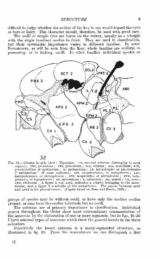
FIG. 32.- Thorax in side view : Tipulidae. cs, cervical sclerites (belonging to neck region) ; PRO, pronotum ; PRS, prescutum ; seT, scutum ; SCL, scutellum; PNT, postscutellum or postnotum ; M, m etanotum ; LT, laterotergite or pleurotergite (" metanotum " of some authors) ; APS, anepisternum, or mesopleuron ; KPS, katepisternum, or sternopleuron ; APM, anepimeron or pterople-uron ; KPM, katepimeron, or hypopleuron; PS, episternum; P, epimeron; ME, m eron; ex, coxa; ABD, abdomen. A figure 2, e .g. APS2, indicates a sclerite belonging to the m esathorax, and a figure 3 a sclerite of the metathorax. The suture between APS2 and APM2 is the pleural suture. (Figure based on Rees and Ferris, 1939.)
groups of species may be without ocelli, or have only the median ocellus present, or may have the ocellar tubercule but no ocelli.
The antennae are of primary importance in classification. Individual genera throughout the Order show most extraordinary ornamentation of the antennae by the elaboration of one or more segments, but in figs. 10-30 I have selected types of antennae which show the general trends in the three suborders.
Primitively the insect antenna is a many-segmented structure, as illustrated in fig. 10. From the musculature we can distinguish a first
I§
IO IX (I). DIPTERA
segment or scape, and a second segment or pedicel, the remaining segments having no individual muscles, and being grouped together as a multisegmented jlagellum. Scape and pedicel vary in shape and size, but the major modifications occur in the flagellum.
In most Nematocera and some Brachycera (e.g. Xylomyia and Xylophagus, figs. 17, 18) the flagellum consists of many similar segments, sometimes elaborately developed, as in the CECIDOMYIIDAE (fig. 16). In more specialized families the segments of the flagellum become fused into a compound "third segment." Intermediate stages of this fusion can be seen in the families SIMULIIDAE, BIBIONIDAE, ScATOPSIDAE and THAUMALEIDAE among Nematocera, and in the families STRATIOMYITDAE, TABANIDAE and some RHAGIONIDAE among Brachycera. In most Brachycera and Cyclorrhapha there is a true third segment, or postpedicel, which does not show any traces of segmentation, and the remaining segments of the flagellum are represented by a style or arista, which often shows by rings that it is of compound origin. The terms " style " and " arista " are rather loosely used ; generally" style "is used for a relatively stout, clearlysegmented appendage which is terminal in position, and "arista" for a more slender, bristle-like, indistinctly-segmented unit, especially if this arises from the dorsal surface of the third segment.
All segments characteristically bear rings, or " whorls " of hairs (figs. 11, 30, 86). These are most developed in the males of some Nematocera (e.g. mosquitoes, CHIRONOMIDAE); in higher Diptera they persist chiefly ventrally, and in the pectinations of the arista seen in some Brachycera and many Cyclorrhapha (fig. 28).
Crampton (1942, p. 25) points out that, in general, Diptera with long antennae have small eyes, and vice versa, as if one sense compensated for diminution of the other. This is not invariably true, since some MYCETOPHILIDAE with long antennae have well-developed eyes, and many Acalypterae, with short antennae, have quite small eyes. The parasitic forms, as might be expected, tend to a reduction of both eyes and antennae (e.g. HIPPOBOSCIDAE, fig. 91).
The feeding habits of Diptera are exceedingly diverse, and the construction of the mouthparts shows a parallel diversity. Some forms, including isolated genera in some families, apparently do not feed in the adult stage, and their mouthparts are reduced and functionless. In others, especially in the bloodsucking and predatory families, the proboscis is developed to a high degree of efficiency. All, however, agree in showing that the component mouthparts of the primitive biting type have been modified into an apparatus for sucking liquids, supplemented perhaps by some form of piercing, cutting or rasping mechanism.
There is not space here to describe more than the most general modifications of the proboscis in Diptera. The labrum, morphologically a part of the head-capsule, forms a roof to the food channel. The ventral surface of the labrum is sometimes regarded as a separate organ, the epipharynx. The hypopharynx forms the floor of the food channel and bears a second tube, the salivary duct. The sides of the food channel are closed by the mandibles and maxillae, where these are present, and the whole assembly is supported and sheathed in a groove of the labium. The latter at its tip is divided into two more or less fleshy lobes, the labella, which show a wide range of development in different families. They are the principal
STRUCTURE 11
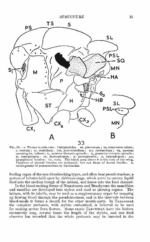
TS 5
A B 33
FIG. 33.-A, Thorax in side view: Calliphoridae. PS, prescutum ; TS, transverse suture; s, scutum; SL, scutellum; PSL, post-scutellum; MN, metanotum; SQ, squama covering HA, haltere ; s1, anterior thoracic spiracle ; s 3, posterior thoracic spiracle; M, mesopleuron; ST, sternopleuron; P, pteropleuron; H, hypopleuron; HB, hypopleural bristles ; ox, coxa. The black area above P is the root of the wing. Positions of pleural bristles are indicated, but not those of dorsal bristles. B, development of postscutellum in Tachinidae.
feeding organ of the non-bloodsucking types, and often bear pseudo-tracheae, a system of tubules held open by chitinous rings, which serve to convey liquid food into the median trough of the labium, and hence into the food channel.
In the blood-sucking forms ofNematocera and Brachycera the mandibles and maxillae are developed into stylets and used as piercing organs. The labium, with its labella, may be used as a supplementary organ for mopping up flowing blood through the pseudotracheae, and in the intervals between blood-meals it forms a sheath for the other mouth-parts. In TABANIDAE the complete proboscis, with stylets ensheathed, is believed to be used for sucking nectar from flowers. Some exotic TABANIDAE have the labium excessively long, several times the length of the stylets, and one field observer has recorded that the whole proboscis may be inserted in the
12 IX (1). DIPTERA
wound, but this is clearly impossible in those forms with either the longest proboscis, or a short one with soft, fleshy labella.
The predatory Brachycera, AsiLIDAE and EMPIDIDAE, use the hypopharynx as a piercing organ, and have the labella reduced and the labium hardened into a horny and conspicuous proboscis. The non-predatory and non-bloodsucking families use chiefly the labium with its labella and pseudotracheal tubes as a sort of sponge for mopping up liquid foods. Often they first exude saliva to macerate the solid food, and in some forms -e.g. CALLIPHORIDAE and MusCIDAE-the labella when folded back expose a number of prestomal teeth which can be used to scratch a surface in search of food, and even to draw blood. Some DoLICHOPODIDAE have carried this process a stage further and envelop smaller insects in their labella, cutting them open with the prestomal teeth and sucking the prey dry before discarding the empty skin. In one exotic Dolichopodid the labella are actually developed into biting jaws.
In the Cyclorrhapha true bloodsucking types have developed in the MusciDAE (including Glossina, the African Tsetse Fly, and the British Stable Fly Stomoxys, and its allies Haematobia and Lyperosia). In these the lost mandibles and maxillae are not recovered, but the labella are reduced and the labium hardened into a stiff auger-like instrument, at the tip of which the prestomal teeth are well developed. The parasitic flies, HIPPOBOSCIDAE and NYCTERIBIIDAE, have an essentially similar mechanism.
There are a number of Diptera which apparently do not feed in the adult stage, and in these the mouthparts are non-functional and sometimes reduced to a mere vestige (e.g. many adult CHIRONOMIDAE). The Acrocerid genus Oncodes possesses mouthparts, but they have no opening to the exterior.
Compact summaries of the mouthparts of biting Diptera are given by Peterson (1916) and Snodgrass (1943).
Associated with the mouthparts are the maxillary palpi, which are an important structure in the classification of Diptera. They lie on each side of the base of the proboscis, and are fairly conspicuous in most groups. Palpi consisting of more than three segments and noticeably drooping indicate Nematocera (CULICIDAE are exceptional in having porrect palpi) ; in the Brachycera and Cyclorrhapha there are never more than three segments, often fewer, and the terminal segment is enlarged and of very diverse form. When the term " palpi " is used in a key to Brachycera or Cyclorrhapha it very often means just the prominent, porrect terminal segment, the one or two smaller basal segments being largely concealed by hairs and bristles.
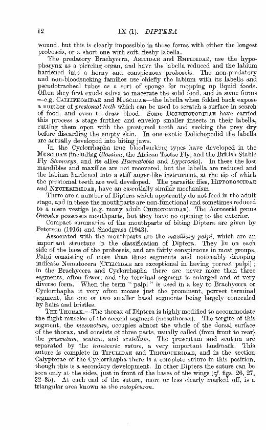
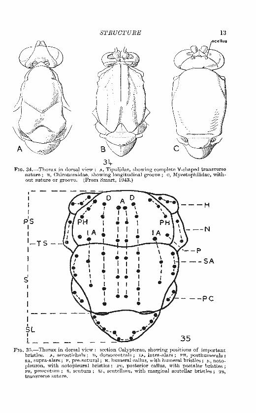
THE THORAX.-The thorax of Diptera is highly modified to accommodate the flight muscles of the second segment (mesothorax). The tergite of this segment, the mesonotum, occupies almost the whole of the dorsal surface of the thorax, and consists of three parts, usually called (from front to rear) the prescutum, scutum, and sctttellum. The prescutum and scutum are separated by the transverse suture, a very important landmark. This suture is complete in TIPULIDAE and TRICHOCERIDAE, and in the section Calypterae of the Cyclorrhapha there is a complete suture in this position, though this is a secondary development. In other Diptera the suture can be seen only at the sides, just in front of the bases of the wings (cf. figs. 26, 27, 32-35). At each end of the suture, more or less clearly marked off, is a triangular area known as the notopleuron.
STRUCTURE 13
34-Fm. 34.-Thorax in dorsal view: A, Tipulidae, showing complete V-shaped transverse
suture ; B, Chironomidae, showing longitudinal groove ; c, Mycetophilidae, with out suture or groove. (From Smart, 1943.)
I I
PS I I 1 ~TS I
I s
I I
SL I L_
0 • I I • I
A D • .. \
I \ \
I • • • I I I I • I
I I • • I I ---SA I I • • I I I I • • ---PC
35 FIG. 35.-Thorax in dorsal view : sect ion Calypterae, showing positions of important
bristles. A, acrostichals ; D , dorsocentrals ; IA, intra-alars ; PH, posthumerals ; SA, supra-alars; P, pre-sutural; H, humeral callus, with humeral bristles; N , notopleuron, with notopleural bristles ; PC, posterior callus, with postalar bristles ; PS, prescutum ; s, scutum ; SL, scutellum, with marginal scutellar bristles ; TS, transverse suture.
14 IX (1) DIPTERA
The pronotwm or tergite of the first thoracic segment is reduced to a hoop-like sclerite, which is often concealed by the overhanging mesonotum. The metanotum, or tergite of the third thoracic segment, is even further reduced to a very narrow band. Many entomologists use the term " metanotum " or " metanotal slopes " or " metanotal callosities " for the region immediately behind, and overhung by, the scutellum (fig. 33 PSL), an area which the morphologists regard as the post-scutellum or postnotum
empodium/
empodium/
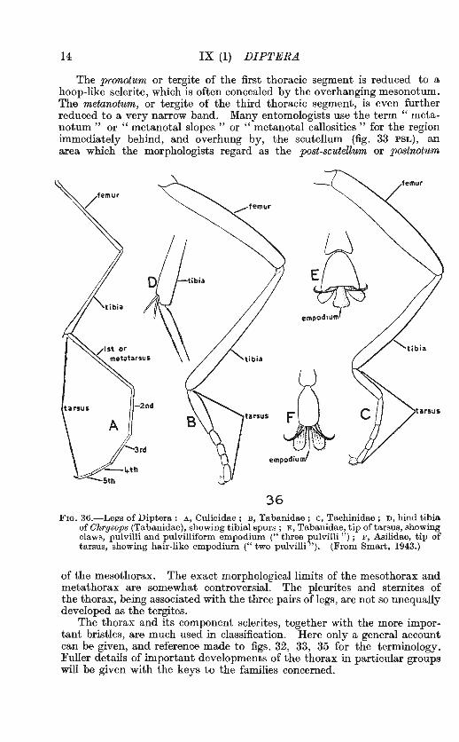
36 FIG. 36.-Legs of Diptera : A, Culicidae ; B, Tabanidae ; c, Tachinidae ; D, hind tibia
of Chrysops (Tabanidae}, showing tibial spurs; E, Tabanidae, tip of tarsus, showing claws, pulvilli and pulvilliform empodium (" three pulvilli ") ; F, Asilidae, tip of tarsus, showing hair-like empodium ("two pulvilli "). (From Smart, 1943.)
of the mesothorax. The exact morphological limits of the mesothorax and metathorax are somewhat controversial. The pleurites and sternites of the thorax, being associated with the three pairs of legs, are not so unequally developed as the tergites.
The thorax and its component sclerites, together with the more important bristles, are much used in classification. Here only a general account can be given, and reference made to figs. 32, 33, 35 for the terminology. Fuller details of important developments of the thorax in particular groups will be given with the keys to the families concerned.
STRUCTURE 15
THE ABDOMEN.-Traces of eleven abdominal segments can be found in some primitive Diptera, but the last two or three are always associated with the genital organs and in consequence are greatly modified. The number of clearly visible segments before the genitalia is usually seven or eight, but may be reduced to as few as four (exceptionally three or even two) in Cyclorrhapha. In this case reduction has also taken place at the base of the abdomen, by fusion of the first segment with the second. Sometimes the first sternite remains distinct after the tergite has been lost, but there are many intermediate stages, and it is important when using descriptions of Cyclorrhapha to look carefully at the first segment, and also to make sure whether the author is counting the visible segments or is making allowance for a possibly evanescent first tergite. Some morphologists claim that the
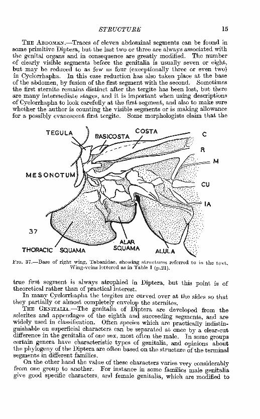
c
R
37
THORACIC
FIG. 37 .-Base of right wing, Tabanidae, showing structures referred to in the text. W'ing-veins lettered as in Table I (p.2l).
true first segment is always atrophied in Diptera, but this point is of theoretical rather than of practical interest.
In many Cyclorrhapha the tergites are curved over at the sides so that they partially or almost completely envelop the sternites.
THE GENITALIA.-The genitalia of Diptera are developed from the sclerites and appendages of the eighth and succeeding segments, and are widely used in classification. Often species which are practically indistinguishable on superficial characters can be separated at once by a clear-cut difference in the genitalia of one sex, most often the male. In some groups certain genera have characteristic types of genitalia, and opinions about the phylogeny of the Diptera are often based on the structure of the terminal segments in different families.
On the other hand the value of these characters varies very considerably from one group to another. For instance in some families male genitalia give good specific characters, and female genitalia, which are modified to
16 IX (1). DIPTERA
suit differem habits of egg-laying, are diagnostic of the genus. In difficult groups it may thus be necessary to have a female specimen to decide the genus and an associated male to determine the species. In nearly related genera even the male genitalia may show no noticeable di.fference, except in species which could be separated with the naked eye. In some groups the genitalia are exserted, and can be studied without dissection, but in others, especially in the smaller families, it is necessary to dissect and mount these structures.
The nomenclature of the parts has not been standardized throughout the Diptera, and there is still doubt about the homologies of some of them. It is therefore not proposed to give any account in this introduction, but where the genitalia are used in separating the British species they will be figured and explained by the author concerned.
Many Diptera show rotation, or twisting of the terminal segments, so that the tergites and sternites of these segments are no longer in line with
38 lA\ Co1
Cu2
FJG. 38.-Wing of Protoplasafitchii (Tany deridae). Veins and cells as in T able I (p. 21).
those of the rest of the abdomen. Sometimes the twisting is obvious without dissection ; often it can only be inferred from the displacement of the genitalia. Care is needed when interpreting descriptions if rotation is suspected, since the terms dorsal and ventral may be used relative to the body as a whole, or to the genitalia.
THE LEGS.-The legs of Diptera are made up of nine obvious segments, which vary in relative size, and one or more of which may be elaborately ornamented with bristles, spines or peculiar processes. Sometimes such ornamentations occur only in one or two species of the genus. Frequently they are confined to one sex, usually the male, and may assist in mating, either directly, or indirectly through courtship behaviour (e.g. DoLICHOPODIDAE).
Fig. 36 shows the typical structure of the legs in Diptera (note that the coxa is omitted and the trochanter barely indicated). The first segment or coxa articulates with the pleurite of the thorax, which it usually resembles in colour and appearance. The fore-coxae are usually longer, more cylindrical, and freer in movement than the middle and hind pairs. The trochanter is small and inconspicuous, but sometimes bears important bristles (e.g. in some BoRBORIDAE). The femur varies widely in size and shape, and may

STRUCTURE 17
~ ~ .. '· M,
l A Cu• MJ+!t
~ R~t-+S. 1,5 \ M,
M,
1A Cu1 M l-t-lt.
h k
Se R,
FIGs. 39-48.-vVings of Diptera, Nematocera. 39. Tipulidae, Limnophila. 40. Anisopodidae, Anisopus. 41. Chironomidae, Procladius. 42. Culicidae, Theobaldia (scales omitted). 43. Simuliidae, Simulium. 44. Mycetophilidae, Symmerus. 45. Bibionidae, Dilophus. 46. Cecidomyiidae, L estremia. 47. Cecidomyiidae, Mayetiola. 48. P sychodidae, Psychoda (hairs omitted). (Figures by P aul Freem an.)
r§§
18 IX (1). DIPTERA
bear bristles, spines, or peculiar processes. The tibia likewise varies in shape, and the number and arrangement of the tibial bristles is much used in classification in the higher Diptera. At their distal end the tibiae may bear one or two movable spurs (fig. 36 n). The tarsus is typically fivesegmented, the first segment usually being longer than any of the others. This segment is often referred to as the metatarsus, though more properly it should be known as the pretarsus or basitarsus. The tarsus may be terminated by a pair of claws, two pad-like pulvilli, and a median structure called an empodium which may resemble the pulvilli, or may be like a bristle. This is the formal terminology : in practice one frequently speaks of Diptera having "two pulvilli," or " three pulvilli " (figs. 36 E, F).
The following convention, devised by Grimshaw (1905), is generally used in describing the legs of Diptera. Each leg is imagined to be extended horizontally at right angles to the body, and in a straight line. Then the surfaces are named dorsal, ventral, anterior and posterior, and these names are applied to the positions of bristles, etc. Thus a posterodorsal row of bristles or a ventral spine on the femora can have only one meaning, whatever position the leg may assume.
THE HALTERES.-The halteres are the vestigial wings of the metathorax, and in some deformed specimens, and in certain mutants of the Fruit-fly Drosophila, they may appear as shrivelled wings. Normally a haltere is a pin-like structure, with a thin stalk and knobbed head, and bears near its base a number of sense-cells of the type known as campaniform sensillae which may register strains set up in the skin of the insect.
The structure and form of the halteres has been studied by a number of workers. Fraenkel (1939) regards them as a form of gyroscope. A gyroscope is a heavy body which is either rotating or vibrating and which, by its inertia, resists attempts to alter the direction of its movement. According to Fraenkel, the two halteres vibrate at the same rate as the wings, and maintain their direction of movement if the fly's body deviates from straight flight. There is then set up at the base of the haltere a twisting of the cuticle, which is detected by the campaniform sensillae and relayed to the fly's brain. It is interesting to note that the same principle is used in the automatic pilot of an aeroplane.*
This is clearly not the whole story. Some Diptera (e.g. some SYRPIDDAE) continue to make a humming noise after they have settled from a flight, and when the wings are seen to be still. The hum appears to arise from vibration of the halteres, i.e. of the metathorax. Since the metathorax bears the hind spiracle it is tempting to suggest that this vibration in some way may help to dispel excess heat generated in flight. The halteres themselves may not have much fanning effect, but they would necessarily vibrate if the metathoracic movements continued.
THE WINGs.-Wing-venation is very important in the classification of the Diptera, and can often be used as a short cut to naming a fly. Verrall (1909, p. 12) gave a key to the wing-venation in certain familes of Brachycera, and, while this cannot be extended to cover all the Diptera, it is hoped that the figures and explanatory notes given here will enable the student to
* This resemblance seems also to have impressed the Sperry Gyroscope Co., makers of this type of automatic pilot. It is stated (J. N. Y. ent. Soc. 55 : 107-113) that they have commissioned a large-scale, working model of a fly's haltere to exhibit .alongside demonstration models of their own products.

STRUCTURE
~' :+:"·'· 1A Cu1 MJ+Lo. Mz
h Se R, R2• 3
~~~ .. 53
Cu1...-JA M .i"*Lo
19
FIGs. 49-58.-Wings of Diptera, Brachycera. 49. Rhagionidae, Ghrysopilus. 50. Stratiomyiidae, Ghloromyia. 51. Tabanidae, Tabanus. 52, Bombyliidae, Bombylius canescens. 53. Scenopinidae, Scenopinus. 54. Asilidae, Dioctria. 55. Therevidae, Thereva. 56. Asilidae, Machimus. 57. Empididae, Empis livida. 58. Acroceridae, Oncodes.
20 IX (1). DIPTERA
recognize many families on sight by the venation. This will materially assist him in tracing them through the key. Unless it is otherwise stated, all remarks about wings apply to the dorsal surface. The same veins can be seen from the ventral surface, but their appearance may be very different, since the ridges and hollows of the wing are now reversed. In any reference to bristles or hairs on a vein it is particularly important to make sure that the right surface is under observation.
The most complete venation in any living fly is shown in certain Tanyderidae (Non-British), one of which is illustrated in fig. 38, and lettered to show the system of names for the veins and cells which is used in this volume. The veins are named after the system of Comstock and Needham (Comstock, 1918), as modified by Tillyard (1919). Under this system the cells also may be named, each taking the name of the vein that forms its anterior border, or the last component of this if it is a compound vein: e.g. the cell behind vein R 1 is cell R 1 , and that behind vein R, + 5 is cell R 5 • Some authors use capital letters for the cells and small letters for the veins, but this system is not universally applied, and it is better always to say " vein R 1 " or " cell R 1 " and leave no doubt which is intended.
Several standard works on Diptera, notably Verrall's British Flies, and much of Lundbeck's Diptera Danica, appeared before the ComstockNeedham system was in general use, and in these are found various modifications of systems attributed to Schiner and Loew. In Table I the Comstock-Needham system and that ofVerrall are given in parallel columns for reference. It will be seen that they are not always exactly equivalent, especially in the cross-veins; what is regarded by one as a cross-vein may be treated by the other as the stem of one of the main veins.
The cells in fig. 38 are numbered to correspond with the names in Table I. The veins are named in all the figures of wings, but it will soon be realized that it is difficult, if not impossible, to be consistent in naming the cells, especially in groups where several veins have faded out or merged, leaving large blank areas of wing. It is always possible to name the cell after the preceding vein, but the space called " cell R 3 " in one group is not necessarily the exact equivalent of that called " cell R 3 " in another group.
Among British flies the two cells 9A and 9B in fig. 38 are always merged into one, and only the PsYCHODIDAE have five separate branches to vein R. The various types of wing venation seen in British Diptera have arisen by reduction from the primitive type. Veins have faded out or coalesced, either from the root--giving rise to a single vein which represents a fusion of the two, such as R 2 + 3-or from the tip, so that two veins meet before reaching the wing margin and continue as a single combined vein, such as Cul and lA. We see that this process has taken place independently and at different rates in the three suborders. Venation is reduced in most Nematocera other than TIPULIDAE and ANISOPODIDAE ; fairly complete, with a discal cell (cell 1st M2) present, in TIPULIDAE, ANISOPODIDAE, most Brachycera and SYRPHIDAE ; reduced and comparatively uniform in most Cyclorrhapba, except SYRPHIDAE. Many groups have a dark area, known as the stigma, lying at or just behind the tip of vein R 1 (figs. 39, 40, 45, 49).
The greatest variety of venation is seen in Nematocera and Brachycera,

STRUCTURE 21
Comstock-Needham {Tillyard's modification).
0 Sctl Se. f Rt
Radial J R 2 + a
Sector Rs l R•+ s M1+• Ma+• Ou1 eu. lA
Number on fig. 38.
1 2.A 2B 3 4.A 4B 5 6 7 8 9.A) 9B J
10 ll 12 13 14 15
ax stem of R.
h r-m
m
base of M4
m-cu
c Sed Sc1 J
Rl Ra} R. R, Rs M1 2nd M2
M a
M. lst M2
R M Cu lA
TABLE I.
Veins .
Verrall.
costal + ambient vein.
mediastinal (auxiliary).
subcosta (first longitudinal). radial (second longitudinal). cubital (third longitudinal). discal (fourth longitudinal). upper branch {postical (fifth lower branch longitudinal) . (shown as a fold but not named.) anal (sixth longitudinal). axillary. praefurca.
Cross-veins.
Cells.
humeral (between veins 0 and Se). discal (middle) (between veins R 5
andM1). postical (between veins M2 and
Ma). lower (small) base of upper branch of postical
vein, (between veins M4 and Cut)·
costal (mediastinal).
subcostal.
marginal .
first submarginal.
second submarginal. first posterior. second posterior. third posterior.
fourth posterior.
fifth posterior (postical). discal. upper (first) basal. lower (second) basal. anal. axillary.
22 IX (l). DIPTERA
' Cu 1+ tA '--..., ______ ..........
Fws. 59-68.-Wings of Diptera, Brachycera and Cyclorrhapha. 59. Dolichopodidae. Dolichopus. 60. Platypezidae, Platypeza. 61. Syrphidae, Eristalis. 62. Ephydridae, Parhydroptera discomyzina. 63. Trypetidae, Euribia stylata (pattern omitted). 64. Phoridae, Diploneura. 65. Piprmculidae, Pipunculus. 66. Coelopidae, Goelopa. 67. Lonchopteridae, Lonchoptera. 68. Drosophilidae, Drosophila (" x " indicates costal breaks).
STRUCTURE 23
where many genera and larger groups may be recognized by peculiarities of venation alone. The closed cell in the middle of the wing-the discal cell or cell lst M2 , numbered ll in all the figures-and the r-m cross-vein are of great importance, and serve as landmarks for the recognition of other veins and cells. In those families where the veins are few, and the venation relatively uniform, use may be made of the relative lengths of certain veins or sections of them.
It is useful to remember that Nematocera have the veins Cu1 and lA running wide apart to the wing-margin, i.e. the anal cell (cell Cu) widely open. Brachycera and Cyclorrhapha, if still retaining these veins, have them approaching or meeting on or before the wing-margin, so that the anal cell may be narrowed at the tip, closed, or stalked (cf. figs. 39-48 with figs. 49-68).
A 69
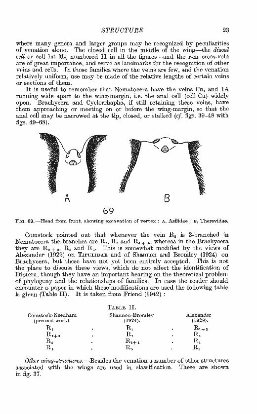
FIG. 69.-Head from front, showing excavati0n of vertex : A, Asilidae ; B, Therevidae.
Comstock pointed out that whenever the vein R 8 is 3-branched in Nematocera the branches are R 2 , Ra and R. + ,, whereas in the Brachycera they are R 2 + 3 , R 4 and R 5 • This is somewhat modified by the views of Alexander (1929) on TIPULIDAE and of Shannon and Bromley (1924) on Brachycera, but these have not yet been entirely accepted. This is not the place to discuss these views, which do not affect the identification of Diptera, though they have an important bearing on the theoretical problem of phylogeny and the relationships of families. In case the reader should encounter a paper in which these modifications are used the following table is given (Table II). It is taken from Friend (1942)
Comstock-N eedham (present work).
R1 R•+ a R. Rs
TABLE II. Shannon-Bromley
(1924).
R1 R. Ra+• Rs
Alexander (1929).
Rt+• Ra R. Rs
Other wing-structures.-Besides the venation a number of other structures associated with the wings are used in classification. These are shown in fig. 37.
24 IX (1). DIPTERA
l
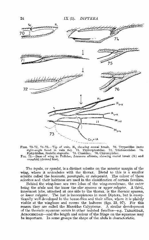
- -- - - ----- ------:Fms. 70-72, 74---76.-Tip of vein, R 1 showing costal break. 70. Trypetidae (note
right-angle bend in vein Se). 71. Thyreophoridae. 72. Trichoscelidae. 74. Ephydridae, Scatella stagnalis. 75. Clusiidae. 76. Chiromyiidae.
FIG. 73.-Base of wing in Psilidae, Loxocera albiseta, showing costal break (X) and crossfold (dotted line).
The tegula, or epaulet, is a distinct sclerite on the anterior margin of the wing, where it articulates with the thorax. Distal to this is a smaller sclerite called the basicosta, parategula, or subepaulet. The colour of these sclerites and their hairiness are used in the classification of certain families.
Behind the wing-base are two lobes of the wing-membrane, the outer being the alula and the inner the alar squama or upper calypter. A third, innermost lobe, attached at one side to the thorax, is the thoracic squama, or lower calypter. The last is inconspicuous in most Diptera, but is exceptionally well developed in the house-flies and their allies, where it is plainly visible at the wingbase and covers the halteres (figs. 33, 97). For this reason they are called the Muscidae Calypterae. A similar development of the thoracic squamae occurs in other isolated families-e.g. TABANIDAE, AcROCERIDAE-and the length and colour of the fringe on the squamae may be important. In some groups the shape of the alula is characteristic.
BIOLOGY AND EARLY STAGESJ 25
BIOLOGY AND EARLY STAGES.
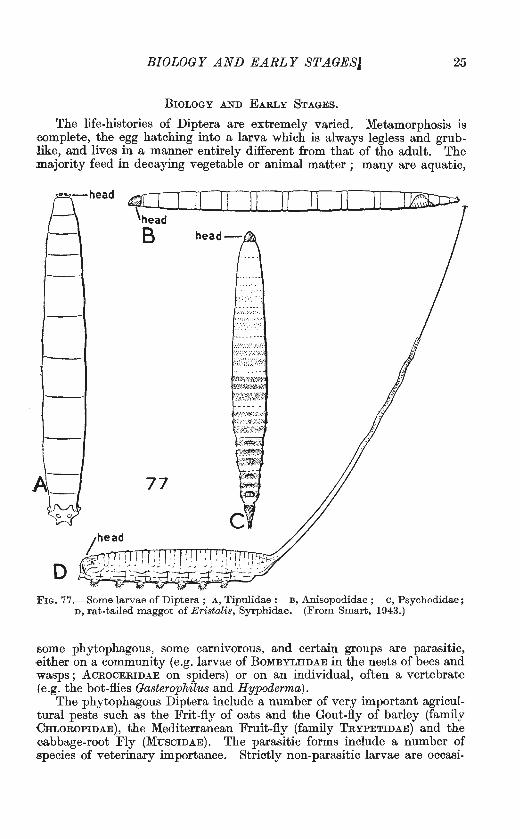
The life-histories of Diptera are extremely varied. Metamorphosis is -complete, the egg hatching into a larva which is always legless and grublike, and lives in a manner entirely different from that of the adult. The majority feed in decaying vegetable or animal matter ; many are aquatic,
~I
-
~- 77 f'J\ c 0
D
11 11 ll
-m:~.lli'(~®.~···
}f,:?.~~1t:~~{1,•,,
;1~JB:~{:~f:~~
··x,\:.:~<,:'~-~-;·x•::.;.:
'•li\Wt.ft;;~·:;~~::..•;
• t.,Z,~·tZ,
ll
F IG. 77 .- Some larvae of Diptera ; A, Tipulidae : B , An isopodidae ; c , Psychodidae; n, rat-tailed maggot of E ristalis, Syrphidae. (From Smart, 1943.)
some phytophagous, some carnivorous, and certain groups are parasit ic, either on a community (e.g. larvae of BoMBYLIIDAE in the nests of bees and wasps; AcROCERIDAE on spiders) or on an individual, often a vertebrate (e.g. the bot-flies Gasterophilus and Hypoderma) .
The phyt ophagous Diptera include a number of very important agricultural pests such as the Frit-fly of oats and the Gout-fly of barley (family CHLoROPIDAE), the Mediterranean Fruit-fly (family TRYPETIDAE) and the cabbage-root Fly (MusciDAE) . The parasitic forms include a number of .species of veterinary importance. Strictly non-parasitic larvae are occasi-
26 IX (1). DIPTERA
posterior spiracle
anterior spiracle
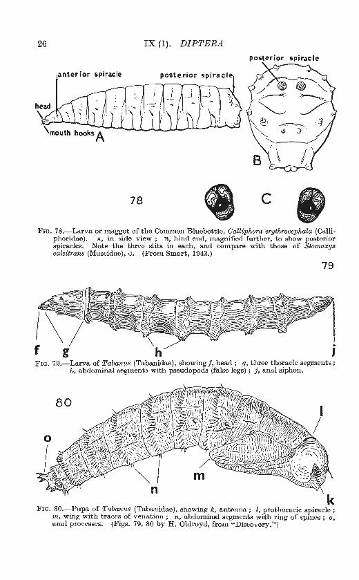
78 c FIG. 78.-Larva or maggot of the Common Bluebottle, Calliphora erythrocephala (Calli
phoridae) . A, in side view ; B, hind end, magnified further, to show posterior spiracles. Note the three slits in each, and compare with those of Stomoxy~t calcitrans (Muscidae), c. (From Smart, 1943.)
79
FIG. 79.-Larva of Tabanus (Tabanidae), showing f, head ; g, three thoracic segments; h, abdominal segments with pseudopods (false legs) ; j, anal siphon.
FIG. 80.-Pupa of Tabanus (Tabanidae), showing k, antenna; l, prothoracic spiracle; m, wing with trace~ of venation ; n, abdominal segments with ring of spines ; o, anal processes. (F1gs. 79, 80 by H. Oldroyd, from "Discovery.")
BIOLOGY AND EARLY STAGES 27
onally introduced into the human body by accident ; the condition thus set up is known as myiasis. The gall-forming CECIDOMYIIDAE of economic importance have recently been reviewed by Barnes (1946), and the Diptera of medical importance are extensively treated by Smart (1943).
It is not possible here to give a key to the larvae and pupae of Diptera. Malloch (1917) has covered Nematocera and Brachycera, and his key is extended by Brues and Melander (1932) to include a few Cyclorrhapha..
84- 85 Fras. 81-83.-Larvae of Diptera in side view. 81. Piophilidae, the Cheese Skipper,
Piophila casei. 82. Sepsidae. 83. Chironomidae, Chironomus, a "blood-worm." Fras. 84-85.-84. Pupariurn of Megaselia, Phoridae; 85, of Drosophila melanogaster,
Drosophilidae. H ead to the right. (From Smart, 1943.)
Johannsen (1934-37) treats the aquatic nematocerous larvae. A model study of the biology and early stages of a family is that of Melin (1923) on the ASILIDAE. James (1947) has given a comprehensive account of the adults and immature stages of flies involved in human myiasis. I have given figures of a few common larvae, but these are by no means representative of the whole.
In general the larvae of the most primitive families have a complete and well-chitinized head-capsule, with well-developed mandibulate mouthparts moving in a horizontal plane (figs. 77 A, B, c). In Brachycera the mouthparts move in a vertical plane, and after the early families the chitinization of the head-capsule is progressively reduced, while the head is partially retracted within the thorax (fig. 79). In Cyclorrhapha the chitinous head
28 IX (1). DIP1'ERA
86
Fra. 86.-Chironomidae. A non·biting midge, female. (From Smart, 1943.)

COLLECTING AND PRESERVING DIPTERA 29
capsule has been lost, and replaced by a secondary development of jointed hooks known as the cephalopharyngeal skeleton, or more commonly as the "mouth-hooks." A typical cyclorrhaphous larva is that of the housefly or blue-bottle, with a pointed fore-end equipped with mouth-hooks and a blunt rear end with two disc-like spiracles (fig. 78). The classification of these larvae is very incomplete, but some families can be recognized by the pattern of the hind spiracles.
Aquatic dipterous larvae may have special respiratory organs. SIMULIIDAE make use of tracheal gills, while CuLICIDAE and some SYRPffiDAE reach the free air by means of a respiratory siphon. Some CmRONOMIDAE (blood-worms) which live in poorly-oxygenated water have haemoglobin in the blood, and possess blood-gills (fig. 83). Many dipterous larvae have pseudopods or false legs on the abdomen (fig. 79).
The pupae of Nematocera and Brachycera, in general, are of the type known as obtect, in which the appendages are visible but are firmly stuck down to the body (fig. 80). Nevertheless these pupae are capable of movement, and can often progress readily by bending the whole trunk. They may be aided in this by spines or other processes which give a purchase against the surrounding medium. Immediately before the emergence of the adult the pupa makes its way to the surface of the soil or wood until it is partly projecting into the free air. According to Hinton (1946) the true pupa is entirely quiescent, and these activities are carried out by the adult insect which, during the first phase of its existence, is still confined within the pupal skin. Final emergence of the adult takes place by breaking through the pupal skin and out of the cocoon, if any.
Many Nematocera construct some form of cocoon, either of silk (some MYCETOPffiLIDAE), or earth (BIBIONIDAE), or use the discarded larval skin as a covering. Among Brachycera some DoLICHOPODIDAE and. EMPIDIDAE form cocoons, either of silk or from the surrounding material.
The pupae of Cyclorrhapha are entirely contained within a smooth, barrel-shaped puparium formed from the last larval skin, which does not reveal on the outside the shape of adult structures such as antennae, wings or legs. Certain larval structures are still visible, notably the mouth-hooks and posterior spiracles. The latter are sometimes borne on paired tubercles, and certain families (e.g. PHORIDAE, DROSOPmLIDAE) may be recognized by having the anterior spiracles borne on long processes like a pair of horns (figs. 84, 85). The lesser house fly, Fannia, and certain ·PHORIDAE have numerous lateral and ventral branched processes. Pupae of this type remain immobile until final emergence of the adult.
CoLLECTING AND PRESERVING DIPTERA. Diptera are found in most varied situations, and may be caught on the
wing with a net, swept from vegetation, or stalked individually with net, tube or pill-box. Small flies may often be swept in large numbers, and the most useful way of handling such a catch is to insert one's head into the net and pick up interesting-looking specimens with a sucking-tube. If the flies are not to be killed immediately, the exit-tube should be stopped, or very soon the flies will crawl out again. A little ethyl acetate on a piece of cotton-wool, held over the mouth of the sucking-tube will immobilize the flies enough to enable them to be transferred to a cyanide killing-·
30 IX (1). DIPTERA
bottle. As an altemative the flies may be shaken into the top of the net, which can be inserted into a large killing-bottle and the dead flies after"' wards sorted.
Diptera for study should always be pinned if possible, unless they are so minute that they need to be mounted on a microscope slide. Diptera are very fragile when dry, and if they are handled the antennae, legs, bristles, hairs and scales are quickly lost, and the specimens cannot be identified.
Fws. 87-88.-87. Psychodidae. A moth-fly. 88. Phoridae. (From Smart, 1943.)
For this reason they should be sorted while still relaxed. Large specimens may be pinned directly on long pins, which will afterwards bear the various labels. Smaller specimens may be pinned on a short stainless steel point through the thorax, slightly to one side of the middle-line, so as to leave the bristles of one side intact. If enough specimens are available some should be pinned sideways through the pleura. Very tiny Diptera may be impaled on the point of the short pin, the headless blunt end being pressed down into the lining of the store-box. By this means the whole upper surface is left intact, and there is less danger of splitting the thorax. A similar result is sometimes achieved by pushing a headed pin upwards through a
OLASSIFIOATION 31
<Jardboard or celluloid mount, and then impaling the fly on the point. This should never be done, because the specimen cannot be removed for re-staging without damage. A surprising number of Diptera on steel points can be stowed away in a small tobacco tin lined with cork or peat, and several such tins can be carried in one pocket.
If large numbers of small Diptera are taken by sweeping it may be very laborious to pin them all, and later they may be found to be nearly all the same species. Such collections may be stored in layers between cellulose wadding (never cotton-wool, the long fibres of which become hopelessly ,entangled in the legs and bristles of Diptera). The wadding is carried in a cardboard box, and is cut into sheets which stack neatly and fill the box without pressure. The method is to cover a sheet of wadding with a single layer of flies, leave them exposed until dry to avoid mould, and then stack ,one on top of another in the cardboard box. During the drying period it is advisable to make sure that the flies are not carried off by ants, nor blown away in a draught.
Diptera should never be placed in any vessel that contains, or has contained, Lepidoptera. Loose scales entangled among the bristles ruin a fly, and cannot be removed without further damage to the specimen.
For permanent storage Diptera already pinned on points may be staged on celluloid or Polyporus strips, and those packed in wadding may be gummed to celluloid points through which a pin can be passed. Care is needed to avoid burying the fly in a mass of adhesive and obscuring all its systematic characters. The proper method is to put a trace of adhesive (celluloid dissolved in amyl acetate to a thin fluid) on the celluloid point and then pick up the specimen with it, using the lightest possible touch. Plastic substitutes for celluloid are not soluble in acetate. Make full notes of locality, date and any other interesting details at the time of capture, and see that the specimen is fully labelled with these as soon as possible.
A fuller account of methods of collecting and preserving is given in Instructions for Collectors: No. 4a--lnsects, by J. Smart (1940, London, British Museum (Nat. Hist.)).
CLASSIFICATION.
It is customary to divide the Diptera into two major groups, Orthorrhapha, in which moulting of larva or pupa takes place by splitting the old skin in a straight or T -shaped slit, and Oyclorrhapha, in which the main slit is circular and detaches a sort of cap. Orthorrhapha are then further divided into the suborders N ematocera and Brachycera by the antenna} structure.
Comparison of adult Diptera, both recent and fossil, indicates that the more correct division is first into Nematocera and "Brachycera" (i.e . all the rest), then" Brachycera" into Brachycera-Orthorrhapha and Brachycera-Cyclorrhapha. Since the method of emergence is of theoretical interest only, and does not help anyone to name a single adult specimen, which is the usual problem, it is simpler to ignore it, and to group the Diptera directly into three suborders, naming t hese, for brevity, Nematocera, Brachycera and Cyclorrhapha.
It is now impossible to write about Diptera without mentioning the names proposed in Meigen's paper of 1800, about which there has been so much controversy. The facts are that many of the generic names most
32 IX (1). DIPTERA
widely used in Diptera are those published by Meigen in a paper of 1803, although he had written an earlier paper in 1800, which proposed a different set of names. There is evidence that Meigen regarded his earlier paper aspremature, and certainly he never used the 1800 names himself. This earlier paper remained forgotten untill908, when it was revived by Hendel, who proposed to alter many well-known names of genera, and the family-
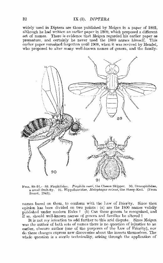
FIGs. 89- 91.-89. Piophilidae. Piophila casei, the Cheese Skipper. 90. Drosophilidae. a small fruit-fly. 91. Hippoboscidae. Melophagus ovinus, the Sheep-Ked. (From Smart, 1943).
names based on them, to conform with the Law of Priority. Since then opinion has been divided on two points : (a) are the 1800 names validly published under modern Rules ? (b) Can these genera be recognized, and if so, should well-known names of genera and families be altered ?
It is not my intention to add further to this arid dispute. Since Meigen was the author of both sets of names there is no question of injustice to an earlier, obscure author (one of the purposes of the Law of Priority), nor do these changes express new discoveries about the insects themselves. The whole question is a sterile technicality, arising through the application of
LIST OF FAMILIES 33
a modern set of rules to works published a century earlier. It is impossible to detect any useful purpose that is served by changing these names.
By this time both sets of names have been so widely used in published work that any serious student of Diptera must know both. In the following List the 1800 names are indicated by an asterisk.
The order of families is based on Kloet and Hincks (1945), with the following differences : GASTEROPHILIDAE are listed as a family of the Acalypterae, not a subfamily of the MusciDAE ; BRAULIDAE are placed next to the CHAMAEMYIIDAE, in accordance with the work of Imms (1942); PALLOPTERIDAE are included with LONCHAEIDAE and DIASTATIDAE with DROSOPHILIDAE.
The status of families in the section Acalypterae is much less definite t han that of other families of Diptera. The characters on which they are separated, such as the convergence or divergence of certain bristles, or the existence of a small fracture in the costa of the wing, seem trivial, though it must be remembered that these characters are artificially selected for convenience in making a key. Two families of Diptera may look quite different when compared under the microscope, and yet it may be extremely difficult to narrow down the difference to a few clear-cut details which can be understood by someone who has never seen either family before. Even so, no two authors agree on the families of the Acalypterae. The arrangement given here is offered not as a final and authoritative .classification, but--in conjunction with the more detailed keys to genera and species to be published later-as a handy means of finding a name for t hese numerous flies.
The families of Diptera recognized in the following pages are listed below :
LIST OF FAMILIES. {* indicat.e8 a family name derived from a generic name proposed by Meigen in 1800.)
Order DIPTERA.
SuboTder 1.-Nematocera.
Tipulidae. Trichoceridae (Petauristidae* ). Anisopodidae (Rhyphidae)
(Phryneidae*). Ptychopteridae (Liriopeidae*). Psychodidae. Culicidae. Chironomidae (Tendipedidae*).
Ceratopogonidae (Heleidae*). Thaumaleidae (Orphnephilidae). Simuliidae (Melusinidae*). Bibionidae. Scatopsidae. Mycetophilidae (Fungivoridae*). Cecidomyiidae (Itoniidae*) .
Suborder 11.- Brachycera.
Stra tiom yiida e. Rhagionidae (Leptidae). Tabanidae. Acroceridae (Cyrtidae). Bombyliidae.
Therevidae . Scenopinidae (Omphralidae*). Asilidae. Empididae. Dolichopodidae.
34 IX (1). DIPTERA
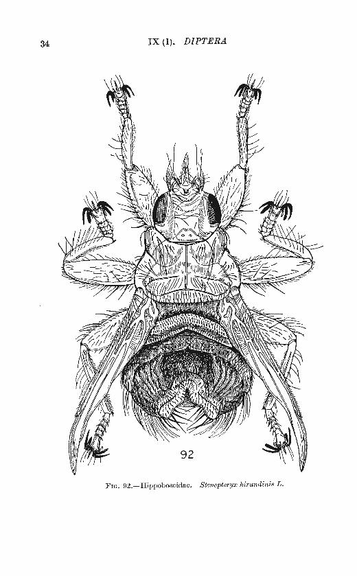
Fw. 92.- H ippoboscidae. Stenepteryx hi1·undinis L .
LIST OF FAMILIES
Suborder JIJ.-Cyclorrhapha. Series Aschiza :
Lonchopteridae (Musidoridae*). Phoridae. Platypezidae (Clythiidae*).
Series Schizophora : Section Acalypterae :
Pipunculidae (Dorilaidae*). Syrphidae.
Coelopidae. Helomyzidae. Trichoscelidae. Chiromyiidae. Clusiidae. Anthomyzidae. Opomyzidae. Tethinidae. Ephydridae. Canaceidae.
35
Gasterophilidae. Conopidae. Platystomidae. Otitidae. Ulidiidae. Piophilidae. Thyreophoridae. Dryomyzidae. N eottiophilidae. Trypetidae. Lonchaeidae.
Borboridae (Cypselidae,* Sphaero-
Lauxaniidae (Sapromyzidae). Micropezidae (Tylidae*). Tanypezidae. Psilidae. Megamerinidae. Sepsidae. Sciomyzidae (Tetanoceridae). Chamaemyiidae (Ochthiphilidae). Braulidae.
Section Pupipara :
Hippoboscidae.
Section Calypterae : Tachinidae (Larvaevoridae*). Calliphoridae.
ceridae). Periscelidae. Asteiidae. Aulacigastridae. Camillidae Drosophilidae (inc. Diastatidae). Agromyzidae. Milichiidae (inc. Carnidae). Odiniidae. Chloropidae.
N ycteribiidae.
Muscidae. Cordyluridae (Scatophagidae).
Although there are one or two families and genera whose affinities are uncertain, the grouping in the foregoing list is a very natural one, and represents the three lines of evolution in the Order, as confirmed by fossil evidence. Nematocera appear in the Permian, and by the early Tertiary had led on to Brachycera and Cyclorrhapha.
N ematocera are, in general, slender, soft-bodied flies with long antennae consisting of many similar segments ; palpi of several segments and often noticeably drooping ; wings with a number of longitudinal veins, but mostly without the conspicuous discal cell (fig. 38) in the middle of the wing. The anal cell, if present, is wide open (fig. 40).
Brachycera are mostly fairly large flies, of stout build ; antennae short, but sometimes showing traces of more than three segments ; pal pi not more than two-segmented, not conspicuously drooping ; wings usually with a very complete venation and with a discal cell (figs. 49-59).
36 IX (l). DIPTERA
Cyclorrhapha include the most highly specialized Diptera, mostly of short, stout build, with short antennae, many bristles, and venation of the type shown in figs. 60-76. There are two lines of relationship, the Acalypterae-a group of small families, some of them ill-defined-and the Calypterae, which include the bristly blowflies, houseflies and related genera. The series Aschiza, including the SYRPHIDAE and several smaller families, is placed between the other Cyclorrhapha and the Brachycera. Crampton (1942) regards the Aschiza as the stem-group from which the other two major groups may have developed. The section Pupipara, composed of parasitic flies which retain the larva within the body of the parent until just before pupation, is a convenient group founded rather on similarity of biology than on true systematic relationship.
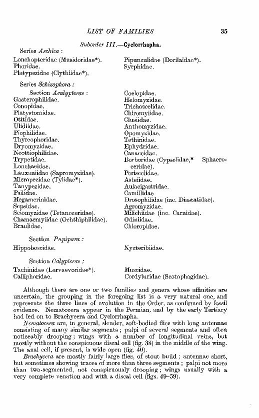
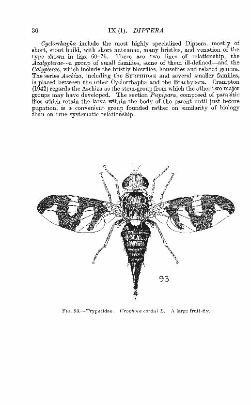
FIG. 93.-Trypetidae. U1·ophora cardui L . A large fruit-fly.
KEY TO FAMILIES 37
KEY TO FAMILIES.
This key is compiled mainly from the following sources : N ematocera, Edwards (1938) ; Brachycera and Aschiza partly after Curran (1934) ; Acalypterae partly based upon Lindner (1925) with many modifications. Grimshaw's key (1934) is a literal translation of Lindner's.
Antennae composed of two basal segments and a flagellum of numerous similar segments (figs. 10-16). In BIBIONIDAE, SIMuLIIDAE and ANISOPODIDAE the segments of the flagellum are contracted, but still distinct (figs. 13-15). Palpi with several segments, often drooping. Anal cell of the wing open, almost never narrowed towards wing-margin (figs. 39-48) .... Suborder Nematocera .... .. 2
Antennae composed of two basal segments and a third compound segment formed by the fusion of the elements of the flagellum. In many STRATIOMYIIDAE, RHAGIONIDAE and TABANIDAE the elements of the flagellum can still be seen (figs. 17- 22). Palpi with l -3 segments, the terminal one enlarged and often porrect (held forwards). Anal cell narrowed towards wing-margin, often closed by the meeting of Cu1 and lA, sometimes very much reduced towards base of wing (figs. 49-68) or absent . ........................................... 16
2 Two anal veins r eaching wing-margin (fig. 39). Mesonotum with a V-shaped furrow dividing prescutum from scutum (fig. 34A) ........ . .. ............ . 3
At most one anal vein reaching wing-margin. Mesonotal V-shaped furrow absent, except in PTYCHOPTERIDAE, where its shape is different .... . .. ....... . ... . . 4
3 Ocelli absent. Vein 2A moderately long .. Crane-flies, daddy long-legs. TrPULIDAE. Ocelli present. Vein 2A very short . . ...... . ... Winte1·-gnats. TRICHOCERIDAE.
4 Ocelli present ................ . ... ... . .... ... ... .. . .. . . .. .. ... . . . ........ 5 Ocelli absent .. ................. .. . . ..... .. .... .. .. .. .... . ............... 9
5 Tibiae spurred at tip ............ . . .... .... .... .. ... . ...... . ............. 6 Tibiae without spurs. Small flies with reduced venation ........ . ............. 8
6 Vein R 8 forking at, or just before r- m crossvein (fig. 40) Window-midges. ANISOPODIDAE (RHYPHIDAE).
Vein R 8 forking (if at a ll) well beyond r-m crossvein (fig. 44) .................. . . 7 7 Antennae placed well below compound eyes, n ear mouth m argin. Antennae as
in fig. 14, short, flagellar segments closely contracted .. March-flies. BIBIONIDAE. Antennae near middle of eyes; as in fig. 12, long, flagellar segments well separated,
with whorls of hairs ..... . ................ Fungus-gnats. MYCETOPHILIDAE. 8 Antennae short (cf. BrBIONIDAE, fig. 14), the segments short, broad, and not easily
distinguished. Bare, usually black flies, with bare wings. Abdomen with only seven distinct segments before the genitalia ................ ScATOPSIDAE.
Antennae usually longer, segments well separated. More hairy flies with at least a conspicuous fringe to the wings. Abdomen with eight segments visible before the genitalia .. . .... . ...... CECIDOMYIIDAE (subfamily LESTREMIINAE).
9 Ten (in som e PsYCHODIDAE eleven) veins or their branches reaching the wing-margin ........ . . . .......... . .... . . . . ...... ... ......... . ........... 10
At most eight veins or their branches reaching the wing-margin . .. . .... ....... 12 10 Main crossveins near or beyond middle of wing. Larger, long-legged flies ...... 11
Main crossveins near base of wing (fig. 48). Small, hairy, mothlike flies (fig. 87) 1'\lloth-flies. PSYCHODIDAE.
11 Vein R 4+ 5 forked. First antenna! segment larger than second. Tibiae with spurs. Flies superficially resembling TIPULIDAE ... . ... ... . . PTYCHOPTERIDAE.
Vein R 4+ 5 simple (fig. 42). First antenna! segment rudimentary, second enlarged. Tibiae without distinct spurs . .. . ... . Mosquitoes, gnats. CULICIDAE.
12 Only four, or even fewer, veins reaching the wing-margin (fig. 47). First two antennal segments about equally long. First segment of t ar si very short
Gall-midges. CECIDOMYIIDAE. At least six, sometimes seven or eight veins reaching wing-margin. First
antenna! segment rudimentary, second more or less enlarged. First tarsal segment nearly always longer than second .. . ........ . .... . . . ............. 13
13 Crossveins near base of wing. Seven veins reaching wing-margin, but vein M simple .. .. ... . .. ................... . ... .. ............. . . THAUMALEIDAE.
Crossveins, when present, near middle of wing ..... . ........ . ... .... . .. .. ... 14
38 IX (1). DIPTERA
14 Wings very broad (fig. 43). Vein M forked. Antennae short and bare (fig. 15) Black-flies. SIMULIIDAE.
Wings not very broad. Antennae hairy, especially in the male . . . . . ........... 15 15 Vein M 1 + 2 forked. Wings superposed fiat over the back when at rest, except in
one species ............................ . Biting-midges. CERATOPOGONIDAE. Vein M 1 + 2 simple. Wings not superposed, except in the genus Podonomus
Non-biting midges. CHIRONOMIDAE. 16 Foot with three pads, the empodium being developed equally with the pulvilli
(fig. 36E) [some DoLICHOPODIDAE and EMPIDIDAE have the empodium so de-veloped, but may be recognized by the venation (figs. 57, 59)] ..... . ....... . 17
Foot with only two pads, the empodium being hairlike or wanting. Some-times pulv illi also are wanting .............. . .... . ... . ...... . ..... .... .. 20
17 F irst few segments of fiagellum fused into a compound " third segment " which clearly shows its component parts (figs. 17, 19, 21, 22) .................... 18
First few segments of fiagellum fused into a compound " third segment " which does not show its component parts (except in Xylophagus, fig. 18), and which usually bears the remaining segments in the form of a style or arista (figs. 20, 24, 25, 28, 29) .. .. ... .. ....... . . ..... . ... . ...... .. ..... . . ... .. 19
18 Squam ae large and conspicuous (fig. 37). Tibiae with spurs, a t least on middle legs (fig. 36D ). Antennae as in figs. 21, 22. Veins R 4 and R, separating into a broad fork across the wingtip (fig. 51). Stoutly-built flies, with large fiat head, and eyes in life usually with brilliantly-coloured spots or bands
Horse-flies, clegs. TABANIDAE. Squam ae usually small. Tibiae without spurs (except Xylomyia). Antennae as in
figs. 17, 19. Venation as in fig. 50 ; note small discal cell, and crowding of veins towards anterior margin of wing. Stoutly-built or slender flies. Eyes in life r arely brilliantly coloured. . . . . . . . . . . . . . . . . . . . . . . . . . . . . . . . STRATIOMYIIDAE.
19 Thoracic squamae very large. Abdomen balloon-like. Flies of globular shape, with a small head consisting almost entirely of the eyes, set very low in front of the thorax. Venation as in fig. 58, or similarly reduced .. AcROCERIDAE (CYRTIDAE)
Thoracic squamae sm all. Abdomen elongate. Slender, very fragile-looking flies, with long, slender legs. Venation as in fig. 49; note short vein Ru which curves forward into costa to enclose stigma .. . ..... RHAGIONIDAE {LEPTIDAE).
20 Frons without ptilinal suture ...... . ............... .. .... . ............... 21 Frons with a ptilinal suture, enclosing the ptilinum (fig. 6) . . ........ . .... . . . . . 31
21 Basal cells long. Anal cell long and pointed, sometimes open. {PLATYPEZIDAE m ay give trouble here, but can be recognized by the venation, fig. 60, especially the peculiar forking of veins M 1 and M 2) . •• ..... •.•...•••.•• . ••.•••••.•. . 22
Second basal and anal cells short ; anal cell, if present, closed by a recurrent veinlet, or with a blunt end (except in EMPIDIDAE, subfamily HYBOTINAE). Never more than four posterior Cells ............. .. .................. . .. 26
22 Veins R 4 and R, not united, reaching the wing-margin separately . ... . . . .. . ... . 23 Veins R 4 and R, fused, so that the radius has only three branches (R1 , R 2 + 3 ,
R 4+ 5) ••••••••••••••••••••••••• •• • • •••• • ••••• ••••• •• •••••• ••••••••••• 29 23 Five posterior cells-i.e. five cells in addition to the discal cell, lying between
R, and Cu 1 (figs. 55, 56; cells 6-10 in fig. 38) . .. .. . ..... . .. . ........ ...... . 24 Fewer than five posterior cells . . .. ... ..... .... .. . .................... . .... 25
24 Vertex sunk between eyes, forming a distinct groove when seen from in front, the ocelli raised on a little island (fig. 69). Eyes of both sexes well separated. F emales with varied forms of ovipositor, but no British species has a circlet of spines .. . . .. . ..... ..... . ...... .. ...... .... .... . Robbe1·-flies . AsiLIDAE.
Vertex not sunk between eyes (fig. 69). Eyes of male touching. F emales with a circlet of spines round the ovipositor . . .. ..... .... ............. THEREVIDAE.
25 Four posterior cells (fig. 52). Furry species, usually with brightly coloured scales forming a distinct p attern on the body. H ead globular, very mobile
B ee-flies. BOMBYLIIDAE . Three posterior cells. Venation as in fig. 53. Very bare flies of small
size ..... . ............ . . . .. ... ... .. . ... . Small window-flies. SCENOPINIDAE. 26 Venation abnormal. Se and R short and strong, running into costa well before
wingtip. Other veins faint, running in parallel lines from R to wing-margin (figs. 64, 88) .................. .... . . .. .. .. .. .. . . ..... . .. ...... PHORIDAE.
Veins Se and R not especially prominent, the following veins diverging in the usual way ... ... .. ...... .. .. . .................................... . 27

KEY TO FAMILIES 39
27 Wings pointed at tip, and with no prominent crossveins in middle (fig. 67) LONCHOPTERIDAE.
Wings rounded at tip, and with at least one crossvein prominent in middle of wing ........ . ... . .... .. ............ . . . ........................... .. . 28
28 First basal cell (cell R) rather long, r-m crossvein being prominent in middle of wing. Often the discal cell and 2nd basal (cell M) are also obvious. Anal cell much shortened or absent (fig . 57). Predatory flies, with proboscis horny and often long .................... . ............ ... .. .. .... .. .... EMPIDIDAE.
First basal cell very short, so that only one crossvein is prominent in middle of wing (this is crossvein m) (fig. 59). Predatory flies, but often with soft, fleshy proboscis. Often metallic and bristly ...................... DoLICHOPODIDAE.
29 Cell R 5 (first posterior) closed by the turning forward of vein M1 parallel to wing margin. Crossvein m closing apex of discal cell is also more or less parallel. Generally a " spurious vein " running obliquely between R and M (fig. 61)
Hover-flies. SYRPHIDAE. Cell R 5 open. Vein M1 may be turned forward, but is never parallel to the
win?:mare:in, an~ ~?es not meet vein R,+ 5, nor is it parallel to crossvein m. No spurwus vein . ............ .. . .. .... ... .. ... ... .... . . ...... .. .. . 30
.30 Arista terminal. Head not outstandingly large or spherical. Hind tarsi and tips of hind tibiae often flattened and dilated. Venation, fig. 60 .... PLATYPEZIDAE.
Arista dorsal. Head outstandingly large, spherical, mostly composed of the eyes in both sexes. Hind tarsi and tibiae not flattened or dilated. Venation, fig. 65 .................................................... PIPUNCULIDAE.
31 Anal cell (cell Cu) extending almost or quite to wing-margin. Proboscis usually long, projecting forward, sometimes bent double. Head more or less inflated (fig. 2) ............................ . ................ . ........ CONOPIDAE.
Anal cell short, not reaching wingmargin, though anal vein (vein lA) may often do so .......... . ........... . . .. ...... . . . . . ... . ... . ............. 32
.32 Normal flies, head n either lying back upon the thorax, nor inserted into a groove in it. Body not markedly flattened for a parasitic life ........ . ... . ......... 33
Abnormal flies, head lying back upon thorax, or inserted into it, or body modified for parasitic life on mammals, birds or bees. Wings often reduced, absent, or of peculiar shape ............................................ 79
33 Second antenna! segment usually without, or with only a short indication of, a dorsal cleft or seam (fig. 29). If a cleft is present, then thoracic transverse suture evident only at sides of thorax, and posterior calli are not marked off by a sutural depression (fig. 27). Thoracic squamae usually vestigial; if somewhat developed, then posterior calli are not differentiated. Mostly small flies, with eyes well separated in both sexes .. . . Section Acalypterae ...... 34
Second antenna! segment always with a distinct dorsal cleft or seam for nearly its whole length (fig. 28). Posterior calli differentiated, and transverse suture of thorax often entire, or at least evident a lmost to centre-line of thorax. Thoracic squamae usually large and concealing the halteres ; if small and vestigial (Cordyluridae), then posterior calli and thoracic suture are well developed. Moderate-sized or small flies, with eyes in male often close together
Section Calypterae ... ... 77 34 Mouthparts vestigial, sunk in very small oral pit
Horse Bot-flies. GASTEROPHILIDAE. Mouthparts well developed, oral opening large ............... . . ... ........ . . 35
35* Costa complete, not even partially interrupted at or near tip of Rl (fig. 66) (PLATYSTOMIDAE, couplet 48, have break near humeral vein (h) but not near Se ; cf. also CLUSIIDAE, couplet 55, genus A cartophthalmus) . ..... ... ............. 36
Costa at least partially interrupted before tip of vein R 1, usually at tip of vein Se if this reaches wing margin ............ . ........... . ..... ... ..... . ..... 49
36 Metathoracic spiracle with at least one long hair in addition to short pubescence (figs. 31A, B) . ............. . . .. ... .. ...... .. .. ......... ... .. ... .. .. .. 37
:Metathoracic spiracle with pubescence only , without any conspicuously longer hairs or bristles (fig. 3 l c). (N.B.-Do not confuse small bristles which may st and on hypopleuron just below spiracle) ............................... 38
* The occurrence and position of costal breaks, referred to in couplets 35 and 49, are comparative characters which need great care. Some families and genera which m ay prove difficult to place are either repeated t wice in the key, or figured (figs. 70-76).
40
37
38
39
40
41
42
43
44
45
46
47*
48
50
IX (1). DIPTERA
Small, bare flies with rounded head. Buccae not strongly produced. Often with black spot at tip of clear wings .. . ... .. ...... .. ... . . ............. SEPSIDAE.
Middle-sized, very bristly flies, with flattened head and protruding buccae. Wings browned, but without an apical spot ............ COELOPIDAE, genus Orygma.
Legs long, thin, stilt-like, tibiae without pre-apical bristles. Abdomen long and slender. First posterior cell (cell R 5) strongly narrowed towards wing m argin, or closed ......... . ... ... . .... ........ . MICROPEZIDAE (inc. TANYPEZIDAE).
Legs not unusually long. Other charact ers do not agree .................. .. 39 Tibiae with dorsal pre-apical bristle (except Coelopa pilipes, couplet 41) ........ 40· Tibiae without dorsal pre-apical bristle ................ . ......... .. .. .. .... 43 Postvertical bristles converging, or even crossed (this pair of bristles stand in the
centre of the occiput, immediately behind the ocellar triangle) ...... ... ..... 41 Post vertical bristles parallel or divergent, rarely absent . .. .. .. ............... 42 Vein lA reaches hind margin of wing as a dark shadow. Anal cell (cell Cu) closed
by a straight crossvein, making an acute angle towards wing margin (fig. 66). Buccae puffed out, with a number of bristles, but without a single distinct vibrissa (fig. 4). Three or four fronto-orbitals, curved outwards. Last tarsal segment broadened .......... ... .... . .. . . .... .... .. Kelpjlies. COELOPIDAE.
Vein lA bluntly shortened. Cell Cu short, outer border convex, never with an acute angle. Two fronto-orbitals, rarely only one. Last tarsal segment not enlarged. . . . . . . . . . . . . . . . . . . . . . . . . . . . . . . . . . . . .. . . . . . . . . . . . . . LAUXANIIDAE
Clypeus retracted, not v isible . . .......... . . . ...... .. ......... ScroMYZIDAE. Clypeus strongly developed and prominent (fig. 5). Wings long and broad, ex-
t ending well beyond the abdomen .......... .. .. .. ..... . .. DRYOMYZIDAE. Hind femora swollen, with two ventral rows of spines. Abdomen long, with a
waist. Anal cell (cell Cu) relatively long and parallel-sided. Vein lA a lmost or quite reaching the wing margin ........................ MEGAMERINIDAE.
Hind femora not swollen and spinose. Anal cell relatively short and broad, not parallel-sided ................................... . ..................... 44
Vein Se not complete, basal part only being visible ........ . ................. 45 Vein Se complete for greater part of its length, even if faint near wing margin .. 45 Ambient vein (continuation of costa) reaches only as far as tip of vein R 4 + 5•
Cell R 5 not narrowed towards wing margin. Cell M (basal) present. Second antenna! segment sometimes projecting like a hood over third segment
PERISCELIDAE. Ambient vein reaches tip of M1• Cell R 5 strongly narrowed t owards wing m argin.
Cell M (basal) absent. Second antenna! segment never projecting like a hood. ASTEIIDAE.
Anal vein well developed. Post verticals divergent, parallel or absent ........ 4 7 Cell Cu (anal) and vein lA almost wanting. Postverticals convergent or absent
CHAMAEMYIIDAE. Vein R 1 bare on dorsal surface. If it is hairy, then at same time cell Cu (anal) is
drawn out into a sharp point, stigm a is narrow, and body-colour shining m etallic. First posterior cell (cell R 5 ) more or less narrowed at tip .. ...... . ... U LIDIIDAE.
Vein R 1 with hairs or bristles on its upper surface. If it is bare, then cell Se is very large, or the eyes are protuberant, or cell R 5 is narrowed at tip ........ 48
Propleural and sternopleural bristles present. Always four supra-alars. Antennae lie in long pits in the face. Vein R 5 ending near tip of wing, so that cell R 5 lies behind the wingtip. Post verticals of normal size, parallel or divergent
0TITIDAE. Propleural bristle absent. Only three supra-alars. Vein R 5 ending well before
tip of wing, so that cell R 5 lies before wingtip. Postvertical bristles very small PLATYSTOi\HDAE.
Break in costa lies some distance before point where R 1 reaches costa (cf. figs. 63, 70-73, 75, 76) ... ..... ..... . .. . . .... .. ....... . .. ... ... .... ......... ... 50
Break in costa lies very close to point where R 1 reaches costa (cf. figs . 68, 74) .. . . 66 Vein Se a lways distinct, separa ted fi·om R 1 , and ending in costa, or abruptly cut
off just before r eaching it (fig. 63) . ......... . . . . . .. . . . . . ..... ... ....... . 51 Vein Se weakly developed, represented partly by a shadow, or entirely vestigial
(cf. TRICHOSCELIDAE and EPHYDRIDAE, genus PaThydr-optem, fig. 62) .. ... . 61
* Homalocephala albita1·sis Zett. (ULIUIIDAE) has R 1 hairy, cell Cu rounded towards the wing margin, no propleurals or sternopleura ls, only two supra-alar·s.
t See footnote to couplet 35.
.51
52
53
54
55
56
.57
58
59
·60
fil
fi2
63
KEY TO FAMILIES 41
Postvertical bristles diverging, parallel or absent ... .............. . .... . . . . . . 52 Postvertical bristles convergent or crossed. Vibrissae present ... ... ......... . 59 Vein Se abruptly bent forward in almost a right angle (figs. 63, 70). Wings often
"pictured," with a banded pattern. Frons usually with inwardly-curved lower fronto-orbital bristles, which stand on an upward extension of the parafacial plates (fig. 7). Ovipositor rigid, non-retractable (fig. 93)
Large fruit-flies. TRYPETIDAE, Vein Se not abruptly bent, though it may be curved towards costa. Parafacial
plates not extending above level of antennae, no lower fronto-orbital bristles (fig. 8). Ovipositor sometimes rigid, wings sometimes pictured .... ........ 53
Vibrissae present ... .. . .. . ..... . . . .. . .. . .. .. .... .. .. ... .. .. . ... .. ...... 54 True vibrissae absent, though a p eristomal bristle may be present. LoNCHAEIDAE. Cell M (second basal) closed apically by a crossvein .. ...... ...... .. . ...... .. . 55 Cell M (second basal) open apically, merged with cell 1st M 2 (discal)
AULACIGASTRIDAE. Vertical plates extending forward, with 2- 4 upper fronto-orbital bristles (fig. 8).
Cells Cu (anal) with a convex outer margin. Vein lA does not reach wing margin, even as a shadow . . ............. .... . . .... . ..... ........ .. . .. CLUSIIDAE.
Vertical plates cut short in front, with not more than one upper fronto-orbital bristle . . .. . . . . ..... . .. . . ... . .... . ............ ..... ... ... ............ 56
Frons of male narrowed to one-quarter or one-fifth of breadth of h ead . Female with ovipositor like a Trypetid (fig. 93). Head flattened-hemispherical, concave behind, eyes in profile semicircular. Vein lA abbreviated, cell Cu (anal) apically convex. Middle tibiae, at most, with preapical bristle .. . .. ... LONCHAEIDAE.
Frons of male not narrowed. Female not with Trypetid-like ovipositor. Head convex behind, eyes rounded . . .... ... .... ..... . . ....... . ........ .. .... 57
Vein R 1 hairy above. Vein lA reaching wing margin . . Nest-flies. NEOTTIOPHILIDAE . Vein R 1 bare above. Vein lA not reaching wing margin .. . . ... .. ... .. .... . . 58 Antennae vertical, lying in deep grooves in the face. Anterior border of frons with
bristles which curve forwards. Scutellum exceptionally long, or at least flattened , with two short projections which bear thick marginal bristles (fig. 71)
THYREOPHORIDAE. Antennae inclined forwards, not lying in deep grooves. Anterior border of frons
without bristles. Scutellum normal (fig. 89) . . Cheese -skippers. PIOPHILIDAE. Vertical plates anteriorly cut short. Vein Se strong, diverging from R 1 towards
tip. Anal cell curved towards wing margin. Vein lA distinct . . HELOMYZIDAE. Vertical plates more or less normally extended forwards. Vein Se rather weakly
developed, though distinct as far as costa, almost parallel with R 1. Vein lA abbreviated . . . ................... .. ............ .. ... .... . . .. . .... .... 60
Propleural bristles present. Tibiae with preapical bristles. Two upper fronto-orbital bristles. Pal pi not abbreviated. (fig. 72) ... .... . . .. . TRICHOSCELIDAE.
Propleural bristles not present. Tibiae without preapical bristles. 2- 3 upper fronto -orbitals. Palpi abbreviated. (fig. 76) .. .. ........... .. . CHIROMYIIDAE.
Second basal and anal cells (cells M and Cu) absent, vein lA absent or weak, vein Se faint, at most indicated basally by a shadow. Costa interrupted only at tip of vein Se. Vertical plates very short and indistinct. Ocellar triangle, on the contrary, usually very large and plate-like, occupying most of frons (fig. 9), which rarely bears strong bristles . .. . .. . ..... .... . . .. . . .. .... CHLOROPIDAE.
Anal cell (cell Cu) and vein lA always present. Cell M (2nd basal) usually so .. 62 Vibrissae and vibrissa! angles distinctly formed . .. . . . . . . . . ....... ...... . ... 63 Vibrissae absent . If the peristomal bristles take their place then only one upper
fronto-orbital is present. All upper fronto-orbitals bent dorsally ........ . . 64 Lower fronto -orbitals present, bowed inwards ; postverticals divergent. Basal part
of ovipositor conical . .. ....... ........ . ..... . ..... ..... ... . . AGROMYZIDAE Lower f'ronto -orbitals absent, postverticals convergent, rar ely absent. Fore
femora usually with a vertical bristle -like spine. Female with inconspicuous ovipositor ......... . . . ......... . ...... .... .... ... . ...... ANTHOMYZIDAE.
Costa ending just beyond tip of v ein R 4 + 5• Parafacial plates extending up to meet vertical plates, and very broad so that they form a bridge across the frons, and almost m eet in the middle line above the ant ennae. AGROMYZIDAE, genus Selachops (not now on British List].
Costa reaching tip of M1 • Parafacials not produced upwards, vertical plates at most half as long as frons, no bridge above antennae ...... . ............... 65
42
65
66
67
68
69
70
7l
72
73
74
75
76
77
IX (1). DIPTERA
Invariably one mesopleural and one sternopleural bristle present (figs. 32 APS, KPS and 33 M, ST). Mesonotum with one dorsocentral before the suture and 2-3 after it (fig. 35 D). Postverticals absent. Always only one upper frontoorbital, bent upwards. Ocellar triangle small. Cells M and Cu (second basal and anal) small. Vein R 2+3 very long, and converging towards tip of R 4+ 5
0POMYZIDAE. Mesopleural and sternopleural bristles absent. Mesonotum almost bare. At
most 2 supra-alars and usually only 2 postsutural dorsocentrals. Postvertical bristles strongly divergent or absent. Ocellar triangle large. Vertical plates small and rudiment.ary. Cells M and Cu (second basal and anal) long and wide. W'ings with a distinctive crossfold running from tip of Se across base ofR2+3 and end of second basal cell (M) (fig. 73) . . . . . . . . . . . . . . . . . . . . . . . . . . . . PsrLIDAE.
One or more lower fronto-orbital bristles are directed inwards .............. 67 No inwardly-directed lower fronto-orbital bristles .......................... 6!} The most anterior upper fronto-orbital bristle is curved upwards. Costa inter-
rupted only before tip of vein R 1 • Vein lA present, even if abbreviated .... 6S The most anterior upper fronto-orbital bristle is curved outwards and forwards.
Costa interrupted twice, after humeral crossvein and before tip of vein R 1 •
Vein lA represented only by a shadow ........... MILICHIDAE (inc. CARNIDAE) .. Vertical plates reach down almost to meet parafacials, and bear two upper fronto
orbital bristles. Abdomen with five visible segm ents before genitalia. Ovi-positor retractable .......................................... 0DINIIDAE .
Vertical plates, with two upper fronto-orbital bristles, lie in upper half of frons. Usually several lower fronto-orbital bristles. Abdomen with six visible segments before genitalia; seventh segment of female is non-retractable conical basis of ovipositor (" Trypetid type ") .............................. AGROMYZIDAE.
Posterior metatarsus shortened and thickened. Branches of vein M beyond the discal cell (1st M 2) often not pigmented ........................ BORBORIDAE.
Posterior metatarsus not shortened and thickened .............. . ........... 70 Cells M and Cu (2nd basal and anal), and vein lA absent .................... 7l At least cell Cu, and usually vein lA present ...................... . ....... 7 4 Vein Se rudimentary .................................................... 72 Vein Se distinct, at least basally .................................. . ..... 73 Ocellar triangle exceptionally large and plate-like, often occupying almost whole
frons down to antennae. Rarely strong bristles on frons. Costa interrupted only at tip of vein Se ................... . .................. CHLOROPIDAE.
Ocellar triangle not exceptionally large. Usual fronto-orbital bristles present. Costa interrupted twice ............................... .. .... . CAMILLIDAE.
Costa distinctly broken in two places (figs. 62, 74). Arista, when feathered, with feathering only on upper surface ......... ... ..... . Shore-flies. EPHYDRIDAE.
Basal interruption of costa, near vein h, indistinct. First posterior cell (cell R 5 }
very long, narrowed at its base. Arista feathered in either one or two rows ASTEIIDAE.
Costa interrupted twice (fig. 68). Often small yellow or yellow-brown flies, with bright red eyes (fig. 90) . . Small fruit-flies. DROSOPHILIDAE (inc. DIASTATIDAE).
Costa interrupted only once, before tip of vein R 1 ....••.....•.•.•.•••..•... 7 5 Ocellar triangle large, extending to anterior margin of frons. Antennae widely
separated at base, third segment conical. Vibrissae absent. Cell Cu (anal) small, vein lA visible only as a short rudiment ............... . CANACEIDAE.
Ocellar triangle not extending more than half-way down frons. Antennae not widely separated at base ............ . .............. . ... . ...... . ....... 76
Vein Se fully developed up to its tip, and separated from R 1 • Postvertical bristles long and divergent. Vertical plates shortened in front. Vein lA distinct, though not reaching wing margin. Larger species, with light orange frons and black occiput ..... .. ......... ... ... . .......... .... ...... THYREOPHORIDAE.
Vein Se not separated from R 1 up to its tip. Postverticals small and convergent. TETHINIDAE.
Hypopleural bristles present, forming an arched row, convex towards spiracle (fig. 33A) ............ . .................... ... ........................ 78
Hypopleural bristles absent, though very small hairs in more or less irregular arrangement may be present House-flies and allies, Yellow dung-flies. MusCIDAE (including ANTHOMYIIDAE
and CoRDYLURIDAE).

78
79
80
REFERENCES 43
Postscutellum present, strongly convex (fig. 33B) TACHINIDAE (including 0ESTRIDAE) (except Gasterophilus and Gephenomyia).
Postscutellum absent or very weakly developed Blow-flies, Deer-bots. CALLIPHORIDAE (including Gephenomyia).
Head very small, capable of being folded back on to dorsum of thorax. Legs long, spider-like. Always wingless. Parasites of bats....... .. . NYCTERIBIIDAE.
Head larger, not folded back on to dorsum of thorax, though it may be inserted into a notch. Wings and halteres present or absent ... . .. ........... . .... 80
Dorsoventrally flattened, head closely inserted into a notch in anterior margin of thorax. Claws very prominent. Long and curved, toothed at base. Wings may be present, reduced, or absent. Parasites of mammals and birds
HIPPOBOSCIDAE. Small (l-It mm.), rounded, wingless, with inconspicuous claws. Living externally
on the bodies of bees ........ ... . . ............ . ..... .... .. . .. BRAULIDAE.
REFERENCES.
ALEXANDER, C. P. 1929. A Comparison of the Systems of Nomenclature that have been Applied to the Radial Field of the vVing in Diptera. Trans. 4th Int. Gongr. Ent. , Ithaca, 2 : 700-707.
BARNES, H. F. 1946. Gall Midges of Economic Importance: vol. I, Root and Vegetable Crops, 104 pp. ; vol. II, Fodder Crops, 160 pp. (other volumes in preparation).
BROMLEY, S. W. 1926. The External Anatomy of the Black Horsefly, Tabanus atratus. Ann. ent. Soc. Amer. 19 : 440- 460.
BRUES, C. T., and MELANDER, A. L. 1932. Classification of Insects. Bull. Mus. comp. Zool., Hm·vard, 73 : 672 pp.
CoLE, F. R. 1927. A Study of the Terminal Abdominal Structures of Male Diptera. Proc. Galif. Acad. Sci. 16 : 397-499.
CoMSTOCK, J. H. 1918. The Wings of Insects. Ithaca. CRAGG, F. W. 1913. Studies on the Mouth Parts and Sucking Apparatus of the
Bloodsucking Diptera, 1-4. Sci. 111em. Med. San. Dept. Gov. India , 57- 60. CRAMPTON, G. C. 1923. The Genitalia of Male Diptera and Mecoptera Compared
with those of Related Insects from the Standpoint of Phylogeny. Trans. Amer. ent. Soc. 48 : 207-225.
1942. Guide to the Insects of Connecticut. VI: Diptera or True Flies, 1 : 10-165. External Morphology.
CURRAN, C. H. 1934. The Families and Generaof N. American Diptera. New York. 512 pp.
DAY, C. D. 1946- British Tachinid Flies : Tachinidae (Larvaevoridae). Northw. Nat . 21 (2), Suppl. and following numbers.
EDWARDS, F. W. 1938. British Short-palped Craneflies; Taxonomy of Adults. Trans. Soc. Brit. Ent. 5 (1) : 1-168.
--, 0LDROYD, H., and SMART, J. 1939. British Bloodsucking Flies . London. 156 pp.
FRAENKEL, G. 1939. The Function of the Halteres of Flies (Diptera). Proc. zool. Soc. Lond. (A) 109 : 69-78 .
FRIEND, R. B. 1942. Guide to the Insects of Connecticut. VI : Diptera or True Flies, 1 : 166-174. Taxonomy-\Ving Venation.
GRAHAM·SMITH, G. S. 1930. Further Observations on the Anatomy and Function of the Proboscis of the Blowfly. Pamsitology 22 : 47-115.
GRIMSHAW, P. H. 1905. On the Terminology of the Leg-bristles of Diptera . Ent. mon. Mag. (2) 16 : 173-176.
--, 1934. Introduction to the Study of Diptera, with a Key for the Identification of Families. P1·oc. roy. Phys. Soc. Edin. 22 : 187-215.
HARDY, G. H. 1939. Miscellaneous Notes on Australian Diptera. V : On Eye.coloration and other Notes. Proc. Linn. Soc. N.S.W. 64: 34--50.
, 1947. The Wing Venation of Syrphidae (Dipt.). Ent. mon. Mag. (4) 9: 142-146.
HENDEL, F. 1908. Nouvelle classification des mouches a deux ailes (Diptera L.). D'apres un plan tout nouveau par J. G. Meigen, Paris an viii (1800 v.s.), mit einem Kommentar herausgegeben von Friedrich Hendel (Wien). Verh. zool.-bot. Ges.
Wien. 58 : 43-69.
44 IX (1). DIPTERA
HENDEL, F. 1928. Die Tierwelt Mitteleuropas II : Zweiflugler, oder Diptera. Allgemeiner Teil : 1- 135.
HrNTON, H. E. 1946. A New Classification of Insect Pupae. Proc. zool. Soc. Lond. 116 : 282-328.
lMMS, A. D. 1942. On Bmula caeca Nitsch and its Affinities. Parasitology, 34 : 88-100 .
. JAMES, M. T. 1947. The Flies that cause Myiasis in Man. U.S. Dept. Agric. Mise. Publ. 631 : 1-175.
JOHANSSEN, 0. A. 1934-1937. Aquatic Diptera. B ull. Cornell Univ. agric. Exp. Sta., Part I, Nemocera, excluding Chironomidae and Ceratopogonidae, 164: 71 pp. Part II, Orthorrhapha-Brachycera and Cyclorrhapha, 177 : 62 pp. ; Part Ill, Chironomidae, subfamilies Tanypodinae, Diamesinae, Orthocladiinae, 205 : 84 pp. Part IV, Chironomidae, subfamily Chironominae ; and Part V, Ceratopogonidae (Lillian C. Thomsen), 210 : 80 pp.
KLOET, G. S., and HINCKS, W . D. 1945. A Check List of B1·itish Insects. Diptera: 327-427.
LAING, G. J. 1935. On the Ptilinum of the Blowfly (Calliphora erythrocephala). Quart. J. micr. Sci. 77 (4) : 497-521.
LINDNER, E., and others. 1923- . Die Fliegen der Palaearktischen Region. Stuttgart. In numerous parts, still appearing at time of going to press.
LUNDBECK, w·. 1907-27. Diptera Danica. 7 vols. In English. MACGILLIVRAY, A. D. 1923. The Anal Veins in the Wings of Diptera. Ent. News
34: 106- 111. MALLOCH, J. R. 1917. A Preliminary Classification of Diptera, exclusive of Pupi
para, Based upon Larval and Pupal Characters, with K eys to Imagines in Certain Families. Part I. Bull. Illinois State Lab. 12 : 161-407.
MEIGEN, J. w. 1800. Nouvelle Classification des Nlouches a deux Ailes (Diptera L.), d'apres un plan tout nouveau. Paris. An. viii (1800 v .s.).
MELIN, D. 1923. Contributions to the Biology, Metamorphosis and Distribution of the Swedish Asilids. Zool. Bidr. Uppsala 8: 1-317.
PETERSON, A. 1916. The Head Capsule and Mouthparts of Diptera. Ill. biol. Monogr. 3 : 62 pp.
REES, B . E., and FERRIS, G. F . 1939. The Morphology of T ipula reesi. Microentomology 4: 143-176 .
.S:EGuY, E., and others. 1923- Faune de Fmnce. Dipteres. SHANNON, R. C. 1923. The Pleural Sclerites of Diptera. Canad. Ent. 55: 219-220.
1924. Some Special Features of the Wings of Diptera. Ins. Insc. Menst. 12: 34-37.
--, and BROMLEY, s. vV. 1924. Radial Venation in the Brachycera. Ibid. 12: 137-140.
SMART, J. 1943. Insects of Medical Importance. London. 266 pp. (2nd Ed., 1948).
1944. An Annotated Bibliography-chronology of Literature and Events Relating to the Generic Names of Meigen, 1800. A nn. Mag. nat. Hist. (11) 11 : 261-272.
SNODGRASS, R. E. 1935. Principles of Insect Morphology . McGraw-Hill. 667 pp. --, 1943. The F eeding Apparatus of Biting and Disease-carrying Flies: A
·wartime Contribution to Medical Entomology. Smithson. mise. Coll. 104 (1) : 1-51. TILLYARD, R . J. 1919. The Panorpoid Complex. Part 3: The Wing-venation.
Proc. Linn. Soc . N .S. W. 44: 637- 718. VERRALL, G. H. 1901. British Flies. VIII; Platypezidae, Pipunculidae, Syrphidae.
London. 812 pp. --, 1909. British Flies. V: Stratiomyiidae, etc. London. 814 pp. WAINW'RIGHT, C. J. 1928. The British Tachinidae. Tmns. ent . Soc. Lond. 76: 139-
250. Supplements, ibid 1932, 80: 405-424; 1940, 90: 411- 448. vVESCHE, W. 1904. The Mouthparts of the Nemocera and Their Relations to Other
Families of Diptera. J. roy. Mie1·. Soc. 1904 : 28-4 7. W IGGLESWORTH, V. B. The P1·inciples of I nsect Physiology. London. 434 pp. ·wrLLIAMS, I. W. 1933. The External Morphology of the Primitive Tanyderid
Dipteron Protoplasa jitchii, with Notes on other Tanyderidae. J. N . Y. ent. Soc. 41 : 1-22.
·wrNGATE, W. J. 1906. A Preliminary List of Durham Diptera, with Analytical Tables. Trans. nat. Hist. Soc. North. Durham, Newcastle, 2 : 417 pp.
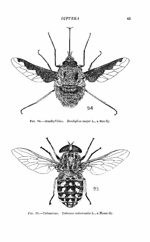
DIPTERA 45
FIG. 94.-Bombyliidae. Bombylius major L., a Bee-fly .
.FIG. 95.-Tabanidae . Tabanus autumnalis L., a Horse-fly.
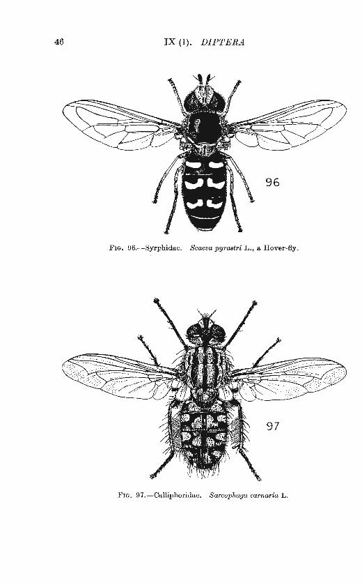
46 IX (1). DIPTERA
FIG. 96.-Syrphidae. Scaeva pyrastri L., a Hover-fly.
F IG. 97. -Calliphoridae. Sarcophaga carnaria L.
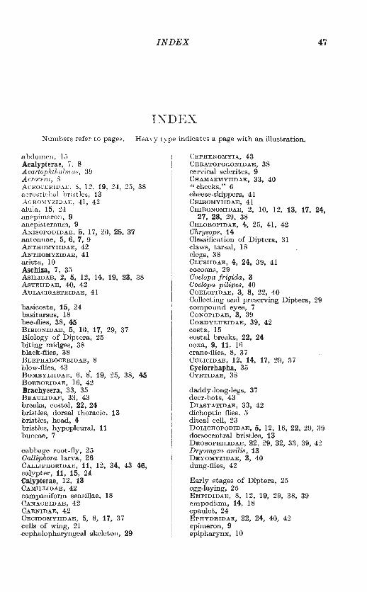
INDEX 47
I~DEX
Numbers refer to pages. Hea\-y t.yp e indicates a page with an illustration.
a bdomen, 15 Acalypterae, 7, 8 Acartophthalnw.s, 39 A crocera, 8 AcRoCERID.U:, 8, 12, 19, 2±, 25, 38 acrost ichal br istles , 13 _-\GR01IYZID"-E, 41, 42 alula, 15, 24 anepimeron, 9 anepisternum, 9 ANISOPODIDAE, 5, 17, 20, 25, 37 antennae, 5, 6, 7, 9 ANTHOMYIIDAE, 42 ANTHOMYZIDAE, 41 arista, 10 Aschiza, 7, 35 ASILIDAE, 2, 5, 12, 14, 19, 23, 38 AsTEIIDAE, 40, 42 AULACIGASTRIDAE, 41
basicosta, 15, 24 basitarsus, 18 bee-flies, 38, 45 BIBIONIDAE, 5, 10, 17, 29, 37 Biology of Diptera, 25 biting midges, 38 black-flies, 38 BLEPHAROCERIDAE, 8 blow -flies, 43 BOMBYLIIDAE, 6, 8', 19, 25, 38, 45 BORBORIDAE, 16, 42 Brachycera, 33, 35 BRAU LIDAE, 33, 43 breaks, costal, 22, 24 bristles, dorsal thoracic, 13 bristles, head, 4 bristles, hypopleural, 11 buccae, 7
cabbage root-fly, 25 Calliphora larva, 26 CALLIPHORIDAE, 11, 12, 34, 43 46, calypter, 11, 15, 24 ·Calypterae, 12, 13 CAMILLIDAE, 42 campaniform sensillae, 18 CANACEIDAE, 42 CARNIDAE, 42 CECIDOMYIIDAE, 5, 8, 17, 37 cells of wing, 21 ·Cephalopharyngeal skeleton, 29
C EPHENOMYIA, 43 CERATOPOGONIDAE, 38 cervical sclerites, 9 CHAMAEMYIIDAE, 33, 40 "cheeks," 6 cheese-skippers, 41 CHIROMYIIDAE, 41 CmRONOMIDAE, 2, 10, 12, 13, 17, 24,
27, 28, 29, 38 CHLOROPIDAE, 4, 25, 41, 42 Chrysops, 14 Classification of Diptera, 31 claws, tarsal, 18 clegs, 38 CLUSIIDAE, 4, 24, 39, 41 cocoons, 29 Coelopa frigida, 3 Coelopa pilipes, 40 COELOPIDAE, 3, 8, 22, 40 Collecting and preserving Diptera, 29 compound eyes, 7 CONOPIDAE, 3, 39 CORDYLURIDAE, 39, 42 costa, 15 costal breaks, 22, 24 coxa, 9, 11, 16 crane-flies, 8, 37 CULICIDAE, 12, 14, 17, 29, 37 Cyclorrhapha, 35 CYRTIDAE, 38
daddy-long-legs, 37 deer-hots, 43 DIASTATIDAE, 33, 42 dichoptic flies, 5 discal cell, 23 DOLICHOPODIDAE, 5, 12, 16, 22, 29, 3!) dorsocentral bristles, 13 DROSOPHILIDAE, 22, 29, 32, 33, 39, 42 Dryomyza anilis, 13 DRYOMYZIDAE, 3, 40 dung-flies, 42
E arly st ages of Diptera, 25 egg-laying, 25 EMPIDIDAE, 8, 12, 19, 29, 38, 39 empodium, 14, 18 epaulet, 24 EPHYDRIDAE, 22, 24, 40, 42 epimeron, 9 epipharynx, 10
48 IX (1). DIPTERA
episternum, 9 epistoma, 6 eye-facets, 7 eyes, 5 eyes, colour of, 8 eyes, compound, 7, 8 eyes, pubescent, 8-9
face, 6 facets, eye, 7 Families, Key to, 37 Families, List of, 33 Fannia, 29 femur, 14 flagellum, 10 frit-fly, 25 frons, 5, 6 frontal lunule, 4, 7 " frontal " suture, 4, 7 fruit-flies (DROSOPHILIDAE), 32, 42 fruit-flies (TRYPETIDAE), 36, 41 fungus-gnats, 37
gall-midges, 37 GASTEROPHILIDAE, 33, 39 Gasterophilus, 25, 43 genitalia, 15 [Glossina], 12 gnats, 37 gout-fly, 25
Haematobia, 12 halteres, 2, 11, 18 head, 3, 4, 5 HELOMYZIDAE, 41 HIPPOBOSCIDAE, 10, 12, 32, 34, 43 holoptic flies, 5 Homalocephala, 40 horse bot-flies, 39 horse-flies, 38, 45 house-flies, 42 hover-flies, 39, 46 humeral bristles, 13 humeral callus, 13 HYBOTINAE, 38 Hypoderma, 25 hypopharynx, 10, 12 hypopleural bristles, 11
intra-alar bristles, 13
"jowls," 7
katepimeron, 9 katepisternum, 9 kelp-flies, 40 Key to Families, 37
tabella, 10 labium, 10, 12 labrum, 10 large fruit-flies (TRYPETIDAE), 41 larvae, 25, 26, 27 laterotergite, 9
LAUXANIIDAE, 40 legs, 14, 16 LEPTIDAE, 38 LESTREMIINAE, 37 List of Families, 33 LONCHAEIDAE, 33, 41 LONCHOPTERIDAE, 22, 39 lunule, frontal, 7 Lyperosia, 12
m andibles, 10 march-flies, 37 maxillae, 10 maxillary palpt, 12 Mediterranean fruit-fly, 25 MEGAMERINIDAE, 40 Melophagus, 32 meron, 9 mesopleuron, 9, 11 mesothorax, 2, 12 m etanotum, 9, 11, 14 "metatarsus," 14, 18 metathorax, 18 MrcROPEZIDAE, 40 MILICHIDAE, 42 mosquitoes, 8, 10, 37 moth-flies, 37 mounting Diptera, 30, 31 mouth-hooks, 29 mouthparts, 10 MusciDAE, 12, 25, 26, 33, 42 MYCETOPHILIDAE, 13, 17, 29, 37
Nematocera, 33, 35 NEOTTIOPHILIDAE, 41 nest-flies, 41 non-biting midges, 38 notop1eural bristles, 13 notopleuron, 12, 13 NYCTERIBIIDAE, 12, 43
occiput, 6 ocelli, 4, 6, 8 0DINIIDAE, 42 0ESTRIDAE, 43 Oncodes, 12 0POMYZIDAE, 42 Orthorrhapha, 31 Orygma, 8, 40 0TITIDAE, 40
p ALLOPTERIDAE, 33 palpi, maxillary, 12 parafacials, 4, 7 parategula, 24 Parhydroptera, 22, 40 pedicel, lO PERISCELIDAE, 40 PHORIDAE, 22, 27, 29, 30, 35, 38. PIOPHILIDAE, 27, 32, 41 PIPUNCULIDAE, 22, 39 plates of head, 4 PLATYPEZIDAE, 22, 38, 39 PLATYSTOMIDAE, 39, 40
pleural suture, 9 pleurotergite, 9 Podonomus, 38 postalar bristles, 13 posterior callus, 13 posthumeral bristles, 13 postpedicel, 10 postscutulum (postnotum), 9, 11, 14 prealar bristles, 13 prescutum, 9, 11, 12, 13 prestomal teeth, 12 pretarsus, 18 proboscis, 10, ll pronotum, 9, 14 Protoplasa, 16 pseudopods, 29 pseudotracheae, ll, 12 PsrLIDAE, 24, 42 PsYCHODIDAE,17, 20,25,30, 37 pteropleuron, 9, 11 ptilinal suture, 4, 7 ptilinum, 7 pulvilli, 14, 18 pupa,26,27 puparium, 7, 27, 29
radial venation, 23 References, 43 RHAGIONIDAE, 5, 10, 19, 38 RHYPHIDAE, 37 robber-flies, 38 rotation of genitalia, 16
scape, 10 SCATOPSIDAE, 10, 37 SCENOPINIDAE, 19, 38 Schizophora, 7, 35 SCIOMYZIDAE, 40 sclerites, cervical, 9 scutellum, 9, 11, 12, 13 scutum, 9, 11, 12, 13 Selaclwps, 41 SEPSIDAE, 8, 27, 40 shore-flies, 42 SIMULIIDAE, 5, 10, 17, 38 small fruit-flies (DROSOPHILIDAE), 42 spiracle, metathoracic, 8, 11 spiracle, prothoracic, 11 spurs, tibial, 14, 18 squamae, 11, 15, 24 stable-fly, 12 staging Diptera, 31
2
INDEX
sternopleuron, 9, 11 Stomoxys, 12, 26 STRATIOMYIIDAE, 5, 10, 19, 38 Structure of Diptera, 2 style, antenna!, 10 subepaulet, 24 supra-alar bristles, 13 suture, "frontal," 4, 7 suture, pleural, 9 suture, ptilinal, 4, 7
49
suture, transverse, 6, 11, 12, 13 SYRPHIDAE, 6, 8, 20, 22, 25, 29, 39,
46
TABANIDAE, 3, 5, 10, ll, 14, 15, 19, 24, 26, 38, 45
TACHINIDAE, 8, 14,43, 46 [TANYDERIDAE), 16, 20 TANYPEZIDAE, 40 tarsus, 14, 16 teeth, prestomal, 12 tegula, 15, 24 TETHINIDAE, 42 THAUMALEIDAE, 8, 10, 37 THEREVIDAE, 19, 23, 38 thorax, 6, 9, 11, 12, 13 THYREOPHORIDAE, 24, 41, 42 tibia, 14, 18 TIPULIDAE, 3, 5, 9, 12, 13, 17, 00, 25,
37 transverse suture, 6, 11, 12, 13 TRICHOCERIDAE, 12 TRICHOSCELIDAE, 24, 41 trochanter, 16 TRYPETIDAE, 4, 22, 24, 25, 36, 41 [tse-tse fly], 12
ULIDIIDAE, 40
veins, names of, 20-21 vertex, 6 vibrissae, 4, 7
window-flies, 38 window midges, 37 wing-cells, 21 wing-veins, 20-21 wings, 15, 16, 17, 18, 19, 22, 24 winter-gnats, 37
Xylomyia,, 5, 10 Xylophagus, 5, 10, 38
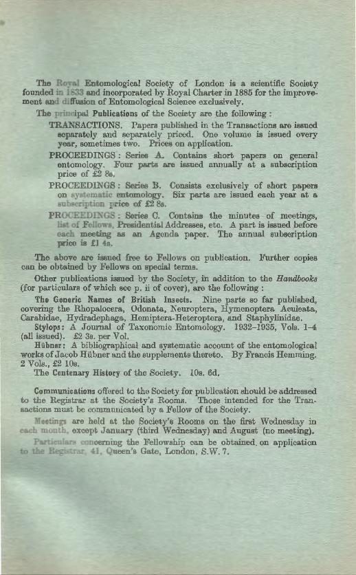
The Ro. Entomological Society of London is a scientific Society founded in 1833 and incorporated by Royal Charter in 1885 for the improvement and diflusion of Entomological Science exclusively.
The pr· ·ipal Publications of the Society are the following: TRA~-SACTIONS. Papers published in the Transactions are issued
separately and separately priced. One volume is issued every year, sometimes two. Prices on application.
PROCEEDINGS : Series A. Contains short papers on general entomology. F our parts are issued annually at a subscription price of £2 8 .
PROCEEDINGS : Series B. Consists exclusively of short papers on syst~matic entomology. Six parts are issued each year at a su cription pr ice of £2 Ss .
.PROCEEDINGS : Series C. Contains the minutes of meetings, · . of Fello s, Presidential Addresses, etc. A part is issued before
eaeh mee ing as an Agenda paper. The annual subscription price is -I 4s.
The above are issued free to Fellows on publication. Further copies can be obtained by Fellows on special terms.
Other publications issued by the Society, in addition to the Handbooks (for particulars of which see p. ii of cover), are the following:
The Generic Names of British Insects. Nine parts so far published, covering the Rhopalocera, Odonata, Neuroptera, Hymenoptera Aculeata, Carabidae, Hydradephaga, Hemiptera-Heteroptera, and Staphylinidae.
Stylops: A Journal of Taxonomic Entomology. 1932-1935, Vols. l-4 (all issued). £2 3s. per Vol.
Hiibner: A bibliographical and systematic account of the entomological works of Jacob Huhner and the supplements thereto. By Francis Hemming. 2 Vols., £2 !Os.
The Centenary History of the Society. !Os. 6d.
Communications offered to the Society for publication should be addressed to the Registrar at the Society's Rooms. Those intended for the Transactions must be communicated by a Fellow of the Society.
Meetings are held at the Society's Rooms on the first Wednesday in e eh month, except January (third Wednesday) and August (no meeting).
P concerning the Fellowship can be obtained_ on application 41, Queen's Gate, London, S.W. 7.
HANDBOOKS FOR THE IDENTIFICATION OF BRITISH INSECTS.
PARTS NOW AVA.ILA.BLE.
I. Part 5 Dermaptera and Ortlwptera. By W. D. Hincks. 20 pp. 3s. 6d.
, 6 Plecoptera. By D. E. Kimmins.
, 9 Ephemeroptera. By D. E. Kimmins.
, 10 Odonata. By F. C. Fraser . . IV. , 3 Goleoptera: Hydradephaga. By F. Balfour-
Browne.
V. , 7 Goleoptera : Goccinellidae and Sphindidae. By R. D. Pope.
, 9. Goleoptera: Lagriidae to Meloidae. By F. D. Buck.
18 pp. 3s. 6d.
18 pp. 3s. 6d.
48 pp. 7s. 6d.
34: pp. 6s. Od.
12 PP· 2s. 6d.
, 12 Goleoptera: Gerambycidae. By E. A. J. Duffy. 18 pp; 3s. 6d;
, 15 Goleoptera: Scolytidae and Platypodidae. By E. A. J. Duffy. 18 pp. 3s. 6d.
VI. , 2(a) Hymenoptera: Symphyta (part). By R. B. Benson. 47 pp. lOs. Od.
, 2(b) Hymenoptera: Symphyta (contd.) Benson.
By R. B.
IX. , 1 Diptera : Introdmtion and Key to Families.
81 pp: 15s. Od.
By H. Oldroyd. 49 pp. 7s. 6d.
, 2 Diptera: Nematocera (part). By R. L. Coe, Paul Freeman, P. F. Mattingly. 216 pp. 20s. Od.
X. , 1 Diptera: Syrphidae. By R. Li Coe. 98 pp. 17s. 6d.
Made and Printed in Great Britain by ADLAltD AND SON, LTD., SOUTH STREET, DORKING, SURREY,