role of ypeb in cortex hydrolysis during germination of bacillus anthracis spores
TRANSCRIPT
1
The role of YpeB in cortex hydrolysis during germination of Bacillus anthracis spores 1
2
3
Casey B. Bernhards and David L. Popham# 4
5
Department of Biological Sciences, Virginia Tech, Blacksburg, VA 24061 6
7
Running Head: Bacillus anthracis YpeB affects spore germination 8
9
# Address correspondence to David L. Popham, [email protected]. 10
11
JB Accepts, published online ahead of print on 14 July 2014J. Bacteriol. doi:10.1128/JB.01899-14Copyright © 2014, American Society for Microbiology. All Rights Reserved.
2
ABSTRACT 12
The infectious agent of the disease anthrax is the spore of Bacillus anthracis. Bacterial 13
spores are extremely resistant to environmental stresses, which greatly hinders spore 14
decontamination efforts. The spore cortex, a thick layer of modified peptidoglycan, contributes 15
to spore dormancy and resistance by maintaining the low water content of the spore core. The 16
cortex is degraded by germination-specific lytic enzymes (GSLEs) during spore germination, 17
rendering the cells vulnerable to common disinfection techniques. This study investigates the 18
relationship between SleB, a GSLE in B. anthracis, and YpeB, a protein necessary for SleB 19
stability and function. Results indicate that sleB and ypeB spores exhibit similar germination 20
phenotypes, and the two proteins have a strict co-dependency for their incorporation into the 21
dormant spore. In the absence of their partner protein, SleB and YpeB are proteolytically 22
degraded soon after expression during sporulation, rather than escaping the developing spore. 23
The three PepSY domains of YpeB were examined for their role in the interaction with SleB. 24
YpeB truncation mutants illustrate the necessity of a region beyond the first PepSY domain for 25
SleB stability. Furthermore, site-directed mutagenesis of highly conserved residues within the 26
PepSY domains resulted in germination defects corresponding to reduced levels of both SleB 27
and YpeB present in the mutant spores. These results identify residues involved in the stability 28
of both proteins and reiterate their co-dependent relationship. By studying GSLEs and 29
interacting proteins, it is hoped that GSLEs will be targets to efficiently activate spore 30
germination and facilitate spore cleanup. 31
32
3
INTRODUCTION 33
Bacterial spores from the Bacillus and Clostridium genera are metabolically dormant and 34
known for their extreme resistance to heat, desiccation, UV radiation, chemicals, and other 35
insults (1-3). These resistance properties allow spores to survive for extended periods of time in 36
the environment and have made eradication from contaminated sites incredibly difficult (4). 37
Spore dormancy and wet heat resistance are largely dependent on spore core dehydration, which 38
is maintained by a thick layer of modified peptidoglycan (PG) known as the cortex (2, 5). While 39
vegetative cell wall PG consists of alternating N-acetylglucosamine (NAG) and N-acetylmuramic 40
acid (NAM) sugars, approximately 50% of NAM residues in the cortex are converted to 41
muramic--lactam, while an additional portion of the NAM side chains are generally cleaved to a 42
single L-alanine (6-11). 43
The spore form of Bacillus anthracis is the etiological agent for all types of anthrax 44
infections: inhalational, gastrointestinal, cutaneous, and the newest described form – injectional 45
anthrax (12, 13). Upon sensing the availability of nutrients, such as when a spore enters a 46
suitable host, spore germination is triggered, causing a chain of events that ultimately result in a 47
vegetative cell capable of producing deadly toxins (1, 12). After germinant contact with 48
receptors at the spore inner membrane, spores release their large pool of Ca2+
-DPA stored in the 49
core, which becomes partially rehydrated through an influx of water (1). This is followed by 50
cortex depolymerization, allowing the spore core to rehydrate to levels necessary for metabolism 51
to resume (14). Spore germination concomitantly results in the loss of resistance properties; 52
thus, triggering this process at a high efficiency is a potentially attractive approach for spore 53
decontamination. 54
Cortex degradation during spore germination is accomplished by germination-specific 55
lytic enzymes (GSLEs) already present within the dormant spore (15, 16). These enzymes 56
exhibit specificity for PG containing the muramic--lactam modification, ensuring that only the 57
4
cortex PG is broken down (17-21). Bacillus anthracis contains the four GSLEs SleB, CwlJ1, 58
CwlJ2, and SleL, where SleB and CwlJ1 are partially redundant enzymes responsible for the 59
majority of cortex hydrolysis (22-25). SleB or CwlJ1 alone is sufficient for cortex hydrolysis, 60
and the absence of both proteins results in spores that are unable to degrade the cortex and 61
complete germination (23-25). Identical roles for SleB and CwlJ have been demonstrated in 62
Bacillus subtilis (26-28) and Bacillus megaterium (29). CwlJ2, a homolog of CwlJ1, appears to 63
play a minor role during B. anthracis spore germination, at best, while SleL further breaks down 64
PG fragments first generated by SleB and/or CwlJ1 (20, 22-25). 65
The arrangement of sleB upstream of ypeB in an operon is highly conserved across 66
Bacillus species, and in a few Clostridiales species possessing sleB (16, 23, 30-32). In the 67
Bacillus species examined, SleB and YpeB are expressed during sporulation from the forespore 68
under the control of G, after which they are translocated across the inner membrane by way of 69
their N-terminal signal sequences (28, 30, 33, 34). Unlike the YpeB signal sequence, which is 70
not predicted to be cleaved, the SleB signal sequence is removed during sporulation and SleB is 71
present in its mature form within the dormant spore (28, 30, 33, 35). The uncleaved signal 72
sequence likely anchors YpeB to the inner membrane, which is supported by studies in B. 73
subtilis that have found both SleB and YpeB associated with the inner membrane of the spore 74
(26). The same study, as well as work in B. cereus, has placed SleB and YpeB at a second 75
location near the outer region of the cortex (26, 30). Not only do SleB and YpeB appear to 76
localize within the same regions of the spore, but it was demonstrated in B. subtilis that YpeB is 77
required for SleB activity and for stable incorporation of SleB into the spore (26, 34). 78
Additionally, the relationship appears to be mutual, where SleB is also needed to stabilize YpeB 79
(36). 80
Compared with YpeB, far more is known about SleB structure and function. At the N-81
terminus of the mature protein, SleB has a PG-binding domain (pfam01471), while the C-82

5
terminus contains a catalytic domain (pfam07486) (21, 33, 37, 38). As anticipated, the N-83
terminal domain plays the dominant role in PG-binding; however, the C-terminal domain 84
appears to be responsible for both SleB lytic transglycosylase activity on cortex PG and its 85
specificity for muramic--lactam (21, 37, 39). Mutational analysis and crystal structure 86
determination of the SleB C-terminal domain in B. anthracis (40) and B. cereus (39) revealed a 87
conserved catalytic glutamate residue and unique substrate-binding cleft thought to mediate 88
specificity for cortex PG. Apart from its signal sequence, the only recognizable features of 89
YpeB are the three putative PepSY domains positioned in the C-terminal 60% of the protein 90
(41). These domains are best characterized in the M4 family of metallopeptidases, where the 91
PepSY domains serve as intramolecular inhibitors of protease activity until the protease is 92
secreted out of the cell (41). Like a large number of other proteins containing PepSY domains, 93
YpeB does not show homology with proteases; thus, the function of the PepSY domains is 94
unknown in YpeB and the other proteins comprising this group (41). 95
The current study investigates the relationship between SleB and YpeB in B. anthracis 96
and demonstrates the contribution of the PepSY domains to this interaction. The data solidify 97
the co-dependent nature of this relationship and reveal that SleB and YpeB are rapidly degraded 98
following expression in the absence of the other protein. The C-terminus of YpeB containing the 99
PepSY domains is critical for SleB stability and function, and certain highly conserved residues 100
within the PepSY domains are important for the stability of both proteins. By understanding 101
GSLEs and regulators of their activity, it may be possible to trigger germination at the stage of 102
cortex hydrolysis, resulting in cells that are readily killed. 103
104
MATERIALS AND METHODS 105
Bacterial strains and general growth conditions. Strains and plasmids used are listed 106
in Table 1. All B. anthracis strains in this study are derived from the Sterne strain 34F2 and 107
6
were grown at 37C in brain heart infusion (BHI; Difco) with 5 g/ml erythromycin or 10 g/ml 108
tetracycline, where appropriate. B. anthracis strains maintaining pBKJ236-derived plasmids 109
extrachromosomally were incubated at 25C. Following plasmid integration into the 110
chromosome (at 42C in most instances), these strains were subsequently grown at 37C. 111
Escherichia coli strains were grown in LB at 37C with 500 μg/ml erythromycin or 100 μg/ml 112
ampicillin for strains involved in plasmid propagation, or 30 g/ml chloramphenicol and 50 113
g/ml ampicillin for strains used in protein overexpression. 114
Mutant construction. The sequences of all primers used during plasmid construction 115
are listed in Table S1. All plasmids created were verified by DNA sequencing. To create a 116
ypeB strain, ypeB and approximately 500-bp flanking each side of the gene were PCR 117
amplified from the B. anthracis chromosome. The PCR product was inserted into the vector 118
pBKJ236 (42) by digesting with the restriction enzymes SacII and NotI and ligating the DNA to 119
create pDPV388. Inverse PCR of the plasmid using primers with BglI restriction sites at the 3 120
ends resulted in a linear PCR product with the majority of ypeB deleted, leaving only three 121
codons from each end of the gene. Subsequent BglI digestion and ligation of the PCR product 122
formed pDPV392. This plasmid containing the ypeB deletion was introduced into B. anthracis 123
using the markerless gene replacement strategy as previously described (42). Gene deletion was 124
verified by PCR amplification and sequencing. 125
The sleB complementation strain published previously (DPBa57) contains an 126
extrachromosomally-maintained pBKJ236 derivative with full-length sleB (pDPV346) (23). In 127
order to achieve more complete complementation of the deletion phenotype by ensuring efficient 128
plasmid partitioning into the forespore during spore formation, pDPV346 was integrated into the 129
sleB chromosome by shifting the temperature to 42C. Plasmid integration through 130
homologous recombination just upstream of sleB in the chromosome was verified with PCR, 131
7
and this new sleB complementation strain was designated DPBa134. To create a ypeB 132
complementation plasmid that includes the native promoter, PCR was performed using the sleB 133
chromosome (23) as template. The resulting PCR product contained the promoter region for the 134
sleB operon, followed by sleB, ypeB, and approximately 500-bp downstream of ypeB. The 135
DNA fragment was cloned into pBKJ236 by digesting with NotI and ligating to form pDPV416. 136
This ypeB complementation plasmid was introduced into the ypeB strain of B. anthracis via 137
conjugation as done in the initial stages of the markerless gene replacement procedure (42). 138
Subsequent plasmid integration within the 500-bp homologous region downstream of ypeB in 139
the chromosome was achieved by shifting the temperature to 42C and was verified by PCR. 140
YpeB truncation and internal deletion mutations were designed using InterPro (43) to 141
predict the locations of PepSY domains, and the Protean application of Lasergene 10.0 142
(DNAStar) to choose truncation/deletion sites based on high residue surface probability and 143
residues not predicted to disrupt protein secondary structure. For the truncation mutations, 144
inverse PCR of the ypeB complementation plasmid was performed using sets of two primers. 145
The tails of the forward primers contained codons encoding a hexahistidine tag followed by a 146
stop codon and a BglII restriction enzyme recognition sequence. The tail of the reverse primer 147
used to create all the truncation mutants also had a BglII site added. The linear PCR products 148
from inverse PCR reactions were digested with BglII and ligated to recircularize the plasmids, 149
generating pDPV421 through pDPV424. The internal deletion mutations were constructed using 150
overlap extension PCR (44), followed by restriction-free cloning (45) to insert the PCR products 151
into the plasmid encoding full-length YpeB with a C-terminal His6-tag (pDPV424). The YpeB 152
truncation and internal deletion plasmids were moved into the ypeB strain of B. anthracis and 153
integrated into the chromosome as described above. The 500-bp homologous region following 154
ypeB in pDPV416 was removed during inverse PCR to create pDPV421-424; thus, these 155
8
plasmids inserted into the ypeB chromosome through homologous recombination upstream of 156
sleB, as confirmed by PCR. 157
In order to predict residues of the YpeB PepSY domains that might be important for 158
protein-protein interactions, the PepSY domain sequences from B. anthracis YpeB were aligned 159
with a PepSY domain consensus sequence (46), other PepSY domain sequences (41, 46, 47), and 160
the YpeB PepSY domain sequences from other Bacillus species using Clustal W sequence 161
alignment software (48). Based on these alignments, highly conserved residues were identified 162
as targets for mutagenesis. Site-directed mutagenesis by overlap extension PCR (44) was used to 163
create point mutations in ypeB. The resulting PCR products were cloned into the ypeB 164
complementation plasmid pDPV416 using restriction-free cloning (45). Successful plasmid 165
construction was verified by screening for the gain or loss of a restriction site in ypeB that was 166
designed as part of the mutagenic primers, followed by DNA sequencing. Plasmids were 167
mobilized into the ypeB strain of B. anthracis and integrated into the chromosome downstream 168
of ypeB as described above for the ypeB complementation plasmid. 169
Spore preparation and decoating. B. anthracis strains were sporulated in modified G 170
broth (49) with appropriate antibiotic at 37C with aeration. After 3 to 4 days, dormant spores 171
were harvested by centrifugation and repeatedly washed with deionized water. Any remaining 172
vegetative cells were heat killed at 65C for 25 min. Spores were further purified with a 50% 173
sodium diatrizoate (Sigma) gradient as described previously (50). Decoated spores were 174
prepared by suspending up to 30 OD units of spores in 1 ml decoating solution (50 mM Tris-HCl 175
pH 8, 8 M urea, 1% SDS, 50 mM DTT). The spores were incubated at 37C for 1 hour and then 176
centrifuged at 8,000 x g for 1 min to remove the decoating solution. The decoating procedure 177
was repeated a second time, followed by five washes with 1 ml deionized water at room 178
9
temperature. Untreated (native) and decoated spores were stored in deionized water at 4C until 179
analysis. 180
Spore and sporangia sample preparation for western blotting. Following spore 181
purification, 7.5 to 10 OD units of dormant, native spores were pelleted, frozen at -80C, and 182
lyophilized. Dried spores were mechanically disrupted with 100 mg 0.1 mm glass beads and 20 183
pulses of 30 sec each at 4,200 rpm using a Wig-L-Bug bead beater. Samples were placed on ice 184
between pulses. Proteins were extracted from the broken material by adding 10 l/OD unit of 185
1X sample loading buffer (62.5 mM Tris-HCl pH 6.8, 2% SDS, 10% glycerol, 5% -186
mercaptoethanol, 0.05% bromophenol blue) and heating to 100°C for 5 min. Extracts were 187
centrifuging at 15,800 x g for 1 min, and volumes of supernatants derived from equal starting 188
OD units of spores were used for western blot analysis. 189
Strains grown in modified G broth with appropriate antibiotic were monitored 190
spectrophotometrically until the OD600 indicated cells had entered stationary phase. This time 191
point, termed t0, coincides with the initiation of sporulation, and t2 through t6 designate 2 through 192
6 hours, respectively, past t0. At hours t2 through t6 of sporulation, 10 ml sporangia samples 193
were collected and centrifuged at 10,000 x g for 10 min at 4C. Pellets were resuspended in 1 ml 194
8 mM NaPO4 pH 7.0 and centrifuged at 15,800 x g for 1 min. The resulting pellets were flash 195
frozen in liquid N2 and lyophilized. Lyophilized sporangia were broken with glass beads and 196
proteins extracted as described above, except that 100 l of 1X sample loading buffer was used. 197
Extract supernatants were used for western blotting using the average OD600 measured for a 198
particular strain from t2 through t6 to adjust sample loading between strains. 199
Germination assays. To assess the rate of spore germination and outgrowth, decoated 200
spores were heat activated at 70C for 30 min and quenched on ice for 5 min. Heat-activated 201
spores were diluted to an OD600 of 0.2 in liquid BHI at 37C to initiate germination, and the 202
10
change in OD600 was measured over time. For germination efficiency assays, decoated spores at 203
an OD600 of 0.2 were heat activated at 70C for 20 min and quenched on ice. Heat activated 204
spores were serially diluted in deionized water, plated on BHI medium without antibiotics, and 205
incubated at 37C overnight. Colonies were counted to determine CFU/OD unit values. 206
Unpaired, two-tailed Student’s t-tests with unequal variance were used for statistical analyses of 207
germination assays. 208
Protein expression and purification. The ypeB gene lacking the first 20 codons was 209
amplified by PCR and inserted into a modified version of pDEST-HisMBP (51) containing a 210
tobacco etch virus (TEV) cleavage site (pDEST-HisMBP-T) using restriction-free cloning (45). 211
The resulting plasmid encoded an N-terminal His6-tagged maltose binding protein (MBP) and 212
YpeB21-446, separated by a TEV cleavage site. Successful plasmid construction was verified by 213
sequencing. The His6-MBP-YpeB21-446 fusion protein was overexpressed in Escherichia coli 214
BL21 (DE3)(pLysS Cmr)(Novagen) grown at 37C until the OD600 reached 1.0, at which point 215
isopropyl--D-thiogalactopyranoside was added to a final concentration of 1 mM and the culture 216
was incubated at 10C for an additional 16 hr. Cells were harvested by centrifugation at 10,000 217
x g for 10 min at 4C, and the pellet was resuspended in 5 ml/g Buffer A (50 mM NaCl, 50 mM 218
Tris-HCl pH 7.5, 5% glycerol, 30 mM imidazole). Resuspended cells were lysed by sonication 219
for 15 min and then centrifuged at 117,000 x g for 1 hr at 4C. Fusion protein in the soluble 220
fraction was purified using a Ni-Sepharose HisTrap HP affinity column (GE Healthcare) 221
equilibrated with Buffer A. Protein was eluted with a linear gradient of 30 to 500 mM imidazole 222
in Buffer A, and elution fractions were dialyzed in Buffer A. Fusion protein was digested with 1 223
mg His6-tagged TEV (S219V) protease (52) per 7 mg fusion protein at 15C for 16 hr, and the 224
mixture was centrifuged at 117,000 x g for 20 min at 4C to remove any precipitated protein. 225
Cleavage of the fusion protein was verified by SDS-PAGE. YpeB21-446 was purified from His6-226
11
MBP and His6-TEV using a second Ni-Sepharose HisTrap HP affinity column as described 227
above. SleB overexpression and purification was performed as described previously (21). 228
Antibody preparation and western blot analysis. Polyclonal anti-SleB and anti-YpeB 229
antibodies were raised in rabbits (Open Biosystems) using purified SleB33-253 (21) or YpeB21-446. 230
SleB, YpeB, and derivatives were detected in western blots on Amersham Hybond-P (PVDF) 231
membranes (GE Healthcare) using BM Blue POD Substrate, Precipitating (Roche) for 232
colorimetric detection. Primary anti-SleB and anti-YpeB antibodies were generally used at 233
1:1,000 and 1:3,000 dilutions, respectively. Secondary goat anti-rabbit-HRP antibodies (Bio-234
Rad) were used at a 1:200,000 dilution. Western blot quantification was performed using Image 235
Lab Software (Bio-Rad). While efforts were made to ensure total protein load was identical for 236
each strain, there is the inherent possibility of slight variations stemming from unequal protein 237
extraction between samples and unequal sample loading. To account for experimental error, 238
relative values were normalized using a highly reproducible non-YpeB background band 239
detected in all strains, including the ypeB strain. Similarly, SleB band intensities were 240
normalized using a background band present on anti-SleB immunoblots. 241
242
RESULTS 243
Germination of ypeB mutant spores. As previously reported by Heffron et al., a 244
homolog of the B. subtilis ypeB gene (57% identity/77% similarity) exists as part of a putative 245
tricistronic operon at locus BAS2561 in the B. anthracis chromosome (23). The ypeB homolog 246
is the second gene in the operon, preceded by sleB (BAS2562) and followed by the open reading 247
frame BAS2560 (23). BAS2560 shows homology to the B. subtilis genes encoding the 248
lipoproteins YlaJ and YhcN, where YlaJ is an uncharacterized spore protein and YhcN plays an 249
unknown role in spore germination or outgrowth (23, 53, 54). In this study, a deletion of the 250
ypeB gene (BAS2561) in the B. anthracis chromosome was made using the markerless gene 251
12
replacement strategy (42). The effects of this ypeB deletion, and of an sleB deletion, are not due 252
to polar effects on expression of BAS2560, because: A) a BAS2560 deletion results in no change 253
in spore germination, specifically in SleB activity (data not shown), B) as shown below, all 254
effects of a ypeB deletion can be complemented by the ypeB gene alone, and C) as shown below 255
and in Heffron et al. (23), all effects of an sleB deletion can be complemented by sleB alone. 256
In assays in which purified spores were germinated in BHI and the change in OD600 257
measured over time, the germination phenotype of ypeB spores was essentially identical to that 258
of sleB spores (23). This could be seen in assays using native spores, in which ypeB and 259
sleB spores produced a shallower germination curve, indicative of a slightly less efficient 260
germination response, and also showed a slower rate of outgrowth (data not shown). The 261
germination defect in ypeB and sleB spores, alike, was more pronounced in assays using 262
decoated spores, where the other GSLEs localized to the outer periphery of the spore are 263
removed or inactivated, and SleB alone is responsible for cortex hydrolysis during germination 264
(Fig. 1A). Both ypeB and sleB decoated spores proceeded through stage one of germination 265
similar to wild-type, where the initial OD decreases as the spores uptake water and release their 266
large deposit of Ca2+
-DPA. Decoated wild-type spores continued to lose nearly half of their 267
initial OD as the cortex peptidoglycan was degraded during stage two of germination, followed 268
by an increase in OD as germination was completed and outgrowth into vegetative cells began. 269
Conversely, germination was arrested for ypeB and sleB decoated spores at the stage of cortex 270
hydrolysis, where ypeB and sleB spores never lost more than 34 percent of their initial OD 271
and did not proceed to outgrowth (Fig. 1A). This germination deficiency is also illustrated in 272
spore plating efficiency assays, where ypeB and sleB decoated spores showed greater than a 273
104-fold reduction in their ability to germinate and form colonies on rich medium, relative to 274
wild-type spores (Table 2). A wild-type germination phenotype was achieved in both 275
13
germination rate and plating efficiency assays using spores from a ypeB complementation strain 276
(DPBa113, ypeB+), in which ypeB, provided on a plasmid downstream of its native promoter, 277
was integrated into the ypeB chromosome. HPLC analyses of muropeptides collected during 278
spore germination revealed the complete absence of SleB-specific lytic transglycosylase products 279
identified by Heffron et al. (23) in both sleB and ypeB spores, with restoration of these peaks 280
during germination of ypeB+ spores (data not shown). Consistent with the results observed from 281
a ypeB deletion in B. subtilis (34, 36), these results indicate YpeB is necessary for SleB lytic 282
activity on spore cortex peptidoglycan in B. anthracis. 283
Stability of SleB and YpeB in spores and sporangia. Not only is YpeB needed for 284
SleB activity, but western blots of ypeB spore extracts demonstrated YpeB is also required for 285
SleB incorporation into the spore (Fig. 2). SleB was essentially not detected in ypeB spore 286
extracts, and this could be complemented. As in B. subtilis (36), this relationship is mutual, 287
where SleB was also needed for the stable incorporation of YpeB into the spore (Fig. 2). 288
Immunoblots of sleB spore extracts revealed significantly diminished levels of YpeB when 289
SleB was absent, which could also be complemented. Miniscule levels of SleB and YpeB 290
detected in western blots of ypeB and sleB spores, respectively, could be attributed either to 291
the expression of the proteins during spore formation followed by rapid proteolysis in the 292
absence of their stabilizing partner, or to escape of the proteins from the developing spore. The 293
former proposal is supported by western blot analyses of whole sporangia samples taken 294
throughout sporulation (Fig. 3). In wild-type and ypeB+ strains, significant amounts of SleB and 295
YpeB were detected beginning 3 hr after the initiation of sporulation (t3)(Fig. 3A and B). This is 296
consistent with previous -galactosidase activity assays performed in B. anthracis which show 297
sleB transcription reaches its peak near t3.5 (23). During spore formation in sleB and ypeB 298
strains, some YpeB and SleB, respectively, could be seen at t3 and t4, but were not strongly 299

14
detected thereafter (Fig. 3A and B). The expression of YpeB at t3 and t4 coincided with the 300
production of large quantities of two YpeB-specific degradation products in sleB sporangia 301
(and to a lesser degree in wild-type and ypeB+ sporangia, data not shown), indicating protein 302
degradation occurred immediately following protein expression (Fig. 3C). Specific SleB 303
degradation products were not observed during sporulation of any of the strains tested (data not 304
shown). The protease(s) responsible for degradation of SleB and YpeB during spore formation 305
are unknown. Elimination of SpoIVB, which is known to be active in the forespore 306
intermembrane space in B. subtilis (55, 56), had no effect on stability of SleB in the absence of 307
YpeB (Fig. S1). Elimination of BAS5314 (HtrC), which in B. subtilis is expressed in the 308
forespore and possesses a signal sequence/membrane anchor (57), had no effect on stability of 309
SleB and YpeB in the absence of the partner protein (C. B. Bernhards, Y. Chen, H. 310
Toutkoushian, and D. L. Popham, unpublished data). 311
Analyses of YpeB PepSY domain truncation mutants. InterPro (43) predicted the 312
presence of three PepSY domains in B. anthracis YpeB from residues 218-282, 291-358, and 313
375-436. To assess the individual contributions of the three putative PepSY domains located at 314
the C-terminus of YpeB, strains were constructed expressing truncated forms of YpeB with a C-315
terminal His6-tag. Plasmids expressing YpeB from its native promoter were integrated into the 316
ypeB chromosome; the resulting strains expressed YpeB with zero PepSY domains (YpeB1-208-317
His6), one PepSY domain (YpeB1-283-His6), two PepSY domains (YpeB1-368-His6), or all three 318
PepSY domains (full-length YpeB)(YpeB1-446-His6). In decoated spore germination rate assays 319
in liquid BHI and in decoated spore plating efficiency assays, spores containing YpeB with zero, 320
one, or two PepSY domains were similar to ypeB spores (Fig. 1B and Table 2). Like ypeB 321
spores, these spores containing truncated versions of YpeB were arrested during germination and 322
showed greater than a 103-fold reduction in plating efficiency in the case of the YpeB1-208-His6 323
mutant, but generally greater than a 104-fold decrease. The reason for the similarity was 324
15
revealed in western blots of dormant spore extracts, which show that YpeB is unstable in many 325
of the truncated forms (Fig. 4A). In fact, YpeB and SleB were both stable only in strains 326
possessing all three YpeB PepSY domains. In spores with highly unstable YpeB (YpeB1-208-His6 327
and YpeB1-368-His6), as anticipated, SleB was also unstable (Fig. 4A). Interestingly, although 328
YpeB with only the first PepSY domain (YpeB1-283-His6) appeared somewhat stable in the anti-329
YpeB western blot, SleB was unstable (Fig. 4A), and these spores had germination phenotypes 330
like those of ypeB spores (Fig. 1B and Table 2). This suggests that the C-terminal region of 331
YpeB, more specifically a region beyond the first PepSY domain, is critical for SleB stability, 332
and thus spore germination. 333
Analysis of YpeB internal deletion mutants. In order to determine what other areas of 334
YpeB may be necessary for YpeB function in stabilizing SleB and SleB activity during 335
germination, internal deletions of different lengths were made between the N-terminal signal 336
sequence and C-terminal PepSY domains. Deletion endpoints were chosen based on software 337
predictions of transitions between secondary structural elements. The created strains expressed 338
YpeB without residues 25-203 (YpeBΔ25-203-His6), 67-203 (YpeBΔ67-203-His6), 119-203 339
(YpeBΔ119-203-His6), or 156-203 (YpeBΔ156-203-His6). Immunoblots of dormant spore extracts 340
revealed that YpeBΔ25-203-His6 and YpeBΔ67-203-His6 are at least moderately stable, while 341
YpeBΔ119-203-His6 and YpeBΔ156-203-His6 are not; however, SleB was not stably incorporated into 342
spores of any of these internal deletion strains (Fig. 4B). Even YpeBΔ25-203-His6, which appears 343
to accumulate in dormant spores to similar levels as full-length YpeB (YpeB1-446-His6), failed to 344
stabilize SleB. Thus, the N-terminal half of YpeB, likely a region between residues 67 and 203, 345
also must play a role in stabilizing SleB. 346
Analyses of YpeB PepSY domain point mutants. As an alternative approach to study 347
the PepSY domains while attempting to maintain YpeB stability, site-directed mutagenesis was 348
performed on selected highly conserved residues within the three PepSY domains. Y254 within 349
16
the first PepSY domain, Y329 in the second PepSY domain, and T377, Y410, and G430, all in 350
the third and last PepSY domain, were selected to be individually changed to alanine. Spores 351
with YpeBY329A
or YpeBT377A
showed no significant decrease in either germination rate in liquid 352
BHI or plating efficiency compared with ypeB+ spores (Figs. 1C and 5). Spores with YpeB
Y254A 353
or YpeBG430A
showed a delay in germination, reaching their lowest OD 40 min and 50 min, 354
respectively, later than ypeB+ spores (Fig. 1C). While YpeB
Y254A and ypeB
+ spores each lost 59-355
60% of their initial OD, YpeBG430A
spores had a shallower germination curve, losing 51% of 356
initial OD (Fig. 1C). Both YpeBY254A
and YpeBG430A
spores showed a slight but significant (P < 357
0.007) decrease in plating efficiency, but less than a 10-fold reduction in CFU/OD was seen (Fig. 358
5). The most dramatic effect on spore germination was seen in YpeBY410A
spores, where spores 359
showed a significant delay in germination and a shallower germination curve (Fig. 1C). 360
YpeBY410A
spores lost only 50% of their initial OD, and this point was reached 105 min after 361
ypeB+ spores lost 59% of their initial OD (Fig. 1C). YpeB
Y410A spores also showed a significant 362
decrease (P < 0.004) in their ability to form colonies on BHI plates with a nearly 100-fold 363
reduction (Fig. 5). 364
The trend seen for the YpeB point mutants in germination rate and plating efficiency 365
assays was mirrored by the levels of YpeB and SleB seen in western blots of dormant spore 366
extracts (Fig. 5). Relative YpeB band intensities for each strain were computed by comparison 367
to the YpeB band from the ypeB+ strain. Similarly, SleB band intensities relative to the SleB 368
band detected in ypeB+ spores were determined. The strains that appeared most similar to ypeB
+ 369
spores in germination rate and plating efficiency assays (YpeBY329A
and YpeBT377A
) contained 370
101 and 99% of the YpeB protein levels and 102 and 93% of the SleB protein levels, 371
respectively, detected in ypeB+ spores. Likewise, YpeB
Y254A and YpeB
G430A spores, which 372
showed minor defects during the other assays tested, possessed 69 and 75% of native YpeB 373
levels and 70 and 65% of native SleB levels, respectively. Immunoblots of YpeBG430A
dormant 374
17
spore extracts also revealed the presence of two stable YpeB-specific degradation products (not 375
shown) that are similar to those observed during sporulation of a sleB strain (Fig. 3C). Of the 376
point mutants, the lowest levels of the two proteins were seen for YpeBY410A
, with 60% YpeB 377
levels and 40% SleB levels of those found in ypeB+ spores. This corresponds well with the more 378
severe deficit in germination phenotypes demonstrated by YpeBY410A
spores. While the 379
correlation between YpeB and SleB band intensities was not perfect, the trend was quite clear, 380
where YpeB abundance, SleB abundance, and germination rate decreased in concert in all strains 381
tested. 382
383
DISCUSSION 384
This study investigated the relationship between SleB and YpeB in B. anthracis for the 385
first time. Consistent with findings in B. subtilis (34), germination of decoated sleB and ypeB 386
spores of B. anthracis was blocked after the initial steps of germination, these spores exhibited 387
greater than a 104-fold decrease in colony forming ability, and lytic transglycosylase products 388
were absent in germinating spores, confirming that YpeB is needed for the lytic activity of SleB 389
on cortex peptidoglycan in vivo. The current work clearly demonstrates that not only is YpeB 390
required for SleB incorporation in the dormant spore, as in B. subtilis (26, 36), but YpeB has a 391
reciprocal requisite for SleB. This mutual dependency was also discovered recently using a B. 392
subtilis strain ectopically-expressed YpeB in a cwlJ sleB ypeB mutant background (36). In that 393
strain, YpeB was only detected in western blots of spore inner membrane fractions if SleB was 394
also expressed (36). It should be noted since only the inner membrane fraction was analyzed in 395
those immunoblots, there’s a possibility the absence of SleB may have resulted in the 396
mislocalization of YpeB. The western blots in the present study utilized entire spore extracts 397
derived from strains in which sleB or ypeB were deleted or expressed from the native operon. 398
Thus, these results provide more conclusive evidence that YpeB is indeed absent in sleB spores. 399
18
Furthermore, it is demonstrated that in the absence of their stabilizing partner, SleB and YpeB 400
are expressed and degraded during early sporulation, rather than simply failing to localize within 401
the developing spore. 402
Li et al. suggested that both the N- and C-terminal domains of YpeB are necessary for 403
SleB lytic activity during spore germination, as a cwlJ sleB ypeB deficient strain of B. subtilis 404
ectopically expressing individual ypeB domains (YpeBN or YpeB
C) with full-length sleB 405
(SleBFL
) could not complement the germination defect in these spores (36). While these results 406
were hampered by an inability to determine if YpeBN or YpeB
C were stably incorporated into the 407
spore, Li et al. did demonstrate that the C-terminus of YpeB fused to its signal sequence 408
(memseg-YpeBC) was stable and yet could not complement when expressed with SleB
FL, 409
indicating the N-terminus of YpeB is needed (36). Results obtained through the current study, in 410
which stretches of residues between the YpeB signal sequence and PepSY domains were deleted, 411
support the necessity of a region within the N-terminal portion of YpeB. This critical region 412
occurs within the span of residues 67 and 203, as YpeBΔ67-203-His6 was moderately stable in the 413
dormant spore yet could not stabilize SleB. Deletion of regions corresponding to the PepSY 414
domains at the C-terminus of YpeB also add to what is known about the portions of YpeB 415
needed for SleB stability. While many of the truncated YpeB variants were unstable, YpeB1-283-416
His6, which contained the first predicted PepSY domain, was moderately stable, yet SleB was 417
not detected in western blots and germination phenotypes resembled those of ypeB spores. 418
These results clearly show that a region beyond the first YpeB PepSY domain is also essential 419
for SleB stabilization. Taken together with the findings by Li et al. (36), it’s apparent that both 420
the N-terminus and PepSY domain-containing C-terminus of YpeB are required for stable 421
incorporation of SleB in the spore. 422
In studying the effects of ypeB point mutations on SleB stability and function, the co-423
dependency between the two proteins observed in sleB and ypeB spores was reinforced. 424
19
There was a strong correlation between SleB and YpeB protein levels present within spores of 425
individual strains, and protein abundance matched well with observed germination phenotypes. 426
This intrinsic co-dependency becomes a complicating factor in determining which YpeB 427
residues are actually important for the interaction with SleB, and which are just important for 428
proper YpeB folding. In the former scenario, alteration of a residue mediating the interaction 429
between YpeB and SleB would destabilize the interaction, resulting in decreased levels of both 430
proteins. In the latter scenario, a mutation effecting YpeB protein folding would likely decrease 431
YpeB stability, subsequently decreasing the stability of SleB. Residue Y410 of YpeB clearly 432
plays a large role in YpeB and SleB stability, and this residue is one of the most highly 433
conserved across PepSY domains (41, 46). While the Y410A substitution had a large effect on 434
spore germination, the tyrosine residues at similar positions in the other two PepSY domains did 435
not prove to be as important. The Y254A substitution in the first PepSY domain resulted in a 436
milder spore germination defect, and the Y329A replacement in the second PepSY domain 437
produced spores that germinated equally as well as ypeB+ spores. Additionally, of the three 438
mutations made in the region encoding the third PepSY domain of YpeB, two resulted in 439
observable phenotypes, indicating the relative importance of this third PepSY domain. The three 440
ypeB point mutations that produced phenotypic changes (Y254A, Y410A, G430A) affect 441
residues that are 100% conserved in an alignment of YpeB proteins from 13 Bacillus species, 442
while those mutations that produced no phenotypic changes were less conserved. This could 443
point to the three PepSY domains being unequal, with the third being the most crucial but the 444
first also playing a role. Without an alternative means of stabilizing YpeB and SleB in vivo, such 445
as via inhibition of proteolysis within the developing sporangium, further in vivo genetic study of 446
the relationship between these proteins is difficult. 447
Analysis of YpeB point mutants also indicated that reduced amounts of SleB in spores 448
not only resulted in reduced germination rates, but also hindered the ability of spores to complete 449
20
outgrowth and form colonies. A similar phenomenon was evident in B. subtilis cwlJ sleB 450
mutants expressing sleB ectopically in addition to a low level of ypeB expression through 451
apparent read-through of an sleB insertion mutation (36). While the amounts of SleB and YpeB 452
present in spores of these B. subtilis strains were not explicitly quantified, immunoblots clearly 453
showed a reduction in SleB and YpeB levels. These same spores exhibited greater than a 50% 454
decrease in colony forming ability (36). One might expect that even limited quantities of SleB 455
within a given spore would allow the spore to germinate, albeit at a slower rate. This finding 456
may suggest heterogeneity within a population of dormant spores from an individual mutant 457
strain with respect to absolute SleB levels present or the portion of SleB capable of being 458
activated. 459
The simplest explanation for how SleB and YpeB are able to stabilize each other is that 460
the two proteins physically interact; yet such a direct interaction has not been demonstrated. Li 461
et al. did not observe an interaction using in vitro affinity pull-down assays involving various 462
forms of B. cereus and B. megaterium SleB and YpeB purified from E. coli and Lactococcus 463
lactis (36). Similarly, in conjunction with the work presented here, affinity pull-down assays 464
were performed using His6-MBP-YpeB1-446 or His6-MBP-YpeB21-446 and untagged SleB33-253, 465
and reciprocally, His6-MBP-SleB33-253 and untagged YpeB21-446. No interaction between SleB 466
and YpeB was observed (data not shown). A logical explanation for the lack of in vitro 467
interaction is that if SleB and YpeB do indeed physically interact, it likely occurs during spore 468
formation as the proteins are co-expressed and translocated in their unfolded forms across the 469
inner forespore membrane via the Sec pathway. Thus, the proteins may need to be co-470
translocated and/or co-folding or, as postulated by Li et al., a membrane might be necessary for 471
the interaction to occur (36). Protein co-expression is likely not the only variable missing from 472
the equation, as assays performed using cell extracts from E. coli in which SleB and YpeB were 473
21
co-expressed, still failed to show an interaction between the two proteins (36), although it’s 474
feasible the N-terminal affinity tags on both proteins impeded an interaction. 475
As YpeB does not have any homologs of known function, one of the major clues in 476
determining the precise nature of the relationship between SleB and YpeB may lie in PepSY 477
domains at the C-terminus of YpeB, which this study has demonstrated are clearly important. 478
While these domains have an unknown function in many proteins, they have been found in the 479
pro-peptide region of M4 class proteases and function as inhibitors of protease activity (41). 480
Another potential model, where SleB and YpeB do not bind and YpeB instead forms an 481
inhibitory interaction with the protease(s) responsible for SleB degradation in the absence of 482
YpeB, fails to explain how SleB stabilizes YpeB. It’s possible PepSY domains are capable of 483
inhibiting a broader range of enzymatic activities, as they belong to a superfamily of bacterial 484
protein domains sharing a -lactamase inhibitor protein (BLIP)-like fold (46). Members of this 485
superfamily have been associated with various inhibitory roles, including inhibition of -486
lactamases, and have also been proposed to mediate protein-protein interactions (46). The 487
potential for PepSY domains to inhibit enzymatic activities outside their known niche makes 488
YpeB a prime candidate for a way by which SleB is held inactive in the dormant spore. It has 489
been demonstrated in B. subtilis that YpeB is cleaved during spore germination (26), so such 490
cleavage could release SleB from the inhibitory interaction with YpeB, thereby activating SleB 491
for cortex degradation. Recent in vitro experiments using various forms of exogenous SleB and 492
YpeB from B. cereus have demonstrated the inhibition of SleB activity in the presence of YpeB 493
(36). While the in vitro experiments performed by Li et al. (36) support an inhibitory role of 494
YpeB, more work is needed to further elucidate the relationship between SleB and YpeB. Such 495
studies should aim to clarify the function of the PepSY domains, and the mechanism by which 496
YpeB, and specifically YpeBN, are able to inhibit SleB activity. YpeB processing during 497
germination should also be investigated to determine if this event is required for SleB activation, 498

22
or merely part of the process whereby unneeded spore proteins are broken down and recycled for 499
new protein synthesis during outgrowth. 500
501
23
ACKNOWLEDGEMENTS 502
Research reported in this publication was supported by the National Institute of Allergy 503
and Infectious Disease of the National Institutes of Health under award number AI060726. The 504
content is solely the responsibility of the authors and does not necessarily represent the official 505
views of the National Institutes of Health. 506
507
24
REFERENCES 508
1. Setlow P. 2003. Spore germination. Curr Opin Microbiol 6:550-556. 509
2. Setlow P. 2006. Spores of Bacillus subtilis: their resistance to and killing by radiation, 510
heat and chemicals. J Appl Microbiol 101:514-525. 511
3. Nicholson WL, Munakata N, Horneck G, Melosh HJ, Setlow P. 2000. Resistance of 512
Bacillus endospores to extreme terrestrial and extraterrestrial environments. Microbiol 513
Mol Biol Rev 64:548-572. 514
4. Gould GW. 2006. History of science--spores. J Appl Microbiol 101:507-513. 515
5. Gerhardt P, Marquis RE. 1989. Spore thermoresistance mechanisms, p. 43-63. In 516
Smith I, Slepecky RA, Setlow P (ed.), Regulation of prokaryotic development. American 517
Society for Microbiology, Washington, D.C. 518
6. Warth AD, Strominger JL. 1969. Structure of the peptidoglycan of bacterial spores: 519
occurence of the lactam of muramic acid. Proc Natl Acad Sci USA 64:528-535. 520
7. Atrih A, Zöllner P, Allmaier G, Foster SJ. 1996. Structural analysis of Bacillus subtilis 521
168 endospore peptidoglycan and its role during differentiation. J Bacteriol 178:6173-522
6183. 523
8. Popham DL, Helin J, Costello CE, Setlow P. 1996. Analysis of the peptidoglycan 524
structure of Bacillus subtilis endospores. J Bacteriol 178:6451-6458. 525
9. Atrih A, Bacher G, Körner R, Allmaier G, Foster SJ. 1999. Structural analysis of 526
Bacillus megaterium KM spore peptidoglycan and its dynamics during germination. 527
Microbiology-UK 145:1033-1041. 528
10. Dowd MM, Orsburn B, Popham DL. 2008. Cortex peptidoglycan lytic activity in 529
germinating Bacillus anthracis spores. J Bacteriol 190:4541-4548. 530
25
11. Atrih A, Foster SJ. 2001. Analysis of the role of bacterial endospore cortex structure in 531
resistance properties and demonstration of its conservation amongst species. J Appl 532
Microbiol 91:364-372. 533
12. Mock M, Fouet A. 2001. Anthrax. Annu Rev Microbiol 55:647-671. 534
13. Sweeney DA, Hicks CW, Cui X, Li Y, Eichacker PQ. 2011. Anthrax infection. Am J 535
Respir Crit Care Med 184:1333-1341. 536
14. Setlow B, Melly E, Setlow P. 2001. Properties of spores of Bacillus subtilis blocked at 537
an intermediate stage in spore germination. J Bacteriol 183:4894-4899. 538
15. Makino S, Moriyama R. 2002. Hydrolysis of cortex peptidoglycan during bacterial 539
spore germination. Med Sci Monit 8:RA119-127. 540
16. Popham DL, Bernhards CB. 2015. Spore Peptidoglycan, p. In Press. In Driks A, 541
Eichenberger P (ed.), The Bacterial Spore: From Molecules to Systems. ASM Press, 542
Washington, D.C. 543
17. Popham DL, Helin J, Costello CE, Setlow P. 1996. Muramic lactam in peptidoglycan 544
of Bacillus subtilis spores is required for spore outgrowth but not for spore dehydration or 545
heat resistance. Proc Natl Acad Sci USA 93:15405-15410. 546
18. Chen Y, Fukuoka S, Makino S. 2000. A novel spore peptidoglycan hydrolase of 547
Bacillus cereus: biochemical characterization and nucleotide sequence of the 548
corresponding gene, sleL. J Bacteriol 182:1499-1506. 549
19. Chen Y, Miyata S, Makino S, Moriyama R. 1997. Molecular characterization of a 550
germination-specific muramidase from Clostridium perfringens S40 spores and 551
nucleotide sequence of the corresponding gene. J Bacteriol 179:3181-3187. 552
20. Lambert EA, Sherry N, Popham DL. 2012. In vitro and in vivo analyses of the Bacillus 553
anthracis spore cortex lytic protein SleL. Microbiology 158:1359-1368. 554
26
21. Heffron JD, Sherry N, Popham DL. 2011. In vitro studies of peptidoglycan binding and 555
hydrolysis by the Bacillus anthracis germination-specific lytic enzyme SleB. J Bacteriol 556
193:125-131. 557
22. Lambert EA, Popham DL. 2008. The Bacillus anthracis SleL (YaaH) protein is an N-558
acetylglucosaminidase involved in spore cortex depolymerization. J Bacteriol 190:7601-559
7607. 560
23. Heffron JD, Orsburn B, Popham DL. 2009. Roles of germination-specific lytic 561
enzymes CwlJ and SleB in Bacillus anthracis. J Bacteriol 191:2237-2247. 562
24. Heffron JD, Lambert EA, Sherry N, Popham DL. 2010. Contributions of four cortex 563
lytic enzymes to germination of Bacillus anthracis spores. J Bacteriol 192:763-770. 564
25. Giebel JD, Carr KA, Anderson EC, Hanna PC. 2009. The germination-specific lytic 565
enzymes SleB, CwlJ1, and CwlJ2 each contribute to Bacillus anthracis spore germination 566
and virulence. J Bacteriol 191:5569-5576. 567
26. Chirakkal H, O'Rourke M, Atrih A, Foster SJ, Moir A. 2002. Analysis of spore 568
cortex lytic enzymes and related proteins in Bacillus subtilis endospore germination. 569
Microbiology 148:2383-2392. 570
27. Paidhungat M, Ragkousi K, Setlow P. 2001. Genetic requirements for induction of 571
germination of spores of Bacillus subtilis by Ca2+
-dipicolinate. J Bacteriol 183:4886-572
4893. 573
28. Moriyama R, Hattori A, Miyata S, Kudoh S, Makino S. 1996. A gene (sleB) encoding 574
a spore cortex-lytic enzyme from Bacillus subtilis and response of the enzyme to L-575
alanine-mediated germination. J Bacteriol 178:6059-6063. 576
29. Setlow B, Peng L, Loshon CA, Li YQ, Christie G, Setlow P. 2009. Characterization of 577
the germination of Bacillus megaterium spores lacking enzymes that degrade the spore 578
cortex. J Appl Microbiol 107:318-328. 579
27
30. Moriyama R, Fukuoka H, Miyata S, Kudoh S, Hattori A, Kozuka S, Yasuda Y, 580
Tochikubo K, Makino S. 1999. Expression of a germination-specific amidase, SleB, of 581
Bacilli in the forespore compartment of sporulating cells and its localization on the 582
exterior side of the cortex in dormant spores. J Bacteriol 181:2373-2378. 583
31. Christie G, Üstok FI, Lu Q, Packman LC, Lowe CR. 2010. Mutational analysis of 584
Bacillus megaterium QM B1551 cortex-lytic enzymes. J Bacteriol 192:5378-5389. 585
32. Popham DL, Heffron JD, Lambert EA. 2012. Degradation of Spore Peptidoglycan 586
During Germination. In Abel-Santos E (ed.), Bacterial Spores: Current Research and 587
Applications. Caister Academic Press, Norwick, UK. 588
33. Moriyama R, Kudoh S, Miyata S, Nonobe S, Hattori A, Makino S. 1996. A 589
germination-specific spore cortex-lytic enzyme from Bacillus cereus spores: cloning and 590
sequencing of the gene and molecular characterization of the enzyme. J Bacteriol 591
178:5330-5332. 592
34. Boland FM, Atrih A, Chirakkal H, Foster SJ, Moir A. 2000. Complete spore-cortex 593
hydrolysis during germination of Bacillus subtilis 168 requires SleB and YpeB. 594
Microbiology 146:57-64. 595
35. Hu K, Yang H, Liu G, Tan H. 2007. Cloning and identification of a gene encoding 596
spore cortex-lytic enzyme in Bacillus thuringiensis. Curr Microbiol 54:292-295. 597
36. Li Y, Butzin XY, Davis A, Setlow B, Korza G, Üstok FI, Christie G, Setlow P, Hao 598
B. 2013. Activity and regulation of various forms of CwlJ, SleB, and YpeB proteins in 599
degrading cortex peptidoglycan of spores of Bacillus species in vitro and during spore 600
germination. J Bacteriol 195:2530-2540. 601
37. Masayama A, Fukuoka H, Kato S, Yoshimura T, Moriyama M, Moriyama R. 2006. 602
Subcellular localization of a germination-specific cortex-lytic enzyme, SleB, of Bacilli 603
during sporulation. Genes & Genet Syst 81:163-169. 604
28
38. Moriyama R, Hattori A, Miyata S, Kudoh S, Makino S. 1996. A gene (sleB) encoding 605
a spore cortex-lytic enzyme from Bacillus subtilis and response of the enzyme to L-606
alanine-mediated germination. J Bacteriol 178:6059-6063. 607
39. Li Y, Jin K, Setlow B, Setlow P, Hao B. 2012. Crystal structure of the catalytic domain 608
of the Bacillus cereus SleB protein, important in cortex peptidoglycan degradation during 609
spore germination. J Bacteriol 194:4537-4545. 610
40. Jing X, Robinson HR, Heffron JD, Popham DL, Schubot FD. 2012. The catalytic 611
domain of the germination-specific lytic transglycosylase SleB from Bacillus anthracis 612
displays a unique active site topology. Proteins 80:2469-2475. 613
41. Yeats C, Rawlings ND, Bateman A. 2004. The PepSY domain: a regulator of peptidase 614
activity in the microbial environment? Trends Biochem Sci 29:169-172. 615
42. Janes BK, Stibitz S. 2006. Routine markerless gene replacement in Bacillus anthracis. 616
Infect Immun 74:1949-1953. 617
43. Hunter S, Jones P, Mitchell A, Apweiler R, Attwood TK, Bateman A, Bernard T, 618
Binns D, Bork P, Burge S, de Castro E, Coggill P, Corbett M, Das U, Daugherty L, 619
Duquenne L, Finn RD, Fraser M, Gough J, Haft D, Hulo N, Kahn D, Kelly E, 620
Letunic I, Lonsdale D, Lopez R, Madera M, Maslen J, McAnulla C, McDowall J, 621
McMenamin C, Mi H, Mutowo-Muellenet P, Mulder N, Natale D, Orengo C, Pesseat 622
S, Punta M, Quinn AF, Rivoire C, Sangrador-Vegas A, Selengut JD, Sigrist CJ, 623
Scheremetjew M, Tate J, Thimmajanarthanan M, Thomas PD, Wu CH, Yeats C, 624
Yong SY. 2012. InterPro in 2011: new developments in the family and domain prediction 625
database. Nucleic Acids Res 40:D306-312. 626
44. Ho SN, Hunt HD, Horton RM, Pullen JK, Pease LR. 1989. Site-directed mutagenesis 627
by overlap extension using the polymerase chain reaction. Gene 77:51-59. 628
29
45. van den Ent F, Löwe J. 2006. RF cloning: A restriction-free method for inserting target 629
genes into plasmids. J of Biochem Biophys Meth 67:67-74. 630
46. Das D, Finn RD, Carlton D, Miller MD, Abdubek P, Astakhova T, Axelrod HL, 631
Bakolitsa C, Chen C, Chiu HJ, Chiu M, Clayton T, Deller MC, Duan L, Ellrott K, 632
Ernst D, Farr CL, Feuerhelm J, Grant JC, Grzechnik A, Han GW, Jaroszewski L, 633
Jin KK, Klock HE, Knuth MW, Kozbial P, Krishna SS, Kumar A, Marciano D, 634
McMullan D, Morse AT, Nigoghossian E, Nopakun A, Okach L, Puckett C, Reyes 635
R, Rife CL, Sefcovic N, Tien HJ, Trame CB, van den Bedem H, Weekes D, Wooten 636
T, Xu Q, Hodgson KO, Wooley J, Elsliger MA, Deacon AM, Godzik A, Lesley SA, 637
Wilson IA. 2010. The structure of BVU2987 from Bacteroides vulgatus reveals a 638
superfamily of bacterial periplasmic proteins with possible inhibitory function. Acta 639
Crystallogr Sect F Struct Biol Cryst Commun 66:1265-1273. 640
47. Gao X, Wang J, Yu DQ, Bian F, Xie BB, Chen XL, Zhou BC, Lai LH, Wang ZX, 641
Wu JW, Zhang YZ. 2010. Structural basis for the autoprocessing of zinc 642
metalloproteases in the thermolysin family. Proc Natl Acad Sci U S A 107:17569-17574. 643
48. Thompson JD, Higgins DG, Gibson TJ. 1994. CLUSTAL W: improving the sensitivity 644
of progressive multiple sequence alignments through sequence weighting, position 645
specific gap penalties and weight matrix choice. Nucl Acids Res 22:4673-4680. 646
49. Kim HU, Goepfert JM. 1974. A sporulation medium for Bacillus anthracis. J Appl 647
Bacteriol 37:265-267. 648
50. Nicholson WL, Setlow P. 1990. Sporulation, germination, and outgrowth, p. 391-450. In 649
Harwood CR, Cutting SM (ed.), Molecular biological methods for Bacillus. John Wiley 650
and Sons Ltd., Chichester, England. 651
30
51. Austin BP, Nallamsetty S, Waugh DS. 2009. Hexahistidine-tagged maltose-binding 652
protein as a fusion partner for the production of soluble recombinant proteins in 653
Escherichia coli. Meth Mol Biol 498:157-172. 654
52. Kapust RB, Tozser J, Fox JD, Anderson DE, Cherry S, Copeland TD, Waugh DS. 655
2001. Tobacco etch virus protease: mechanism of autolysis and rational design of stable 656
mutants with wild-type catalytic proficiency. Protein Eng 14:993-1000. 657
53. Kuwana R, Kasahara Y, Fujibayashi M, Takamatsu H, Ogasawara N, Watabe K. 658
2002. Proteomics characterization of novel spore proteins of Bacillus subtilis. 659
Microbiology 148:3971-3982. 660
54. Bagyan I, Noback M, Bron S, Paidhungat M, Setlow P. 1998. Characterization of 661
yhcN, a new forespore-specific gene of Bacillus subtilis. Gene 212:179-188. 662
55. Cutting S, Driks A, Schmidt R, Kunkel B, Losick R. 1991. Forespore-specific 663
transcription of a gene in the signal transduction pathway that governs Pro-K processing 664
in Bacillus subtilis. Genes & Dev. 5:456-466. 665
56. Wakeley PR, Dorazi R, Hoa NT, Bowyer JR, Cutting SM. 2000. Proteolysis of 666
SpolVB is a critical determinant in signalling of Pro-K processing in Bacillus subtilis. 667
Mol Microbiol 36:1336-1348. 668
57. Fabret C, Hoch JA. 1998. A two-component signal transduction system essential for 669
growth of Bacillus subtilis: implications for anti-infective therapy. J Bacteriol 180:6375-670
6383. 671
672 673
31
FIGURE LEGENDS 674
FIG. 1. Effects of a variety of ypeB mutations on germination and outgrowth of decoated B. 675
anthracis spores. Decoated spores were heat activated and germinated in BHI medium at 37°C. 676
Data shown are averages of results from three independent spore preparations; error bars are 677
omitted for clarity. (A) Germination and outgrowth are blocked in decoated ΔypeB spores. 678
Wild-type (), sleB (), ypeB (), and ypeB complementation (ypeB+,) strains were 679
analyzed. Germination of sleB and ypeB spores was statistically indistinguishable (P > 0.23), 680
as was germination of wild-type and ypeB+ spores (P > 0.08). Both sleB and ypeB spores 681
were significantly different (P < 0.04) from wild-type and ypeB+ spores during stage two of 682
germination from 45-95 min. (B) Truncations removing YpeB PepSY domains block decoated 683
spore germination. ypeB (), YpeB1-208-His6 (), YpeB1-283-His6 (), YpeB1-368-His6 (), 684
YpeB1-446-His6 (), and ypeB+ () strains were analyzed. Germination of ypeB
+ and YpeB1-446-685
His6 spores is not statistically different (P > 0.08); likewise, germination of ypeB and YpeB1-686
283-His6 spores is statistically indistinguishable (P > 0.49). YpeB1-208-His6 and YpeB1-368-His6 687
spores do not differ (P > 0.05) from ypeB spores during germination, except at 55 min, and 688
from 45-55 min, respectively (P < 0.05). Germination of YpeB1-208-His6, YpeB1-283-His6, and 689
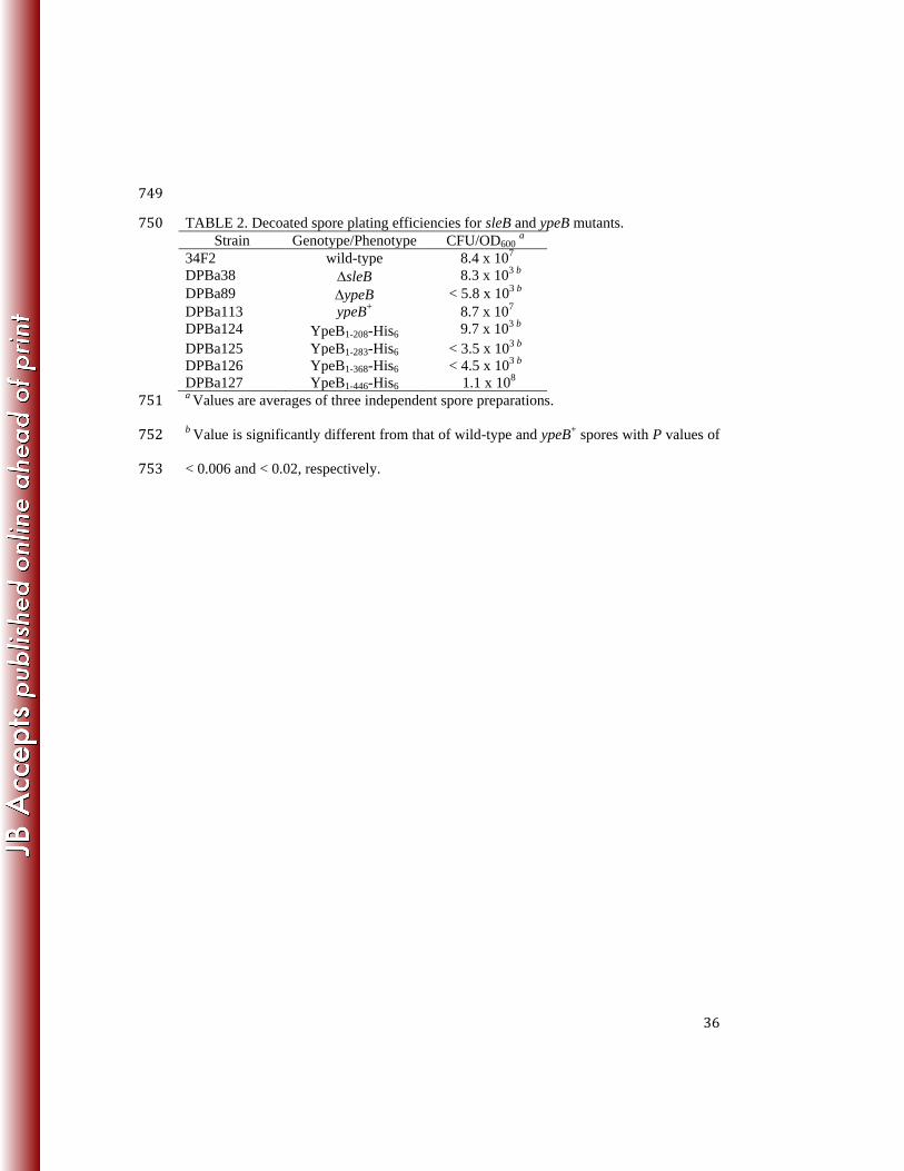
YpeB1-368-His6 spores is not statistically different (P > 0.11) from 4 min onward. (C) Point 690
mutations in YpeB PepSY domain conserved residues slow decoated spore germination. ypeB 691
(), YpeBY254A
(), YpeBY329A
(), YpeBT377A
(), YpeBY410A
(), YpeBG430A
(), and 692
ypeB+ () strains were analyzed. Statistical analysis of ypeB point mutants is complex due to 693
the greater variability of germination rates of decoated spore preparations, as well as the fact that 694
spores of some strains are initiating outgrowth while others are still germinating. Focusing on 695
stage two of germination, between 45 and 95 min, YpeBY329A
and YpeBT377A
spores are not 696
significantly different from ypeB+ spores (P > 0.38). Germination of YpeB
Y254A spores from 80-697
32
90 min, YpeBY410A
spores from 45-55 min and 70-95 min, and YpeBG430A
spores at 50 min and 698
from 75-95 min, are significantly different from ypeB+ spores (P < 0.05). 699
700
FIG. 2. B. anthracis SleB and YpeB are co-dependent for incorporation into dormant spores. 701
Dormant wild-type (WT), sleB, sleB complementation (sleB+), ypeB, and ypeB 702
complementation (ypeB+) spores were mechanically disrupted and proteins extracted with sample 703
loading buffer for western blot analysis. Blots were probed with anti-YpeB antibodies (top) and 704
anti-SleB antibodies (bottom). The predicted molecular weights of mature SleB lacking its 705
signal sequence and YpeB are 24.1 and 50.0 kDa, respectively. The positions of molecular 706
weight marker proteins (not shown) are indicated on the left. 707
708
FIG. 3. B. anthracis SleB and YpeB are degraded during spore formation in the absence of their 709
partner protein. Wild-type (WT), sleB, ypeB, and ypeB complementation (ypeB+) strains were 710
grown in modified G broth at 37C, and samples collected during sporulation were used for 711
western blot analysis. Values t2 through t6 indicate hours since the initiation of sporulation. (A) 712
Sporangia probed with anti-SleB antibodies. (B) Sporangia probed with anti-YpeB antibodies. 713
(C) Sporangia from the sleB strain probed with anti-YpeB antibodies. The positions of 714
molecular weight marker proteins (kDa) (not shown) are indicated on the left. 715
716
FIG. 4. Effects of ypeB truncation and internal deletion mutations on stability of YpeB and SleB. 717
Dormant spores were mechanically disrupted and proteins extracted with sample loading buffer 718
for western blot analysis. Samples were probed with anti-YpeB antibodies (top panels) and anti-719
SleB antibodies (bottom panels). Rightward pointing arrowheads indicate YpeB derivatives. 720
The positions of molecular weight marker proteins (not shown) are indicated on the left. (A) 721
33
Truncations removing YpeB PepSY domains destabilize both YpeB and SleB. YpeB1-208-His6, 722
YpeB1-283-His6, YpeB1-368-His6, and YpeB1-446-His6 have predicted molecular weights of 24.3, 723
32.5, 42.1, and 50.9 kDa, respectively. (B) Internal deletions in the YpeB N-terminal domain 724
destabilize SleB. Lanes intervening between lanes 5 and 6 were removed for clarity. YpeBΔ25-725
203-His6, YpeBΔ67-203-His6, YpeBΔ119-203-His6, YpeBΔ156-203-His6, and YpeB1-446-His6 have 726
predicted molecular weights of 30.6, 35.5, 41.2, 45.6, and 50.9 kDa, respectively. 727
728
FIG. 5. Protein stability and plating efficiency in YpeB point mutant spores. Dried, dormant 729
ypeB, YpeBY254A
(Y254A), YpeBY329A
(Y329A), YpeBT377A
(T377A), YpeBY410A
(Y410A), 730
YpeBG430A
(G430A), and ypeB complementation (ypeB+) spores were broken, and extracted 731
proteins were analyzed through western blots with anti-SleB antibodies (-SleB) or anti-YpeB 732
antibodies (-YpeB). Band intensities were quantified and are depicted as levels of YpeB (white 733
bars) and SleB (gray bars) present in spores, relative to the native protein levels found in ypeB+ 734
spores, which were set at 100%. Spores of the same strains were heat activated, serially diluted, 735
and plated on BHI medium. Following incubation, colonies were counted and % native 736
CFU/OD values (striped bars) were determined by comparison to the CFU/OD value of ypeB+ 737
spores, which was set at 100%. Plating efficiency and western blot quantification data shown are 738
averages from three independent spore preparations with error bars representing the standard 739
deviation. Western blots pictured are representative results from one of the replicates. 740
741
34
TABLE 1. Bacterial strains and plasmids742 Strain or plasmid Relevant genotype/phenotype a Construction b Source
E. coli
DPVE13 BL21 (DE3) pLysS (Cmr) Novagen
DPVE440 pDPV426 (His6-MBP-YpeB21-446 Ampr) Cmr pDPV426DPVE13 This study
B. anthracis
Sterne 34F2 pXO1+ pXO2- P. Hanna
DPBa38 sleB (23)
DPBa57 sleB, pDPV346 (sleB+ Err) (23)
DPBa89 ypeB pDPV39234F2 This study
DPBa113 ypeB::pDPV416 (ypeB+ Err) pDPV416DPBa89 This study
DPBa124 ypeB::pDPV421 (YpeB1-208-His6 Err) pDPV421DPBa89 This study
DPBa125 ypeB::pDPV422 (YpeB1-283-His6 Err) pDPV422DPBa89 This study
DPBa126 ypeB::pDPV423 (YpeB1-368-His6 Err) pDPV423DPBa89 This study
DPBa127 ypeB::pDPV424 (YpeB1-446-His6 Err) pDPV424DPBa89 This study
DPBa134 sleB::pDPV346 (sleB+ Err) DPBa57 integration This study
DPBa143 ypeB::pDPV435 (YpeBY410A Err) pDPV435DPBa89 This study
DPBa144 ypeB::pDPV436 (YpeBG430A Err) pDPV436DPBa89 This study
DPBa148 ypeB::pDPV432 (YpeBT377A Err) pDPV432DPBa89 This study
DPBa149 ypeB::pDPV433 (YpeBY254A Err) pDPV433DPBa89 This study
DPBa150 ypeB::pDPV434 (YpeBY329A Err) pDPV434DPBa89 This study
DPBa158 ypeB::pDPV448 (YpeB25-203-His6 Err) pDPV448DPBa89 This study
DPBa159 ypeB::pDPV449 (YpeB67-203-His6 Err) pDPV449DPBa89 This study
DPBa160 ypeB::pDPV450 (YpeB19-203-His6 Err) pDPV450DPBa89 This study
DPBa161 ypeB::pDPV451 (YpeB156-203-His6 Err) pDPV451DPBa89 This study
Plasmids
pBKJ236 Err ori(Ts) (42)
pBKJ223 Tetr Ampr, Pamy-I-SceI (42)
pDEST-HisMBP-T His6-MBP, Ampr Cmr F. Schubot
pDPV346 sleB+ pBKJ236::sleB (23)
pDPV388 ypeB+ pBKJ236::ypeB This study
pDPV392 ypeB pBKJ236::ypeB This study
pDPV416 ypeB+ pBKJ236::sleB ypeB This study
pDPV421 YpeB1-208-His6 pBKJ236::sleB ypeB1-208-His6 This study
pDPV422 YpeB1-283-His6 pBKJ236::sleB ypeB1-283-His6 This study
pDPV423 YpeB1-368-His6 pBKJ236::sleB ypeB1-368-His6 This study
pDPV424 YpeB1-446-His6 pBKJ236::sleB ypeB1-446-His6 This study
pDPV426 His6-MBP-YpeB21-446 pDEST-HisMBP-T::ypeB21-446 This study
pDPV432 YpeBT377A pBKJ236::sleB ypeBT377A This study
pDPV433 YpeBY254A pBKJ236::sleB ypeBY254A This study
pDPV434 YpeBY329A pBKJ236::sleB ypeBY329A This study
pDPV435 YpeBY410A pBKJ236::sleB ypeBY410A This study
pDPV436 YpeBG430A pBKJ236::sleB ypeBG430A This study
pDPV448 YpeB25-203-His6 pBKJ236::sleB ypeB25-203-His6 This study
pDPV449 YpeB67-203-His6 pBKJ236::sleB ypeB67-203-His6 This study
pDPV450 YpeB119-203-His6 pBKJ236::sleB ypeB119-203-His6 This study
pDPV451 YpeB156-203-His6 pBKJ236::sleB ypeB156-203-His6 This study a Cm
r, chloramphenicol resistance; Amp
r, ampicillin resistance; Er
r, erythromycin resistance; 743
ori(Ts), temperature-sensitive origin of replication; Tetr, tetracycline resistance. 744

35
b Strains were constructed by conjugation or electroporation. The designation preceding the 745
arrow is the plasmid, while the designation following the arrow is the recipient strain. Single 746
strain designations indicate the existing plasmid was integrated into the chromosome.747
748
36
749
TABLE 2. Decoated spore plating efficiencies for sleB and ypeB mutants. 750 Strain Genotype/Phenotype CFU/OD600
a
34F2 wild-type 8.4 x 10⁷
DPBa38 sleB 8.3 x 10³ b
DPBa89 ypeB < 5.8 x 10³ b
DPBa113 ypeB+ 8.7 x 10
7
DPBa124 YpeB1-208-His6 9.7 x 10³ b
DPBa125 YpeB1-283-His6 < 3.5 x 10³ b
DPBa126 YpeB1-368-His6 < 4.5 x 10³ b
DPBa127 YpeB1-446-His6 1.1 x 10⁸
a Values are averages of three independent spore preparations. 751
b Value is significantly different from that of wild-type and ypeB
+ spores with P values of 752
< 0.006 and < 0.02, respectively. 753