role of mri in assessment and diagnosis of axial spondyloarthritis lebanese society of rheumatology...
TRANSCRIPT
Role of MRI in Assessment and Diagnosis of Axial Spondyloarthritis
Lebanese Society of Rheumatology 2009 Nov 07
Ulrich Weber MD, Rheumatology
Balgrist University Hospital, Zurich, Switzerland
Disclosure
Nothing to disclose
No advisory board memberships
Funding of the project Whole Body MRI in SpA
• Walter L. and Johanna Wolf Foundation, Zurich, Switzerland
• Foundation for Scientific Research at the University of Zurich, Switzerland
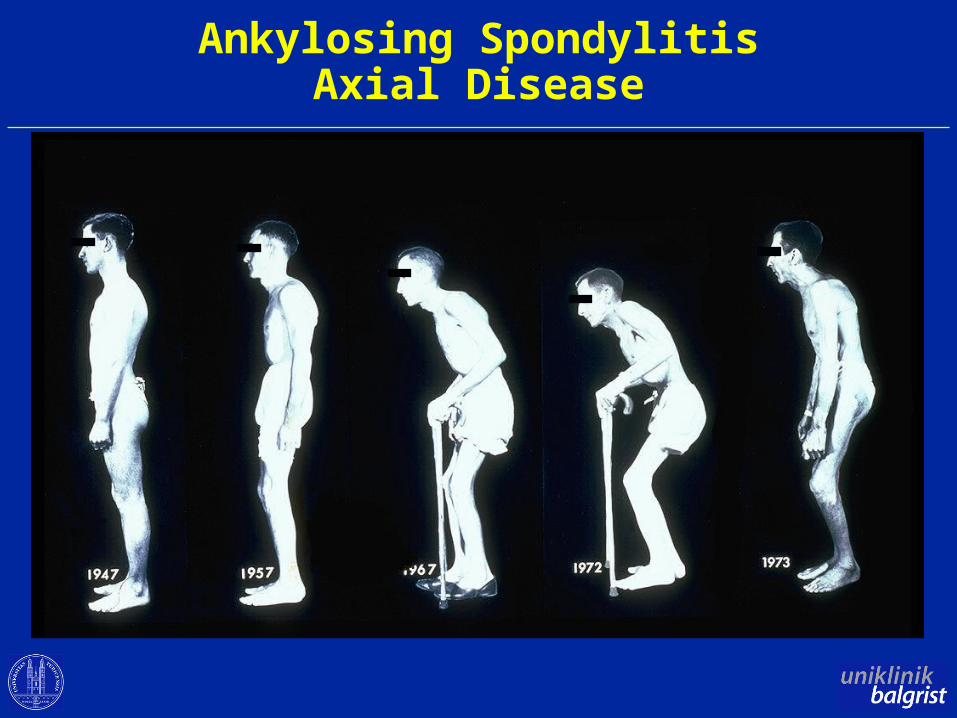
Ankylosing SpondylitisAxial Disease
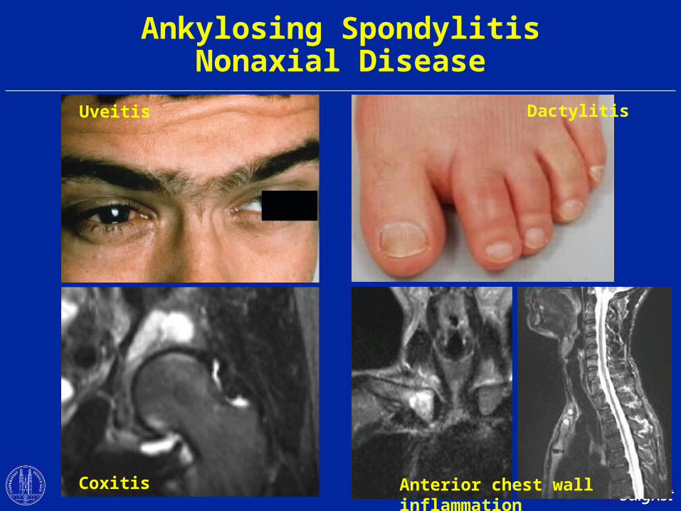
Ankylosing SpondylitisNonaxial Disease
Uveitis Dactylitis
Anterior chest wall inflammationCoxitis
Objectives
• Role of MRI in early diagnosis of axial SpA
• Whole body MRI – a promising MRI variant
• Emerging roles of MRI in axial SpA
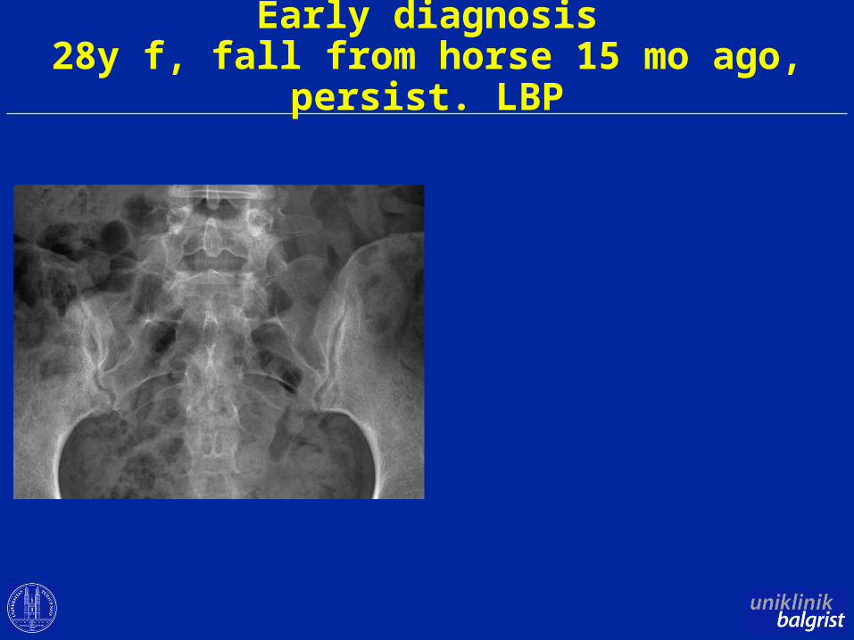
Early diagnosis28y f, fall from horse 15 mo ago, persist. LBP
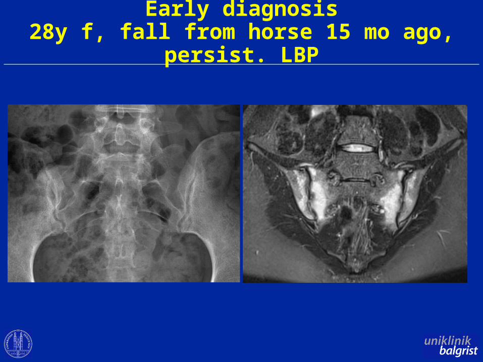
Early diagnosis28y f, fall from horse 15 mo ago, persist. LBP
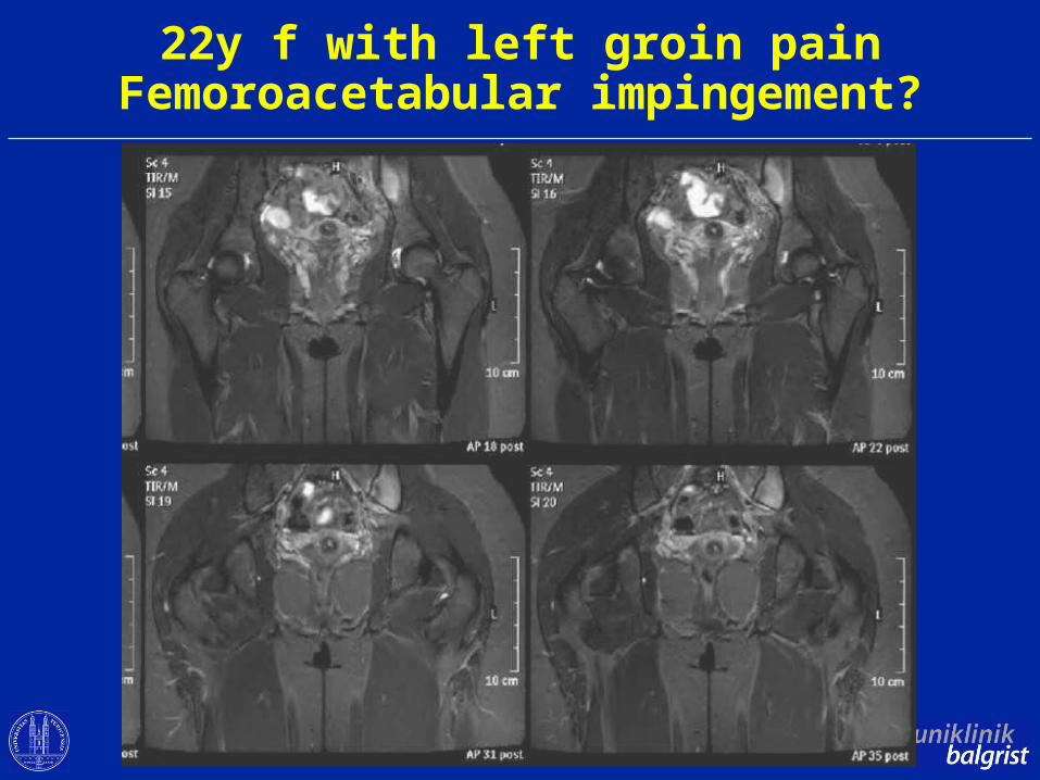
22y f with left groin painFemoroacetabular impingement?
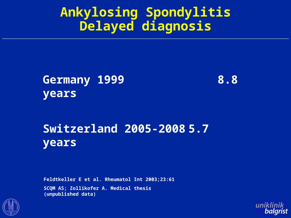
Ankylosing SpondylitisDelayed diagnosis
Germany 1999 8.8 years
Switzerland 2005-2008 5.7 years
Feldtkeller E et al. Rheumatol Int 2003;23:61
SCQM AS; Zollikofer A. Medical thesis (unpublished data)
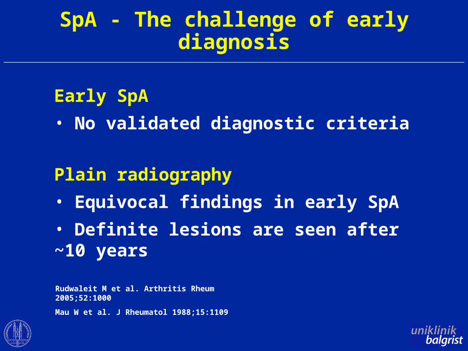
SpA - The challenge of early diagnosis
Early SpA
• No validated diagnostic criteria
Plain radiography
• Equivocal findings in early SpA
• Definite lesions are seen after ~10 years
Rudwaleit M et al. Arthritis Rheum 2005;52:1000
Mau W et al. J Rheumatol 1988;15:1109
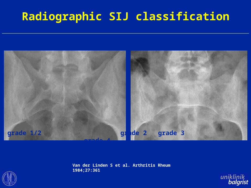
Radiographic SIJ classification
grade 1/2 grade 2 grade 3 grade 4
Van der Linden S et al. Arthritis Rheum 1984;27:361
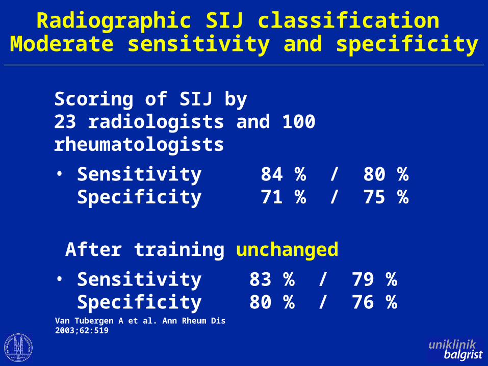
Radiographic SIJ classification Moderate sensitivity and specificity
Scoring of SIJ by23 radiologists and 100 rheumatologists
• Sensitivity 84 % / 80 % Specificity 71 % / 75 %
After training unchanged
• Sensitivity 83 % / 79 % Specificity 80 % / 76 %
Van Tubergen A et al. Ann Rheum Dis 2003;62:519
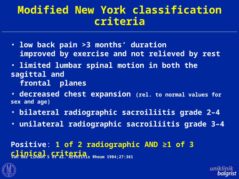
Modified New York classification criteria
• low back pain >3 months‘ duration improved by exercise and not relieved by rest
• limited lumbar spinal motion in both the sagittal and frontal planes
• decreased chest expansion (rel. to normal values for sex and age)
• bilateral radiographic sacroiliitis grade 2–4
• unilateral radiographic sacroiliitis grade 3–4
Positive: 1 of 2 radiographic AND ≥1 of 3 clinical criteria
Van der Linden S et al. Arthritis Rheum 1984;27:361
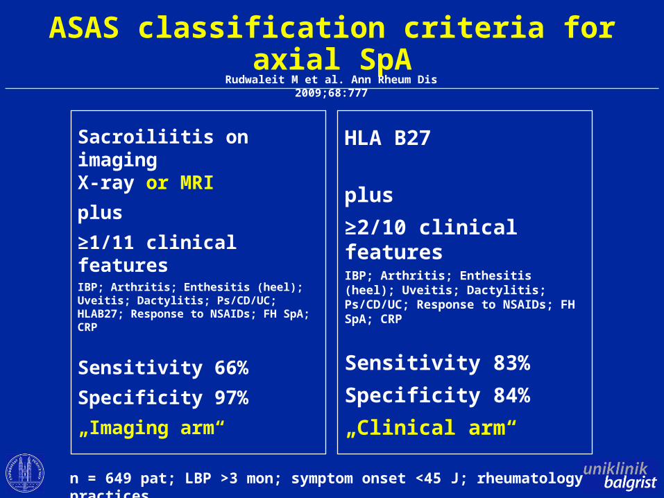
ASAS classification criteria for axial SpA
Sacroiliitis on imagingX-ray or MRI
plus
≥1/11 clinical featuresIBP; Arthritis; Enthesitis (heel); Uveitis; Dactylitis; Ps/CD/UC; HLAB27; Response to NSAIDs; FH SpA; CRP
Sensitivity 66%
Specificity 97%
„Imaging arm“
HLA B27
plus
≥2/10 clinical featuresIBP; Arthritis; Enthesitis (heel); Uveitis; Dactylitis; Ps/CD/UC; Response to NSAIDs; FH SpA; CRP
Sensitivity 83%
Specificity 84%
„Clinical arm“
n = 649 pat; LBP >3 mon; symptom onset <45 J; rheumatology practices
Rudwaleit M et al. Ann Rheum Dis 2009;68:777
ASAS classification criteria for axial SpA
MRI equivalent to plain X-ray
however:
What is a positive MRI?
in the spine?
in the SIJ?
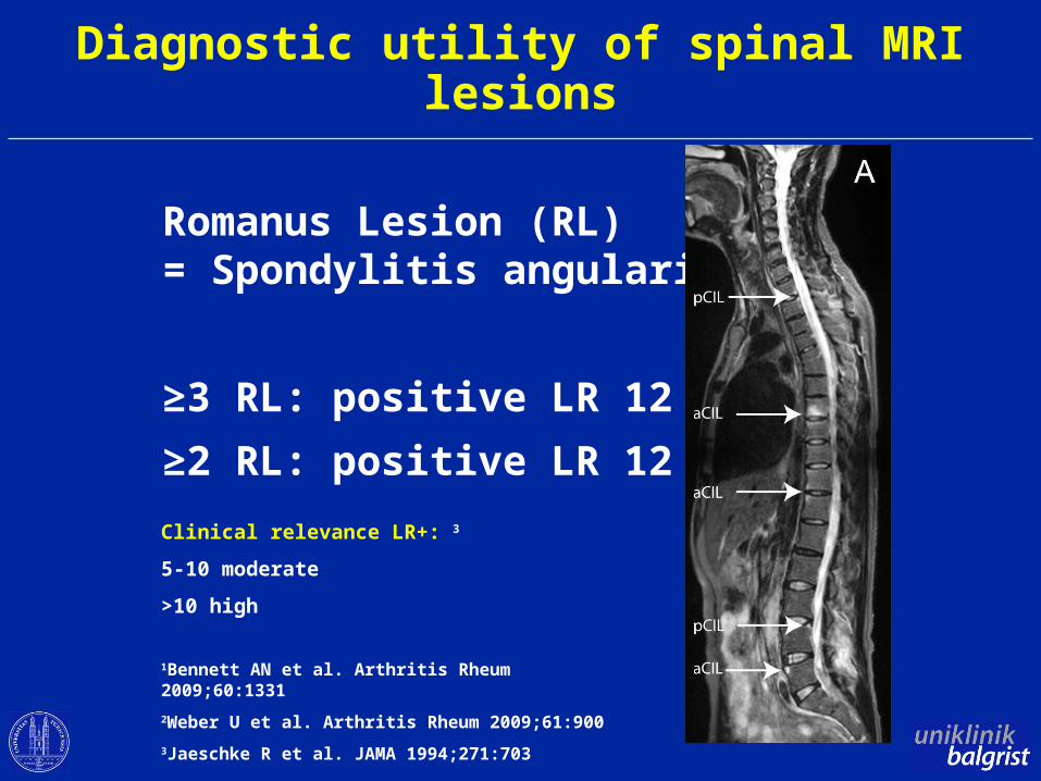
Diagnostic utility of spinal MRI lesions
Romanus Lesion (RL)= Spondylitis angularis
≥3 RL: positive LR 12 1
≥2 RL: positive LR 12 2
1Bennett AN et al. Arthritis Rheum 2009;60:1331
2Weber U et al. Arthritis Rheum 2009;61:900
3Jaeschke R et al. JAMA 1994;271:703
Clinical relevance LR+: 3
5-10 moderate
>10 high
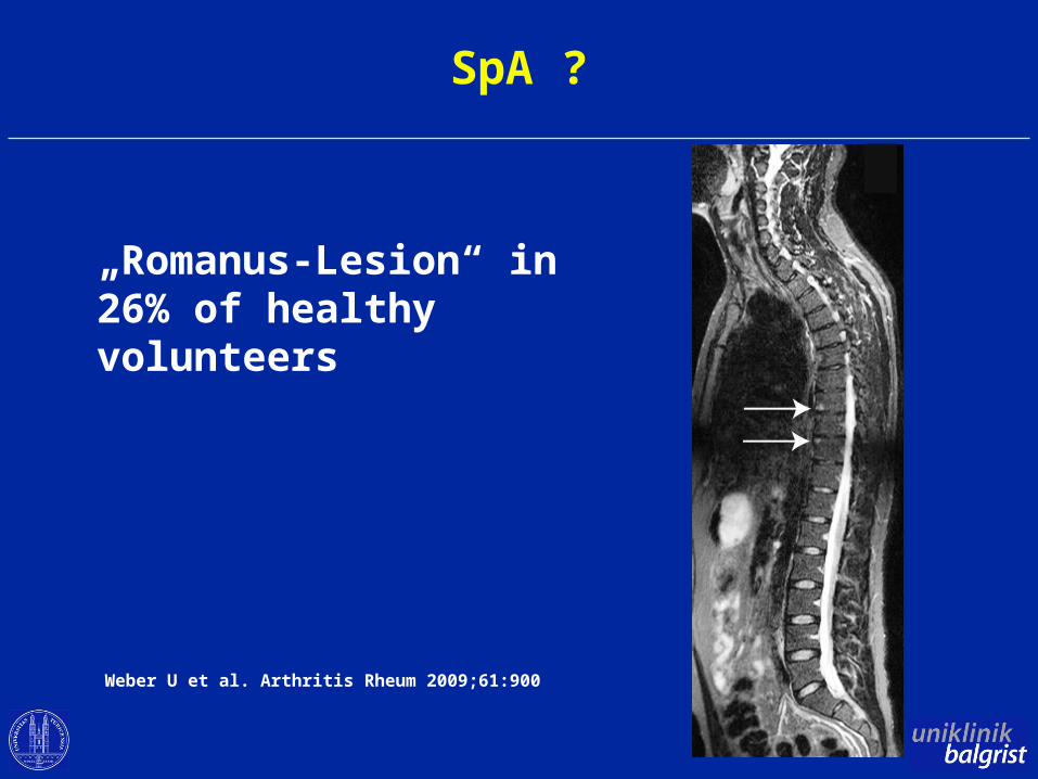
SpA ?
„Romanus-Lesion“ in 26% of healthy volunteers
Weber U et al. Arthritis Rheum 2009;61:900
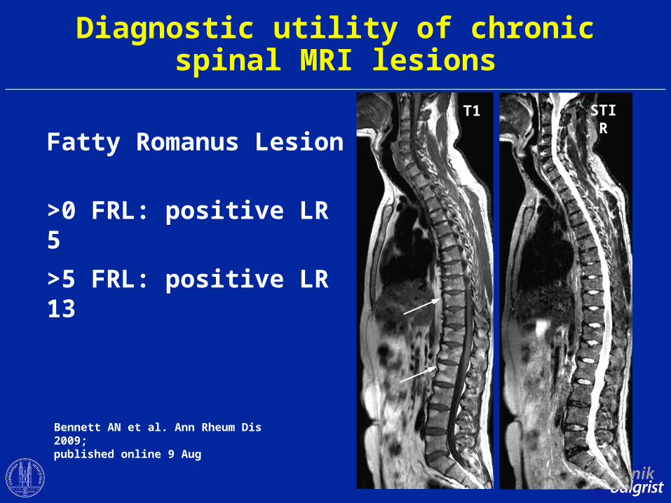
Diagnostic utility of chronicspinal MRI lesions
Fatty Romanus Lesion
>0 FRL: positive LR 5
>5 FRL: positive LR 13
Bennett AN et al. Ann Rheum Dis 2009;published online 9 Aug
T1 STIR
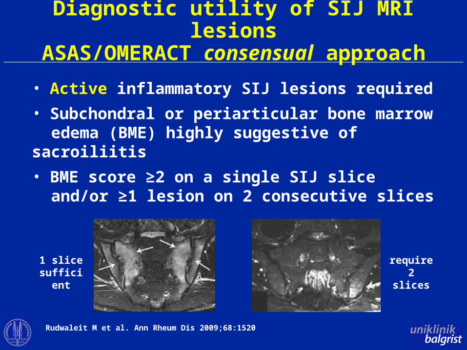
Diagnostic utility of SIJ MRI lesionsASAS/OMERACT consensual approach
• Active inflammatory SIJ lesions required
• Subchondral or periarticular bone marrow edema (BME) highly suggestive of sacroiliitis
• BME score ≥2 on a single SIJ slice and/or ≥1 lesion on 2 consecutive slices
1 slice sufficient
require 2 slices
Rudwaleit M et al. Ann Rheum Dis 2009;68:1520
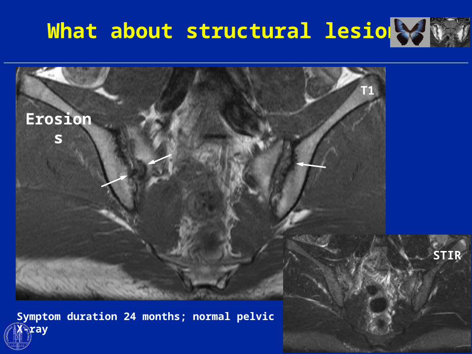
What about structural lesions?
Symptom duration 24 months; normal pelvic X-ray
T1
STIR
Erosions
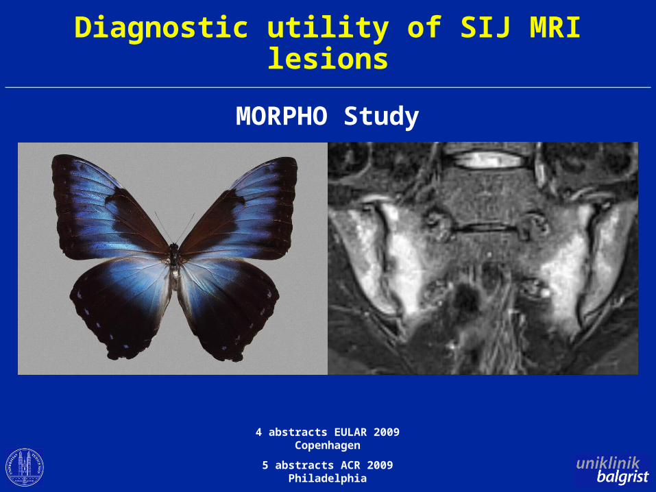
Diagnostic utility of SIJ MRI lesions
MORPHO Study
4 abstracts EULAR 2009 Copenhagen
5 abstracts ACR 2009 Philadelphia
Objectives of MORPHO program
• To assess the diagnostic utility of SIJ MRI by - MRI sequences used in routine practice - comparison with appropriate controls
• To assess the relative contribution of T1 (structural lesions) versus STIR (acute lesions) to assess diagnostic utility
• To define a „positive“ MRI for SpA using a data driven approach
MORPHO Methodology
187 subjects / patients
All ≤45 years old
All patients with inflammatory back pain≤10 years duration
Subjects– 59 asymptomatic healthy volunteers (HV)– 26 patients with non-specific back pain (NSBP)– 77 patients with SpA (met modified NY criteria)– 25 patients with inflammatory back pain
(did not meet modified NY criteria)
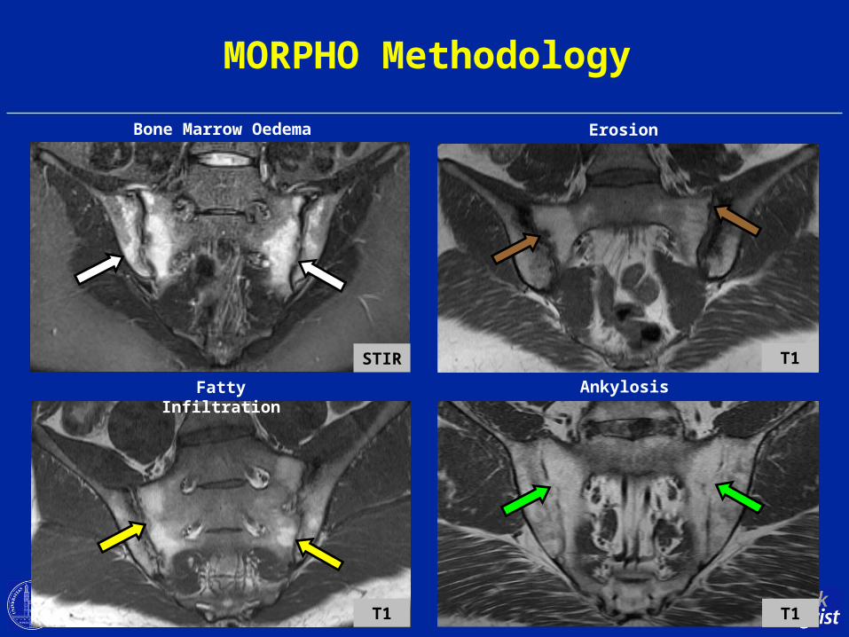
MORPHO Methodology
STIR
Bone Marrow Oedema Erosion
T1
Ankylosis
T1
Fatty Infiltration
T1
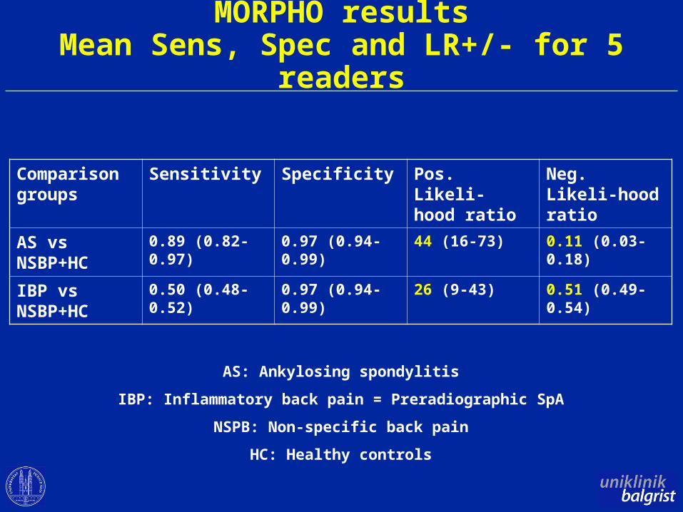
MORPHO resultsMean Sens, Spec and LR+/- for 5 readers
Comparison groups
Sensitivity Specificity Pos. Likeli-hood ratio
Neg. Likeli-hood ratio
AS vs NSBP+HC
0.89 (0.82-0.97) 0.97 (0.94-0.99) 44 (16-73) 0.11 (0.03-0.18)
IBP vs NSBP+HC
0.50 (0.48-0.52) 0.97 (0.94-0.99) 26 (9-43) 0.51 (0.49-0.54)
AS: Ankylosing spondylitis
IBP: Inflammatory back pain = Preradiographic SpA
NSPB: Non-specific back pain
HC: Healthy controls
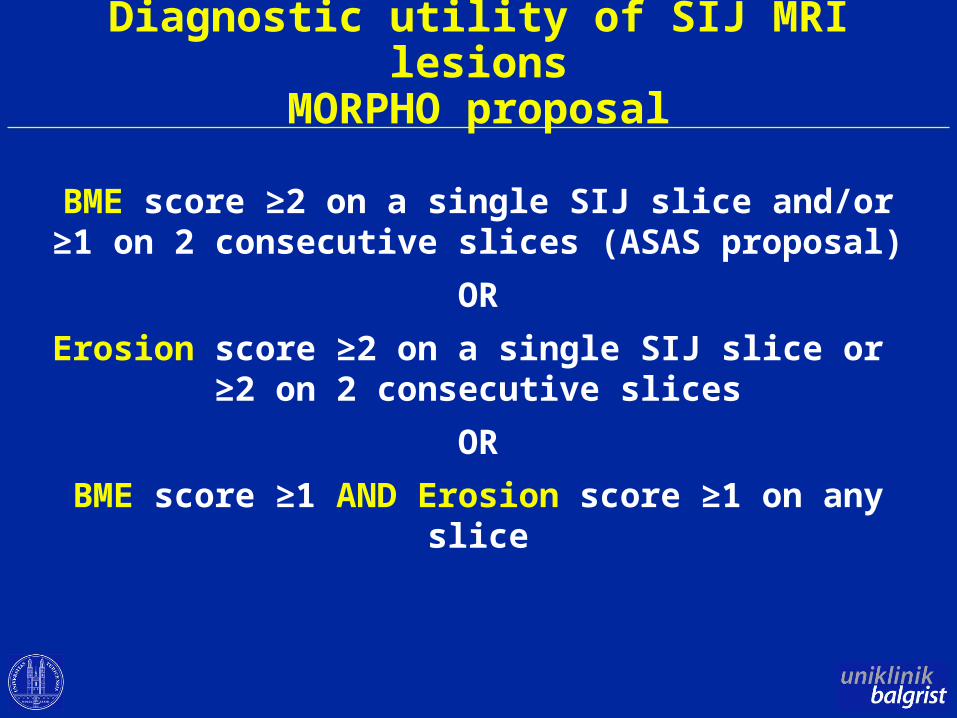
Diagnostic utility of SIJ MRI lesionsMORPHO proposal
BME score ≥2 on a single SIJ slice and/or≥1 on 2 consecutive slices (ASAS proposal)
OR
Erosion score ≥2 on a single SIJ slice or ≥2 on 2 consecutive slices
OR
BME score ≥1 AND Erosion score ≥1 on any slice
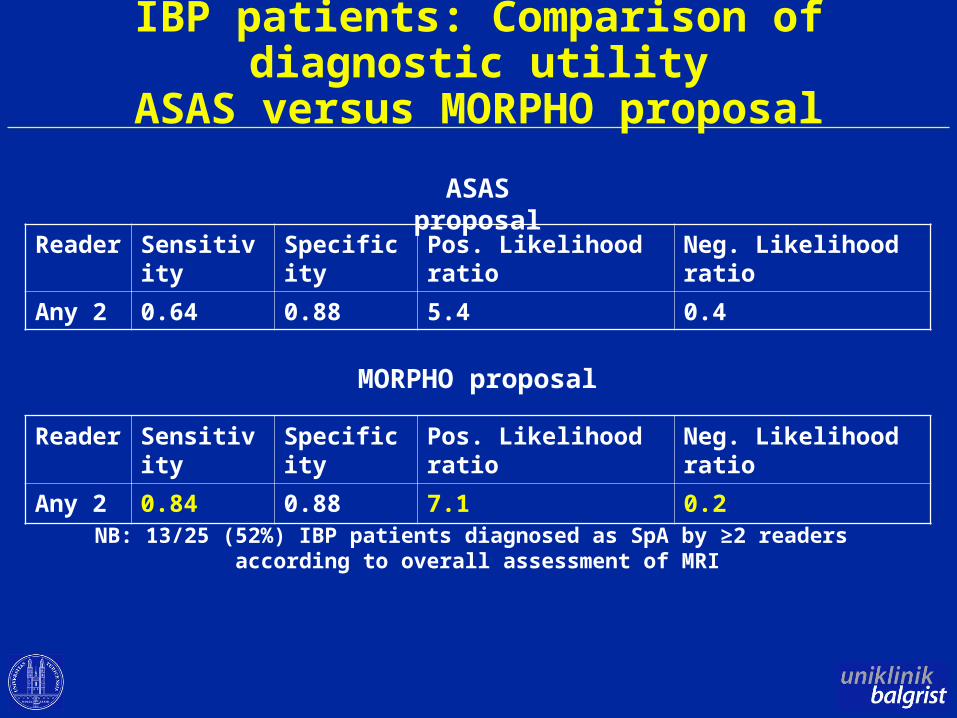
IBP patients: Comparison of diagnostic utilityASAS versus MORPHO proposal
Reader Sensitivity Specificity Pos. Likelihood ratio Neg. Likelihood ratio
Any 2 0.64 0.88 5.4 0.4
Reader Sensitivity Specificity Pos. Likelihood ratio Neg. Likelihood ratio
Any 2 0.84 0.88 7.1 0.2
ASAS proposal
MORPHO proposal
NB: 13/25 (52%) IBP patients diagnosed as SpA by ≥2 readers according to overall assessment of MRI
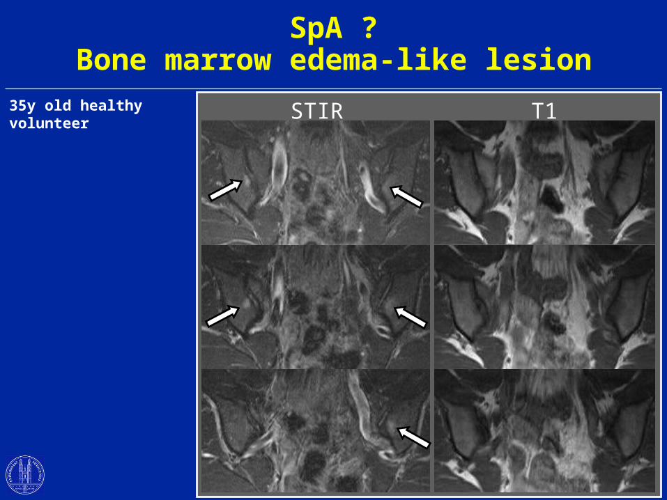
SpA ?Bone marrow edema-like lesion
STIR T135y old healthy volunteer
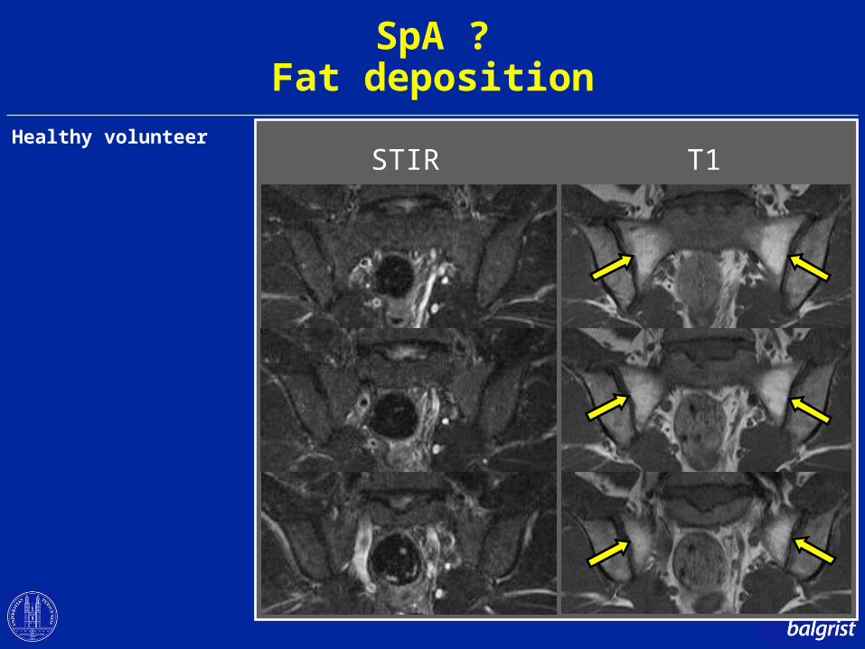
SpA ?Fat deposition
STIR T1Healthy volunteer
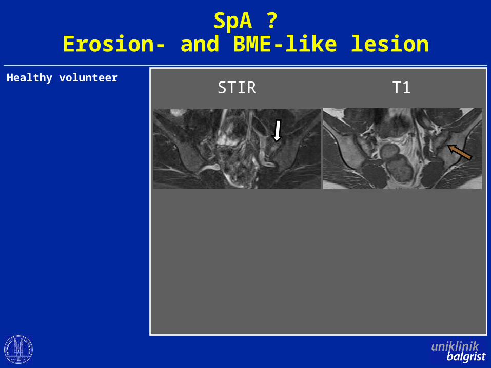
SpA ?Erosion- and BME-like lesion
STIR T1Healthy volunteer
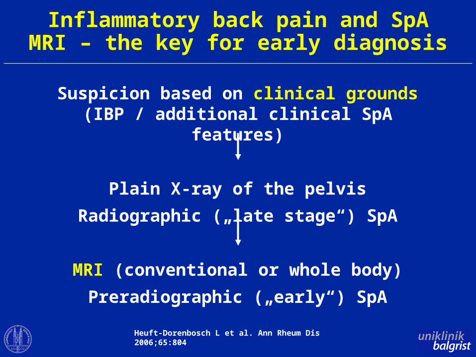
Inflammatory back pain and SpAMRI – the key for early diagnosis
Suspicion based on clinical grounds(IBP / additional clinical SpA features)
Plain X-ray of the pelvis
Radiographic („late stage“) SpA
MRI (conventional or whole body)
Preradiographic („early“) SpA
Heuft-Dorenbosch L et al. Ann Rheum Dis 2006;65:804
Objectives
• Role of MRI in early diagnosis of axial SpA
• Whole body MRI – a promising MRI variant
• Emerging roles of MRI in axial SpA
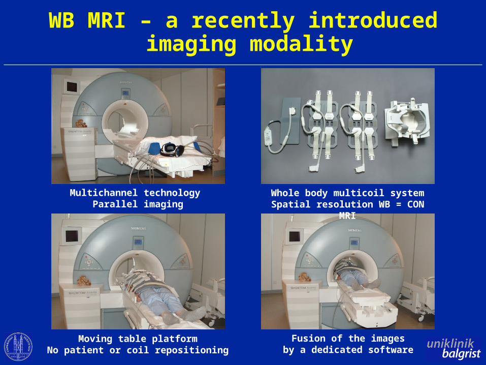
WB MRI – a recently introduced imaging modality
Multichannel technology Parallel imaging
Whole body multicoil systemSpatial resolution WB = CON MRI
Moving table platformNo patient or coil repositioning
Fusion of the imagesby a dedicated software
WB MRI in ASPractical issues
Examination time30 minutes including patient positioning
Reporting time15 minutes for a trained reader
Costsabout 1.5 times the expense for CON MRI(in billing systems based on the amount of time needed for
a particular exam)
Additional imaging of lower extremitiespotential objective measure for enthesitisadditional examination time of 20 minutes
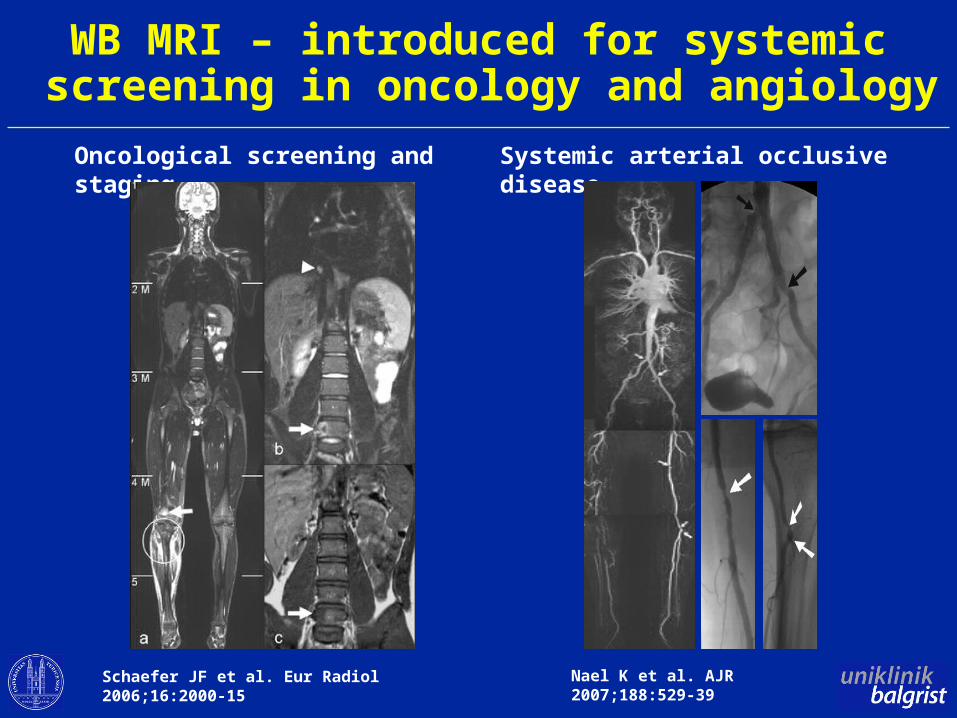
WB MRI – introduced for systemic screening in oncology and angiology
Systemic arterial occlusive disease
Nael K et al. AJR 2007;188:529-39
Oncological screening and staging
Schaefer JF et al. Eur Radiol 2006;16:2000-15
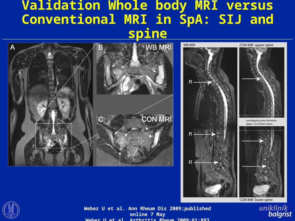
Validation Whole body MRI versus Conventional MRI in SpA: SIJ and spine
Weber U et al. Ann Rheum Dis 2009;published online 7 MayWeber U et al. Arthritis Rheum 2009;61:893
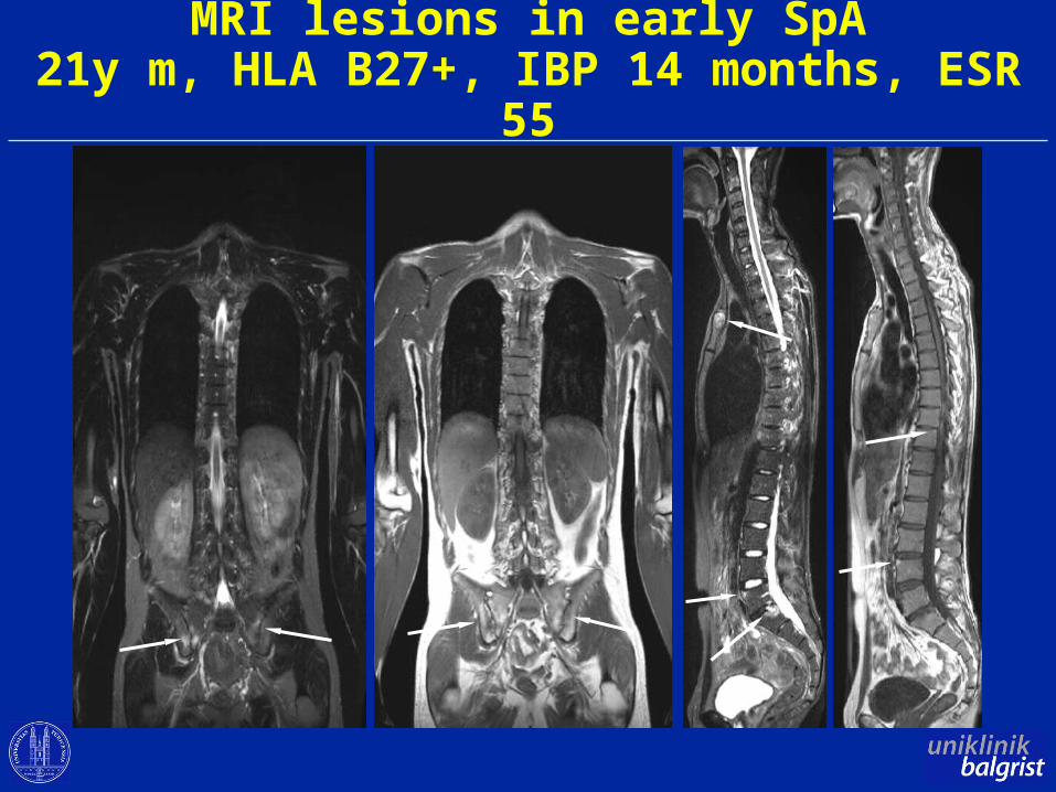
MRI lesions in early SpA21y m, HLA B27+, IBP 14 months, ESR 55
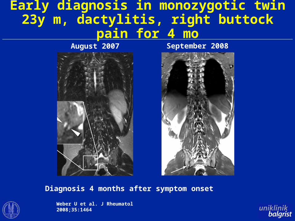
Early diagnosis in monozygotic twin23y m, dactylitis, right buttock pain for 4 mo
August 2007 September 2008
Diagnosis 4 months after symptom onset
Weber U et al. J Rheumatol 2008;35:1464
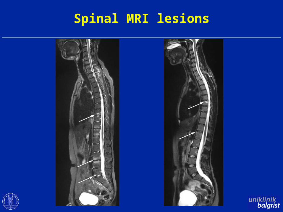
Spinal MRI lesions
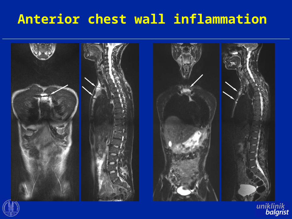
Anterior chest wall inflammation
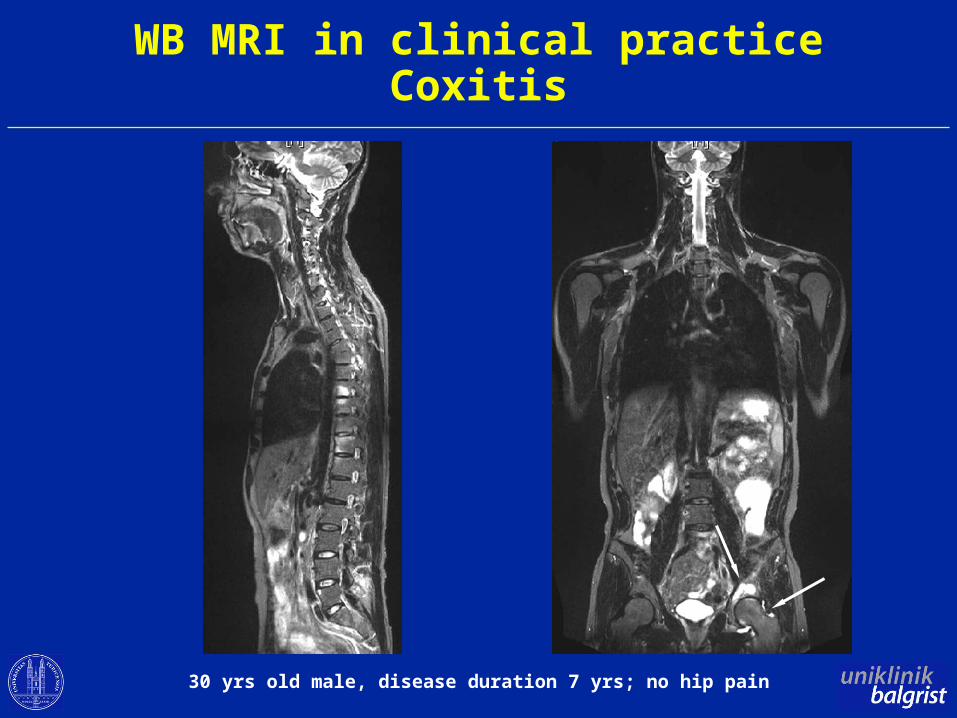
WB MRI in clinical practiceCoxitis
30 yrs old male, disease duration 7 yrs; no hip pain
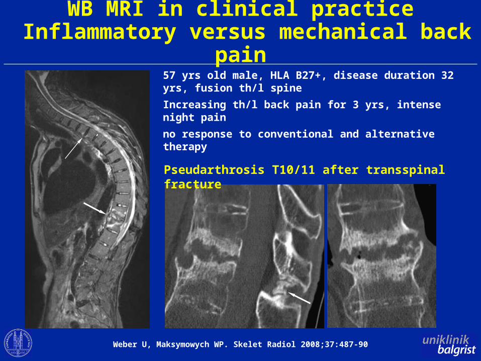
WB MRI in clinical practice Inflammatory versus mechanical back pain
57 yrs old male, HLA B27+, disease duration 32 yrs, fusion th/l spine
Increasing th/l back pain for 3 yrs, intense night pain
no response to conventional and alternative therapy
Pseudarthrosis T10/11 after transspinal fracture
Weber U, Maksymowych WP. Skelet Radiol 2008;37:487-90
Objectives
• Role of MRI in early diagnosis of axial SpA
• Whole body MRI – a promising MRI variant
• Emerging roles of MRI in axial SpA
Inflammatory MRI spinal lesions Predictive for new syndesmophytes
Prospective observational cohort, follow-up after 24 months by plain X-ray and MRI
New syndesmophytes developed significantly more frequently in vertebral corners with inflammation (14.3%) than in those without inflammation (2.9%) seen on baseline MRI (p<0.003)
Maksymowych WP et al. Arthritis Rheum 2009;60:93
Baraliakos X et al. Arthritis Res Ther 2008;10:R104
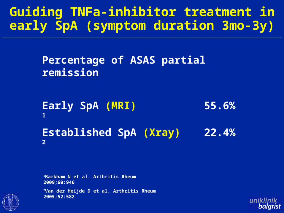
Guiding TNFa-inhibitor treatment in early SpA (symptom duration 3mo-3y)
Percentage of ASAS partial remission
Early SpA (MRI) 55.6% 1
Established SpA (Xray) 22.4% 2
1Barkham N et al. Arthritis Rheum 2009;60:946
2Van der Heijde D et al. Arthritis Rheum 2005;52:582
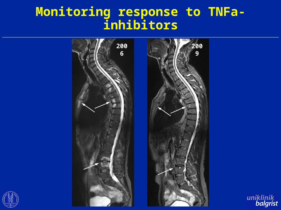
Monitoring response to TNFa-inhibitors
2006 2009
Disease activityMRI versus clinical/laboratory parameters
• No correlation of MRI activity parameters with clinical and laboratory activity in various study designs (cross-sectional, cohort and interventional studies)
• MRI may reflect other aspects of disease activity than the ones expressed by clinical and laboratory parameters
Puhakka KB et al. Rheumatology 2004;43:234Maksymowych WP et al. Arthritis Rheum 2007;57:501Lambert RG et al. Arthritis Rheum 2007;56:4005Weber U et al. Arthritis Rheum 2009;61:893
Roles of MRI in axial SpASummary
• Confirmation of SpA diagnosis suspected on clinical grounds (preradiographic stage)
• Diagnostic MRI thresholds both for SIJ and spine needed
• Emerging role for guiding treatment and predicting disease course
Acknowledgement
• Radiology Balgrist Juerg Hodler Marco Zanetti Christian Pfirrmann
• Rheumatology Balgrist Rudolf Kissling
• Walter Maksymowych, Edmonton Robert Lambert, Edmonton
• Anne Grethe Jurik, Aarhus Anna Zejden, Aarhus
• Mikkel Ostergaard, Copenhagen Susanne Pedersen, Copenhagen
• Asim Khan, Cleveland
• Kaspar Rufibach, Zurich
• Rahel Kubik, Baden
• Stefan Duewell, Frauenfeld
Discussion
White-browed Robin (pair)
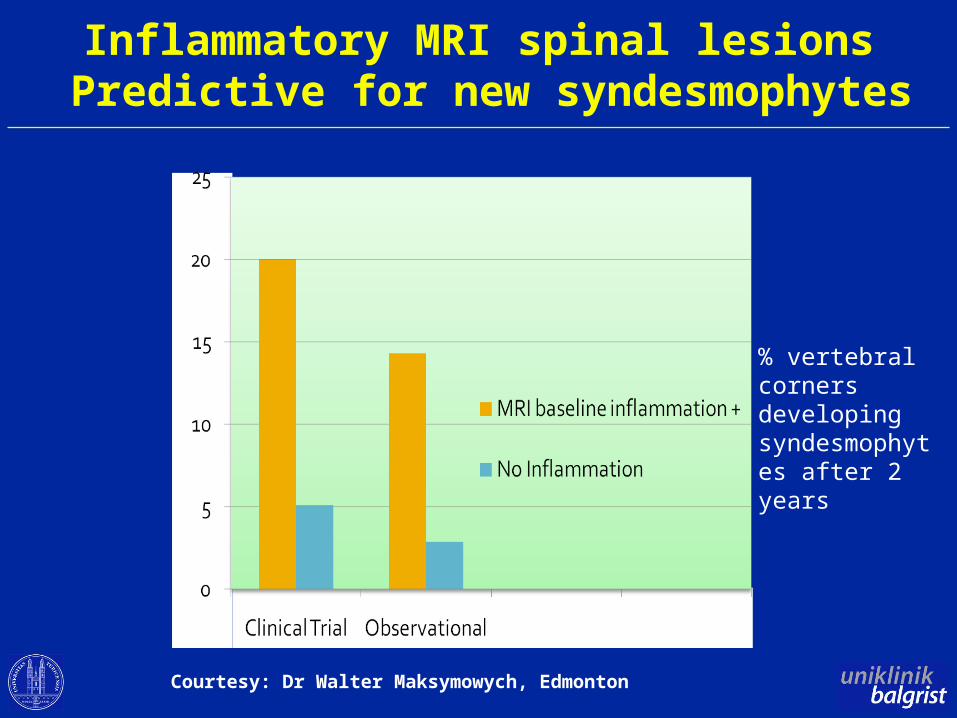
% vertebral corners developing syndesmophytes after 2 years
Inflammatory MRI spinal lesions Predictive for new syndesmophytes
Courtesy: Dr Walter Maksymowych, Edmonton