role of dicer and drosha for endothelial microrna...
TRANSCRIPT

Role of Dicer and Drosha for Endothelial MicroRNAExpression and Angiogenesis
Angelika Kuehbacher, Carmen Urbich, Andreas M. Zeiher, Stefanie Dimmeler
Abstract—MicroRNAs (miRNAs) are small noncoding RNAs that regulate gene expression by binding to the cellulartranscript leading to translational repression or degradation of the target mRNA. Dicer and Drosha are the miRNAprocessing enzymes that are required for the maturation of miRNAs. Here, we investigated the role of Dicer and Droshafor angiogenesis. Endothelial cells were transfected with siRNA against Dicer and Drosha to inhibit miRNA biogenesis.Genetic silencing of Dicer and Drosha significantly reduced capillary sprouting of endothelial cells and tube formingactivity. Migration of endothelial cells was significantly decreased in Dicer siRNA–transfected cells, whereas DroshasiRNA had no effect. Silencing of Dicer but not of Drosha reduced angiogenesis in vivo. Next, we attempted to identifymiRNAs expressed in endothelial cells. A screening analysis of 168 human miRNAs using real-time PCR revealed thatmembers of the let-7 family, mir-21, mir-126, mir-221, and mir-222 are highly expressed in endothelial cells. Dicer andDrosha siRNA reduced lef-7f and mir-27b expression. Inhibitors against let-7f and mir-27b also reduced sproutformation indicating that let-7f and mir-27b promote angiogenesis by targeting antiangiogenic genes. In silico analysisof predicted targets for let-7 cluster identified the endogenous angiogenesis inhibitor thrombospondin-1. Indeed, Dicerand Drosha siRNA significantly increased the expression of thrombospondin-1. Taken together, transient reduction ofthe miRNA-regulating enzyme Dicer impairs angiogenesis in vitro and in vivo, whereas Drosha siRNA induced a minorantiangiogenic effect in vitro and was not effective in vivo. The let-7 family and mir-27b appear to be attractive targetsfor modulating angiogenesis. (Circ Res. 2007;101:59-68.)
Key Words: angiogenesis � endothelial cells � gene expression
MicroRNAs (miRNAs) are a new class of highly con-served, noncoding small RNAs that regulate gene
expression on the posttranscriptional level by binding to thetranscript, leading to translational repression or degradationof the target mRNA.1 There is increasing evidence thatmiRNAs are involved in various biological processes, such ascardiogenesis, skeletal muscle proliferation and differentia-tion, brain morphogenesis, oncogenesis, and hematopoieticlineage differentiation.2–6
miRNAs are generated in a 2-step processing pathwaymediated by 2 major enzymes, Dicer and Drosha, whichbelong to the class of RNAse III endonucleases. Drosha ispart of a multiprotein complex, the microprocessor, whichmediates the nuclear processing of the primary miRNAs intostem–loop precursors of approximately 60 to 70 nucleotides(pre-miRNA).7 The treatment of Hela cells with RNA inter-ference against Drosha results in the strong accumulation ofprimary miRNAs and the reduction of pre-miRNAs andmature miRNAs.7 Exportin-5 mediates the nuclear export ofcorrectly processed miRNA precursors in a Ran-GTP–depen-dent manner.8 In the cytoplasm, the pre-miRNA is cleaved byDicer into the mature 22 nucleotide miRNA.1 Dicer was origi-nally recognized for its role in generating small interfering
(si)RNAs9 and was later shown to be also involved in miRNAmaturation.10 The mature miRNA incorporates as single-stranded RNAs into a ribonucleoprotein complex, known as theRNA-induced silencing complex.11 This complex directs themiRNA to the target mRNA, which leads either to translationalrepression or degradation of the target mRNA.1
Although the function of Drosha in vivo is so far unknown,Dicer plays a crucial role in vertebrate development. Dicer-deficient mice die early in embryonic development, betweenembryonic day 12.5 and 14.5, showing an impaired bloodvessel and yolk sac formation.12 Zebrafish Dicer mutantembryos display abnormal morphogenesis during gastrula-tion, brain formation, somitogenesis, and heart development.4
One recent study indicated that mir-221 and mir-222modulate the angiogenic properties of human umbilical veinendothelial cells (HUVECs).13 However, the function ofmiRNAs in endothelial cell biology is unclear. Therefore, weinvestigated the role of the major miRNA-regulating enzymesDicer and Drosha for angiogenic functions of endothelialcells. Our results demonstrate that Dicer and Drosha arecritical regulators of endothelial sprout and network forma-tion in vitro, whereas only Dicer siRNA significantly blockedangiogenesis in vivo.
Original received December 20, 2006; resubmission received April 10, 2007; revised resubmission received May 8, 2007; accepted May 21, 2007.From Molecular Cardiology, Department of Internal Medicine III, University of Frankfurt, Frankfurt, Germany.Correspondence to Stefanie Dimmeler, PhD, Molecular Cardiology, Department of Internal Medicine III, University of Frankfurt, Theodor-Stern-Kai
7, 60590 Frankfurt, Germany. E-mail [email protected]© 2007 American Heart Association, Inc.
Circulation Research is available at http://circres.ahajournals.org DOI: 10.1161/CIRCRESAHA.107.153916
59
by guest on July 3, 2018http://circres.ahajournals.org/
Dow
nloaded from
by guest on July 3, 2018http://circres.ahajournals.org/
Dow
nloaded from
by guest on July 3, 2018http://circres.ahajournals.org/
Dow
nloaded from
by guest on July 3, 2018http://circres.ahajournals.org/
Dow
nloaded from
by guest on July 3, 2018http://circres.ahajournals.org/
Dow
nloaded from
by guest on July 3, 2018http://circres.ahajournals.org/
Dow
nloaded from
by guest on July 3, 2018http://circres.ahajournals.org/
Dow
nloaded from

Materials and MethodsCell CulturePooled HUVECs were purchased from Cambrex and cultured inendothelial basal medium (EBM; Cambrex) supplemented withhydrocortisone, bovine brain extract, epidermal growth factor, and10% FCS (GIBCO) until the third passage. After detachment withtrypsin, cells were grown in 6-cm culture dishes for at least 40 hours.
RNA InterferenceFor siRNA-mediated gene knockdown, HUVECs were grown to60% to 70% confluence and transfected with GeneTrans II (Mo-BiTec) according to the protocol of the manufacturer. Two differentsiRNA duplexes for Dicer and Drosha have previously been de-scribed.7,14,15 If not stated otherwise, cells were transfected withDicer and Drosha siRNA I (Dicer I: 5�-UGCUUGAAGCAG-CUCUGGA-3�; Dicer II: 5�-UUUGUUGCGAGGCUGAUUC-3�;Drosha I: 5�-AACGAGUAGGCUUCGUGACUU-3�; Drosha II: 5�-AAGGACCAAGUAUUCAGCAAG-3�). A nonrelated scrambledsiRNA was used as a control (5�-UCAAGAAGCCAAGGAUAAU-3�or 5�-CAACAUCACUUUAAGGAAG-3�). For inhibition of spe-cific miRNAs, HUVECs were grown to 60% to 70% confluencebefore transfection with the specific inhibitors. 2�-O-Methyl anti-sense oligoribonucleotides against mir-27b (5�-GCAGAACUUAG-CCACUGUGAA-3�) and let-7f (5�-AACUAUACAAUCUACUACCUCA-3�) were synthesized by VBC Biotech, and a concentra-tion of 200 nmol/L was transfected with GeneTrans II (MoBiTec)according to the protocol of the manufacturer.
Western Blot AnalysisFor Western blot analysis, HUVECs were lysed in a lysis buffer(20 mmol/L Tris [pH 7.4], 150 mmol/L NaCl, 1 mmol/L EDTA,1 mmol/L EGTA, 1% Triton, 2.5 mmol/L sodium pyrophosphate,1 mmol/L �-glycerophosphate, 1 mmol Na3VO4, 1 �g/mL leupeptin,and 1 mmol/L phenylmethylsulfonyl fluoride) for 20 minutes on ice.After centrifugation for 15 minutes at 20 000g (4°C), the proteincontent of the samples was determined according to the Bradfordmethod. Nuclear and cytoplasmic extracts from HUVECs wereprepared using NE-PER nuclear and cytoplasmic kit (Pierce) accord-ing to the protocol of the manufacturer. Equal amounts of proteinwere loaded onto sodium dodecyl sulfate–polyacrylamide gels andblotted onto poly(vinylidene difluoride) membranes. Western blotswere performed by using antibodies directed against Dicer (mousemonoclonal anti-Dicer, 1:500; Abcam), Drosha (rabbit polyclonalanti-Drosha, 1:1000; Upstate), Topo I (rabbit polyclonal anti-Topo I,1:250; Santa Cruz Biotechnology), Hsp70 (rabbit polyclonal anti-Hsp70, 1:500; Upstate), thrombospondin (TSP)-1 (mouse monoclo-nal anti-TSP, 1:250; Laboratory Vision), or tubulin (mouse mono-clonal anti-tubulin, 1:1500; Dianova).
Migration AssayTo determine the migration of endothelial cells, HUVECs weredetached with trypsin, harvested by centrifugation, resuspended in500 �L of endothelial basal medium with 0.1% BSA, counted andplaced in the upper chamber of a modified Boyden chamber (5�104
cells per chamber; pore size, 8 �m; BD Biosciences) coated with 2.5�g/mL fibronectin. The chamber was placed in a 24-well culturedish containing endothelial basal medium with 0.1% BSA andhuman vascular endothelial growth factor (VEGF) (50 ng/mL). Afterincubation for 5 hours at 37°C, the nonmigrating cells on the upperside of the chamber were mechanically removed and the remainingcells on the lower side were fixed with 4% formaldehyde. Forquantification, cell nuclei were stained with 4�,6-diamidino-phenylidole. Migrating cells on the bottom side of the chamber werecounted manually in 5 random microscopic fields.
Tube Formation AssayHUVECs (7�104) were cultured in a 12-well plate (Greiner) coatedwith 200 �L of Matrigel Basement Membrane Matrix (BD Bio-sciences). Tube length was quantified after 24 hours by measuring
the cumulative tube length in three random microscopic fields witha computer-assisted microscope using the program KS300 3.0(Zeiss).
Spheroid-Based Angiogenesis AssayEndothelial cell spheroids of defined cell number were generated asdescribed previously.16,17 In vitro angiogenesis was quantified bymeasuring the cumulative length of the sprouts that had grown out ofeach spheroid with or without human basic fibroblast growth factor(30 ng/mL) using a digital imaging software (Axioplan, Zeiss)analyzing 10 spheroids per experimental group and experiment.
Reverse-Transcription PCRTotal RNA was isolated using the RNeasy Mini Kit (Qiagen).Afterward, 1 �g of RNA from each sample was reverse transcribedinto cDNA and subjected to conventional PCR. Primer sequences forPCR were: Dicer, 5�-CAAGTGTCAGCTGTCAGAACTC-3� and5�-CAATCCACCACAATCTCACATG-3�; Drosha, 5�-CACCTGT-TCTAGCAGCTCAGAC-3� and 5�-CTCCTCCCACTGAAGCAT-ATTG-3�. To assess the differential miRNA expression in scram-bled, Dicer siRNA–transfected, and Drosha siRNA–transfectedHUVECs, we isolated total RNA using TRIzol (Invitrogen) accord-ing to the protocol of the manufacturer. RT-PCR was performedusing the mirVana quantitative RT-PCR miRNA detection kit(Ambion) and primer sets specific for amplification of hsa-mir-27band hsa-let-7f (Ambion) (1 cycle: 3 minutes at 95°C; 20 cycles: 15seconds at 95°C, 30 seconds at 60°C).
MTT Viability AssayAssessment of cell viability was performed using the MTT [3-(4,5-dimethylthiazol-2-yl)-2,5-diphenyl-2H-tetrazolium bromide] assay.Twenty-four and 48 hours after transfection, 0.5 mg/mL MTT wasadded to each well and cells were incubated for 4 hours at 37 C. Cellswere washed with PBS and lysed 30 minutes at room temperaturewith lysis buffer (40 nmol/L HCl in isopropanol). Absorbance wasphotometrically measured at 550 nm.
ImmunocytochemistryHUVECs grown on a cover slip were fixed in 4% paraformaldehydefor 15 minutes. After permeabilization with 0.25% Triton X-100, thecells were blocked in 2% BSA for 30 minutes at room temperatureand incubated with antibodies against Dicer (mouse monoclonalanti-Dicer, 1:50; Abcam), Drosha (rabbit polyclonal anti-Drosha,1:50; Upstate), phospho– histone H3 (anti-Phospho-H3 AlexaFluor 488-conjugated, 1:10; Cell Signaling), SYTOX Blue fornuclear staining (SYTOX Blue, 1:1000; Invitrogen), and Phalloi-din for F-actin staining (Alexa Fluor 633-conjugated Phalloidin,1:300; Invitrogen).
In Vivo Matrigel Plug AssayThis assay was performed as described previously18 with the follow-ing modifications: HUVECs were transfected with siRNA againstDicer and Drosha or scrambled oligonucleotides as described above.Eighteen hours after transfection, cells were labeled with cell trackerCM-Dil (Invitrogen), were detached, washed, and counted. Cells(1�106) were resuspended in 30 �L of PBS and mixed with 500 �Lof Matrigel Basement Membrane Matrix (BD Biosciences) contain-ing 15 U of heparin (Sigma-Aldrich). The cell–Matrigel mixture wasinjected subcutaneously into 6- to 8-week-old female athymic nudemice (Harlan) along the abdominal midline. After 7 days, bloodvessel infiltration in Matrigel plugs was quantified by analysis oflectin or CD31-stained sections using microscopy. For hemoglobinanalysis, the Matrigel plug was removed after 7 days and homoge-nized in 130 �L of deionized water. After centrifugation, thesupernatant was used in the Drabkin assay (Sigma-Aldrich) tomeasure hemoglobin concentration. Stock solutions of hemoglobinare used to generate a standard curve. Results are expressed relativeto total protein in the supernatant.
60 Circulation Research July 6, 2007
by guest on July 3, 2018http://circres.ahajournals.org/
Dow
nloaded from

MiRNA Expression AnalysisFor determination of global baseline miRNA expression inHUVECs, total RNA was isolated using TRIzol. The expression of168 mature human miRNAs in HUVECs was profiled using real-time PCR (DNAvision, Gosselies, Belgium). Gene expression datawere normalized to let-7a. The relative expression was determinedfor each of the 168 miRNAs using the formula 2��Ct.
To assess the differential miRNA expression in scrambled, DicersiRNA–transfected, and Drosha siRNA–transfected HUVECs, weisolated total RNA using TRIzol. The expression analysis of 344miRNA was performed by a miRNA-profiling service (Exiqon)using miRCURY LNA arrays.
Statistical AnalysisData are expressed as mean�SEM. Three or more treatment groupswere compared by 1-way ANOVA followed by post hoc analysis
adjusted with a least-significant-difference correction for multiplecomparisons (SPSS Inc). Results were considered statistically sig-nificant when P�0.05.
ResultsExpression and Localization of Dicer and Droshain Endothelial CellsBecause little is known about Dicer and Drosha in endothelialcells, we first assessed the expression and localization of the2 enzymes using immunocytochemistry and Western blotanalysis. Dicer was predominantly localized in the cytoplasm,whereas the localization of Drosha was restricted to thenucleus of endothelial cells (Figure 1A and 1B). To determinewhether Dicer and Drosha localization might be different in
Figure 1. Expression and localization of Dicer and Drosha in endothelial cells. A, Dicer and Drosha localization was assessed by immu-nocytochemistry. Dicer and Drosha staining is shown in red fluorescence, phospho-H3 is used as proliferation marker (green), cytoskel-etal staining is performed by F-actin (blue), and nuclei (Sytox blue) are shown in white. B, Nuclear and cytoplasmic extracts were pre-pared as described in Materials and Methods. Western blot analysis was performed using antibodies directed against Dicer, Drosha,Hsp70, and Topo I. C and D, HUVECs were transfected with siRNAs targeting Dicer and Drosha or scrambled oligonucleotides. C,RT-PCR analysis of Dicer and Drosha mRNA expression after 24 hours. A representative gel is shown. GAPDH serves as loading con-trol. D, Forty-eight hours after transfection, cells were lysed and subjected to Western blot analysis with antibodies against Dicer andDrosha. An antibody directed against tubulin was used as loading control.
Kuehbacher et al Role of Dicer and Drosha in Endothelial Cells 61
by guest on July 3, 2018http://circres.ahajournals.org/
Dow
nloaded from

proliferating cells, we additionally identified proliferatingcells by phospho–histone-H3 staining. However, the prolif-eration status had no influence on the localization of the 2enzymes (Figure 1A and 1B).
Role of Dicer and Drosha for Sprouting, TubeFormation, and Migration of Endothelial CellsTo investigate the role of Dicer and Drosha for angiogenesis,we measured endothelial sprouting and network formationafter silencing of Dicer and Drosha expression with siRNA(Figure 1C and 1D). Western blotting confirmed the efficientand specific suppression of Dicer and Drosha by the respec-tive siRNA oligonucleotides (Figure 1D). The reduction ofDicer and Drosha significantly inhibited basal and basicfibroblast growth factor–stimulated endothelial cell sproutformation, as measured by capillary sprouting in a 3Dcollagen-embedded spheroid culture assay (Figure 2A and2B), whereas VEGF-induced sprout formation was selec-tively blocked by Dicer siRNA (Figure I in the online datasupplement, available at http://circres.ahajournals.org). Next,we tested whether the combined silencing of Dicer andDrosha further suppressed sprout formation. However, noadditive effect was detected compared with the single reduc-tion of Dicer or Drosha gene expression (supplemental FigureIIA and IIB). As a control for the specificity of Dicer andDrosha silencing, a second Dicer and Drosha siRNA wasgenerated that reduced capillary sprout formation to a similarextent as compared with Dicer and Drosha siRNA I (data notshown). Moreover, suppression of Dicer and Drosha led to a
significant impairment of network-forming activity using aMatrigel assay (Figure 3A and 3B). To address whether thereduction of Dicer and Drosha contributes to reduced endo-thelial cell migration, we performed a Boyden chambermigration assay. Whereas Dicer siRNA-mediated knockdownsignificantly decreased the migratory capacity, Droshaknockdown had no effect on cell migration (Figure 3C). Totest whether the reduction of endothelial cell sprouting andtube formation was secondary to a nonspecific effect on cellgrowth or apoptosis, we measured cell viability using a MTTassay. As shown in Figure 3D, Dicer and Drosha knockdowndid not impair cell viability after 24 hours. After 48 hours,Dicer siRNA transfection slightly reduced viability, whereasDrosha siRNA transfection did not affect viability (Figure3E). To test whether an induction of cell death mediates theantiangiogenic effect of Dicer siRNA, apoptosis was blockedby the addition of the caspase inhibitor zVAD. However, thereduced sprout-forming activity of Dicer siRNA–transfectedendothelial cells was not improved by zVAD addition (datanot shown), indicating that the inhibition of sprout formationis independent of the induction of cell death.
Dicer Is Required for In Vivo AngiogenesisNext, we assessed the effects of Dicer on in vivo angiogen-esis. Because Dicer-deficient mice are embryonic lethal andthus not available for the study, we subcutaneously injectedsiRNA-transfected HUVECs in a Matrigel plug into nudemice and assessed the sprout formation. Vessel-like structureswere significantly reduced in Matrigel plugs with Dicer
Figure 2. Dicer and Drosha regulate en-dothelial cell sprouting. A and B, HUVECswere transfected with Dicer and DroshasiRNA or scrambled oligonucleotides. Aspheroid assay was performed to analyzebasal endothelial sprouting capacity. A,Representative spheroids are shown. B,Analysis of endothelial sprouting capacitywith or without basic fibroblast growthfactor (bFGF) (30 ng/mL) stimulation. En-dothelial sprouting capacity is given ascumulative sprout length per spheroid.Data are shown as means�SEM (per-centage scrambled without stimulation);n�3 to 4.
62 Circulation Research July 6, 2007
by guest on July 3, 2018http://circres.ahajournals.org/
Dow
nloaded from

Figure 3. Effect of Dicer and Drosha on tube formation, migration, and cell viability. A through G, HUVECs were transfected with Dicer andDrosha siRNA or scrambled oligonucleotides. A and B, HUVECs were seeded on a “growth factor–enriched” Matrigel Basement MembraneMatrix (BD Biosciences) 24 hours after transfection. Representative micrographs and statistical summary of the tube-forming activity. Lengthof capillary-like structures was measured by light microscopy after 24 hours in a blinded fashion. Data are means�SEM (percentage scram-bled); n�4. C, Cell migration was measured using a modified Boyden chamber. Cells were seeded in the upper chamber of a modified Boy-den chamber 24 hours after transfection. Endothelial cell migration was assessed using VEGF (50 ng/mL) as a chemoattractant. Data aremeans�SEM (percentage scrambled); n�3. D and E, Cell viability was measured using a MTT viability assay after 24 (D) or 48 (E) hours.Data are means�SEM (percentage scrambled); n�3. F and G, The in vivo Matrigel plug assay was performed as described in Materials andMethods. Data are means�SEM (percentage scrambled); n�6 (F) and n�3 (G) per group.
Kuehbacher et al Role of Dicer and Drosha in Endothelial Cells 63
by guest on July 3, 2018http://circres.ahajournals.org/
Dow
nloaded from

∆
Figure 4. miRNA expression profile in HUVECs. The expression of 168 human miRNAs was profiled using real-time PCR. Comparativeanalysis was performed for 92 miRNAs, detected in all 3 samples. Raw data were normalized to let-7a and converted using theformula 2��Ct (relative expression). Data represent miRNAs that are highly expressed in HUVECs (relative expression�100). Data aremeans�SEM; n�3.
64 Circulation Research July 6, 2007
by guest on July 3, 2018http://circres.ahajournals.org/
Dow
nloaded from

siRNA–transfected HUVECs (Figure 3F). Silencing of Diceradditionally reduced Matrigel plug hemoglobin concentra-tions indicating that the blood supply is reduced (Figure 3G).In contrast, Drosha siRNA transfection did not significantlyaffect sprouting angiogenesis and hemoglobin concentrationsof Matrigel plugs in vivo (Figure 3F and 3G).
Dicer and Drosha Regulate the Expression ofmiRNAs in Endothelial CellsTo assess the specific involvement of miRNAs in endothelialcell function, we analyzed the expression of 168 humanmiRNAs in HUVECs. miRNAs with a relative expression(2��Ct) of �100 are listed in Figure 4. In particular, miR-21,miR-126, mir-221, and mir-222 are highly expressed inendothelial cells. In addition, several members of the let-7family revealed a high abundance in HUVECs. To investigatethe role of Dicer and Drosha in miRNA expression inendothelial cells, we analyzed the expression of 344 humanmiRNAs after knockdown with siRNA targeting Dicer andDrosha using miRNA array (Figure 5 and the Table). Theexpression of 202 miRNAs was below the detection limit ofthe assay. The selective analysis of the effect of Dicer andDrosha downregulation revealed that the expression of 2miRNAs was reduced to more than 30% by Dicer knock-down, 29 miRNAs were reduced to more than 30% by Droshaknockdown, and 31 miRNAs were regulated by both en-zymes. The reduction in miRNA expression after Dicer andDrosha knockdown was confirmed by RT-PCR for mir-27band let-7f (Figure 6A and 6B). To test the functional effect ofmir-27b and let-7f, the miRNA were blocked by specificinhibitors. Inhibition of mir-27b and let-7f significantlyreduced in vitro sprout formation (Figure 6C).
Dicer and Drosha Silencing Induces theUpregulation of TSP-1 ExpressionIn silico prediction of potential target genes using miRandasoftware revealed that the let-7 miRNA family may regulatethe expression of TSP-1, which was recently identified as anendogenous angiogenesis inhibitor. Therefore, we assessedthe expression of TSP-1 after Dicer and Drosha siRNAtransfection. As shown in Figure 6D and 6E, the reduction ofDicer and Drosha by siRNA transfection significantly in-creased the expression of TSP-1 to 263�68% in DicersiRNA–transfected cells and to 358�95% in Drosha siRNA–transfected cells, respectively. However, depletion of TSP-1expression by siRNA did not rescue the impaired sproutformation induced by Dicer or Drosha deficiency (supple-mental Figure III), indicating that additional targets areinvolved to fully explain the angiogenesis inhibition. Because
let-7f in comparison to other let-7 family members was mostprofoundly downregulated by Dicer and Drosha, we investi-gated the effect of a let-7f inhibitor on TSP-1 expression.However, inhibition of let-7f induced only a minor, notsignificant, increase in TSP-1 expression to 115�18% (n�6),suggesting that additional miRNAs are involved in theregulation of TSP-1.
Effect of Dicer and Drosha Silencing on CytokineExpression and Akt SignalingFinally, we elucidated the effect of Dicer and Drosha on theexpression of angiogenic growth factors by using a humancytokine array. Although Dicer siRNA significantly sup-pressed angiogenesis, most of the proangiogenic cytokineswere increased in Dicer-silenced endothelial cells (supple-mental Figure IVA). Moreover, no significant difference inthe expression of the VEGF receptor 2 was detected (supple-mental Figure IVB). In contrast, phosphorylation of Akt wasselectively inhibited in Dicer siRNA compared with DroshasiRNA–treated cells (supplemental Figure IVC), indicatingan interference of Dicer with Akt activation.
DiscussionAlthough several studies have dissected the role of miRNAsin controlling embryonic development, myogenesis and car-diogenesis, the involvement of miRNAs in vascular signalingand function is less clear. Using transient silencing of themiRNA processing enzymes, the present study demonstratesthat Dicer and Drosha are involved in the regulation ofangiogenesis in vitro. Silencing of Dicer expression addition-ally impaired in vivo angiogenesis, whereas depletion ofDrosha did not exert significant effects. Profiling of highlyexpressed miRNAs in endothelial cells provides evidence thatmiRNA let-7f and hsa-mir-27b are significantly downregu-lated by Drosha and Dicer siRNA. In accordance with insilico prediction of putative let-7 targets, our data suggest thatDicer and Drosha regulate the angiogenesis-inhibitor TSP-1via let-7 family members.
Interestingly, the knockdown of Dicer exerted more pro-found effects on endothelial sprout formation in vitro and invivo compared with Drosha. To exclude that the distinctbiological response is caused by a methodological problem,control experiments were performed to confirm a similarefficiency of the downregulation on protein and mRNA level.Moreover, the findings were reproduced using a secondsiRNA sequence excluding an nonspecific effect of one of thesequences. The genetic downregulation of Drosha efficientlyreduced the expression of �42% of the detected miRNAs,indicating that the minor phenotypic alteration in Drosha
≤≤
≤
Figure 5. Role of Dicer and Drosha inmiRNA expression. HUVECs were trans-fected with Dicer and Drosha siRNA orscrambled oligonucleotides. The expres-sion analysis of 344 human miRNAs wasperformed using miRCURY LNA arrays.
Kuehbacher et al Role of Dicer and Drosha in Endothelial Cells 65
by guest on July 3, 2018http://circres.ahajournals.org/
Dow
nloaded from
siRNA–transfected cells is not related to an insufficientreduction of Drosha enzymatic activity. An explanation forthe distinct effects of Dicer versus Drosha may be related toother biological properties of Dicer, namely its role in theformation and maintenance of heterochromatin,19 whichmight also affect endothelial cell functions independent onthe reduction of miRNA processing. The minor role ofDrosha compared with Dicer on sprouting might alternativelybe explained by a concomitant effect on antiapoptotic orproangiogenic genes, which are less affected by DicersiRNA. A downregulation of miRNAs targeting antiapoptoticor proangiogenic genes might lead to an upregulation ofprotective and proangiogenic proteins in Drosha-silencedendothelial cells and may partially compensate for the in-crease in angiogenesis inhibitors. It is well established thatangiogenesis is regulated by a balance of pro- or antiangio-genic genes. A disturbance of this critical balance by inter-fering with various miRNAs, each of which inhibiting trans-lation or transcription of multiple target mRNAs, may wellexplain differences in the phenotype.
Profiling of miRNA expression in endothelial cells, as ithas been done in the present study, additionally provides a
tool to identify potentially important miRNAs in endothelialcell biology. Strikingly, the relative expression of �60% ofthe miRNAs was below the detection limit, indicating that themajority of the miRNAs are downregulated in endothelialcells. These data were selectively confirmed by demonstrat-ing that several miRNAs, such as miR-142-3p, were notdetected by RT-PCR (data not shown). This is consistent withthe concept that miRNAs are differentially expressed duringdevelopment and control lineage commitment.4,6,20,21 A celltype–specific regulation of miRNA expression may contrib-ute to tissue and cell type–specific patterning of gene expres-sion. Therefore, the data of the present miRNA array analysisallow for selectively evaluating the function and regulation ofendothelial cell–enriched miRNAs in future studies. Indeed,the high expression of miR-221 is in accordance with a recentstudy demonstrating that miR-221 may affect the angiogenicproperties of endothelial cells by regulating c-kit, the receptorfor the cytokine stem cell factor.13 Consistently, c-kit expres-sion was significantly increased in Dicer siRNA– and DroshasiRNA–transfected cells (supplemental Figure V).
Surprisingly, the downregulation of Drosha and Dicer onlyaffected a subset of miRNAs. Based on the assumption thatthese 2 enzymes are essential for miRNA processing, onewould have expected to see a downregulation of the majorityof miRNAs. However, in accordance with our data, othersalso reported that only a subset of miRNAs is reduced afterDicer downregulation (55%22 and 15% to 59%4). The incom-plete inhibition of miRNAs might be explained by an insuf-ficient downregulation of the enzymes or a higher stability ofsome of the miRNAs exceeding the transient effect of Droshaor Dicer downregulation by siRNA. Finally, the data shouldbe interpreted with caution because of the limitation of themiRNA array technology. Although the assays preferentiallydetect mature miRNAs, the discrimination of primary, pre-,and mature miRNAs requires confirmation by Northernblotting. The low sensitivity of Northern blot, however, limitsits general use.
By using in silico prediction of targets for the highlyexpressed and Drosha/Dicer downregulated miRNA let-7cluster, we identified several potentially interesting genesincluding TSP-1. TSP-1 is a potent endogenous inhibitor ofangiogenesis.23 Upregulation of TSP-1 in endothelial progen-itor cells contributes to impaired vasculogenesis in diabeticmice.24 Consistent with a predicted role of the let-7 cluster forTSP-1 expression, Drosha and Dicer silencing augmentedTSP-1. Given that TSP-1 is a potent angiogenesis inhibitor,its upregulation might contribute to the impairment of angio-genesis after Drosha or Dicer downregulation. A let-7finhibitor, however, increased TSP-1 expression only �15%,indicating that other factors (eg, other let-7 family members)may, in part, compensate for the loss of let-7f. Indeed, otherlet-7 family members target TSP-1 as predicted by in silicoanalysis. Moreover, TSP-1 is also repressed by the miR-17 to92 miRNA cluster.25
In summary, our data provide evidence for an importantrole of Dicer and Drosha for endothelial cell functions. Theprofound impairment of in vitro angiogenesis in Dicer-deficient endothelial cells is consistent with findings thatwere recently published during the revision of the present
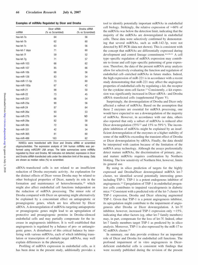
Examples of miRNAs Regulated by Dicer and Drosha
miRNADicer siRNA
(% vs Scrambled)Drosha siRNA
(% vs Scrambled)
hsa-let-7a 64 56
hsa-let-7b 62 60
hsa-let-7c 63 56
hsa-let-7 days 71 60
hsa-let-7f 59 52
hsa-let-7g 71 57
hsa-miR-103 66 62
hsa-miR-107 85 66
hsa-miR-10b 68 56
hsa-miR-126 83 54
hsa-miR-17–5p 73 60
hsa-miR-193a 67 42
hsa-miR-21 98 50
hsa-miR-22 73 45
hsa-miR-221 66 54
hsa-miR-23a 90 56
hsa-miR-24 87 64
hsa-miR-26a 93 53
hsa-miR-27b 64 ND
hsa-miR-29a 82 58
hsa-miR-30c 72 56
hsa-miR-331 42 34
hsa-miR-365 81 60
hsa-miR-486 163 62
HUVECs were transfected with Dicer and Drosha siRNA or scrambledoligonucleotides. The expression analysis of 344 human miRNAs was per-formed using miRCURY LNA arrays. The table summarizes some miRNAsregulated by Dicer and Drosha. ND indicates not detected; expression in Dicerand Drosha siRNA-transfected cells under the detection limit of the assay. Dataare shown as median ratios (%) vs scrambled.
66 Circulation Research July 6, 2007
by guest on July 3, 2018http://circres.ahajournals.org/
Dow
nloaded from

study.26 The angiogenesis suppressive effect of Dicer andDrosha downregulation may in part be related to the augmen-tation of the angiogenesis inhibitor TSP-1. However, theprofound difference between Dicer and Drosha silenced cellsis more likely attributable to the selective impairment of Aktsignaling in Dicer siRNA–treated cells. Because a variety ofmiRNAs are highly expressed and dysregulated by Droshaand/or Dicer siRNA and each miRNA has multiple pro- andantiangiogenic targets, further studies are required to dissectthe complex process of posttranscriptional regulation of geneexpression during angiogenesis.
AcknowledgmentsWe thank Andrea Knau, Nicole Konecny, and Tino Roexe forexcellent technical help.
Sources of FundingThis study was supported by the Deutsche Forschungsgemeinschaft(FOR501 [Di600/6-3]). A.K. is supported by the Alfried KruppStiftung (S.D.). C.U., A.M.Z., and S.D. belong to the EuropeanVascular Genomics Network, a Network of Excellence supported bythe European Community’s sixth Framework Programme for Re-search Priority 1 “Life sciences, genomics and biotechnology forhealth” (contract no. LSHM-CT-2003-503254).
Figure 6. TSP-1 as a miRNA-regulated downstream target. A and B, HUVECs were transfected with Dicer and Drosha siRNA or scram-bled oligonucleotides. The expression of mir-27b and let-7f was analyzed 48 hours after transfection using RT-PCR. A, A representativegel is shown. The small nuclear ribonucleoprotein U6 serves as loading control. B, Quantitative analysis. Data are means�SEM; n�5.C and D, Spheroid sprouts were detected 72 hours after transfection with 2�-O-methyl antisense oligoribonucleotides against mir-27band let-7f. Data are means�SEM; n�4. Representative spheroids are shown. E and F, Forty-eight hours after transfection, cells werelysed and subjected to Western blot analysis with an antibody against TSP-1. An antibody directed against tubulin was used as loadingcontrol. E, A representative Western blot is shown. F, Quantitative analysis. Data are means�SEM; n�6.
Kuehbacher et al Role of Dicer and Drosha in Endothelial Cells 67
by guest on July 3, 2018http://circres.ahajournals.org/
Dow
nloaded from

DisclosuresNone.
References1. Bartel DP. MicroRNAs: genomics, biogenesis, mechanism, and function.
Cell. 2004;116:281–297.2. Zhao Y, Samal E, Srivastava D. Serum response factor regulates a
muscle-specific microRNA that targets Hand2 during cardiogenesis.Nature. 2005;436:214–220.
3. Chen JF, Mandel EM, Thomson JM, Wu Q, Callis TE, Hammond SM,Conlon FL, Wang DZ. The role of microRNA-1 and microRNA-133 inskeletal muscle proliferation and differentiation. Nat Genet. 2006;38:228–233.
4. Giraldez AJ, Cinalli RM, Glasner ME, Enright AJ, Thomson JM,Baskerville S, Hammond SM, Bartel DP, Schier AF. MicroRNAsregulate brain morphogenesis in zebrafish. Science. 2005;308:833–838.
5. Esquela-Kerscher A, Slack FJ. Oncomirs - microRNAs with a role incancer. Nat Rev Cancer. 2006;6:259–269.
6. Chen CZ, Li L, Lodish HF, Bartel DP. MicroRNAs modulate hemato-poietic lineage differentiation. Science. 2004;303:83–86.
7. Lee Y, Ahn C, Han J, Choi H, Kim J, Yim J, Lee J, Provost P, RadmarkO, Kim S, Kim VN. The nuclear RNase III Drosha initiates microRNAprocessing. Nature. 2003;425:415–419.
8. Lund E, Guttinger S, Calado A, Dahlberg JE, Kutay U. Nuclear export ofmicroRNA precursors. Science. 2004;303:95–98.
9. Bernstein E, Caudy AA, Hammond SM, Hannon GJ. Role for a bidentateribonuclease in the initiation step of RNA interference. Nature. 2001;409:363–366.
10. Grishok A, Pasquinelli AE, Conte D, Li N, Parrish S, Ha I, Baillie DL,Fire A, Ruvkun G, Mello CC. Genes and mechanisms related to RNAinterference regulate expression of the small temporal RNAs that controlC. elegans developmental timing. Cell. 2001;106:23–34.
11. Hammond SM, Boettcher S, Caudy AA, Kobayashi R, Hannon GJ.Argonaute2, a link between genetic and biochemical analyses of RNAi.Science. 2001;293:1146–1150.
12. Yang WJ, Yang DD, Na S, Sandusky GE, Zhang Q, Zhao G. Dicer isrequired for embryonic angiogenesis during mouse development. J BiolChem. 2005;280:9330–9335.
13. Poliseno L, Tuccoli A, Mariani L, Evangelista M, Citti L, Woods K,Mercatanti A, Hammond S, Rainaldi G. MicroRNAs modulate theangiogenic properties of HUVEC. Blood. 2006;108:3068–3071.
14. Hutvagner G, McLachlan J, Pasquinelli AE, Balint E, Tuschl T, ZamorePD. A cellular function for the RNA-interference enzyme Dicer in thematuration of the let-7 small temporal RNA. Science. 2001;293:834–838.
15. Chendrimada TP, Gregory RI, Kumaraswamy E, Norman J, Cooch N,Nishikura K, Shiekhattar R. TRBP recruits the Dicer complex to Ago2 formicroRNA processing and gene silencing. Nature. 2005;436:740–744.
16. Korff T, Augustin HG. Integration of endothelial cells in multicellularspheroids prevents apoptosis and induces differentiation. J Cell Biol.1998;143:1341–1352.
17. Diehl F, Rossig L, Zeiher AM, Dimmeler S, Urbich C. The histonemethyltransferase MLL is an upstream regulator of endothelial cell sproutformation. Blood. 2006;109:1472–1478.
18. Potente M, Urbich C, Sasaki K, Hofmann WK, Heeschen C, Aicher A,Kollipara R, DePinho RA, Zeiher AM, Dimmeler S. Involvement ofFoxO transcription factors in angiogenesis and postnatal neovascular-ization. J Clin Invest. 2005;15:2382–2392.
19. Volpe TA, Kidner C, Hall IM, Teng G, Grewal SI, Martienssen RA.Regulation of heterochromatic silencing and histone H3 lysine-9 meth-ylation by RNAi. Science. 2002;297:1833–1837.
20. Sempere LF, Freemantle S, Pitha-Rowe I, Moss E, Dmitrovsky E,Ambros V. Expression profiling of mammalian microRNAs uncovers asubset of brain-expressed microRNAs with possible roles in murine andhuman neuronal differentiation. Genome Biol. 2004;5:R13.
21. Wienholds E, Kloosterman WP, Miska E, Alvarez-Saavedra E, Berezikov E,de Bruijn E, Horvitz HR, Kauppinen S, Plasterk RH. MicroRNA expressionin zebrafish embryonic development. Science. 2005;309:310–311.
22. Cummins JM, He Y, Leary RJ, Pagliarini R, Diaz LA Jr, Sjoblom T,Barad O, Bentwich Z, Szafranska AE, Labourier E, Raymond CK,Roberts BS, Juhl H, Kinzler KW, Vogelstein B, Velculescu VE. Thecolorectal microRNAome. Proc Natl Acad Sci U S A. 2006;103:3687–3692.
23. Iruela-Arispe ML, Bornstein P, Sage H. Thrombospondin exerts an anti-angiogenic effect on cord formation by endothelial cells in vitro. ProcNatl Acad Sci U S A. 1991;88:5026–5030.
24. Ii M, Takenaka H, Asai J, Ibusuki K, Mizukami Y, Maruyama K, YoonYS, Wecker A, Luedemann C, Eaton E, Silver M, Thorne T, LosordoDW. Endothelial progenitor thrombospondin-1 mediates diabetes-induced delay in reendothelialization following arterial injury. Circ Res.2006;98:697–704.
25. Dews M, Homayouni A, Yu D, Murphy D, Sevignani C, Wentzel E, FurthEE, Lee WM, Enders GH, Mendell JT, Thomas-Tikhonenko A. Augmen-tation of tumor angiogenesis by a Myc-activated microRNA cluster. NatGenet. 2006;38:1060–1065.
26. Suarez Y, Fernandez-Hernando C, Pober JS, Sessa WC. Dicer dependentmicroRNAs regulate gene expression and functions in human endothelialcells. Circ Res. 2007;100:1164–1173.
68 Circulation Research July 6, 2007
by guest on July 3, 2018http://circres.ahajournals.org/
Dow
nloaded from

Angelika Kuehbacher, Carmen Urbich, Andreas M. Zeiher and Stefanie DimmelerRole of Dicer and Drosha for Endothelial MicroRNA Expression and Angiogenesis
Print ISSN: 0009-7330. Online ISSN: 1524-4571 Copyright © 2007 American Heart Association, Inc. All rights reserved.is published by the American Heart Association, 7272 Greenville Avenue, Dallas, TX 75231Circulation Research
doi: 10.1161/CIRCRESAHA.107.1539162007;101:59-68; originally published online May 31, 2007;Circ Res.
http://circres.ahajournals.org/content/101/1/59World Wide Web at:
The online version of this article, along with updated information and services, is located on the
http://circres.ahajournals.org/content/suppl/2007/05/31/CIRCRESAHA.107.153916.DC1Data Supplement (unedited) at:
http://circres.ahajournals.org//subscriptions/
is online at: Circulation Research Information about subscribing to Subscriptions:
http://www.lww.com/reprints Information about reprints can be found online at: Reprints:
document. Permissions and Rights Question and Answer about this process is available in the
located, click Request Permissions in the middle column of the Web page under Services. Further informationEditorial Office. Once the online version of the published article for which permission is being requested is
can be obtained via RightsLink, a service of the Copyright Clearance Center, not theCirculation Researchin Requests for permissions to reproduce figures, tables, or portions of articles originally publishedPermissions:
by guest on July 3, 2018http://circres.ahajournals.org/
Dow
nloaded from

0
20
40
60
80
100
120
scr Dicer Drosha
Cum
ulat
ive
spro
utle
ngth
(%) v
s. s
cr
P < 0.05 P < 0.05
n.s.
siRNA
Supplementary Figure 1
Supplementary Figure 1: HUVEC were transfected with siRNA against Dicer and Drosha orscrambled oligonucleotides. Quantitative analysis of endothelial sprouting in response to VEGF stimulation. Data are mean±SEM, n=4.
CIRCRESAHA/2006/147090

Supplementary Figure 2
Dicer
Drosha
GAPDH
siRNA scrDice
r I
Drosha I
H 2ODice
r I/
DroshaI
A B
Cum
ulat
ive
spro
utle
nght
(%) v
s. s
cr
siRNA0
20
40
60
80
100
120
scr Dicer Drosha Dicer/Drosha
P < 0.05
P < 0.05
P < 0.05
Supplementary Figure 2: HUVEC were transfected with siRNA against Dicer and Droshaor a combination of both siRNAs. Scrambled siRNA was used as control. A) RT-PCR analysis of Dicer and Drosha mRNA expression 18 h after transfection. A representative gel is shown. GAPDH serves as loading control. B) Quantitative analysis of endothelialsprouting capacity in response to bFGF-stimulation. Data are mean ±SEM, n=4.
CIRCRESAHA/2006/147090

Supplementary Figure 3
scr
Dicer/T
SP-1
TSP-1
TSP-1
Tubulin
siRNA
Drosha
/TSP-1
A
B
0
20
40
60
80
100
120
scr
Dicer
Drosh
a
Dicer/T
SP-1Dro
sha/T
SP-1
Cum
ulat
ive
spro
utle
ngth
(%) v
s. s
cr
Supplementary Figure 3: HUVEC were transfected with siRNA against Dicer, Drosha or TSP-1 or a combination of two different siRNAs. Scrambled siRNA was used as control. A) 18 hours after transfection, cells were lysed and subjected to western blot analysis with an antibody againstthrombospondin-1. Tubulin was used as loading control. A representative western blot is shown. B) Quantitative analysis of endothelial sprouting in response to bFGF stimulation. Data are mean ±SEM, n=3.
n.s.
n.s.
CIRCRESAHA/2006/147090

Supplementary Figure 4
0
50
100
150
200
250
Ang-2 BDNF DKK-4 EGF IGF-I PDGF-BB
PIGF TPO VEGF
Cyt
okin
eex
pres
sion
(%) v
s. s
cr
scr
Dicer
Drosha
A
0
20
40
60
80
100
120
scr Dicer Drosha
KD
R e
xpre
ssio
n(%
) vs.
scr
siRNA
n.s. n.s.
n.s.
B
0
20
40
60
80
100
120
140
160
scr Dicer DroshasiRNA
Phospho-Akt
Akt
siRNA scr
Drosha
Dicer
C
Supplementary Figure 4: HUVEC were transfected with siRNA against Dicer and Drosha orscrambled oligonucleotides. A) 24 h after transfection, cells were starved for 20 h in EBM + 0.05 % BSA. Supernatants were collected and concentrated 10-fold using Vivaspin columns. 10x supernatants were subjected to a human cytokine antibody array (Ray Bio) according to theinstructions of the manufacturer. Quantitative analysis of cytokine expression is shown. Data aremean±SEM, n=3. B) KDR expression was assessed by FACS staining. Quantitative analysis is shown. Data are mean±SEM, n=3. C) To assess Akt phosphorylation, cells were lysed and subjected to western blot analysis using an antibody against phospho-Akt. Total Akt was used as loading control. A representative western blot and quantitative analysis is shown. Data are mean±SEM, n=3.
CIRCRESAHA/2006/147090
Akt
pho
spho
ryla
tion
(%) v
s. s
cr

Supplementary Figure 5
0
50
100
150
200
250
300
350
scr Dicer Drosha
c-ki
texp
ress
ion
(%)
vs. s
cr
P < 0.05 P < 0.05
P < 0.05
siRNA
Supplementary Figure 5: HUVEC were transfected with siRNA against Dicer and Drosha or scrambledoligonucleotides. The expression of c-kit was detected by Western blot analysis. Tubulin was used as loading control. Quantitative analysis is shown. Data are mean±SEM, n=3.
CIRCRESAHA/2006/147090