role of clostridium perfringens alpha, beta, epsilon and...
TRANSCRIPT
Role of Clostridium perfringens Alpha, Beta,Epsilon and Iota toxins in Enterotoxemia ofmonogastrics and Ruminants
Mariano E. Fernandez-Miyakawa and Leandro M. Redondo
ContentsIntroduction . . . . . . . . . . . . . . . . . . . . . . . . . . . . . . . . . . . . . . . . . . . . . . . . . . . . . . . . . . . . . . . . . . . . . . . . . . . . . . . . . . . . . . . 2Clostridium perfringens Major Toxins and Enterotoxemias . . . . . . . . . . . . . . . . . . . . . . . . . . . . . . . . . . . . . 3
Alpha Toxin (CPA) . . . . . . . . . . . . . . . . . . . . . . . . . . . . . . . . . . . . . . . . . . . . . . . . . . . . . . . . . . . . . . . . . . . . . . . . . . . . 4Beta Toxin (CPB) . . . . . . . . . . . . . . . . . . . . . . . . . . . . . . . . . . . . . . . . . . . . . . . . . . . . . . . . . . . . . . . . . . . . . . . . . . . . . 7
Epsilon Toxin (ETX) . . . . . . . . . . . . . . . . . . . . . . . . . . . . . . . . . . . . . . . . . . . . . . . . . . . . . . . . . . . . . . . . . . . . . . . . . . . . . 11Structure and Mode of Action . . . . . . . . . . . . . . . . . . . . . . . . . . . . . . . . . . . . . . . . . . . . . . . . . . . . . . . . . . . . . . . . 11Associated Diseases . . . . . . . . . . . . . . . . . . . . . . . . . . . . . . . . . . . . . . . . . . . . . . . . . . . . . . . . . . . . . . . . . . . . . . . . . . . 12Intestinal Effects . . . . . . . . . . . . . . . . . . . . . . . . . . . . . . . . . . . . . . . . . . . . . . . . . . . . . . . . . . . . . . . . . . . . . . . . . . . . . . . 14Systemic Effects . . . . . . . . . . . . . . . . . . . . . . . . . . . . . . . . . . . . . . . . . . . . . . . . . . . . . . . . . . . . . . . . . . . . . . . . . . . . . . . 15Iota Toxin (ITX) . . . . . . . . . . . . . . . . . . . . . . . . . . . . . . . . . . . . . . . . . . . . . . . . . . . . . . . . . . . . . . . . . . . . . . . . . . . . . . . 18
Further Directions . . . . . . . . . . . . . . . . . . . . . . . . . . . . . . . . . . . . . . . . . . . . . . . . . . . . . . . . . . . . . . . . . . . . . . . . . . . . . . . . . 21Cross-References . . . . . . . . . . . . . . . . . . . . . . . . . . . . . . . . . . . . . . . . . . . . . . . . . . . . . . . . . . . . . . . . . . . . . . . . . . . . . . . . . 22References . . . . . . . . . . . . . . . . . . . . . . . . . . . . . . . . . . . . . . . . . . . . . . . . . . . . . . . . . . . . . . . . . . . . . . . . . . . . . . . . . . . . . . . . 23
AbstractClostridium perfringens produce several virulence factors to increase coloniza-tion and improve nutrient availability as well. Enterotoxins are among thesevirulence factors, and while some only have local effects, others can act at adistance from the bacterial colonization site. Enterotoxemias are defined asdiseases caused by toxins generated in the intestine and absorbed into systemiccirculation with systemic consequences. The term enterotoxemia is generallyused indistinctly to name enteric and/or systemic diseases, but under the defini-tion of enterotoxemia, several aspects of intestinal bacterial diseases include
M.E. Fernandez-Miyakawa (*) • L.M. Redondo (*)Instituto de Patobiologia - Centro de Investigación en Ciencias Veterinarias y Agronómicas,Instituto Nacional de Tecnología Agropecuaria, Castelar, Buenos Aires, Argentina
Consejo Nacional de Investigaciones Científicas y Técnicas, Ciudad Autónoma de Buenos Aires,Argentinae-mail: [email protected]; [email protected]
# Springer Science+Business Media Dordrecht 2016P. Gopalakrishnakone et al. (eds.), Microbial Toxins, Toxinology,DOI 10.1007/978-94-007-6725-6_16-1
1
pathogenesis and toxin modes of action. The present aim is to describe someenterotoxemia-associated toxins, focusing on those which clearly produce sys-temic and enteric effects, as well as those commonly thought to produceenterotoxemia but remain questionable upon further consideration of the existingevidence.
KeywordsClostridium perfringens • Enterotoxemia • Enteritis • Alpha toxin • Beta toxin •Epsilon toxin • Iota toxin
Introduction
Toxin production by bacteria represents one of the most potent and widespreadmechanisms of pathogenicity. Enteropathogenic bacteria, like Clostridiumperfringens, produce several virulence factors that include adhesins and entero-toxins, which increase mucosal colonization and cause cell alterations as well.Enterotoxins are produced by bacteria in the lumen of the gastrointestinal tract,and while some of these toxins have only local digestive effects, others can havesystemic effects and act at distance from the bacterial colonization site. By defini-tion, enterotoxemias are diseases produced by enterotoxins absorbed into systemiccirculation, which acts on distant organs such as the brain.
Although different toxinogenic bacteria can produce enterotoxemias, the ordinaryuse of the term “enterotoxemia” is a synonym of clostridial intestinal disease. Thebacterial genus Clostridium includes several enteric pathogens, and while it is truethat many clostridial intestinal diseases are mediated by specific toxins with anintestinal origin, most of them are doubtfully or incorrectly called enterotoxemias.In particular, C. perfringens is a widely spread enteropathogen with an intimidatingtoxin arsenal; many of these toxins are produced in the intestinal lumen. While someof these toxins act only locally, others act both locally and systemically. Despiteenterotoxemia being generally used indistinctly to name clostridial/C. perfringensenteric and/or systemic diseases, enterotoxemia includes several pathogenic aspectsof intestinal bacterial diseases and toxin modes of action.
For example, epsilon toxin (ETX) is produced by intestinal C. perfringens type Dand generates an enterotoxemia in sheep, since in this species ETX exerts damage ondistant organs such as the brain and lungs, with no evident intestinal alterations. Ingoats the same ETX, also secreted by type D strains, causes enterocolitis withoutsystemic effects. Therefore, precisely defined enterotoxemias, meaning diseasesproduced by systemically acting toxins absorbed from the intestines, should belimited to a particular group of diseases caused by certain bacterial toxins indetermined animal species under particular conditions.
Under these concepts, a true enterotoxemia should be defined as a disease causedby an individual toxin with the ability to induce both intestinal and systemic effectsand where the intestine alterations contribute to the onset of systemic disease. These
2 M.E. Fernandez-Miyakawa and L.M. Redondo
systemic diseases of intestinal origin are mainly produced by an identified, andusually potent, protein toxin. From a strict point of view, an “enterotoxemic” toxin,when present in the intestinal lumen, should induce mild to severe modifications ofthe intestinal functions, with or without the requirement of a determined host orenvironment conditions. Intestinal changes should favor toxin translocation into thesystemic bloodstream. Once in the circulatory system, the toxin should reach targetorgans, such as kidney or brain, causing damage.
Among enterotoxemic toxins, C. perfringens ETX and E. coli Shiga toxin (STX)are two of the most potent toxins known, responsible for important diseases withmajor economic and health impacts. The following pages aim to review the role ofC. perfringens major toxins in associated diseases, questioning disease definition ofthose commonly thought to produce enterotoxemia.
Clostridium perfringens Major Toxins and Enterotoxemias
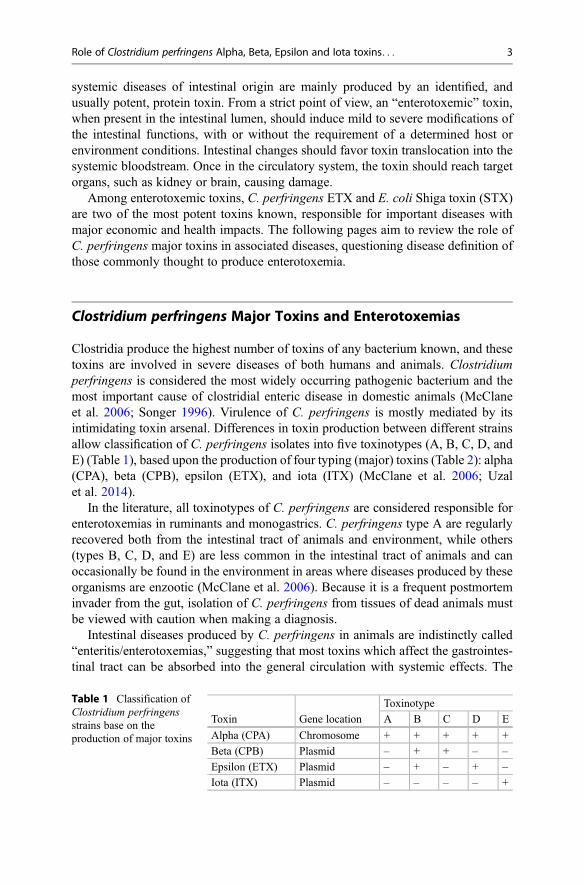
Clostridia produce the highest number of toxins of any bacterium known, and thesetoxins are involved in severe diseases of both humans and animals. Clostridiumperfringens is considered the most widely occurring pathogenic bacterium and themost important cause of clostridial enteric disease in domestic animals (McClaneet al. 2006; Songer 1996). Virulence of C. perfringens is mostly mediated by itsintimidating toxin arsenal. Differences in toxin production between different strainsallow classification of C. perfringens isolates into five toxinotypes (A, B, C, D, andE) (Table 1), based upon the production of four typing (major) toxins (Table 2): alpha(CPA), beta (CPB), epsilon (ETX), and iota (ITX) (McClane et al. 2006; Uzalet al. 2014).
In the literature, all toxinotypes of C. perfringens are considered responsible forenterotoxemias in ruminants and monogastrics. C. perfringens type A are regularlyrecovered both from the intestinal tract of animals and environment, while others(types B, C, D, and E) are less common in the intestinal tract of animals and canoccasionally be found in the environment in areas where diseases produced by theseorganisms are enzootic (McClane et al. 2006). Because it is a frequent postmorteminvader from the gut, isolation of C. perfringens from tissues of dead animals mustbe viewed with caution when making a diagnosis.
Intestinal diseases produced by C. perfringens in animals are indistinctly called“enteritis/enterotoxemias,” suggesting that most toxins which affect the gastrointes-tinal tract can be absorbed into the general circulation with systemic effects. The
Table 1 Classification ofClostridium perfringensstrains base on theproduction of major toxins
Toxin Gene location
Toxinotype
A B C D E
Alpha (CPA) Chromosome + + + + +
Beta (CPB) Plasmid – + + – –
Epsilon (ETX) Plasmid – + – + –
Iota (ITX) Plasmid – – – – +
Role of Clostridium perfringens Alpha, Beta, Epsilon and Iota toxins. . . 3
scientific evidence and abundance of experimental and clinical evidence, togetherwith the molecular Koch’s postulate for enterotoxemia and ETX produced byC. perfringens type B and D, is strong (Garcia et al. 2013). However, the role ofother C. perfringens major toxins in enteritis/enterotoxemia awaits full experimentalconfirmation.
Alpha Toxin (CPA)
Alpha toxin (CPA) is produced by all toxinotypes of C. perfringens. Although it hasbeen generally assumed that type A strains produce higher amounts compared withthe other toxinotypes, Fernandez-Miyakawa et al. (2005) described that othertoxinotypes can produce also comparatively high levels of CPA in vitro. Type A isthe most commonly found toxinotype in nature, and it is usually isolated from theintestine of apparently healthy humans and animals (McClane et al. 2006). Becauseof high prevalence in the intestines of healthy individuals, controversy exists aboutthe real pathogenic role of this toxinotype. An exception to this uncertainty, how-ever, is necrotic enteritis in poultry, where there is considerable evidence thatC. perfringens type A is responsible for the disease (Prescott et al. 2016). As typeA strains produce only CPA as a major toxin, a similar controversy exists about therole of this toxin in type A-associated diseases. While several studies with differentapproaches define a clear association between CPA and histotoxic diseases ofhumans and other animals (Flores-Díaz and Alape-Girón 2003), the role of CPA inenteric disease is not fully understood. Currently many nontyping toxins aredescribed for pathogenic type A strains, like NetB (Keyburn et al. 2008).
Structure and Mode of ActionCPA has phospholipase C activity and a calcium-dependent phospholipid bindingdomain (Sakurai et al. 2004). This toxin is related to several Clostridium sp.phospholipases C (Flores-Díaz and Alape-Giron 2003) and other bacterialphospholipases C, including Zn2+�metalloenzymes from Bacillus cereus andListeria monocytogenes. CPA is the most toxic among bacterial phospholipases C
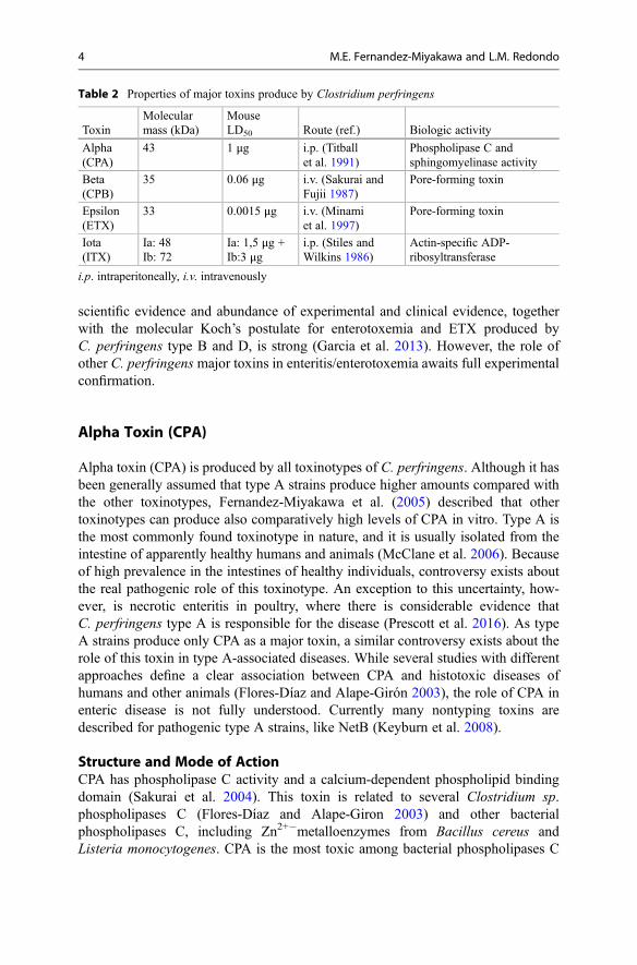
Table 2 Properties of major toxins produce by Clostridium perfringens
ToxinMolecularmass (kDa)
MouseLD50 Route (ref.) Biologic activity
Alpha(CPA)
43 1 μg i.p. (Titballet al. 1991)
Phospholipase C andsphingomyelinase activity
Beta(CPB)
35 0.06 μg i.v. (Sakurai andFujii 1987)
Pore-forming toxin
Epsilon(ETX)
33 0.0015 μg i.v. (Minamiet al. 1997)
Pore-forming toxin
Iota(ITX)
Ia: 48Ib: 72
Ia: 1,5 μg +Ib:3 μg
i.p. (Stiles andWilkins 1986)
Actin-specific ADP-ribosyltransferase
i.p. intraperitoneally, i.v. intravenously
4 M.E. Fernandez-Miyakawa and L.M. Redondo
and has platelet-aggregating, hemolytic, cytotoxic, and myotoxic activities (Titballet al. 1999).
CPA displays both lecithinase and sphingomyelinase activity and degrades thetwo major components of the outer leaflet of eukaryotic plasma membranes in thepresence of calcium ions (Sakurai et al. 2004). Hydrolysis of cell membranephospholipids by CPA results in damage on the cell membrane and activates severalother membrane and internal cell mechanisms that contribute to cytotoxicity (Titballet al. 1991). For example, CPA activates the arachidonic cascade resulting in theformation of thromboxanes, leukotrienes, and prostaglandins, which activate theinflammation cascade and produce vasoconstriction (Flores-Díaz and Alape-Girón2003).
Associated DiseasesA condition like enterotoxemia in lambs associated with C. perfringens type A,known as “yellow lamb disease,” has been clinically characterized by depression,anemia, icterus, and hemoglobinuria, with animals dying after a clinical course of6–12 h (Songer 1998). Necropsy findings include pale and friable liver and spleenand the presence of red urine in the urinary bladder (Songer 1998). Histopathologicalchanges include periacinar necrosis of the liver, splenic congestion, nephrosis withhemoglobin casts, pulmonary congestion, and edema (Songer 1998). A similarcondition has been reported in goats and calves (Songer 1996). It is generallyassumed that most clinical signs and lesions are due to the effects of CPA, and it isconsidered a virulence factor in cases of presumed enterotoxemia.
CPA and C. perfringens type A strains have been, and are still frequently, blamedfor enteritis, abomasitis, and/or enterotoxemia in cattle (Lebrun et al. 2010), horses,goats, and pigs (Songer 1996). The most important gross changes observed innatural cases are characterized by hemorrhage and ulceration of the abomasalmucosa and necrohemorrhagic enteritis of the jejunum, ileum, and sometimescolon (Songer 1996). In pigs, infection by type A strains is an important cause ofenteritis in neonatal pigs. Affected animals develop watery diarrhea that continues inuntreated pigs for about 5 days (Songer and Uzal 2005). At necropsy, the smallintestine is flaccid, thin walled, and often gas filled with watery contents devoid ofblood. Microscopic lesions can include mild necrotizing enterocolitis and equallymild villous atrophy (Songer and Uzal 2005), but microscopic examination oftenreveals no lesions. Unfortunately little information about pathogenesis of typeA-associated diseases has come to light. Although CPA may be the determinantvirulence factor as the experimental evidence suggest, most in vivo studies havebeen based on the use of crude preparations of CPA which can contain otherpotentially toxic molecules.
Avian necrotic enteritis is an important veterinary disease because of the eco-nomic consequences for poultry producers (Van Immerseel et al. 2004). This diseasewas initially attributed to CPA produced by specific type A strains. However, arecent work has described that CPA null mutants retained full virulence in a chickendisease induction model. These data suggest that CPA is not responsible for disease,likely involving another toxin or virulence factor (Keyburn et al. 2008). The
Role of Clostridium perfringens Alpha, Beta, Epsilon and Iota toxins. . . 5
discovery of the pore-forming toxin NetB, and the growing evidence supporting anessential role in the ability of chicken C. perfringens isolates to cause necroticenteritis, has been a paradigm shift in understanding the pathogenesis of necroticenteritis (Keyburn et al. 2008).
Intestinal EffectsAlthough CPA’s role in histotoxic diseases is clear (Flores-Díaz and Alape-Girón2003), its role in enteric disease is not fully understood. Previous works describedintestinal alterations after CPA treatment, suggesting an important role for this toxinin the pathogenesis of C. perfringens type A-induced enteritis or abomasitis. How-ever, the information available is contradictory.
Different studies report that CPA induces morphologic alterations in the small andlarge bowels of ruminants. In lambs, a crude preparation of CPA inoculated intoligated small intestinal loops induced fluid accumulation. Further works report thatCPA-inoculated colonic loops of lambs developed physiological and morphologicalchanges (Fernandez-Miyakawa and Uzal 2005). Ovine ileal and colonic loopsincubated with CPA retained more fluid than control loops, and there was a mildto moderate multifocal infiltration of neutrophils into the lamina propria and sub-mucosa. In vitro measurements of water transport also revealed inhibition of netepithelial water absorption in ileum and colon incubated with CPA on the mucosalside (Fernandez-Miyakawa and Uzal 2005). Experiments from the same group incattle also report fluid accumulation in small and large intestine loops (Morriset al. 2012). In CPA-treated ileal loops, morphological alterations include shorteningand blunting of villi, together with epithelial cell detachment and hemorrhage of thelamina propria. Congestion and mild hemorrhage in the lamina propria wereobserved in the colon loops. Subsequent studies with null cpa/pfo mutants showthat cpa complementation was necessary for C. perfringens type A strains to inducehistological damage to bovine intestinal loops (Valgaeren et al. 2013). These resultssuggest that CPA could be responsible, or at least play an important role, in entericdiseases in ruminants.
Experiments in monogastric animal models also report CPA-induced histologicand physiologic alterations. Intragastric inoculation of CPA to neonatal pigletscauses edema of villi and neutrophilic inflammation of the small intestine (Songerand Uzal 2005), although this toxin inoculated into ligated intestinal loops of adultpigs does not produce substantial lesions or fluid loss. Explants of rabbit smallintestine incubated in vitro, with CPA, cause detachment of the epithelial cells at thevilli tips. Injection of CPA into small intestinal loops of rats causes neutrophilicenteritis. In birds, intraduodenal infusion or oral inoculation of CPA induces necrosisof the small intestinal epithelium. Examination of the CPA effect upon the electro-physiology of chicken jejunum reveals electrogenic secretion of anions, probably bystimulation of chloride secretion. CPA also diminishes electrogenic cotransport ofNa+/glucose from the mucosa to the serosa (Rehman et al. 2006).
Results from other works, intended to explain pathogenesis of C. perfringenstoxins different from CPA, involving isogenic mutants, suggest that this toxin is notimportant in the pathogenesis of intestinal diseases. For instance, neither
6 M.E. Fernandez-Miyakawa and L.M. Redondo
enterotoxigenic type A cpe null mutants nor type C cpb null mutants that stillproduce CPA induce damage to the intestinal mucosa in rabbit ileal loops (Sayeedet al. 2008). However, it is important to consider that wild-type strains expressingadditional virulence factors are probably poor CPA producers.
Systemic EffectsCPA can induce several in vivo systemic alterations which depend on its capacity tohydrolyze membrane phospholipids (Titball et al. 1999). CPA decreases cardiaccontractility; increases capillary permeability; and induces platelet aggregation,hemolysis, and myonecrosis, and it is lethal for different animal species (Titballet al. 1999). The CPA-mediated hydrolysis of phospholipids has different conse-quences, depending upon concentrations. At high concentrations, extended phos-pholipid degradation causes membrane disruption and cytolysis (Titball et al. 1999).With low concentrations, CPA causes a limited hydrolysis of phosphatidylcholineand sphingomyelin, generating second messengers like diacylglycerol and ceramide.This triggers various signal transduction pathways and leads to the uncontrolledproduction of several intercellular mediators (Titball et al. 1999).
Several works suggest that CPA is a major virulence factor in C. perfringens-mediated gas gangrene/malignant edema. Studies using purified CPA report thatintramuscular injection in mice causes myonecrosis and reproduces many of thehistopathological features of gas gangrene (Bunting et al. 1997). Further works showthat immunization with the recombinant C-terminal domain of CPA protects micefrom challenge with C. perfringens vegetative cells. Final evidence comes fromworks with a C. perfringens mutant strain, in which the cpa gene has beeninactivated by homologous recombination. This CPA-deficient mutant was unableto produce gas gangrene (Awad et al. 1995). Furthermore, the CPA-deficient strainrecovers virulence upon complementation with a plasmid carrying the wild-type cpagene (Awad et al. 1995).
Yellow lamb disease has been considered a CPA-associated, enterotoxemia-likesyndrome (Songer 1998; Uzal et al. 2014) because of the intravascular hemolysisand capillary damage, platelet aggregation, hepatic necrosis, and cardiac effectsobserved in lambs. Although these findings are consistent with the action of acirculating hemolytic toxin such as CPA, the disease is very rare and there are notmany published reports of similar findings in other ruminant species. The absence ofevidence about confirmatory clinical cases, the fact that disease was not reproduced,and Koch’s postulates have not been satisfied do not allow one to define a role forCPA in enterotoxemia. Further work is needed to demonstrate CPA translocation togeneral bloodstream and define the mechanisms involved in CPA intestinalalterations.
Beta Toxin (CPB)
Beta toxin (CPB) is produced by C. perfringens types B and C, and it is associatedwith diseases in several animal species. Type B isolates often cause fatal
Role of Clostridium perfringens Alpha, Beta, Epsilon and Iota toxins. . . 7
hemorrhagic dysentery in sheep, and possibly in other species, while type C isolatescause enteritis necroticans (also called Darmbrand or Pig-bel) in humans andnecrotic enteritis and/or enterotoxemias in almost all livestock species. Both typesB and C animal disease are often accompanied by sudden death or acute neurologicalsigns (McClane et al. 2006; Songer 1996). The experimental evidence employing arabbit intestinal loop model and oral or intestinal inoculation models of mice showthat CPB is an important virulence factor of type B and C strains, as described byusage of purified CPB or isogenic null mutants of type C isolates (Fernandez-Miyakawa et al. 2007; Sayeed et al. 2008).
Structure and Mode of ActionCPB belongs to a β pore-forming toxin family, which includes Staphylococcusaureus alpha toxin, leukocidin, and gamma toxin. The cpb gene encodes a prototoxinof 336 amino acids that includes a 27 amino acids signal sequence removed duringsecretion, resulting in a mature toxin of �35 kDa (Sakurai and Duncan 1978).Purified CPB is thermolabile and highly sensitive to protease treatment in vitroand in vivo (Sayeed et al. 2008; Uzal et al. 2009).
CPB is known to shift to a multimeric complex in vitro; these CPB polymers formpores in the membrane of susceptible cell lines. After CPB binds to specificreceptors, it forms an oligomeric pore of 228 kDa that is linked to its cytotoxiceffect. Those channels induce K+ efflux and Ca2+, Na+, and Cl� influxes, which thenproduce cell swelling and lysis (Nagahama et al. 2003a), and can be related toneuromuscular effects observed in type C-associated diseases (Shaturskyet al. 2000). Receptor binding and pore formation is related to membrane cholesterolin lipid raft microdomains of HL-60 cells (Nagahama et al. 2003b). Site-directedmutagenesis emphasizes the importance of pore formation for CPB-associatedcytotoxicity, since the mutation of arginine 212 to glutamine or glutamic acidreduces CPB lethality and channel formation (Steinthorsdottir et al. 1998). Incuba-tion of porcine endothelial cells with CPB results in the typical biochemical andmorphological behaviors of cells that had died due to necrotic cell death(Schumacher et al. 2013). Other effects of purified CPB include the release ofTNF-alpha and IL-1 β, as well as activation of tachykinin NK1 receptors by a stillunknown mechanism (Nagahama et al. 2008; Nagahama et al. 2003b).
Associated DiseasesCPB is elaborated by C. perfringens type B and C strains and is the essentialvirulence factor involved in type C hemorrhagic enteritis (McClane et al. 2006;Uzal et al. 2009). Toxinotype C strains produce lethal infections ranging fromnecrohemorrhagic enterocolitis to enterotoxemia in pigs, horses, cattle, sheep, andgoats. Even though mature animals can contract such illness, young animals are mostvulnerable (Songer 1996). Piglets seem to be the most sensitive to type C infectiousdiseases (Songer and Uzal 2005), although similar infections occur in newborncalves, foals, and goats (Songer 1998). In unvaccinated herds, mortality can reach100 %, causing significant economic losses (Songer 1996).
8 M.E. Fernandez-Miyakawa and L.M. Redondo
During type C infection, vegetative cells proliferate in the small bowel andelaborate toxins (McClane et al. 2006), which causes extensive hemorrhagic intes-tinal lesions leading to the passage of CPB from the small bowel into systemiccirculation. Neurological symptoms such as tetanic contraction and opisthotonoshave been recognized in affected animals prior to death (Uzal et al. 2009), and thoseneurological symptoms are attributed to CPB elaborated in the gastrointestinal tract.In naturally occurring hemorrhagic enteritis in piglets, CPB binds to vessel endo-thelial cells in the enteric mucosa (Schumacher et al. 2013).
In humans, type C strains induce food-borne necrotizing enterocolitis (alsonamed as Darmbrand or Pig-bel), which is an endemic disease in the highlands ofPapua New Guinea (McClane et al. 2006; Uzal et al. 2009). Darmbrand, also knownas “fire-belly,” was a severe human illness that occurred in malnourished people ofnorthern Germany in post-World War II (McClane et al. 2006). The disease wasoften fatal because of necrotic inflammation of the small intestine, especially in thejejunum. Pig-bel is historically the type C infection most strongly related to humandisease. In the highlands of Papua New Guinea, the disease occurs in individualsafter the ingestion of insufficiently cooked pork during certain ritualistic ceremonies(McClane et al. 2006; Uzal et al. 2009). Affected individuals present with seriousbloody diarrhea, abdominal pain, distension, and emesis. As Pig-bel results from anincreased consumption of pork, it is proposed that the illness is related to the intakeof a high-protein food, together with decreased trypsin activity either due to chronicprotein nutritional deficiency (Darmbrand or Pig-bel) or ingestion of food withtrypsin inhibitors like sweet potatoes (Pig-bel). Each of these risk factors plays arole in the persistence of CPB in the alimentary tract during infection ofC. perfringens type C. At the present, cases of necrotizing enterocolitis associatedwith type C isolates has been described in several countries (Matsuda et al. 2007). Inthose cases, risks factors include delayed gastric emptying and reduced intestinalmobility, diabetes, and other pancreatic diseases. Immunohistochemical studies oftissue samples from a diabetic patient who died of necrotizing enterocolitis showedendothelial binding of CPB to enteric lesion sites (Matsuda et al. 2007).
At necropsy, the predominant lesions are most frequently observed in smallintestine of most species, but the cecum and colon can sometimes be involved.Gross and histological lesions are similar in all segments of the intestine. Grosslesions in peracute cases consist of diffuse or segmental and extensive necrotizingand hemorrhagic enteritis, with emphysema and bloody gut contents (Songer 1996;Uzal et al. 2014). In acute and subacute cases, fibrinonecrotic enteritis with thepresence of pseudomembranes is usually observed.
Histologically, the hallmark of C. perfringens type C infection is hemorrhagicnecrosis of the intestinal wall, which starts in the mucosa but usually progresses intoall layers of the intestine. Fibrin thrombi occluding superficial arteries and veins ofthe lamina propria and submucosa are characteristic of this condition (Songer andUzal 2005; Uzal et al. 2009).
Role of Clostridium perfringens Alpha, Beta, Epsilon and Iota toxins. . . 9

Intestinal EffectsEvidence from natural type C enteritis cases, together with experimental works, hasdemonstrated that CPB is the main virulence factor of C. perfringens type C strains.In piglets, naturally occurring C. perfringens type C enteritis consists of CPBbinding to vascular endothelium in lesion sites, suggesting that CPB could causevascular necrosis, hemorrhage, and subsequent hypoxic necrosis (Popescuet al. 2011). In a piglet jejunal loop model, CPB was recognized in microvascularendothelial cells in intestinal villus in sites with necrohemorraghic lesion develop-ment. A direct binding of CPB to endothelial cells might induce damage to endo-thelial cells and vascular necrosis, contributing to the pathogenesis of type Cnecrotizing enterocolitis (Popescu et al. 2011).
Studies from the McClane group evaluated the role of CPB in the pathogenesis oftype C disease, using isogenic CPB null mutants of a type C strain (CN3685)(Sayeed et al. 2008; Uzal et al. 2009; Vidal et al. 2008). As type C isolates typicallyproduce CPA, CPB, and perfringolysin O, single and double mutants of these toxinswere also used for this study. In rabbit intestinal loops, inoculation of wild-typeCN3685 induced necrosis of villous tips, which showed that there was early intes-tinal epithelial injury. On the other hand, the cpb null mutant induced neitherintestinal necrosis nor accumulation of bloody fluid in rabbit intestinal loops(Vidal et al. 2008). Additionally, complementing the cpb null mutant to recoverCPB production notably elevated intestinal pathogenesis. Mutants of CN3685 thatdid not produce CPA or perfringolysin O retained sufficient virulence, as probed inrabbit ileal loops. Furthermore, purified CPB in the presence of trypsin inhibitorreproduced the intestinal injury of wild-type CN3685, and this damage was blockedby anti-CPB antibody (Sayeed et al. 2008; Uzal et al. 2009). Taking these findingsaltogether, the above studies suggest that CPB plays a crucial role in the pathogen-esis of type C isolates.
Systemic EffectsThe lethality of type C infection has long been attributed to toxin(s) absorption fromthe intestine to the circulation. Several studies have proven that purified CPB ishighly lethal for mice (Fisher et al. 2006; Sakurai and Duncan 1978; Shaturskyet al. 2000; Steinthorsdottir et al. 1998), with a calculated LD50 of 3.2 μg/kg of bodyweight when administered intravenously (Sakurai and Fujii 1987). However, type Cisolates typically produce at least three, and sometimes up to five, different lethaltoxins (Fisher et al. 2006). The role of CPB in type C lethality was clarified by meansof single and double isogenic mutants of a type C strain (CN3685) in a mouseintravenous injection lethality model (Sayeed et al. 2008). In this work, a wild-typeC strain was 100 % lethal, yet an isogenic cpb null mutant showed largely decreasedlethality (25 %), and the isogenic CN3685 double mutant, unable to produce CPA orperfringolysin O, exhibited only a modest reduction in lethality (76 %). Also, in aprevious work, lethality induced by type C culture supernatants or purified CPBcould be completely blocked by preincubation with a monoclonal anti-CPB anti-body, but not one against CPA (Fisher et al. 2006).
10 M.E. Fernandez-Miyakawa and L.M. Redondo
In natural type C diseases, CPB originates in the intestine but often systemicalterations are observed, probably due to absorption of toxins into the generalcirculation (enterotoxemia). Type C cultures or purified CPB reproduced neurolog-ical signs and lethality in mice after intragastric and intraduodenal challenge (Uzalet al. 2009). It is evident that CPB produced in the intestines of infected animals canreach the general bloodstream, causing systemic alterations. Further works areneeded to clarify CPB translocation mechanisms, but is possible that extensivedamage to the intestinal epithelium might allow CPB and other toxins to translocateinto the circulation to induce systemic/lethal effects.
The molecular basis of CPB-induced neurological signs during type C disease isunder investigation. CPB attacks control of the autonomic nervous system and thencauses arterial contraction by catecholamine liberation, thus elevating blood pres-sure. Other works describe CPB causing substance P liberation, an agonist oftachykinin NK1 receptor, involved in subsequent neurogenic plasma extravasation(Nagahama, et al. 2003b). Furthermore, substance P liberated by CPB from sensoryneurons causes the liberation of TNF-α, and these agents are responsible for plasmaextravasation (Nagahama et al. 2008). These results indicate that CPB has a direct, orindirect, effect upon the central and peripheral nerves.
Epsilon Toxin (ETX)
Epsilon toxin (ETX) is synthesized by Clostridium perfringens types B and D. TypeB is rare and is the causative agent of lamb dysentery which is endemic in somecountries such as the UK (Songer 1996). Sheep and goats affected by type Denterotoxaemia could have a disease ranging from a peracute form with neurologicalsigns and sudden death up to a chronic disease including hemorrhagic diarrhea andcolitis (Songer 1998). The cause for this variation in clinical signs remains unknown.ETX contributes with CPB to the pathogenesis of toxinotype B (Fernandez-Miyakawa et al. 2007), and it is considered the main virulence factor of toxinotypeD (Garcia et al. 2013).
Structure and Mode of Action
ETX is expressed as a prototoxin from C. perfringens vegetative cells. To exertsignificant cytotoxic activity, the secreted prototoxin must be proteolyticallyprocessed, which increases its activity nearly 1,000-fold (Freedman et al. 2014).Purified trypsin or chymotrypsin can activate ETX prototoxin in vitro (Freedmanet al. 2014), as well as other proteases present in the intestines of mammals (elastase,enteropeptidase, and carboxypeptidases) (Freedman et al. 2014).
Specific activity of ETX is also observed in cultured cells. Only very few celllines, including renal cell lines from various species such as MDCK (dog),mpkCCDcl4 (mouse), and to a lesser extent the human leiomyoblastoma (G-402)cells, are sensitive to ETX (Popoff 2011). ETX binding to the surface of sensitive
Role of Clostridium perfringens Alpha, Beta, Epsilon and Iota toxins. . . 11
cells is mediated by a specific membrane receptor. ETX cytotoxicity can beprevented by the prior administration of the prototoxin or formalin-inactivatedprototoxin (Manni et al. 2015), probably by competitive binding, supporting theexistence of specific receptor sites.
Binding of ETX to its receptor leads to the formation of large membranecomplexes. These complexes correspond to the heptamerization of toxin moleculeswithin the membrane and pore formation (Popoff 2011). The structure of an ETXpore has been defined as a cone shape (Popoff 2011), and thus its insertion in lipidbilayer might be favored by a specific lipid membrane organization. Therefore,although ETX does not directly bind to a lipid receptor, the lipid composition andphysical properties of membrane influence ETX pore formation and insertion intothe membrane. ETX cytotoxicity is associated with a rapid loss of intracellular K+,and an increase in Cl� and Na+, whereas the increase in Ca2+ occurs later. Inaddition, the loss of viability also correlates with the entry of propidium iodide,indicating that ETX forms large pores in the cell membrane. Although ETX causesrapid cell death by necrosis, cell signaling leading to necrosis is not yet fullyunderstood.
Associated Diseases
ETX produced by types B and D strains is responsible for a highly fatalenterotoxemia in livestock (Uzal et al. 2014). Type D strains and ETX produceacute, subacute, or chronic diseases in sheep, characterized by sudden death orneurologic and respiratory signs, including blindness, opisthotonos, convulsions,bleating, frothing from the mouth, and recumbency with paddling immediatelybefore death. Digestive clinical manifestations like diarrhea are occasionallyobserved, although this is not a common clinical presentation in sheep (Uzal et al.2004). In goats, type D produces acute, subacute, or chronic disease as well. Theacute form occurs more frequently in young, unvaccinated animals and is clinicallysimilar to the acute disease in sheep (Songer 1998). The subacute form is morefrequently seen in adult goats, (Songer 1998; Uzal et al. 2004) vaccinated or not, andis characterized by hemorrhagic diarrhea, abdominal discomfort, severe shock,opisthotonos, and convulsions. The disease may result in death 2–4 days afteronset, but some animals recover (Garcia et al. 2013; Songer 1998).
Most cases of type D enterotoxemia in ruminants are related to sudden changes indiet, usually those rich in highly fermentable carbohydrates (Popoff 2011). Suchalimentary conditions induce a perturbation in the microbial balance in the gut andmassive passage into the small intestine of undigested fermentable carbohydrates,like starch, which are normally metabolized in the rumen and represent an excellentsubstrate for bacterial growth together with ETX production. Other predisposingfactors which cause intestinal stasis will contribute to the accumulation ofC. perfringens vegetative cells and ETX in the intestinal loops.
Gross lesions may be absent in some cases of acute or subacute type D ovineenterotoxemia, and lack of such lesions should not, therefore, be considered
12 M.E. Fernandez-Miyakawa and L.M. Redondo
sufficient to rule out this condition. Intestinal changes, although rarely present,consist of hyperemic small intestine mucosa with slight to marked red fluid contents.Colitis may occur, but it is not a consistent finding in sheep enterotoxemia. Patho-gnomonic gross changes in sheep are rarely observed in brain and consist ofherniation of the cerebellar vermis (cerebellar coning) in acute or subacute casesand focal symmetrical encephalomalacia in chronic cases. The focal symmetricalencephalomalacia is characterized by dark hemorrhagic foci in corpus striatum,thalamus, midbrain, and cerebellar peduncles, as well as white matter (Garciaet al. 2013; McClane et al. 2006; Songer 1998). Kidney lesions, from which oneof the common names of the disease is derived (pulpy kidney disease), is likely apostmortem change (Popoff 2011). It is widely believed that autolysis occurs fasterin animals dying of type D/ETX enterotoxemia versus other causes, but no datasupport or refute this contention. This change in kidney is highly subjective, andrelying upon the presence of lesions can lead to diagnostic errors. Gross changes inacute caprine type D enterotoxemia are similar to those in the ovine disease. In thechronic form of the disease in goats, fibrino-hemorrhagic colitis with occasionalinvolvement of the distal portion of the small intestine seems to be the mostconsistent lesion described (Uzal et al. 2004). A combination of necropsy findingstypical of acute and chronic forms of disease is frequently seen in subacute forms.Neither the so-called pulpy kidney has been reported in caprine enterotoxemia norhas cerebellar herniation or focal symmetrical encephalomalacia.
Microscopic changes in the brain of sheep with type D infection are unique andpathognomonic, although they are not present in all cases (Buxton and Morgan1976; Uzal et al. 2004). The most consistent change, observed in approximately90 % of cases, is perivascular proteinaceous edema (microangiopathy) in the brain(Buxton and Morgan 1976), which presents as acidophilic accumulations of proteinsurrounding small- and medium-sized arteries and veins (Uzal et al. 2004). Theselesions are first evident a few hours after onset of clinical signs. Apparently, no othercondition in sheep produces this highly proteinaceous perivascular edema in brain,and this change is therefore diagnostic for type D enterotoxemia. In chronic disease,necrosis of white matter, grossly known as focal symmetrical encephalomalacia, canbe observed (Uzal et al. 2004). This lesion is usually multifocal and characterized bydegeneration of white matter, hemorrhage, as well as astrocyte and axonal swelling.Perivascular edema, with necrosis of brain parenchyma, are always bilateral andsymmetrical, and they have been described most frequently in corpus striatum,thalamus, midbrain, cerebellar peduncles, and cerebellar white matter (Uzalet al. 2004). These areas are not exclusively affected, and lesions can sometimesbe seen in other parts of the brain, such as cortex and hippocampus (Uzal et al. 2004).Most descriptions of microscopic lesions in the brain of sheep with enterotoxemiaare based on experimental inoculations (Uzal et al. 2004), and there is little infor-mation about distribution of lesions in natural cases. These changes are a valuablecriterion for diagnosing enterotoxemia, particularly when intestinal content is notavailable for examination for ETX. However, histologic changes are useful indica-tors of enterotoxemia, but the absence of these lesions does not preclude a diagnosisin sheep. In goats, there are few descriptions of histologic changes in type D
Role of Clostridium perfringens Alpha, Beta, Epsilon and Iota toxins. . . 13
enterotoxemia, and changes in brain are not considered a consistent feature ofcaprine enterotoxemia.
Intestinal microscopic alterations in subacute and chronic type D enterotoxemiasin goats are characterized by fibrinonecrotic (pseudomembranous) colitis, with largenumbers of intralesional Gram-positive bacilli (Songer 1998; Uzal 2004). However,this lesion is suggestive but not specific, and it cannot be used to establish adefinitive diagnosis of enterotoxemia. No significant histologic changes are usuallyfound in the intestines of sheep dying from enterotoxemia (Songer 1998). Histologicchanges were not observed in kidneys of experimentally inoculated lambsnecropsied immediately after death (Popoff 2011), supporting suggestions thatthese lesions are due to postmortem changes. Thus, microscopic lesions in kidneyshould not be considered a diagnostic indicator of ovine or caprine enterotoxemia.
Intestinal Effects
The sequential events leading to clinical enterotoxemia start with the elaboration ofprototoxic ETX in the gut, which is activated by intestinal trypsin or ametalloproteinase produced by C. perfringens (Minami et al. 1997). ETX accumu-lates in the intestinal lumen. In sheep, accumulation of ETX up to 102–103 mouselethal dose/mL remarkably do not induce any associated clinical signs ofenterotoxemia. In experimental mice and rat intestinal loops, ETX at a concentrationof 103 mouse lethal dose/ml and higher are necessary to produce accumulation offluid in the intestinal lumen, decrease in transepithelial electrical resistance, and anincrease in macromolecule passage across the intestinal barrier (Goldsteinet al. 2009). The absence of histological and ultrastructural changes in the smallintestinal epithelium, and the indirect detection of macromolecules in the gapjunction between enterocytes of this intestinal section, suggest increased passageof relative high molecular-weight molecules through the paracellular pathway inde-pendent of morphological damage (Goldstein et al. 2009). The only lesions observedin the small intestine are perivascular edema and apoptotic cells in the lamina propia(Fernandez-Miyakawa and Uzal 2003). In the colon, obvious damage to the epithe-lial mucosa has been observed in mice, rats, sheep, goats, cows, etc. (Fernandez-Miyakawa and Uzal 2003; Garcia et al. 2013). Although binding of ETX has beenobserved on enterocytes at the top of small intestinal villi and surface colonocytes oflarge intestine (Goldstein et al. 2009), the precise mechanism of ETX-dependentpermeability of the small intestinal barrier and epithelial damage of the colon isunknown. It has been postulated that massive ETX absorption from the intestineoccurs paracellularly after changes in the organization of the tight junction, producedby ETX (Goldstein et al. 2009). Subsequently, ETX absorption into the generalcirculation can occur from the small and large intestines (Losada-Eaton et al. 2008).However, clinical and experimental evidence from mice, sheep, and goats (Garciaet al. 2013) suggest that during lethal and acute disease, most of the ETX absorptionoccurs in the small intestine. It seems that the toxin also modifies gastrointestinalmotility (Losada-Eaton and Fernandez-Miyakawa 2010), apparently by an indirect
14 M.E. Fernandez-Miyakawa and L.M. Redondo

action via the nervous system. Inhibition of gastrointestinal motility could be a keyevent for the progression of enterotoxemia, potentiating bacterial overgrowth andtoxin accumulation in the intestinal lumen to lethal levels which finally boosts toxinabsorption.
Systemic Effects
EndotheliumOnce ETX gains access to the systemic circulation, several organs are at risk ofdamage, including brain, lungs, and kidneys (Popoff 2011; Uzal et al. 2004). Theaccumulated evidence suggests that ETX targets endothelial cells and alters theintegrity of the vascular barrier. It has been observed that ETX binds to the luminalsurface of the endothelium of most blood vessels after intravenous injection of mice(Soler-Jover 2004, 2007). Edema and petechia have been observed in several tissuesfrom naturally or experimentally intoxicated animals (Garcia et al. 2013; Uzalet al. 2004). It has been evidenced that a direct interaction of ETX with endothelialcells increases the vascular permeability of rat mesentery microvessels (Adamsonet al. 2005). Also, intradermal ETX injection produces an increased permeability ofskin vessels. The observation of necrotic cells and gaps in endothelium a fewminutes after toxin exposure shows that ETX modifies directly the integrity of theendothelial barrier by destroying cells rather than causing disassembly of theintercellular junctions (Adamson et al. 2005). However, primary cultures of endo-thelial cells obtained from various animal species are not sensitive to ETX (Uzalet al. 1999). Although it is possible that these primary culture cells obtained from amajor vessel could lose the specific ETX receptor (Uzal et al. 1999), significantmorphological and functional differences exist between cells obtained from differentvascular endothelium (Popoff 2011). On the other hand, culturing endothelial cellsin vitro deprive them of their microenvironment which could be important for ETXeffects (Goldstein et al. 2009). Therefore, the action of ETX on endothelial cells andthe posterior increase of endothelial barrier permeability appear to be one of thecrucial steps of intoxication in exposed animals.
KidneyClostridium perfringens type D/ETX enterotoxemia in lambs has been calledtraditionally “pulpy kidney disease” (McClane et al. 2006). Rapid postmortemautolysis of kidneys is characteristic of lamb type D enterotoxemia and is lessevident in sheep and other animal species. Immediately after death, only a variabledegree of congestion in the kidneys is observable in lambs intoxicated with ETX,which is more pronounced at 2 h, and after 4 h post mortem, kidneys showinterstitial hemorrhage between tubules and degeneration of the proximal tubuleepithelium. Similar findings were described in mice injected with ETX-GFP(fluorescent-labeled ETX), which showed severe kidney alterations. The latterinclude hemorrhagic medullae and selective degeneration of distal tubules con-gestion and hemorrhage in the medulla, as well as severe degeneration of the distal
Role of Clostridium perfringens Alpha, Beta, Epsilon and Iota toxins. . . 15
tubule epithelium (Soler-Jover 2004). It has been shown that ETX binds specifi-cally to the basolateral surface of distal tubule epithelial cells of many species(Soler-Jover 2004), which is in agreement with the degenerative changes observedin this epithelium. These findings suggest that an ETX receptor is expressed onrenal distal tubules of mammalian species, including human. ETX also binds to theluminal surface of proximal tubules, although in a nonspecific manner, probably asa result of toxin filtration by the glomerules (Soler-Jover 2004; Tamai et al. 2003).It is also supported by experiments of nephrectomy in mice that shortens the timeto death of animals injected with ETX, suggesting that the kidneys play a protec-tive role by trapping the toxin from the circulation and eliminating it from theorganism (Tamai et al. 2003). Also, the few cultured cell lines that are sensitive toETX are kidney-derived cells like MDCK. Taken together, these results stronglysuggest that the kidney is one of the main target organs for ETX. However, Uzalet al. (2004) stated that histology of the kidney should not be a diagnostic indicatorin sheep enterotoxemia, following the hypothesis that the so-called pulpy kidneylesion is a postmortem phenomenon.
BrainFollowing systemic dissemination, ETX also reaches the central nervous system.The brain is the second organ, after the kidneys, where ETX accumulates massively(Popoff 2011). It has been described that ETX-GFP can be detected on the luminalsurface of the vascular endothelium, after the intravenous injection of mice (Soler-Jover et al. 2007). Once ETX reaches brain blood vessels, ETX induces alterations toendothelial cells, altering the integrity of the blood–brain barrier, permitting not onlyits own passage but also that of other macromolecules like serum albumin.ETX-induced alterations in the blood–brain barrier, and the concomitant extravasa-tion of macromolecules, are extremely fast when lethal ETX concentrations areinjected in mice (<1–2 min), and it is reduced (about 20 min) when less than onelethal dose is injected. Diffusion of ETX into the brain parenchyma is greater thanthat of albumin, which remains confined around the damaged vessels (Soler-Joveret al. 2007).
ETX then causes cerebral microvascular endothelial damage by severe, diffusevasogenic edema and a marked increase in intracranial pressure (Finnie 2003).Cerebral lesions of acute and subacute C. perfringens type D/ETX enterotoxemiahave been produced experimentally in sheep (Buxton and Morgan 1976), mice(Fernandez-Miyakawa et al. 2007; Finnie 2003), and rats (Finnie 2003). It seemsthat formation and the spread of edema depends on both ETX concentration and thetime between ETX application and observation (Buxton and Morgan 1976; Finnie2003; Uzal et al. 1997). When high doses of ETX cross the blood–brain barrier, thedisease is very severe and leads to quick death. By contrast, when low doses of ETXare applied, the lethal outcome is delayed and numerous brain lesions develop(Fernandez-Miyakawa et al. 2007; Finnie 2003). Widening of the perivascularspace is the most evident early change, observed as soon as 1 h after intraperitonealinjection of a sublethal dose in mice, progressing to stenosis of the capillary lumen(Finnie 2003). Perivascular edema is mainly distributed in white matter and
16 M.E. Fernandez-Miyakawa and L.M. Redondo
accompanied by swelling of the perivascular astrocytic cells, predominantly in thecerebellum, a sensitive site for the induction of early central nervous system damage(Finnie 2003). Swelling is also observed in axon terminals and dendrites, with themyelin sheath being distended by edema (Morgan et al. 1975). In animals exposed tocomparatively low doses of toxin, focal, bilaterally symmetrical encephalomalaciamay also develop after a more delayed clinical progress. The localization andseverity of cell damage seems dependent upon ETX doses, the time between ETXinjection and animal euthanasia (Finnie 2003; Miyamoto et al. 2000), as well as onthe repetition of ETX injection (Finnie 2003; Uzal et al. 2004). Altered neurons arescattered among apparently normal nerve cells in the cerebral cortex, hippocampus,thalamus, basal ganglia, and cerebellum. In rats, injection of ETX at a sublethal doseseems to cause neuronal damage predominantly in the hippocampus (Miyamotoet al. 2000), a brain region that plays important roles in the consolidation ofinformation from short- to long-term memory and spatial navigation.
The damage induced by ETX against neural cells could be produced by direct andindirect cellular actions of ETX. Indirectly, as stated above, alterations of cells inbrain tissue may be a consequence of impaired blood–brain vessels that leads tovasogenic edema and reduced perfusion of the tissues and, therefore, to tissuehypoxia and cell necrosis. Directly after ETX spreads into the brain parenchyma,there are effects upon neurons and other cell types. It is highly probably that acombination of both processes might be involved.
Direct action of ETX on neuronal tissue is suggested by the observation ofbilateral symmetry of the damaged brains. It has been shown that ETX accumulatesin brain areas that have been described as the main sites of histological changes inanimals suffering the action of ETX (Soler-Jover et al. 2007).
Examination of the cellular localization of ETX in mouse cerebellum has revealedtoxin binding to the cell body of cerebellar granule cells, which are glutamatergicneurons. This identification is confirmed by the observation that ETX colocalizeswith specific granule cell markers, such as the alpha-6-GABAA receptor subunit orpotassium channel subunit Kv3.1b. In the granule cells layer of the cerebellar cortex,ETX colocalizes with MAP-2 (microtubules-associated protein-2) denoting thatETX concentrate not only the somata but also the dendritic trees of granule cells.It is significant that ETX binds to the cell body of granule cells or other target cells,but not to axons or nerve terminals. This finding suggests that a specific ETXinteraction with a cell body membrane receptor must occur. It has been found thatETX-GFP binds to only a subset of astrocytes and microglia cells and is cytotoxic(Soler-Jover et al. 2007). A more detailed analysis of ETX binding to mousecerebellum has identified granule cells and oligodendrocytes, but not Purkinjecells and astrocytes. ETX binding to myelin probably accounts for ETX stainingof oligodendrocytes, which are involved in myelin synthesis in contrast to astro-cytes, which participate in blood–brain barrier function, regulation of local pH andelectrolytes, and probably recapture of neurotransmitter.
It has been shown that ETX specifically targets the myelin-forming cells of thecentral nervous system and oligodendrocytes, leading to cell death. ETX also bindsto the myelin structure of the peripheral nervous systems of mammals (Dorca-
Role of Clostridium perfringens Alpha, Beta, Epsilon and Iota toxins. . . 17
Arévalo et al. 2008). However, myelin does not seem to be the primary target of ETXsince intravenously injected toxin in mice does not show a correlation between theETX staining pattern and myelin-containing structures. The selectivity of ETX foroligodendrocytes is remarkable, as other cells of the CNS are unaffected. Thepossibility that ETX can act on oligodendrocytes, thereby causing demyelination,agrees with the concept of a direct action of ETX (Wioland et al. 2015). Moreover, itsuggests that for certain cell types such as oligodendrocytes, ETX can act withoutforming pores, namely through the activation of an undefined receptor-mediatedpathway (Wioland et al. 2015). Albeit, no direct interaction between ETX and MALhas been evidenced, recent studies with knock-out mice suggest that ETX-inducedoligodendrocyte death is dependent on expression of MAL (Linden et al. 2015).
Both in vitro and in vivo experimental approaches describe different and probablycomplementary mechanisms. However, to understand the pathogenesis ofETX-induced neural damage, it is important to consider that cellular manifestations(binding, cell damage or death) caused by ETX and cell types affected by this toxinultimately depend on the actual concentration of ETX in neural tissue. The actualconcentration of ETX in neural tissue still remains unknown and may benonhomogenous. Future works should be directed to describe the limiting steps ofETX passage through the different epithelial barriers.
Iota Toxin (ITX)
Iota toxin (ITX) is a clostridial binary toxin produced by C. perfringens type Estrains. These toxins have a common structure consisting of two independent proteincomponents that are not covalently linked, one being the binding component(Ib,100 kDa) and the other an enzymatic component (Ia, 45 kDa) (Stiles and Wilkins1986). Both components are required for biological activity. Although ITX is onlyproduced by C. perfringens type E strains, structurally related binary toxins arewidely spread among other species of enterotoxic clostridia and Bacillus species(Stiles et al. 2014). Clostridial binary toxins include CDT from C. difficile (Gericet al. 2006), CST from C. spiroforme (Stiles and Wilkins 1986), and C2 fromC. botulinum (Ohishi et al. 1980). It has been generally assumed, but not directlyproven, that the pathogenesis of type E enteritis/enterotoxemia largely involves ITX.
Structure and Mode of ActionThe binding component of ITX (Ib) recognizes a cell-surface receptor and mediatesinternalization of the enzymatic component (Ia) into the cytosol. Alone, eachcomponent is nontoxic, but together, Ia and Ib are cytotoxic to various culturedcells, lethal to mice, and dermonecrotic in guinea pigs (Sakurai and Kobayashi 1995;Stiles and Wilkins 1986).
Cell intoxication starts with the binding of Ib component to cell-surface receptor,the lipolysis-stimulated lipoprotein receptor (LSR) (Papatheodorou et al. 2011)which forms clusters in lipid rafts following binding of Ib. It appears that lipidrafts are necessary for heptamer formation, although monomeric Ib components can
18 M.E. Fernandez-Miyakawa and L.M. Redondo
evidently bind to receptors located outside of these microdomains. Ib heptamerstrigger the internalization of Ia into the cell by receptor-mediated endocytosis. Afterinternalization of the receptor-Ib-Ia complex, an acidic pulse in endocytic vesiclesinduces Ib heptamers to form ion permeable channels, allowing the enzymaticcomponent to escape into the cytosol (Nagahama and Yamaguchi 2004; Sakuraiet al. 2009). Once in the cytosol, Ia component mediates ADP-ribosylation ofG-actin at Arg-177 (Stiles et al. 2014) which causes accumulation of G-actin mono-mers leading to depolymerization of actin filaments. Cytoskeleton disorganizationincludes alterations of the intermediate filaments, and whereas the microtubules arenot directly affected by Ia, changes in actin filaments have indirect consequencesupon microtubule regulation (Schwan et al. 2009).
The final result of actin depolymerization includes changes in morphology(rounding); inhibition of migration and activation of leucocytes (Stiles et al. 2014);inhibition of smooth muscle contraction; and impairment of endocytosis, exocytosis,and cytokinesis. Deregulation of the cytoskeleton by ITX also induces disorganiza-tion of tight and basolateral intercellular junctions, with a subsequent increase inpermeability of cultured intestinal cell monolayers (Richard et al. 2002). Afterinducing nonreversible cytopathic effects on eukaryotic cells, ITX triggers delayed,caspase-dependent death of epithelial cells (Hilger et al. 2009).
Associated DiseasesITX is solely produced by C. perfringens type E strains which, since its firstdescription more than 50 years ago, has been considered an uncommon cause ofenteritis. However, recent reports suggest that this toxinotype could be more com-mon than previously considered (Songer and Miskimmins 2004). C. perfringenstype E strains are associated with enteritis in cattle (Redondo et al. 2013; Songer andMiskimmins 2004), rabbits (Baskerville et al. 1980), lambs (Songer 1996), and dogs(McClane et al. 2006). Disease in ruminants is characterized by hemorrhagic enter-itis that is often fatal, affecting calves and lambs. Lesions observed at necropsyreveal hyperemia and edema in the intestinal mucosa and abomasum, with foci ofhemorrhage, acute inflammation, as well as submucosal edema (Redondoet al. 2013; Songer and Miskimmins 2004). In rabbits, clinical signs include diarrheaand weight loss (Baskerville et al. 1980). At the moment, only one report from Japandescribes the occurrence of type E-associated foodborne disease in humans. In thiswork the authors describe the isolation of a new kind of type E strain which canproduce both ITX and enterotoxin (Miyamoto et al. 2011). Subsequent works from adifferent group, also from Japan, describes foodborne outbreaks caused byC. perfringens strains; however, these strains produce novel binary enterotoxinsdifferent from ITX, BEC, and CPILE (Yonogi et al. 2014). Further work is needed todetermine if these strains should be classified as novel type E strains.
Available information about C. perfringens type E pathogenesis is scarce, and themechanisms involved in the onset of this disease are poorly understood. Despite thelack of information about ITX, the actual information of the intestinal effects of otherclostridial binary toxins may suggest an important role for ITX in type E-associateddigestive diseases.
Role of Clostridium perfringens Alpha, Beta, Epsilon and Iota toxins. . . 19
Intestinal EffectsITX and C. perfringens type E strains seem to be associated solely with often fatalhemorrhagic enteritis in different animal species (Uzal et al. 2010). Actual informa-tion regarding the systemic or intestinal effects of ITX on mammals is scanty, basedupon descriptions of clinical cases (Baskerville et al. 1980; Redondo et al. 2013;Songer and Miskimmins 2004). In ruminants, type E disease is clinically character-ized by severe diarrhea and sudden death. Necropsy findings include abomasal andsmall intestinal hyperemia and edema, with multifocal mucosal hemorrhage, acuteinflammation, and edema of the submucosa (Redondo et al. 2013; Songer andMiskimmins 2004).
Evidence of the importance of ITX as a virulence factor in C. perfringens type Eenteritis was first provided when a relatively crude toxoid of C. spiroforme wasshown to protect rabbits against type E hemorraghic enteritis (Songer 1996). Recentwork with different binary toxins (CDT from C. difficile and BEC fromC. perfringens) shows that this group of toxins can induce fluid accumulation inrabbit intestinal loops (Geric et al. 2006; Yonogi et al. 2014), suggesting anenterotoxic effect. More recent work shows that the intraintestinal inoculation ofpurified ITX into ligated loops of mice reproduces the microscopic alterations foundin natural cases of type E hemorraghic enteritis in cattle (Redondo et al. 2015).Treatment of ileal loops with low concentrations of toxin causes necrosis in theenterocytes of the villus tips and degenerative changes among the enterocytes of themiddle region of the villi. Ileal loops treated with higher concentrations of toxinreveal sloughing of the epithelium.
Several works describe changes in intestinal fluid and ion permeability inducedby ITX and other binary toxins (Redondo et al. 2015; Geric et al. 2006); however, noexisting works describe changes in macromolecular permeability. Actindepolimeryzation induced by ITX causes disruption of the actin cytoskeleton andintercellular junctions (Richard et al. 2002). The morphological changes and poste-rior detachment of enterocytes could cause increased permeability observed inadvanced stages of type E enteritis. This damage to the intestinal epithelium mightallow ITX and other toxins to be translocated into the circulation to induce systemiceffects.
Systemic EffectsInformation about changes produced by systemically circulating ITX in mammals issparse. To differentiate between enterotoxemia and enteritis is essential for clearlydefining systemic effects produced by a suspected enterotoxin such as ITX.According to previous studies, ITX is lethal when administered intravenously orintraperitoneally to mice (Sakurai and Kobayashi 1995). The reported LD50 for ITX(above 1 μg/mouse) is relatively high compared to other toxins that produce “true”enterotoxemia (i.e., ETX 0.0015 μg/mouse) (Minami et al. 1997), raising questionsabout the role of ITX in systemic clinical signs and death described in natural casesof type E disease.
20 M.E. Fernandez-Miyakawa and L.M. Redondo
Sakurai and Kobayashi (1995) propose that injection of separate components ofITX (Ia and Ib over time) will reproduce lethal or dermonecrotic properties of ITX.Such data suggest that individual components can be distributed throughout thebloodstream until they bind to their specific cell receptor, in the case of Ib, or areceptor/Ib complex for Ia. In vitro experiments show that ITX binds to different celllines (Stiles et al. 2000), including kidney derived (Vero andMDCK) or lung derived(MRC-5). However, further work is needed to define the association between thein vitro results and the origin of the macroscopic lesions observed in natural cases ofITX-associated disease.
Further Directions
Clostridium perfringens is an important etiological agent of a wide range of diseasesin humans and animals. The ability of these bacteria to cause such a diverse range ofdiseases is closely related to differential production of toxins/virulence factors.Among the diverse virulence factors, toxins probably represent the most powerfuland fast-acting way to affect target cells. Bacterial toxins with effects upon thegastrointestinal tract have a broad variety of structures (single-chain, oligomeric orbinary toxins) and modes of action, including signal transduction through themembrane, pore formation, alteration of a particular mechanism of eukaryoticcells, that then leads to cell death. The mechanism of cell intoxication produced bythe toxins reviewed in the present work is well documented and, in some cases, fullyunderstood. However, relations between these cell changes and clinical disease is notas well documented, and further works would be necessary to define the validation ofthose mechanisms in host species.
In C. perfringens, it is clear that there are many different toxins capable ofproducing enteritis and/or enterotoxemia. Although each toxin has its own particu-larities with different surface receptors, trafficking routes, and translocation com-partments, most of these toxins share a common mechanism of action. With theexception of CPA, which is also an enzyme, the most important C. perfringens toxinsare pore-forming toxins. A brief analysis highlights an apparent redundancy in thefunction of these toxins and promptly becomes interesting to determine the origin ofthese convergent evolutionary processes. The study of these toxins from an evolu-tionary perspective is a valuable tool since it is possible that functional and structuraldata from one pore-forming toxin may be often applicable to other members of thegroup. Consequently, the study of a relatively minor toxin may have an impact oneconomically or socially important diseases produced by other toxins. Such com-parative studies have the potential to offer new insights upon the modes of action ofthese toxin groups.
On the other hand, it is interesting that toxins like ETX share several commonfunctional features with toxins from other bacterial genera, as Shiga toxins ofEscherichia coli. These toxins produce analogous damage in the same target organsbut own different structures and cellular intoxication mechanisms. The comparativestudy of both toxins from an evolutionary point of view can provide innovative
Role of Clostridium perfringens Alpha, Beta, Epsilon and Iota toxins. . . 21
concepts about the host-pathogen interactions. For example, during their evolution,enteropathogenic bacteria have developed protein toxins that affect the digestivesystems of several vertebrates, obtaining benefits inherent to enterotoxicity, as moreefficient gut colonization and outgrowing gut microbiota. In contrast, it is not veryobvious why toxins produced by enteric bacteria can act on distant organs such asthe brain, with such a specific mode of action. Together with the lack of an obviousadvantage for the bacteria, these toxins are codified in mobile genetic elementswhich represent a high metabolic cost for host bacteria. For these reasons, it isevident that production of enterotoxemic toxins is favored by natural selection. But,what is the underlying selective pressure for such evolution? A possible answer isthat common features between ETX and Shiga toxin result as alternative responsesfrom two different bacteria, as C. perfringens and E. coli, in a similar ecologicalniche defined by the same challenges.
A possible interpretation for the maintenance of highly expensive, metabolicvirulence factors in pathogenic bacteria is the possibility of increased transmissionrate between infected and susceptible hosts. Some of the physical or behavioralmanifestations of disease, for example diarrhea, are probably consequences ofpathogen survival strategies. Eventually such pathogens could acquire a combina-tion, or just one particular, characteristic with lethal consequences for the host.Although pathogen transmission could partially explain the production of ETX byC. perfringens or STX by E. coli, this is an extremely simplified point of view andoverlooks the complexity of host-pathogens relationship.
Furthermore, it is not evident how systemic alterations described for ETX, andprobably for the other reviewed toxins, can be related to an increase in bacterialtransmission. Also, it is difficult to define the relations between highly potent toxinsand biological fitness of microorganisms. Considering that most of the toxinsdescribed in this chapter are present in mobile genetic elements, it would be possiblethat natural selection discerns more positively a potent specific virulence factor thanthe whole bacterium, which is only a temporary reservoir. These mobile elements,with an inherent high cost for bacterial cells, would produce an important benefitonly under certain conditions. This new perspective can probably help improveactual strategies for control and prevention of enterotoxemic diseases.
Cross-References
▶Clostridium perfringens Epsilon Toxin: Structural and Mechanistic Insights▶Clostridium perfringens Iota Toxin: A Successfully Shared Template for CommonEnteric Pathogens
▶Role of Clostridium perfringens Toxins in Necrotic Enteritis in Poultry
22 M.E. Fernandez-Miyakawa and L.M. Redondo
References
Adamson RH, Ly JC, Fernandez-Miyakawa ME, Ochi S, Sakurai J, Uzal F, Curry FE. Clostridiumperfringens epsilon-toxin increases permeability of single perfused microvessels of rat mesen-tery. Infect Immun. 2005;73(8):4879–87.
Awad MM, Bryant AE, Stevens DL, Rood JI. Virulence studies on chromosomal alpha-toxin andtheta-toxin mutants constructed by allelic exchange provide genetic evidence for the essentialrole of alpha-toxin in Clostridium perfringens mediated gas gangrene. Mol Microbiol. 1995;15(2):191–202.
Baskerville M, Wood M, Seamer J. Clostridium perfringens type E enterotoxaemia in rabbits. VetRec. 1980;107(1):18–9.
Bunting M, Lorant DE, Bryant AE, Zimmerman GA, McIntyre TM, Stevens DL, PrescottSM. Alpha toxin from Clostridium perfringens induces proinflammatory changes in endothelialcells. J Clin Invest. 1997;100(3):565–74.
Buxton D, Morgan KT. Studies of lesions produced in the brains of colostrum deprived lambs byClostridium welchii (C. perfringens) type D toxin. J Comp Pathol. 1976;86(3):435–47.
Dorca-Arévalo J, Soler-Jover A, Gibert M, Popoff MR, Martín-Satué M, Blasi J. Binding of ε-toxinfrom Clostridium perfringens in the nervous system. Vet Microbiol. 2008;131(1–2):14–25.
Fernandez-Miyakawa ME, Uzal FA. The early effects of Clostridium perfringens type D epsilontoxin in ligated intestinal loops of goats and sheep. Vet Res Commun. 2003;27(3):231–41.
Fernandez-Miyakawa ME, Uzal FA. Morphologic and physiologic changes induced by Clostridiumperfringens type A alpha toxin in the intestine of sheep. Am J Vet Res. 2005;66(2):251–5.
Fernandez-Miyakawa ME, Sayeed S, Fisher DJ, Poon R, Adams V, Rood JI, McClane BA,Saputo J, Uzal FA. Development and application of an oral challenge mouse model for studyingClostridium perfringens type D infection. Infect Immun. 2007;75(9):4282–8.
Finnie JW. Pathogenesis of brain damage produced in sheep by Clostridium perfringens type Depsilon toxin: a review. Aust Vet J. 2003;81(4):219–21.
Fisher DJ, Fernandez-Miyakawa ME, Sayeed S, Poon R, Adams V, Rood JI, Uzal FA, McClaneBA. Dissecting the contributions of Clostridium perfringens type C toxins to lethality in themouse intravenous injection model. Infect Immun. 2006;74(9):5200–10.
Flores-Díaz M, Alape-Girón A. Role of Clostridium perfringens phospholipase C in the pathogen-esis of gas gangrene. Toxicon. 2003;42(8):979–86.
Freedman JC, Li J, Uzal FA, McClane BA. Proteolytic processing and activation of Clostridiumperfringens epsilon toxin by caprine small intestinal contents. mBio. 2014;5(5):e01994–14.doi:10.1128/mBio.01994-14.
Garcia JP, Adams V, Beingesser J, Hughes ML, Poon R, Lyras D, Hill A, McClane BA, Rood JI,Uzal FA. Epsilon toxin is essential for the virulence of Clostridium perfringens type D infectionin sheep, goats, and mice. Infect Immun. 2013;81(7):2405–14.
Geric B, Carman RJ, Rupnik M, Genheimer CW, Sambol SP, Lyerly DM, Gerding DN, JohnsonS. Binary toxin-producing, large clostridial toxin-negative Clostridium difficile strains areenterotoxic but do not cause disease in hamsters. J Infect Dis. 2006;193(8):1143–50.
Goldstein J, Morris WE, Loidl CF, Tironi-Farinati C, McClane BA, Uzal FA, Fernandez-MiyakawaME. Clostridium perfringens epsilon toxin increases the small intestinal permeability in miceand rats. PLoS One. 2009;4(9):e7065.
Hilger H, Pust S, von Figura G, Kaiser E, Stiles BG, Popoff MR, Barth H. The long-lived nature ofClostridium perfringens iota toxin in mammalian cells induces delayed apoptosis. InfectImmun. 2009;77(12):5593–601.
Keyburn AL, Boyce JD, Vaz P, Bannam TL, Ford ME, Parker D, DiRubbo A, Rood JI, MooreRJ. NetB, a new toxin that is associated with avian necrotic enteritis caused by Clostridiumperfringens. PLoS Pathog. 2008;4(2):e26.
Lebrun M, Mainil JG, Linden A. Cattle enterotoxaemia and Clostridium perfringens: description,diagnosis and prophylaxis. Vet Rec. 2010;167(1):13–22.
Role of Clostridium perfringens Alpha, Beta, Epsilon and Iota toxins. . . 23
Linden JR, Ma Y, Zhao B, Harris JM, Rumah KR, Schaeren-Wiemers N, Vartanian T. Clostridiumperfringens epsilon toxin causes selective death of mature oligodendrocytes and central nervoussystem demyelination. mBio. 2015;6(3):e02513–4.
Losada-Eaton DM, Fernandez-Miyakawa ME. Clostridium perfringens epsilon toxin inhibits thegastrointestinal transit in mice. Res Vet Sci. 2010;89(3):404–8.
Losada-Eaton DM, Uzal FA, Fernandez-Miyakawa ME. Clostridium perfringens epsilon toxin isabsorbed from different intestinal segments of mice. Toxicon. 2008;51(7):1207–13.
Manni MM, Sot J, Goñi FM. Interaction of Clostridium perfringens epsilon-toxin with biologicaland model membranes: a putative protein receptor in cells. Biochim Biophys Acta Biomembr.2015;1848(3):797–804.
Matsuda T, Okada Y, Inagi E, Tanabe Y, Shimizu Y, Nagashima K, Sakurai J, Nagahama M, TanakaS. Enteritis necroticans “pigbel” in a Japanese diabetic adult. Pathol Int. 2007;57(9):622–6.
McClane BA, Uzal FA, Fernandez-Miyakawa ME, Lyerly DM, Wilkins TD. The enterotoxicclostridia. In: Dworkin M, Falkow S, Rosenberg E, Schleifer KH, Stackebrandt E, editors. Inthe prokaryotes. New York: Springer US; 2006.
Minami J, Katayama S, Matsushita O, Matsushita C, Okabe A. Lambda-toxin of Clostridiumperfringens activates the precursor of epsilon-toxin by releasing Its N- and C-terminal peptides.Microbiol Immunol J. 1997;41(7):527–35.
Miyamoto O, Sumitani K, Nakamura T, Yamagami S, Miyata S, Itano T, Negi T, OkabeA. Clostridium perfringens epsilon toxin causes excessive release of glutamate in the mousehippocampus. FEMS Microbiol Lett. 2000;189(1):109–13.
Miyamoto K, Yumine N, Mimura K, Nagahama M, Li J, McClane BA, Akimoto S. Identification ofnovel Clostridium perfringens type E strains that carry an iota toxin plasmid with a functionalenterotoxin gene. PLoS One. 2011;6(5):e20376.
Morris WE, Dunleavy MV, Diodati J, Berra G, Fernandez-Miyakawa ME. Effects of Clostridiumperfringens alpha and epsilon toxins in the bovine gut. Anaerobe. 2012;18(1):143–7.
Morgan KT, Kelly BG, Buxton D. Vascular leakage produced in the brains of mice by Clostridiumwelchii type D toxin. J Comp Pathol. (1975);85, 461–466.
Nagahama M, Yamaguchi A. Binding and internalization of Clostridium perfringens iota-toxin inlipid rafts. Infect Immun. 2004;72(6):3267–75.
Nagahama M, Hayashi S, Morimitsu S, Sakurai J. Biological activities and pore formation ofClostridium perfringens beta toxin in HL 60 cells. J Biol Chem. 2003a;278(38):36934–41.
Nagahama M, Morimitsu S, Kihara A, Akita M, Setsu K, Sakurai J. Involvement of tachykininreceptors in Clostridium perfringens beta-toxin-induced plasma extravasation. Br J Pharmacol.2003b;138(1):23–30.
Nagahama M, Kihara A, Kintoh H, Oda M, Sakurai J. Involvement of tumour necrosis factor-alphain Clostridium perfringens beta-toxin-induced plasma extravasation in mice. Br J Pharmacol.2008;153(6):1296–302. doi:10.1038/bjp.2008.9.
Ohishi I, Iwasaki M, Sakaguchi G. Purification and characterization of two components of botuli-num C2 toxin. Infect Immun. 1980;30(3):668–73.
Papatheodorou P, Carette JE, Bell GW, Schwan C, Guttenberg G, Brummelkamp TR, AktoriesK. Lipolysis-stimulated lipoprotein receptor (LSR) is the host receptor for the binary toxinClostridium difficile transferase (CDT). Proc Natl Acad Sci. 2011;108(39):16422–7.
Popescu F, Wyder M, Gurtner C, Frey J, Cooke RA, Greenhill AR, Posthaus H. Susceptibility ofprimary human endothelial cells to C. perfringens beta-toxin suggesting similar pathogenesis inhuman and porcine necrotizing enteritis. Vet Microbiol. 2011;153(1–2):173–7.
Popoff MR. Epsilon toxin: a fascinating pore-forming toxin. FEBS J. 2011;278(23):4602–15.Prescott JF, Parreira VR, Mehdizadeh Gohari I, Lepp D, Gong J. The pathogenesis of necrotic
enteritis in chickens: what we know and what we need to know. Rev Avian Pathol.2016;9457:1–21.
Redondo LM, Farber M, Venzano A, Jost BH, Parma YR, Fernandez-Miyakawa ME. Sudden deathsyndrome in adult cows associated with Clostridium perfringens type E. Anaerobe.2013;20:1–4.
24 M.E. Fernandez-Miyakawa and L.M. Redondo
Redondo LM, Diaz Carrasco JM, Redondo Ea, Delgado F, Fernandez Miyakawa ME. Clostridiumperfringens Type E Virulence Traits Involved in Gut Colonization. Plos One. 2015;10(3),e0121305.
Rehman H, Awad WA, Lindner I, Hess M, Zentek J. Clostridium perfringens alpha toxin affectselectrophysiological properties of isolated jejunal mucosa of laying hens. Poult Sci. 2006;85(7):1298–302.
Richard JF, Mainguy G, Gibert M, Marvaud JC, Stiles BG, Popoff MR. Transcytosis of iota-toxinacross polarized CaCo-2 cells. Mol Microbiol. 2002;43(4):907–17.
Sakurai J, Duncan CL. Some properties of beta-toxin produced by Clostridium perfringens typeC. Infect Immun. 1978;21(2):678–80.
Sakurai J, Fujii Y. Purification and characterization of Clostridium perfringens beta toxin. Toxicon.1987;25(12):1301–10.
Sakurai J, Kobayashi K. Lethal and dermonecrotic activities of Clostridium perfringens lota toxin:biological activities induced by cooperation of two nonlinked components. Microbiol Immunol.1995;39(4):249–53.
Sakurai J, Nagahama M, Oda M. Clostridium perfringens alpha-toxin: characterization and Modeof Action. J Biochem. 2004;136:569–74.
Sakurai J, Nagahama M, Oda M, Tsuge H, Kobayashi K. Clostridium perfringens iota-toxin:structure and function. Toxins. 2009;1(2):208–28.
Sayeed S, Uzal FA, Fisher DJ, Saputo J, Vidal JE, Chen Y, Gupta P, Rood JI, McClane BA. Betatoxin is essential for the intestinal virulence of Clostridium perfringens type C disease isolateCN3685 in a rabbit ileal loop model. Mol Microbiol. 2008;67(1):15–30.
Schumacher VL, Martel A, Pasmans F, Van Immerseel F, Posthaus H. Endothelial binding of betatoxin to small intestinal mucosal endothelial cells in early stages of experimentally inducedClostridium Perfringens type C enteritis in pigs. Vet Pathol. 2013;50(4):626–9.
Schwan C, Stecher B, Tzivelekidis T, van Ham M, Rohde M, Hardt WD, Wehland J, AktoriesK. Clostridium difficile toxin CDT induces formation of microtubule-based protrusions andincreases adherence of bacteria. PLoS Pathog. 2009;5(10):e1000626.
Shatursky O, Bayles R, Rogers M, Jost BH, Songer JG, Tweten RK. Clostridium perfringens beta-toxin forms potential-dependent, cation-selective channels in lipid bilayers. Infect Immun.2000;68(10):5546–51.
Soler-Jover A. Effect of epsilon toxin-GFP on MDCK cells and renal tubules in vivo. J HistochemCytochem. 2004;52(7):931–42.
Soler-Jover A, Dorca J, Popoff MR, Gibert M, Saura J, Tusell JM, Serratosa J, Blasi J, Martín-SatuéM. Distribution of Clostridium perfringens epsilon toxin in the brains of acutely intoxicatedmice and its effect upon glial cells. Toxicon. 2007;50(4):530–40.
Songer JG. Clostridial enteric diseases of domestic animals. Clin Microbiol Rev. 1996;9(2):216–34.Songer JG. Clostridial diseases of small ruminants. Vet Res. 1998;29(3–4):219–32.Songer JG, Miskimmins DW. Clostridium perfringens type E enteritis in calves: two cases and a
brief review of the literature. Anaerobe. 2004;10(4):239–42.Songer JG, Uzal FA. Clostridial enteric infections in pigs. J Vet Diagn Invest. 2005;17(6):528–36.Steinthorsdottir V, Fridriksdottir V, Gunnarsson E, Andresson OS. Site-directed mutagenesis of
Clostridium perfringens beta-toxin: expression of wild-type and mutant toxins in Bacillussubtilis. FEMS Microbiol Lett. 1998;158(1):17–23.
Stiles BG, Wilkins TD. Clostridium perfringens iota toxin: synergism between two proteins.Toxicon. 1986;24(8):767–73.
Stiles BG, Hale ML, Marvaud JC, Popoff MR. Clostridium perfringens iota toxin: binding studiesand characterization of cell surface receptor by fluorescence-activated cytometry. Infect Immun.2000;68(6):3475–84.
Stiles BG, Pradhan K, Fleming J, Samy R, Barth H, Popoff MR. Clostridium and Bacillus binaryenterotoxins: bad for the bowels, and eukaryotic being. Toxins. 2014;6(9):2626–56.
Role of Clostridium perfringens Alpha, Beta, Epsilon and Iota toxins. . . 25
Tamai E, Ishida T, Miyata S, Matsushita O, Suda H, Kobayashi S, Sonobe H, OkabeA. Accumulation of Clostridium perfringens epsilon-toxin in the mouse kidney and its possiblebiological significance. Infect Immun. 2003;71(9):5371–5.
Titball RW, Leslie DL, Harvey S, Kelly D. Hemolytic and sphingomyelinase activities of Clostrid-ium perfringens alpha-toxin are dependent on a domain homologous to that of an enzyme fromthe human arachidonic acid pathway. Infect Immun. 1991;59(5):1872–4.
Titball RW, Naylor CE, Basak AK. The α-toxin. Anaerobe. 1999;5(2):51–64.Uzal FA. Diagnosis of Clostridium perfringens intestinal infections in sheep and goats. Anaerobe.
2004;10(2):135–43.Uzal FA, Glastonbury JR, Kelly WR, Thomas R. Caprine enterotoxaemia associated with cerebral
microangiopathy. Vet Rec. 1997;141(9):224–6.Uzal FA, Rolfe BE, Smith NJ, Thomas AC, Kelly WR. Resistance of ovine, caprine and bovine
endothelial cells to Clostridium perfringens type D epsilon toxin in vitro. Vet Res Commun.1999;23(5):275–84.
Uzal FA, Saputo J, Sayeed S, Vidal JE, Fisher DJ, Poon R, Adams V, Fernandez-Miyakawa ME,Rood JI, McClane BA. Development and application of new mouse models to study thepathogenesis of Clostridium perfringens type C Enterotoxemias. Infect Immun. 2009;77(12):5291–9.
Uzal FA, Freedman JC, Shrestha A, Theoret JR, Garcia J, Awad MM, Adams V, Moore RJ, Rood JI,McClane BA. Towards an understanding of the role of Clostridium perfringens toxins in humanand animal disease. Future Microbiol. 2014;9(3):361–77.
Valgaeren B, Pardon B, Goossens E, Verherstraeten S, Schauvliege S, Timbermont L, Ducatelle R,Deprez P, Van Immerseel F. Lesion development in a new intestinal loop model indicates theinvolvement of a shared Clostridium perfringens virulence factor in haemorrhagic enteritis incalves. J Comp Pathol. 2013;149(1):103–12.
Van Immerseel F, De Buck J, Pasmans F, Huyghebaert G, Haesebrouck F, Ducatelle R. Clostridiumperfringens in poultry: an emerging threat for animal and public health. Avian Pathol. 2004;33(6):537–49.
Vidal JE, McClane BA, Saputo J, Parker J, Uzal FA. Effects of Clostridium perfringens beta-toxinon the rabbit small intestine and colon. Infect Immun. 2008;76(10):4396–404.
Wioland L, Dupont JL, Doussau F, Gaillard S, Heid F, Isope P, Pauillac S, Popoff MR, Bossu JL,Poulain B. Epsilon toxin from Clostridium perfringens acts on oligodendrocytes withoutforming pores, and causes demyelination. Cell Microbiol. 2015;17(3):369–88.
Yonogi S, Matsuda S, Kawai T, Yoda T, Harada T, Kumeda Y, Gotoh K, Hiyoshi H, Nakamura S,Kodama T, Iida T. BEC, a novel enterotoxin of Clostridium perfringens found in human clinicalisolates from acute gastroenteritis outbreaks. Infect Immun. 2014;82(6):2390–9.
26 M.E. Fernandez-Miyakawa and L.M. Redondo