rna sequence and 2d structure features required for ... · 1 rna sequence and 2d structure features...
TRANSCRIPT

1
RNA SEQUENCE AND 2D STRUCTURE FEATURES REQUIRED FOR EFFICIENT SUBSTRATE MODIFICATION BY THE SACCHAROMYCES CEREVISIAE
RNA:Ψ-SYNTHASE PUS7P Urban, Alan, Isabelle Behm-Ansmant, Christiane Branlant and Yuri Motorin*
From Laboratoire de Maturation des ARN et Enzymologie Moléculaire, UMR 7567 CNRS-UHP Nancy I, Nancy Université, Vandoeuvre-les-Nancy Cedex, France
Running head : RNA recognition by the yeast Pus7p
Address correspondence to : Yuri MOTORIN, Laboratoire Maturation des ARN et Enzymologie Moléculaire, Nancy Université, UMR 7567 CNRS-UHP Nancy I, Faculté des Sciences, BP 239, 54506 Vandoeuvre-les-Nancy Cedex, France, Tel. +33(0)3 8368 4316, FAX. +33(0)3 8368 4307, e-mail: [email protected]
The RNA:pseudouridine (Ψ) synthase Pus7p of Saccharomyces cerevisiae is a multisite-specific enzyme that is able to modify U13 in several yeast tRNAs, U35 in the pre-tRNATyr
(GΨA), U35 in U2 snRNA and U50 in 5S rRNA. Pus7p belongs to the universally conserved TruD-like family of RNA:Ψ-synthases found in Bacteria, Archaea and Eukarya. While several RNA substrates for yeast Pus7p have been identified, specificity of their recognition and modification has not been studied. However, conservation of an 7 nt–long sequence including the modified U residue in all natural Pus7p substrates suggested importance of these nucleotides for Pus7p recognition and/or catalysis. Using site-directed mutagenesis we designed a set of RNA variants derived from the yeast tRNAAsp(GUC), pre-tRNATyr(GΨA) and U2 snRNA and tested their ability to be modified by Pus7p in vitro. We demonstrated that the highly conserved U-2 and A+1 residues (as referred to modified U0) are crucial identity elements for efficient modification by Pus7p. Nucleotide substitutions at other surrounding positions (-4, -3, +2, +3) have only a moderate effect. Surprisingly, the identity of the nucleotide immediately 5’ to the target U0 residue (position -1) is not important for efficient modification. Alteration of tRNA 3D structure had no detectable effect on Pus7p activity at position 13. However, our results suggest that the presence at least of one stem-loop structure including or close to the target U nucleotide is required for Pus7p-catalyzed modification. RNA:pseudouridine(Ψ)-synthases catalyze the post-transcriptional U to Ψ conversion in RNAs. Extensive studies during the last ten years
allowed the discovery and characterization of almost the complete set of bacterial and yeast enzymes responsible for this modification in tRNAs, rRNAs and snRNAs. All known RNA:Ψ-synthases share a set of conserved amino acid sequence motifs and can be grouped into 6 distinct families based on the degree of amino acid sequence homology : families related to TruA, TruB, RluA, RsuA, TruD and PsuX, respectively (1-3). The S. cerevisiae RNA:Ψ-synthase Pus7p belongs to the recently described TruD-related family of proteins whose members are conserved in all kingdoms of life, but have no apparent sequence homology with the other RNA:Ψ-synthases. However, strong homologies were found at the level of the 3D structure of the catalytic site (2). Pus7p was initially fished out in a high-throughput screening aiming to characterize U2 snRNA-specific pseudouridylation activity. By this approach, Ψ35-forming activity in U2 snRNA was attributed to the YOR243c ORF, designed as PUS7 gene (4). Later on, by analysis of in vivo RNA modification defects resulting from deletion of the PUS7 gene and by in vitro tests using the recombinant protein, we identified other targets of the Pus7p enzyme (U13 in several yeast cytoplasmic tRNAs and U35 in the intron-containing pre-tRNATyr(GΨA)) (5). Recently, the activity of Pus7p at position 50 in 5S rRNA was also demonstrated (6). The activity of the E. coli homologue of the yeast Pus7p (TruD protein) (2) was also characterized. Since U35 in the bacterial tRNATyr(GUA) is not converted to Ψ and since snRNAs do not exist in these organisms, the activity of the bacterial TruD enzyme seems to be restricted to U13 conversion in tRNAs (2). TruD-like proteins are also present in Archaea.
http://www.jbc.org/cgi/doi/10.1074/jbc.M807986200The latest version is at JBC Papers in Press. Published on December 29, 2008 as Manuscript M807986200
Copyright 2008 by The American Society for Biochemistry and Molecular Biology, Inc.
by guest on January 19, 2020http://w
ww
.jbc.org/D
ownloaded from
2
However, their activity and specificity have not been studied up to now. The rules governing RNA substrate recognition by RNA:Ψ-synthases and RNA:modification enzymes in general, have only been elucidated in a few cases (for review, (7). Some of the characterized RNA:Ψ-synthases have a rather strict substrate specificity and are only able to modify one position in only one type of cellular RNA, as for instance tRNAs or even in only one cellular RNA, like rRNA. This is the case for several of the characterized yeast RNA:Ψ-synthases: Pus5p modifies a unique position in the mitochondrial 21S rRNA (8), Pus6p modifies only position 31 in both cytoplasmic and mitochondrial tRNAs (9), while Pus8p and Pus9p modify position 32 in the cytoplasmic and mitochondrial tRNAs, respectively (10). Finally, Pus4p, that converts the universally conserved U55 residue into a Ψ residue in all elongator tRNAs (11) is the only one well studied example of RNA:Ψ-synthase acting at a unique position in tRNAs. It was demonstrated that this strict substrate specificity depends on the universally conserved G53UUCNANNC60 sequence in most tRNAs and on the particular 3D structure of the TΨC loop, which is stabilized by a reverse Hoogsteen interaction between residues U54 and A58. Mutations, introduced at any of the conserved positions, completely abolished or considerably reduced the modification efficiency (12). Very similar results were also obtained for bacterial TruB (13). The substrate specificity of other characterized yeast RNA:Ψ-synthases Pus5p, Pus6p, Pus8p and Pus9p enzymes was not studied in details, so that the sequence and structure requirements for activity of these enzymes are not known. In contrast to the yeast RNA:Ψ-synthases Pus4p, Pus5p, Pus6p, Pus8p and Pus9p, both Pus1p and Pus7p are peculiar in acting on different types of substrates: 8 positions in tRNAs and position 44 in U2 snRNA for Pus1p (14-16) and position 13 in cytoplasmic tRNAs, 35 in the pre-RNAtTyr(GΨA), 35 in U2 snRNA and 50 in 5S rRNA for Pus7p (4-6). One can ask the question how RNA:Ψ-synthase Pus7p can recognize such different substrates with a high degree of specificity. These substrate RNAs have different size, sequence, and also different 2D and 3D structures. RNA substrate specificity of yeast Pus1p was not systematically studied, but, taken together,
the previously published and unpublished observations indicate that Pus1p modifies multiple accessible uridines in a flexible segments between two RNA helices or even at the extremity of the helix (14-16). In some cases the preference for purine residue immediately 3’ to the modified U was observed, but this tendency seems not be general for all Pus1p substrates. RNA regions modified by Pus7p also show no obvious common features at the level of the 2D structure, U13 in cytoplasmic tRNAs, U35 in the pre-tRNATyr(GΨA) and U50 in 5S rRNA are located in helical regions according to the 2D structures proposed for these RNAs (Fig. 1) (17-19). In tRNAs, the stability of these helices are quite low. For yeast 5S rRNA, several alternative 2D structures have been proposed (see Fig. 1A, (20,21). The base-pairing patterns differ in the region covering Ψ50, this nucleotide appears to be unpaired in the unified 2D structure proposed for eukaryotic 5S rRNA (22). In contrast, U35 in U2 snRNA is located in a single-stranded RNA region (23,24) (Fig. 1A). On the other hand, inspection of the Pus7p RNA targets in S. cerevisiae revealed a high conservation of an 7-nt long sequence including the target U residue (5). The newly identified Pus7p target in 5S rRNA (U50) is also surrounded by the same conserved sequence (6) (Fig. 1B). Site-directed mutagenesis of the plant A. thaliana pre-tRNATyr(GΨA), which also contains an intron and is pseudouridylated at position 35 had revealed the importance of the U33N34U35A36Pu37 sequence for efficient Ψ35 formation in a wheat germ and HeLa extracts (25). To test whether the sequence recognition is a general feature of the eukaryal Pus7-like enzymes and if the conserved residues have an equal importance in different types of RNA substrates, we produced variants of each of the yeast Pus7p substrates (tRNAAsp(GUC), pre-tRNATyr(GΨA) and U2 snRNA) with base substitutions at positions -4, -3, -2, -1, +1, +2 or +3 and compared the activity of the recombinant Pus7p on these variant RNAs. As the cytoplasmic tRNAAla(ICG), which contains the conserved 7-nt long sequence including position 13, except for a C to U substitution at position -2, is not pseudouridylated at position 13 in vivo, we tested whether it can be modified in vitro after C11U substitution. Several mitochondrial tRNAs also have a sequence around the U13 residue which fits to the consensus sequence of the
by guest on January 19, 2020http://w
ww
.jbc.org/D
ownloaded from

3
Pus7p substrates. Therefore, we also tested the in vitro activity of recombinant Pus7p on these tRNAs. Finally, we produced several variants of the tRNAAsp(GUC), pre-tRNATyr(GΨA) and U2 snRNA substrates of Pus7p that were designed in order to modify their 2D and 3D structures. Then, we tested the activity of the recombinant Pus7p on these RNA variants. Altogether, the data obtained for all 3 types of Pus7p RNA substrates tested in this study demonstrate that two highly conserved residues around the target U are most important for Pus7p modification and that other conserved or semi-conserved residues have a modulator effect on the activity, depending on the identity of the RNA. Furthermore, the presence of some stable RNA 2D structure elements was found to reinforce the activity, while a highly compact 3D structure may decrease the action of Pus7p.
Experimental procedures
Strains and plasmids- The haploid yeast strain BY4742 MATα his3∆1 leu2∆0 lys2∆0 ura3∆0 from the EUROSCARF collection was used as a source of genomic DNA for PCR amplification. The plasmids used for in vitro transcription of tRNAAsp(GUC) and its mutants ∆TΨ-SL, MC3 and MC4 were kindly provided by C. Florentz (IBMC, Strasbourg, France). The construct bearing the sequence of S. cerevisiae pre-tRNATyr(GΨA) under the control of T7 promoter was described previously (5). Plasmid pT7U2Sc, kindly provided by P. Fabrizio, was used for in vitro transcription of S. cerevisiae U2 snRNA fragment 1-139 (14). Other sequences of tRNA genes used in the study were PCR-amplified and inserted into SmaI site of pUC19, along with T7 promoter sequence at 5’ and BstNI (MvaI) restriction site at 3’. Deletion of the 14-nt intron from pre-tRNATyr(GΨA) construct was performed by PCR, using the following primers: 5’-ATCTTGAGATCGGGCGTTCGACTCGCCCCCG-3’ and 5’- TTACAGTCTTGCGCCTTAAACCAACTTGG-3’. Amplified linear DNA was self-ligated and used to transform competent E. coli cells. DNA matrices for in vitro transcription of minisubstrates and U2 snRNA truncated variants were produced by PCR or annealing of complementary DNA oligonucleotides. The forward primers used for PCR amplification generated the T7 RNA polymerase promoter. All
variants of tRNAAsp(GUC), pre-tRNATyr(GΨA) and U2 snRNA with point mutations were made by site-directed mutagenesis using Quick Change Kit (Stratagene, USA). The sequences of all the generated recombinant plasmids were verified by DNA sequencing. In vitro T7 RNA polymerase transcription with or without incorporation of the appropriate [α-32P]-NTP and purification of the resulting RNA transcripts by electrophoresis on denaturing gel were performed as previously described (5,26). Cloning, expression and purification of the recombinant His6-Pus7p - Preparation of the pET28-PUS7 plasmid used for expression of the recombinant S. cerevisiae Pus7p was described previously (5). N-terminally His6-tagged Pus7p protein was expressed in E. coli strain BL21(DE3)-RIL (Stratagene, USA). The transformed cells were grown during 5 h in auto-induction ZYM-5052 media (27) at 37°C followed by 20 h at 20°C. Cells where then harvested and disrupted in buffer A (20 mM Tris–HCl pH 7.5, 1 M NaCl) by a 2 x 2 min sonication at 4°C using a Branson-250 sonicator (50% duty cycle, Output 6). The lysate was cleared by centrifugation (20000g for 30 min) and was directly applied to a column of Ni-Sepharose (Amersham Bioscience, France) charged with Ni2+. The column was first washed with buffer A and than 50 mM imidazole in Buffer A, the protein is eluted using 500 mM imidazole in the same buffer. Fractions containing the Pus7p were pooled, concentrated to 10 mg/ml in a buffer containing 10 mM Tris–HCl pH 7.5, 300 mM NaCl using Vivaspin YM-30 (Vivascience, France) concentration cell and stored at -80°C in 50% glycerol. In vitro activity tests using nearest neighbour approach- The enzymatic activity of the recombinant Pus7p was assayed in 100 mM Tris-HCl buffer, pH 8.0, containing 100 mM ammonium acetate, 5 mM MgCl2, 2 mM DTT and 0.1 mM EDTA. All RNA substrates, diluted in 9 µl of the reaction buffer, were renatured by incubation for 5 min at 80°C, followed by 20 min incubation at room temperature. Two different ratios of [Pus7p]/[RNA] were used, in the first series of tests (excess of Pus7p) we used only [32P]-labeled RNA substrate (1 nM final concentration), in the second series of tests (excess of RNA) we used the same amount of [32P]-labeled RNA but complemented by 10 pmol of non-radiolabeled RNA transcript (final
by guest on January 19, 2020http://w
ww
.jbc.org/D
ownloaded from

4
concentration of 1 µM). The reaction was initiated by addition of purified Pus7p diluted in the reaction buffer: 1 µM (conditions of Pus7p excess) or 10 nM (excess of RNA substrate). After 90 min of incubation at 30°C, the modified RNAs were phenol-extracted, ethanol-precipitated and digested overnight by RNase T2 (0.01 U/µl) in 50 mM ammonium acetate buffer pH 4.6. The resulting 3’-nucleotide monophosphates were fractionated by two-dimensional chromatography on thin layer cellulose plates (Polygram CEL 400, Macherey-Nagel, Germany). An isobutyric acid: ammonia 25%: water (66:1:33/v:v:v) mixture was used for the first dimension, while the second dimension was done in 2-propanol: HCl 37%: water (68:17.6:14.4/v:v:v). Assignment of nucleotides was based on previously published maps (28). Radioactive spots were quantified on a PhosphoImager instrument (Typhoon 9410, Amersham Bioscience) using the ImageQuant software.
RESULTS Importance of the conserved sequence in RNA substrates for Pus7p activity in vitro. The sequence alignment of the identified S. cerevisiae Pus7p substrates (Fig. 1B) revealed the strict conservation of a U residue at positions -2 and an A residue at position +1 (as referred to the modified uridine). A G or a C residue is found at position -3, a purine residue at both positions -4 and +2 and a pyrimidine residue at position +3. In contrast, any nucleotide can be found at position -1, suggesting that the identity of this residue is not important for Pus7p activity. In order to test the influence of this conserved sequence on Pus7p activity, a set of tRNAAsp(GUC), pre-tRNATyr(GΨA) and 5’-terminal regions of U2 snRNA variants were produced. The 5’-terminal region of U2 snRNA (positions 1 to 139), which contains all the functional elements of U2 snRNA, was previously shown to be sufficient for Pus7p activity (4). Versions of these 3 RNAs with any nucleotide at positions -4, -3, -2, +1 and +2 were produced by in vitro transcription. Variant RNAs with a pyrimidine to purine substitution at position +3 were also generated. In addition, one variant with a base substitution at position -1 was used as a control. The recombinant Pus7p was produced as previously described (5) and its activity on the variant and WT RNAs was tested both in the
presence of a large excess of RNA substrate (1 µM of RNA for 10 nM of enzyme) and in conditions where the enzyme was in large excess (about 1 nM of labeled RNA alone for 1 µM of enzyme). The level of RNA modification by Pus7p was measured by the nearest-neighbour approach after a long incubation period (90 min). This incubation time was selected based on the observation that complete modification of each of the WT RNAs was obtained after about 30 min of incubation at the same conditions. If some of the conserved residues in the RNA substrate were important for Pus7p activity, after their replacement we expected to detect a decreased level of Ψ formation after 90 min of incubation. For each variant RNA, the identity of the [α-32P]NTP used for transcription was defined according to the identity of the residue at position +1 as referred to the modified uridine. After incubation with the enzyme, the RNAs were digested with RNase T2 and the released 3’-phosphate mononucleotides (3’-NMPs) were fractionated by 2D thin layer chromatography. The yield of U to Ψ conversion was evaluated by measurement of the radioactivity in the fractionated 3’-NMPs, as previously described (5). Quantification of the formed Ψ residue is less accurate for U2 snRNA as compared to tRNAAsp(GUC) and pre-tRNATyr(GΨA), because of its length and a high occurrence of UA dinucleotides in this RNA. To verify that the observed Ψ residue formed in the WT RNAs indeed corresponded to the expected position of U to Ψ conversions, control experiments were performed with variants bearing U to C substitution of the target U (U13C tRNAAsp(GUC), U35C pre-tRNATyr(GΨA) and U35C snRNA U2 5’-terminal region transcripts). As expected, no significant formation οf Ψ residue was detected in these mutant RNAs after a 90-min incubation with Pus7p (supplementary data). These results confirmed the strict specificity of Pus7p for a single position within all these substrates and thus we proceeded to definition of the identity elements required for this RNA recognition specificity. Residues at positions -2 and +1 play a crucial role for U to Ψ conversion by Pus7p. In vitro modification of the WT and variants of tRNAAsp(GUC) and pre-tRNATyr(GΨA) by recombinant Pus7p was performed both in the excess of RNA substrate (Table I) and enzyme (Pus7p) (Fig. 2). All tests were done in triplicate and the mean values of the U to Ψ conversion
by guest on January 19, 2020http://w
ww
.jbc.org/D
ownloaded from
5
level, together with the standard deviation, for each RNA substrate are given in Table I and in Figure 2. The autoradiograms of the representative 2D TLC obtained in one of these three series of experiments are shown as examples in supplementary data (Figure S1). At both experimental conditions, a major observation is that the yield of Ψ formation strongly depended on the position of the base substitution. Secondly, nearly identical results were obtained when using an excess of substrate or an excess of enzyme (Fig. 2). This might reflect the occurrence of structural heterogeneity of some of the variant RNAs with only one part of the molecules being able to be modified. As base substitutions in the tRNAAsp(GUC) were expected to alter the tRNA 2D structure, we tested whether the presence of a compensatory mutations at position 24 in the D-stem could compensate the strong decrease of Ψ formation found for base substitutions at position 11 (position -2 in the conserved sequence). As shown in Table I, restoration of a Watson-Crick base pair between nucleotides 11 and 24 in D-stem did not increase the Pus7p activity. Therefore, modification of the sequence and not of RNA structure is responsible for the observed effect. The levels of Ψ formation in both tRNA transcripts bearing base substitutions at both positions -2 and +1 were nearly identical to those formed in control molecules (U13C substitution in tRNAAsp(GUC) and U35C substitution in the pre-tRNATyr(GΨA)). These data clearly indicated that base substitutions at positions -2 and +1 in both tRNAAsp(GUC) and pre-tRNATyr(GΨA) almost completely abolish U to Ψ conversion by yeast Pus7p. The same strong negative effect of base substitutions at positions -2 and +1 was also found in U2 snRNA (see supplementary data). Hence, interaction with residues U-2 and A+1 in any RNA substrate is probably required to achieve one of the steps in the overall Pus7p catalytic process. These data are in perfect agreement with the strict conservation of U-2 and A+1 in all natural RNAs modified by Pus7p in vivo (Fig. 1B), and with previous data obtained for modification of the A. thaliana pre-tRNATyr(GΨA) in a wheat germ extract (25). Base substitutions at the four other positions, which are less strictly conserved (R-4, G/c-3, R+2, Y+3), had much less dramatic effects on the yield of U to Ψ conversion in all the three substrates tested, (Table I and Fig. S1 ABC in
supplementary data). However, mutations at positions -3 and -4 had almost opposite effects in tRNAAsp(GUC) and pre-tRNATyr(GΨA). This may be linked to their respective RNA 2D structures. Indeed, the base -3 in tRNAAsp(GUC) is base-paired with the nucleotide in the D-stem, while nucleotide -3 in the pre-tRNATyr(GΨA) is in the single stranded region. On the other hand, position -4 in tRNAAsp(GUC) is not involved in the interaction with other nucleotides, while the base-pairing of the nucleotide at position -4 in the pre-tRNATyr(GΨA) is expected to stabilize the RNA 2D structure (18) (Fig. 1A). In consequence, substitution of a nucleotide at position -3 in tRNAAsp(GUC) had greater effect on Pus7p activity than substitution at position -4, while a more important effect is observed when nucleotide at position -4 was mutated in the pre-tRNATyr(GΨA). Interestingly, substitution of residue A+2 had a marked negative effect in both the tRNAAsp(GUC) and pre-tRNATyr(GΨA). In addition, in both substrates, the A to U substitution had the lowest negative effect while, the A to C substitution had the greatest negative effect (Table I, Fig. 2). As expected, mutation at position -1, where any one of the four possible nucleotides may be found in Pus7p RNA substrates, had no effect on the yield of modification of both the tRNAAsp(GUC) and pre-tRNATyr(GΨA). This was also the case for the pyrimidine to purine substitution at position +3 (Table I, Fig. 2). Restoring of the consensus sequence converts some tRNAs into Pus7p substrates. Only three of the yeast cytoplasmic tRNAs, which have a U residue at position 13 are not substrates of Pus7p (Fig. 3A). Interestingly, these three tRNAs (tRNAAla(IGC) and two isoacceptors of tRNAArg(mcm5UCU), (29)) carry a C instead of a U residue at position 11 (position -2 in the consensus sequence of Pus7p substrates). Based on the above data, we expected that a C11U mutation in these tRNAs will convert them into Pus7p substrates. To test this hypothesis, we used the tRNAAla(IGC) as a model. The activity of the recombinant Pus7p was tested both on a WT tRNAAla(IGC) transcript and on a transcript with the C11U substitution. As illustrated in Figure 3B, whereas an insignificant level of Ψ formation was detected in the WT RNA transcript, the C11U variant was almost completely modified (U to Ψ conversion of
by guest on January 19, 2020http://w
ww
.jbc.org/D
ownloaded from

6
95±3%). These data further demonstrated the requirement of a U residue at position -2 for Pus7p activity. Recombinant Pus7p can modify mitochondrial tRNAs with U-2, U0 and A+1 residues. Interestingly, in spite that no Ψ13 residue was detected in yeast mitochondrial tRNAs (29), the sequence found for several of them around residue U13 fits to the consensus sequence of Pus7p RNA substrates (Fig. 3A). The absence of U13 pseudouridylation in mitochondria could therefore be due either to inability of Pus7p to act on mitochondrial tRNAs, because of differences in 2D structures as compared to cytoplasmic tRNAs, or simply due to the absence of Pus7p in mitochondria. To clarify this point, we tested the in vitro activity of Pus7p on one of the mitochondrial tRNAs having an U at position 13 surrounded by the sequence which is conserved in Pus7p substrates, namely, tRNATrp(UCA). A significant level of in vitro modification of this tRNA by Pus7p was obtained (77±2%), showing that the absence of Ψ13 modification in mitochondrial tRNAs is probably due to the absence of Pus7p in mitochondria. This hypothesis is in agreement with the very low probability for a mitochondrial localization of Pus7p (30), which was also found when using various computer programs (PSORT I and II, SubLoc, ESLPred and YPLS) developed for the prediction of protein subcellular localization (31-34). Ψ13 formation in tRNAAsp(GUC) does not depend on correct tRNA 3D structure but is increased by the presence of RNA helices. In the tRNAAsp(GUC) the residues -4 (A9), +1 (A14) and +2 (A15) are involved in the stabilization of the RNA 3D structure; A9 is involved in the A9-A23-U12 triple-base pair, A14 forms the conserved U8-A14 interaction, and A15 forms the semi-conserved purine-pyrimidine Levitt base pair A15-U48 (17,35). The residue A14 (+1) is particularly important for efficiency of Pus7p modification. However, since the same requirement was also found for pre-tRNATyr(GΨA) and U2 snRNA, the strong negative effect upon the replacement of residue +1 is probably not related to 3D tRNA structure. In line of this, the mutation at position -4 (A9) has almost no influence on the efficiency of modification while its substitution affects tRNA 3D folding. To verify that tRNA 3D structure is indeed not important for Pus7p activity and to test for a possible effect of the tRNA 2D structure on this
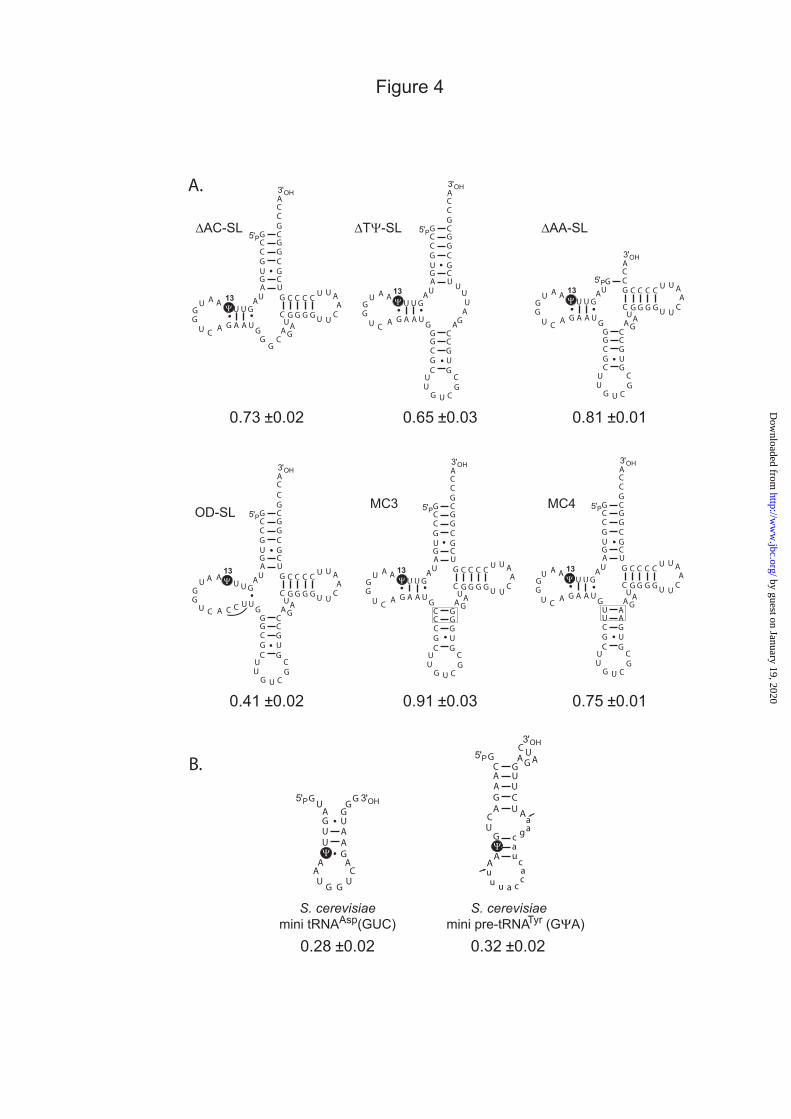
activity, we produced a large series of tRNAAsp(GUC) variants. The 3D tRNA structure was disrupted to different extent in all these variants, most important perturbation was introduced in the variant ∆TΨ-SL (Fig. 4A) where the TΨ-stem-loop, which interacts with the D-loop, was deleted. In the variants ∆AC-SL and ∆AA-SL (Fig. 4A) the anticodon stem-loop and acceptor stem, respectively, were missing. In MC3 and MC4 variants the anticodon stem was either slightly stabilized (MC3) or slightly destabilized (MC4). For each of these RNAs, 3 independent series of in vitro modification experiments were performed in the excess of RNA as compared to Pus7p (Fig. 4A). As already demonstrated above, point mutations that destabilize the tRNA D-stem have only a limited effect on the Pus7p activity (Table I and Fig. 2). To go one step further in the destabilization of this stem, the sequence of its 3’-strand was completely mutated in variant OD-SL (Fig. 4A). Finally, two minisubstrates corresponding to the D stem-loop of tRNAAsp(GUC) and to the anticodon stem-loop containing the intron of the pre-tRNATyr(GΨA), respectively, were produced (Fig. 4B). Altogether, the results of these experiments with all 8 RNA variants produced (Fig. 4) confirmed that the tRNA 3D structure is not really required for Pus7p activity. The deletion of the acceptor stem (∆AA-SL) had almost no effect on the activity, while elimination of one of the stem-loop structures other than the D stem-loop (∆AC-SL, ∆TΨ-SL) or the destabilization of the anticodon stem loop (MC4) only slightly decreased the yield of modification. A stronger negative effect was observed when the conformation of D stem-loop where modification occurs was altered (level of Ψ residue formed in OD-SL is divided by 2) and a quite low, but detectable, level of modification was obtained for the two mini-substrates (28 and 32%) (Fig. 4B). We noticed a slight increase of Ψ formation upon stabilization of the anticodon stem (variant MC3). These data suggested that at least one stem-loop structure, containing the residue to be modified, is required for Pus7p activity. In addition, the presence of other stable stem-loops, which may facilitate the overall folding of the tRNA, probably also favours Pus7p activity. Ψ35 can be formed in the mature tRNATyr(GΨA) in vitro. Alignment of the sequence surrounding
by guest on January 19, 2020http://w
ww
.jbc.org/D
ownloaded from
7
Ψ35 in the mature S. cerevisiae tRNATyr(GΨA) with the sequences containing Ψ13 in Pus7p tRNA substrates, reveals that intronless tRNATyr(GΨA) fits almost perfectly to the consensus sequence of Pus7p RNA substrates (Fig. 5A). The only difference is the substitution of the pyrimidine at position +3 by an A residue. As shown above, substitution of this pyrimidine at position +3 by an A residue in both tRNAAsp(GUC) and pre-tRNATyr(GΨA) had no marked effect on the yield of U to Ψ conversion by Pus7p in vitro. Therefore, one might expect that the recombinant Pus7p is able to modify the mature tRNATyr (GΨA) in vitro. To verify this hypothesis, modification experiments were performed in parallel on the precursor and the mature tRNATyr(GΨA), using the excess of RNA substrate. The yield of modification for both RNA transcripts was tested after incubation with either 1 nM, 10 nM or 1 µM of the recombinant Pus7p. Interestingly, whereas 81% of U35 was converted into Ψ35 in the pre-tRNATyr(GΨA) upon incubation with 10 nM Pus7p, no significant Ψ35 formation was detected in the mature RNA under these conditions (Fig. 5A). However, when the protein concentration was increased to 1 µM, even mature tRNATyr(GΨA) transcript was modified to a significant extent (63%). The modification did occur at position 35, since it was not observed in the same transcript bearing U35C substitution (Fig. 5B). How one may explain the difference in modification of tRNAAsp(GUC) variants and mature tRNATyr(GΨA)? Indeed, A+3 variants of tRNAAsp(GUC) or pre-tRNATyr(GΨA) are modified by 10 nM Pus7p to 76-79% (Table I), whereas mature tRNATyr(GΨA), which has the same sequence surrounding U35, remains unmodified. One possible explanation could be the highly constrained 3D structure of the anticodon loop containing U35 in mature tRNATyr(GΨA). Indeed, the residue U33 (position -2) in the anticodon loop is known to be involved in a peculiar ribose-phosphate backbone folding that is present in some tRNA (U33-turn). The stability of this U33-turn has been attributed to the formation of three interactions involving U33: one hydrogen bond formed by the 2’-OH of residue U33 and the N-7 of purine residue at position 35, a second hydrogen bond formed between the N3-H of residue U33 and the phosphate of the nucleotide N36, and the stacking of residue U33 on the residue 35 (35). Therefore, the residue U33 that
is the important U-2 residue in the conserved sequence of Pus7p substrates might have an inappropriate conformation in the anticodon loop of the mature tRNATyr (GΨA). The other residue essential for Pus7 activity (position +1 in the Pus7 conserved sequence, A36 in mature tRNATyr (GΨA)) might also be poorly accessible in the anticodon loop. If this was the case, insertions of residues between positions 31 and 32 in the anticodon loop would abolish the 3D structure constraints and therefore reinforce Pus7p activity. Indeed, we observed a significant Ψ35 formation in both +A and +AA variants of mature tRNATyr(GΨA) upon their incubation in the presence of 10 nM Pus7p. These two variants are modified to 61% and 58%, respectively (Fig. 5C). In contrast, the A38U substitution in the loop that restored the complete Pus7p consensus sequence did not allow Ψ35 formation in the mature tRNATyr(GΨA) when the same enzymatic conditions were used (10 nM of Pus7p enzyme) (Fig. 5C). These data strongly suggest that the 3D structure of the anticodon loop in mature tRNATyr(GΨA) strongly limits its modification by Pus7p. The presence of 2D structural motifs is required for efficient modification of U2 snRNA in vitro. In the 5’-terminal region of yeast U2 snRNA, residue U35 is flanked by two stem-loop structures I and IIa. Stem-loop IIa is immediately followed by a third stem-loop structure IIb and a pseudoknot can be formed between the terminal stem-loop IIa and the single-stranded segment located downstream from stem-loop IIb (Fig. 6 A). An additional potential target uridine surrounded by a consensus sequence is present in yeast U2 snRNA at position 56, however, this nucleotide is not modified neither in vivo, nor in vitro, using the recombinant Pus7p. To test the importance of the U2 snRNA 2D structure for Pus7p activity, we first eliminated the sequence downstream from position 85 in order to disrupt the pseudoknot structure (variant U2:1-85). Then, the stem-loop structures I and IIb were individually or simultaneously deleted (variants U2:1-85 ∆I, U2:1-85 ∆IIb and U2:1-85 ∆I∆IIb) and all three stem-loop structures were eliminated in the RNA variant U2:28-47 (Fig. 6B). Disruption of the pseudoknot structure only slightly decreased the yield of Ψ35 formation. In agreement with previous data (4), elimination of stem-loop structure I also had only a limited effect on the yield of Ψ35 formation,
by guest on January 19, 2020http://w
ww
.jbc.org/D
ownloaded from
8
whereas deletion of stem-loop IIb alone or together with stem-loop I decreased the level of modification by a factor of about 2. In addition, Pus7p did not modify the RNA variant U2:28-47 without any double-stranded region. Therefore, altogether the data obtained on both U2 snRNA and the tRNAAsp(GUC) strongly suggested that the presence of stable 2D structure motifs increases the level of in vitro activity of Pus7p.
DISCUSSION In this study, we performed a deep characterization of the RNA sequence and structure requirements needed for modification of three distinct RNA substrates by the yeast RNA:Ψ-synthase Pus7p. The data obtained are as much important as yeast Pus7p is presently the RNA:Ψ-synthase with the largest spectrum of identified RNA substrates (tRNA, pre-tRNATyr(GΨA), U2 snRNA and 5S rRNA). Our data obtained in vitro point out the importance of a given sequence in a flexible RNA segment for efficient Pus7p recognition and modification. However, both in vitro and in vivo, many potential U to Ψ conversion sites are not modified by Pus7p. Even if the sequence of these RNA segments is suitable for recognition, their flexibility may not be sufficient for correct access of the target uridine into Pus7p active site. In addition, such potential target uridines may by buried inside of stable RNA structures or covered by numerous associated proteins in vivo. The competition between the cognate and near-cognate RNA substrates may also influence the outcome of the reaction in the cell. All these parameters probably play an important role in the choice of RNA substrates by Pus7p. Point sequence determinants are required for Pus7p activity on any of its substrates. Sequence alignments of all yeast Pus7p substrates reveals that the target U residue is located in an 7-long nt sequence with three strictly conserved residues (the target U residue, U at position -2 and A at position +1), the other residues being semi- or partially conserved, except the nucleotide at position -1. Here, we demonstrate that the three strictly conserved residues are absolutely required for Pus7p activity on three of its substrates (tRNAAsp(GUC), pre-tRNATyr(GΨA) and U2 snRNA). Both purine-purine and pyrimidine-pyrimidine replacements at positions -2 and +1, respectively, abolish the activity. Therefore we show that the U33N34U35A36
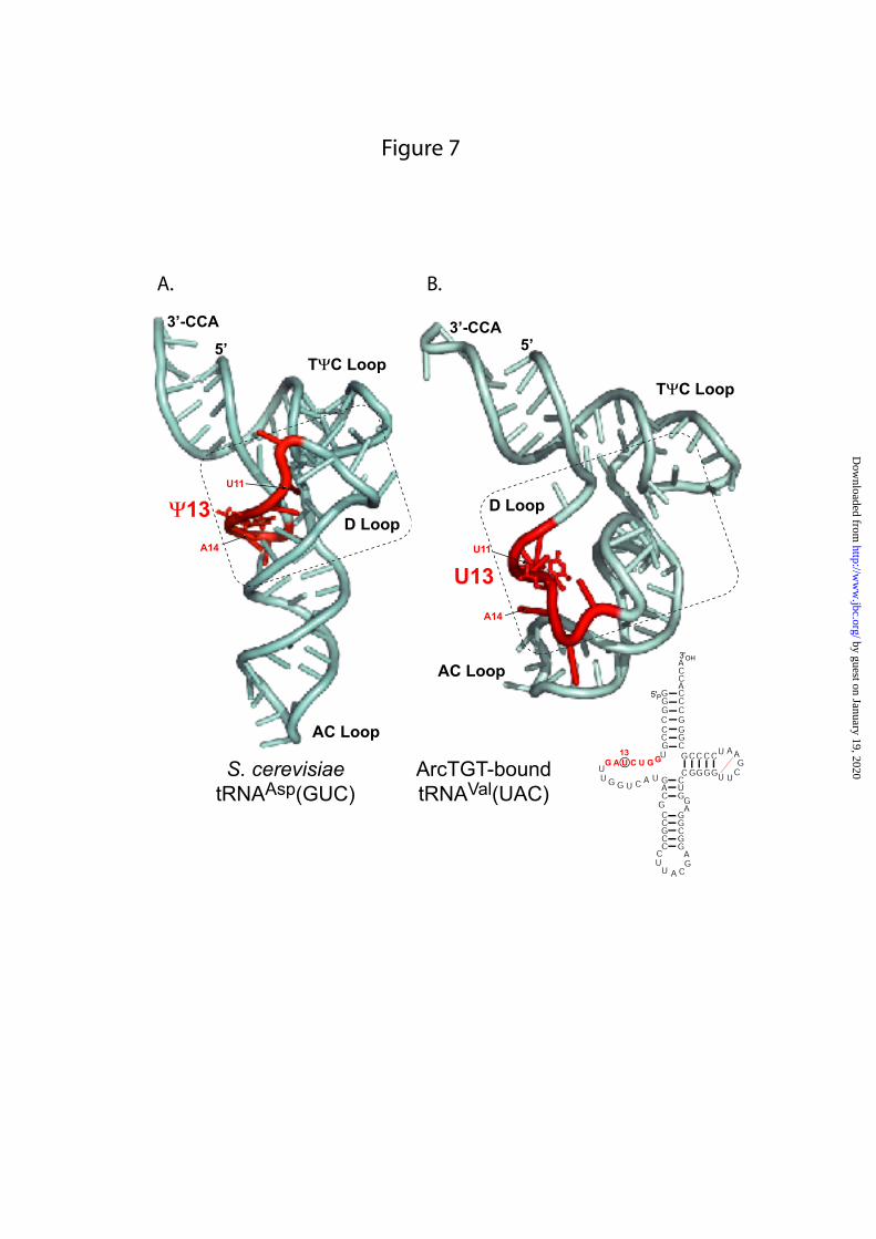
sequence previously found to be required for Ψ35-forming activity in plant pre-tRNATyr(GΨA) is needed for all Pus7p substrates. These residues may be necessary for stabilization of “active” RNA substrate conformation, as well as for stable anchoring of the RNA substrate onto the enzyme, or for catalytic activity. Residue at position +2 in the consensus sequence is important for the yield of modification, but is not essential as are residues U-2 and A+1. The identities of the other partially conserved residues of the 7-nt sequence only have a moderate effect on the modification yield and these effects vary from one substrate to another, probably due to differences in their respective RNA 2D structures. In the present in vitro tests, we compared the efficiency of variant RNA modification using an end-point measurement. These differences in modification efficiency may be even higher in terms of initial modification rate. Remodeling of the tRNA structure is expected to occur for Ψ13 formation by Pus7p. In the L-shaped canonical 3D structure of tRNAs, the essential U-2 and A+1 residues in RNA substrates are not accessible for the enzyme. Both of them are involved in base-pair interactions: U11 (U-2) base pairs with A24 in the D stem, while A14 (A+1) forms a tertiary interaction with U8. Furthermore, they are buried into the molecule due to global tRNA 3D structure. Thus, the isomerization of U to Ψ at position 13 requires considerable structural remodelling. The 3D structure of the complex formed by a tRNA and another RNA modification enzyme acting on a residue in the D-loop has been solved, namely, the complex formed by the archaeal tRNA-guanine transglycosylase (TGT) which converts G at the position 15 into archaeosine in many archaeal tRNAs and one of its tRNA substrate Fig. 7 (36). This tRNA carries a U11, U13 and A14 residues like Pus7p substrates. In the 3D structure, it fits into the enzyme active site in an open conformation called the lambda-tRNA (λ-tRNA) conformation (36). This conformation is characterized by the opening of the D-stem loop and the interaction of its 3’-strand with the variable loop (36). By inspection of yeast tRNA sequences we noticed that from three to five contiguous and non-contiguous base pairs can always be formed between the 3’-strand of the D-stem and the variable loop, so that all Pus7p tRNA substrates may be able to adopt λ-shaped structure (data not shown). In this conformation,
by guest on January 19, 2020http://w
ww
.jbc.org/D
ownloaded from

9
the 5’-strand of the D-stem containing U11 and U13 would be free of base-pair interactions and these two essential residues as well as residue A14 would become easily accessible as compared to the canonical L-shaped tRNA structure (Fig. 7). Moreover, the conformation of the E. coli TruD active site observed in the crystal does not allow fitting the tRNA substrate in canonical L-form, while the alternative λ-form can be accommodated (37). Taken these arguments together, one can guess that by interaction with its tRNA substrate, Pus7p converts the L-shaped tRNA into a λ-like shaped tRNA so that the three identity elements (U11, U13 and A14) become available allowing U13 to Ψ13 conversion by the catalytic Asp256 residue. The intron in tRNATyr(GΨA) precursor may avoid 3D structural constraints limiting Pus7p activity on the mature tRNATyr(GΨA). The presence on an intron in pre-tRNATyr(GΨA) as well as the presence of a Ψ35 residue in mature tRNATyr(GΨA) are general features of Eukarya. Previous work of J. Abelson’s group (38) demonstrated that the absence of the intervening sequence in the yeast RNATyr ochre suppressor gene (SUP6) leads to the absence of Ψ35 modification in the mature tRNATyr(GΨA) in vivo. Later on, studies performed by RNA microinjection in X. laevis oocytes showed that Ψ35 could be formed in both X. laevis and Drosophila pre-tRNATyr(GΨA), in spite of the strong sequence and length differences of the introns of these two precursors (39-41). The conclusion of this study was that the tRNA:Ψ35-synthase needs the intron for tRNATyr(GΨA) modification, but that Ψ formation is not dependent on the size and sequence of the intervening sequence. Similar observation was made for the plant pre-tRNATyr(GΨA): the presence of an intron is required to get modification and only the identity of residues U33, A36 and A37 have a strong influence on the activity (25). Further studies of Ψ35 formation in plant tRNATyr pointed out the moderate importance of C32 (position -3) and the length of the intron, the minimal size still supporting modification was found to be of 7 nt (42). Here we show that the recombinant Pus7p can form Ψ35 residue in the mature tRNATyr(GΨA) but only when used at rather high concentration (1 µM), which is probably much superior to that found in vivo. Indeed, experimental measurements have indicated that Pus7p is
moderately expressed in S. cerevisiae; its estimated amount is about 4000 molecules/cell (see http://yeastgfp.ucsf.edu/). We could markedly increase the activity of Pus7p on the mature tRNATyr(GΨA) by insertion of one or two A residues in the anticodon loop, suggesting that the low activity of Pus7p on the mature tRNATyr(GΨA) is probably due to the inability of Pus7p to disrupt the 3D structure of the tRNATyr(GΨA) anticodon loop. The presence of an intron in the anticodon loop of all eukaryal pre-tRNATyr(GΨA) probably relieves the 3D structural constraints and therefore allows Pus7p activity on this tRNAs. Therefore, Pus7p has probably the capability to remodel the L-shaped structure of the tRNAs into the more open λ-form, but is not able to change quite rigid conformation of the anticodon loop of mature tRNA. In this case, the solution retained is the course of evolution for modification at position 35 was the insertion of an intron in the pre-tRNATyr(GΨA). Stable double-stranded RNA regions are required for Pus7p activity. Whereas tight 3D structure may be a handicap for Pus7p activity as seen for tRNATyr(GΨA), a substrate without any double-stranded region is not modified by Pus7p as evidenced for the single-stranded U2 snRNA segment 28 to 47 containing the conserved 7-nt long sequence. More generally, all the mutations that decreased the number of stem-loop structures in RNA substrate diminished the modification level upon incubation with Pus7p. This was the case for variants of both tRNAAsp(GUC) (∆TΨ-SL, ∆AC-SL and ∆AA-SL) and 5’-terminal region of U2 snRNA (U2:1-85∆I, U2:1-85 ∆IIb, U2:1-85 ∆I∆IIb and U2:28-47). Similarly, minisubstrates corresponding to the D stem-loop of tRNAAsp(GUC) or the anticodon stem-loop of the pre-tRNATyr(GΨA) were modified at a low level. In the case of U2 snRNA, deletion of one stem-loop structure IIb had a particularly strong effect on RNA modification. Mutation at position -4 in the pre-tRNATyr(GΨA) which disrupts the intron structure also had a strong negative effect as well as disruption of the D stem-loop structure in tRNAAsp(GUC). Hence, either Pus7p has numerous specific binding sites for recognition of each of its substrates in their integrity or more likely, it contains binding sites able to recognize one stem-loop structure including or very close to the target residue and site recognizing with a looser specificity. The specific recognition of
by guest on January 19, 2020http://w
ww
.jbc.org/D
ownloaded from

10
substrate may be ensured by interaction with the three point identity determinants and one stem-loop structure. The presence of poor specific recognition sites might reinforce the stability of the RNA-protein complex and therefore the yield of the reaction. Numerous Pus7p orthologs have been identified in all kingdoms of life (2). Comparison of these proteins reveals an increase of size from Bacteria to Archaea and a greater one from Archaea to Eukarya. This size increase may be linked to the presence of additional domains needed to accommodate a larger number of RNA substrates in Eukarya compared to Bacteria (pre-tRNATyr(GΨA), 5S rRNA and U2 snRNA). Alternatively, these additional domains may help to maintain the specificity of eukaryotic Pus7p and to avoid unproductive modification of non-cognate RNA substrates. Further studies are required to verify these hypotheses. Inspection of the amino acid sequence of Pus7p-like enzymes does not reveal the presence of domain belonging to the double-stranded RNA binding domain family (DSRBD) that might explain a reinforced binding in the presence of several stem-loop structures. We did not detect strong sequence homologies between the Pus7p-like proteins outside of the catalytic domain. Further structural analysis of Pus7p is required to understand how this enzyme recruits a large variety of substrates and accommodates them into its active site. Mitochondrial yeast tRNAs carry the conserved sequence of Pus7p substrates but are not modified in vivo. Interestingly, 6 out of 17 yeast mitochondrial tRNAs carry a U13 residue with a surrounding sequence that fits perfectly to the consensus sequence of Pus7p substrates. Although we show that Pus7p can modify them in vitro, they do not carry a Ψ13 residue in vivo. This is probably explained by the absence of
import of Pus7p in the mitochondria. Indeed, expression of GFP-Pus7p fusions revealed that its location is mainly nuclear (43). Thus, pre-tRNATyr(GΨA), 5S rRNA and probably other tRNAs are modified by Pus7p in the nuclear compartment, since only a small fraction of GFP-Pus7p protein was detected in the cytoplasm (43). Functional importance of Pus7p-catalyzed modification of cellular RNAs. The exact functional roles of Ψ50 in 5S rRNA and Ψ13 in several tRNA are not clearly established; however, these pseudouridine residues may stabilize the local RNA conformation and/or favour interactions with protein partners. In contrast, more experimental data were accumulated for importance of Ψ35 in eukaryotic tRNATyr(GΨA). First of all, the conformation of the anticodon loop of tRNATyr(GΨA) is stabilized by the presence of Ψ35 (44,45). In addition, the presence of Ψ35 in the middle anticodon position of tRNATyr influences its suppressor activity towards stop codons UAG and UAA (41). These data show that Ψ35 plays an important role in the stabilization of the codon-anticodon interaction between mRNA and tRNATyr(GΨA) and thus increases its translation efficiency. On the other hand, Ψ35 in U2 snRNA should play an important role in the efficiency of the first step of the splicing reaction (46). Structural data clearly indicate that Ψ35 in U2 snRNA induces particular conformation of the mRNA-U2 snRNA duplex and favor the extra-helical conformation of the branch-point adenosine (47,48). Hence, there are strong arguments explaining the need for a cellular Pus7p activity (at least production of functional U2 snRNA and tRNATyr(GΨA)).
REFERENCES
1. Koonin, E. V. (1996) Nucleic Acids Res. 24, 2411-2415 2. Kaya, Y., and Ofengand, J. (2003) RNA 9, 711-721 3. Roovers, M., Hale, C., Tricot, C., Terns, M. P., Terns, R. M., Grosjean, H., and
Droogmans, L. (2006) Nucleic Acids Res. 34, 4293-4301 4. Ma, X., Zhao, X., and Yu, Y. T. (2003) EMBO J. 22, 1889-1897 5. Behm-Ansmant, I., Urban, A., Ma, X., Yu, Y. T., Motorin, Y., and Branlant, C. (2003)
RNA 9, 1371-1382 6. Decatur, W. A., and Schnare, M. N. (2008) Mol. Cell. Biol. 28, 3089-3100 7. Hur, S., Stroud, R. M., and Finer-Moore, J. (2006) J. Biol. Chem. 281, 38969-38973
by guest on January 19, 2020http://w
ww
.jbc.org/D
ownloaded from
11
8. Ansmant, I., Massenet, S., Grosjean, H., Motorin, Y., and Branlant, C. (2000) Nucleic Acids Res. 28, 1941-1946
9. Ansmant, I., Motorin, Y., Massenet, S., Grosjean, H., and Branlant, C. (2001) J. Biol. Chem. 276, 34934-34940
10. Behm-Ansmant, I., Grosjean, H., Massenet, S., Motorin, Y., and Branlant, C. (2004) J. Biol. Chem. 279, 52998-53006
11. Becker, H. F., Motorin, Y., Planta, R. J., and Grosjean, H. (1997) Nucleic Acids Res. 25, 4493-4499
12. Becker, H. F., Motorin, Y., Sissler, M., Florentz, C., and Grosjean, H. (1997) J. Mol. Biol. 274, 505-518
13. Gu, X., Yu, M., Ivanetich, K. M., and Santi, D. V. (1998) Biochemistry 37, 339-343 14. Massenet, S., Motorin, Y., Lafontaine, D. L., Hurt, E. C., Grosjean, H., and Branlant,
C. (1999) Mol. Cell. Biol. 19, 2142-2154 15. Motorin, Y., Keith, G., Simon, C., Foiret, D., Simos, G., Hurt, E., and Grosjean, H.
(1998) RNA 4, 856-869 16. Behm-Ansmant, I., Massenet, S., Immel, F., Patton, J. R., Motorin, Y., and Branlant,
C. (2006) RNA 12, 1583-1593 17. Moras, D., Comarmond, M. B., Fischer, J., Weiss, R., Thierry, J. C., Ebel, J. P., and
Giege, R. (1980) Nature 288, 669-674 18. Swerdlow, H., and Guthrie, C. (1984) J. Biol. Chem. 259, 5197-5207 19. Kiparisov, S., Petrov, A., Meskauskas, A., Sergiev, P. V., Dontsova, O. A., and
Dinman, J. D. (2005) Mol. Genet. Genomics 274, 235-247 20. Nishikawa, K., and Takemura, S. (1974) FEBS Lett. 40, 106-109 21. Smith, M. W., Meskauskas, A., Wang, P., Sergiev, P. V., and Dinman, J. D. (2001)
Mol. Cell. Biol. 21, 8264-8275 22. Szymanski, M., Barciszewska, M. Z., Erdmann, V. A., and Barciszewski, J. (2002)
Nucleic Acids Res. 30, 176-178 23. Branlant, C., Krol, A., Ebel, J. P., Lazar, E., Haendler, B., and Jacob, M. (1982)
EMBO J. 1, 1259-1265 24. Ares, M., Jr. (1986) Cell 47, 49-59 25. Szweykowska-Kulinska, Z., and Beier, H. (1992) EMBO J. 11, 1907-1912 26. Jiang, H. Q., Motorin, Y., Jin, Y. X., and Grosjean, H. (1997) Nucleic Acids Res. 25,
2694-2701 27. Studier, F. W. (2005) Protein Expr. Purif. 41, 207-234 28. Keith, G. (1995) Biochimie 77, 142-144 29. Sprinzl, M., and Vassilenko, K. S. (2005) Nucleic Acids Res. 33, D139-140 30. Claros, M. G., and Vincens, P. (1996) Eur. J. Biochem. 241, 779-786 31. Bhasin, M., and Raghava, G. P. (2004) Nucleic Acids Res. 32, W414-419 32. Drawid, A., and Gerstein, M. (2000) J. Mol. Biol. 301, 1059-1075 33. Hua, S., and Sun, Z. (2001) Bioinformatics 17, 721-728 34. Nakai, K., and Horton, P. (1999) Trends Biochem. Sci. 24, 34-36 35. Quigley, G. J., and Rich, A. (1976) Science 194, 796-806 36. Ishitani, R., Nureki, O., Nameki, N., Okada, N., Nishimura, S., and Yokoyama, S.
(2003) Cell 113, 383-394 37. Ericsson, U. B., Nordlund, P., and Hallberg, B. M. (2004) FEBS Lett. 565, 59-64 38. Johnson, P. F., and Abelson, J. (1983) Nature 302, 681-687 39. Choffat, Y., Suter, B., Behra, R., and Kubli, E. (1988) Mol. Cell. Biol. 8, 3332-3337 40. van Tol, H., and Beier, H. (1988) Nucleic Acids Res. 16, 1951-1966 41. Zerfass, K., and Beier, H. (1992) Nucleic Acids Res. 20, 5911-5918
by guest on January 19, 2020http://w
ww
.jbc.org/D
ownloaded from
12
42. Pienkowska, J., Michalowski, D., Krzyzosiak, W. J., and Szweykowska-Kulinska, Z. (2002) Biochim. Biophys. Acta 1574, 137-144
43. Huh, W. K., Falvo, J. V., Gerke, L. C., Carroll, A. S., Howson, R. W., Weissman, J. S., and O'Shea, E. K. (2003) Nature 425, 686-691
44. Auffinger, P., and Westhof, E. (1998) J. Biomol. Struct. Dyn. 16, 693-707 45. Auffinger, P., and Westhof, E. (2001) RNA 7, 334-341 46. Yang, C., McPheeters, D. S., and Yu, Y. T. (2005) J. Biol. Chem. 280, 6655-6662 47. Newby, M. I., and Greenbaum, N. L. (2002) Nat. Struct. Biol. 9, 958-965 48. Lin, Y., and Kielkopf, C. L. (2008) Biochemistry 47, 5503-5514
FOOTNOTES We thank C. Florentz and R. Giegé (CNRS, Strasbourg, France) for providing plasmids containing the yeast tRNAAsp and its variants, P. Fabrizio for providing plasmid pT7U2Sc. This work was supported by laboratory funds from the CNRS and the “Ministère de la jeunesse, de l’éducation nationale et de la recherche”. A. Urban was a pre-doctoral fellow from the “Ministère de la jeunesse, de l’éducation nationale et de la recherche”. The abbreviation used are : PUS – PseudoUridine Synthase,
by guest on January 19, 2020http://w
ww
.jbc.org/D
ownloaded from

13
TABLES
Table I : Effect of mutations in tRNAAsp and pre-tRNATyr transcripts on their modification by recombinant Pus7p in catalytic reaction conditions.
WT 0.84 ±0.03 WT 0.83 ±0.02A9C 0.74 ±0.06 A31C 0.46 ±0.03A9G 0.65 ±0.05 A31G 0.41 ±0.03A9U 0.70 ±0.06 A31U 0.48 ±0.02
G10A 0.56 ±0.03 C32A 0.84 ±0.04G10C 0.71 ±0.03 C32G 0.69 ±0.02G10U 0.64 ±0.03 C32U 0.74 ±0.03U11A 0.01 ±0.05U11C 0.04 ±0.01U11G 0.02 ±0.02
U11A/A24U 0.02 ±0.03U11C/A24G 0.01 ±0.02U11G/A24C 0.01 ±0.03
A14C 0.05 ±0.01 A36C 0.09 ±0.05A14G 0.05 ±0.02 A36G 0.03 ±0.01A14U 0.02 ±0.01 A36U 0.00 ±0.01A15C 0.24 ±0.02 A37C 0.22 ±0.01A15G 0.38 ±0.03 A37G 0.31 ±0.02A15U 0.46 ±0.03 A37U 0.39 ±0.02
tRNAAsp(GUC) pre-tRNATyr(GΨA)Ψ quantification
(mol Ψ/mol of tRNA)Ψ quantification
(mol Ψ/mol of tRNA)
0.79 ±0.03 U38A 0.76 ±0.02
Ψ U13C
+1
+2
+3 U16A
0.00 ±0.01 U35C
U33G 0.06 ±0.01
0.81 ±0.03
0.03 ±0.01
-1 U12C 0.81 ±0.03 U34C
0.03 ±0.01
U33C 0.04 ±0.01
Position
-4
-3
-2U33A
Modification level is expressed in mole of pseudouridine per mole of tRNA after 90 min incubation with the recombinant Pus7p at 30°C as described in Materials and Methods (the excess of RNA substrate). The identity of each mutation is indicated on the left of the measured ratio. Each value (mean ±confidence interval) was obtained from at least three independent experiments.
by guest on January 19, 2020http://w
ww
.jbc.org/D
ownloaded from
14
FIGURE LEGENDS
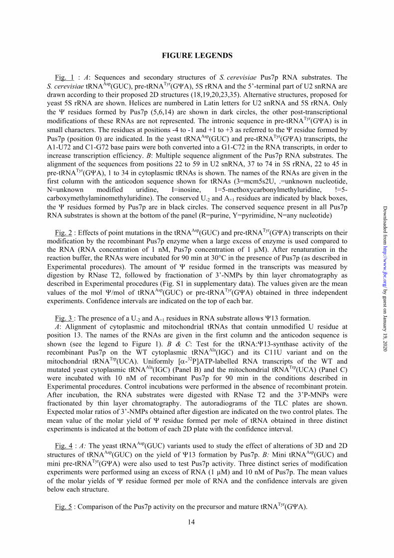
Fig. 1 : A: Sequences and secondary structures of S. cerevisiae Pus7p RNA substrates. The S. cerevisiae tRNAAsp(GUC), pre-tRNATyr(GΨA), 5S rRNA and the 5’-terminal part of U2 snRNA are drawn according to their proposed 2D structures (18,19,20,23,35). Alternative structures, proposed for yeast 5S rRNA are shown. Helices are numbered in Latin letters for U2 snRNA and 5S rRNA. Only the Ψ residues formed by Pus7p (5,6,14) are shown in dark circles, the other post-transcriptional modifications of these RNAs are not represented. The intronic sequence in pre-tRNATyr(GΨA) is in small characters. The residues at positions -4 to -1 and +1 to +3 as referred to the Ψ residue formed by Pus7p (position 0) are indicated. In the yeast tRNAAsp(GUC) and pre-tRNATyr(GΨA) transcripts, the A1-U72 and C1-G72 base pairs were both converted into a G1-C72 in the RNA transcripts, in order to increase transcription efficiency. B: Multiple sequence alignment of the Pus7p RNA substrates. The alignment of the sequences from positions 22 to 59 in U2 snRNA, 37 to 74 in 5S rRNA, 22 to 45 in pre-tRNATyr(GΨA), 1 to 34 in cytoplasmic tRNAs is shown. The names of the RNAs are given in the first column with the anticodon sequence shown for tRNAs (3=mcm5s2U, .=unknown nucleotide, N=unknown modified uridine, I=inosine, 1=5-methoxycarbonylmethyluridine, !=5-carboxymethylaminomethyluridine). The conserved U-2 and A+1 residues are indicated by black boxes, the Ψ residues formed by Pus7p are in black circles. The conserved sequence present in all Pus7p RNA substrates is shown at the bottom of the panel (R=purine, Y=pyrimidine, N=any nucleotide)
Fig. 2 : Effects of point mutations in the tRNAAsp(GUC) and pre-tRNATyr(GΨA) transcripts on their
modification by the recombinant Pus7p enzyme when a large excess of enzyme is used compared to the RNA (RNA concentration of 1 nM, Pus7p concentration of 1 µM). After renaturation in the reaction buffer, the RNAs were incubated for 90 min at 30°C in the presence of Pus7p (as described in Experimental procedures). The amount of Ψ residue formed in the transcripts was measured by digestion by RNase T2, followed by fractionation of 3’-NMPs by thin layer chromatography as described in Experimental procedures (Fig. S1 in supplementary data). The values given are the mean values of the mol Ψ/mol of tRNAAsp(GUC) or pre-tRNATyr(GΨA) obtained in three independent experiments. Confidence intervals are indicated on the top of each bar.
Fig. 3 : The presence of a U-2 and A+1 residues in RNA substrate allows Ψ13 formation. A: Alignment of cytoplasmic and mitochondrial tRNAs that contain unmodified U residue at
position 13. The names of the RNAs are given in the first column and the anticodon sequence is shown (see the legend to Figure 1). B & C: Test for the tRNA:Ψ13-synthase activity of the recombinant Pus7p on the WT cytoplasmic tRNAAla(IGC) and its C11U variant and on the mitochondrial tRNATrp(UCA). Uniformly [α-32P]ATP-labelled RNA transcripts of the WT and mutated yeast cytoplasmic tRNAAla(IGC) (Panel B) and the mitochondrial tRNATrp(UCA) (Panel C) were incubated with 10 nM of recombinant Pus7p for 90 min in the conditions described in Experimental procedures. Control incubations were performed in the absence of recombinant protein. After incubation, the RNA substrates were digested with RNase T2 and the 3’P-MNPs were fractionated by thin layer chromatography. The autoradiograms of the TLC plates are shown. Expected molar ratios of 3’-NMPs obtained after digestion are indicated on the two control plates. The mean value of the molar yield of Ψ residue formed per mole of tRNA obtained in three distinct experiments is indicated at the bottom of each 2D plate with the confidence interval.
Fig. 4 : A: The yeast tRNAAsp(GUC) variants used to study the effect of alterations of 3D and 2D
structures of tRNAAsp(GUC) on the yield of Ψ13 formation by Pus7p. B: Mini tRNAAsp(GUC) and mini pre-tRNATyr(GΨA) were also used to test Pus7p activity. Three distinct series of modification experiments were performed using an excess of RNA (1 µM) and 10 nM of Pus7p. The mean values of the molar yields of Ψ residue formed per mole of RNA and the confidence intervals are given below each structure.
Fig. 5 : Comparison of the Pus7p activity on the precursor and mature tRNATyr(GΨA).
by guest on January 19, 2020http://w
ww
.jbc.org/D
ownloaded from

15
A: The sequence around position 35 in the mature cytoplasmic tRNATyr(GΨA) fits to the consensus sequence established for all Pus7p substrates. B: Modification of the tRNATyr(GΨA) precursor and mature tRNATyr(GΨA) using different concentration of the recombinant Pus7p for the incubation (1 nM, 10 nM and 1 µM). The modified transcripts were analysed as described in the legend to Figure 2. The molar amounts of Ψ residue formed per mole of tRNA are indicated at the bottom of the 2D plates. A control assay was done with 1 µM of Pus7p and the tRNATyr U35C variant. C: Modification of tRNATyr(GΨA) variants with an extended anticodon loop or a A38U point mutation. One (+A) or two (+AA) adenosine residues were inserted between nucleotides 31 and 32 (indicated by arrow). Nucleotide A38 (+3) mutated into a U residue is indicated by an arrow. The results of in vitro modification in the presence of 10 nM Pus7p are given on the bottom of the autoradiograms.
Fig. 6 : Presence of stem loop structures IIa and IIb is essential for U2 snRNA modification by
Pus7p. A: Schematic drawing of the 2D structure of the yeast U2 snRNA fragment (U2:1-139). The stem-loops I, IIa and IIb are indicated and the double arrow shows the interaction responsible for formation of the pseudoknot structure. The insert shows the modification of WT U2 snRNA in the presence of 10 nM Pus7p. B: The molar amounts of Ψ residue formed per mole of RNA after Pus7p-catalyzed modification of U2 snRNA truncated variants. The secondary structures and the molar yield of Ψ35 formation are given for each variant.
Fig. 7 : Residues Ψ13 and A14 are exposed at the surface of the λ-shaped tRNA observed in the
crystal structure of ArcTGT-tRNA complex (A) (36) as compared to the canonical L-shaped tRNA structure (B). Ribbon 3D structure models and the 2D secondary structure of the λ-shaped tRNAVal(UAC) are shown. The region corresponding to the 8-nt conserved sequence found in all Pus7p tRNA substrates is coloured in red. Positions of U11, Ψ13 and A14 are shown.
Fig. S1 Supplementary Data 1 A, B & C: In vitro U to Ψ modification of tRNAAsp(GUC), pre-tRNATyr(GΨA) and U2 snRNA and
their variants by recombinant Pus7p. Uniformly [α-32P]NTP-labelled RNA transcripts of each WT and variants RNAs were incubated with 10 nM of the recombinant Pus7p in the conditions described in Experimental procedures. Control incubations were performed in the absence of the recombinant protein. The incubated RNA substrates were digested with RNase T2 and the 3’P-MNPs were fractionated by thin layer chromatography. The autoradiograms of the TLC plates are shown.
by guest on January 19, 2020http://w
ww
.jbc.org/D
ownloaded from
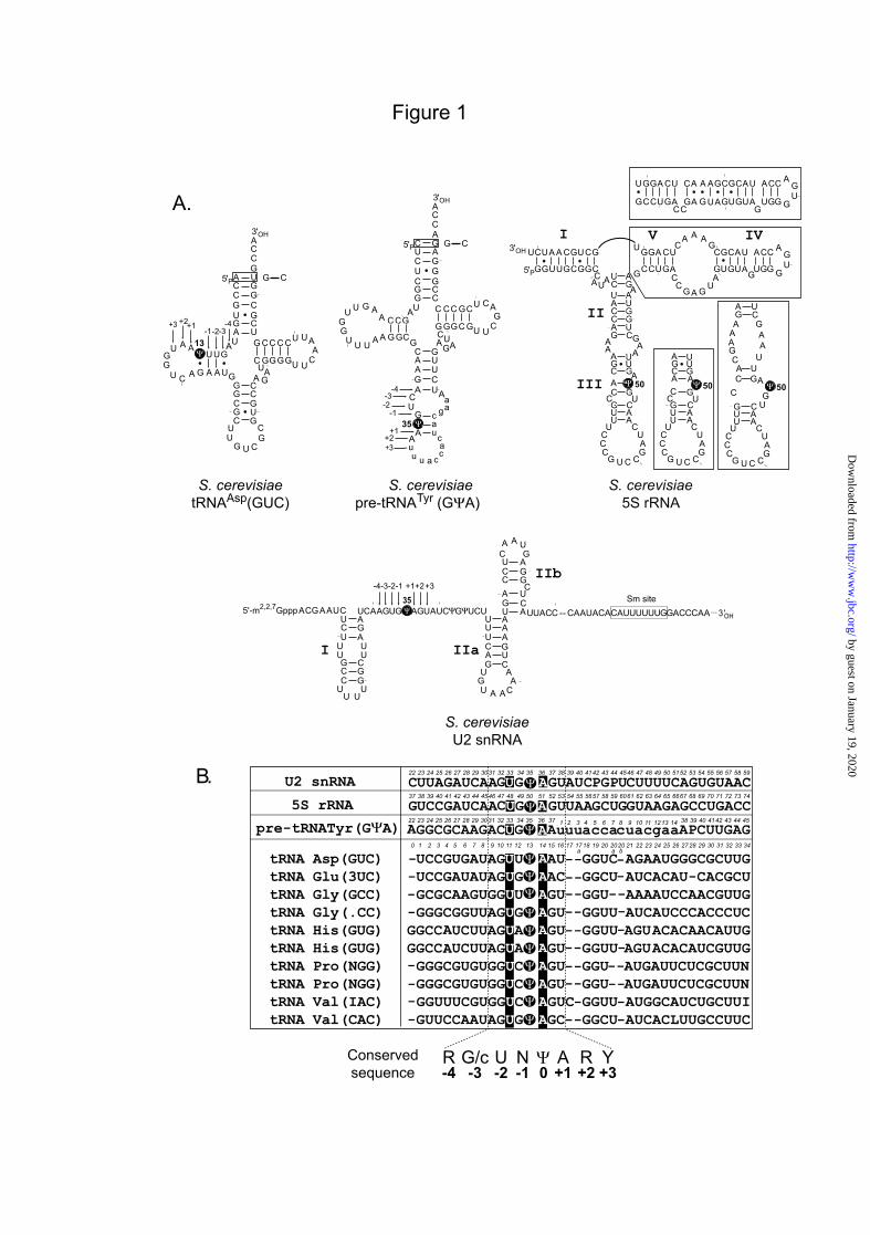
Figure 1
tRNA Asp(GUC) UCCGUGAUAGUU AAU GGUC AGAAUGGGCGCUUGtRNA Glu(3UC) UCCGAUAUAGUG AAC GGCU AUCACAU-CACGCUtRNA Gly(GCC) GCGCAAGUGGUU AGU GGU AAAAUCCAACGUUGtRNA Gly(.CC) GGGCGGUUAGUG AGU GGUU AUCAUCCCACCCUCtRNA His(GUG) GGCC UCUUAGUA AGU GUU AGUACACAACAUUGtRNA His(GUG) GGCC UCUUAGUA AGU GUU AGUACACAUCGUUGtRNA Pro(NGG) GGGCGUGUGGUC AGU GGU AUGAUUCUCGCUUNtRNA Pro(NGG) GGGCGUGUGGUC AGU GGU AUGAUUCUCGCUUNtRNA Val(IAC) GGUUUCGUGGUC AGUC GGUU AUGGCAUCUGCUUItRNA Val(CAC) GUUCCAAUAGUG AGC GGCU AUCACLUUGCCUUC
-----------------
--
-------
GG
------
AA
----
----
GGCGCAAGACUG Au acca uacgaaAPCUUGAGuu cA
UUAGAUCAAGUG AGU CPGP CUUUUCAGUGUAACAU UC
A
U2 snRNA
1 2 3 4 5 6 7 8 9 10 11 1213 14
15 16 17 18 19 20
22 23 24
17a
25 26 27 28 29 3031 32 33 34
2020a b
35 36 37 38 39 40 4142 43 44 4546 47 48 49 50 5152 53 54 55 56 57
22 23 24 25 26 27 28 29 30 31 32 33 3421
58 59
22 23 24 25 26 27 28 29 3031 32 33 34 35 36 37 38 39 40 4142 43 44 45
1 2 3 4 5 6 7 8 9 10 11 12 13 140
Conservedsequence +3
Y-3
G/c-2U
-1N
+1A
-4R
+2R
5S rRNA UCCGAUCAACUG AGU AGCU GUAAGAGCCUGACCUA GG37 38 39 40 41 42 43 44 4546 47 48 49 50 51 52 53 54 55 5657 58 59 6061 62 63 64 65 6667 68 69 70 71 72 73 74
B.
A.
S. cerevisiaetRNAAsp(GUC)
3'OH
-4-3-2
+1 +2
AA 13
5'P
AA A
A
A
A
C
C
C C
CC
G
U
A
GG
G
GUU
U U
U
U
U U
G
U
GG
C
U
G
GC
G
CCA
A
UUG GC GGU
A
CG
C
GU
G
GC
G
CG
UU
G U CGC
CC
A
G
-1 +3
G C
0
pre-tRNATyr(GΨA)
S. cerevisiaeU2 snRNA
... 3 'OH
Sm site35-4-3-2 +1 +2
GA
A
U
CU
U
U
UG
C
U UU
GG
CCC
G
UU U
U
AA
AUU
U
UC
GAG
C
AA
AA
GU
GCA
C
AC
UG
C
AUCA A U
G
I IIa
IIb-1 +3
U GCUAAGCA5'-m2,2,7Gppp CAA UG AGUAUCΨG UΨ CU UUACC -- AC AUA AC AC UUUUUUGGACCCAA
S. cerevisiae5S rRNA
GGUUGCGGCC
AUAUCUACCAG
AA
AGCAC
CGUU
CU
CC
G U C CGA
UC
AAC
UGU
AGUUA
AGC
UGGUAAGA GCCUGA
CC
G A GU
AGUGUA
GUGG G
UG
ACCAUACGCG
AAAC
UCAGGUGCUGCAAUCU3'OH
50
5'P
AGCAC
CGUU A
AC
UG
UAGUU
50
AGA
AAG
CA
C
CGUU
CU
CC
G U C CGA
UC
AAC U
GUAG
UUAA
GCU
50
CU
CC
G U C CGA
UC
GCCUGACC
GA GUAGUGUAG
UGG GUG
ACCAUACGCGAAACUCAGGU
I
II
III
V IV
3'OH
-4-3-2
+1 +2
AG
5'P
AA G
A
G
G
CC
C C
UC
G
U
C
GG
A
GCC
UC
U
C
U C
U
C
AG
C
G
C
GG
G
GCG
A
UUG GG CGUA
UU
G
UC
G
AA
C
AG
CU
CC
A
A
AU
UU
G
AA
u uu
u
a
ac
c
c
ca
gaa
A
35-1
+3
G C
S. cerevisiaepre-tRNATyr (GΨA)
by guest on January 19, 2020http://w
ww
.jbc.org/D
ownloaded from
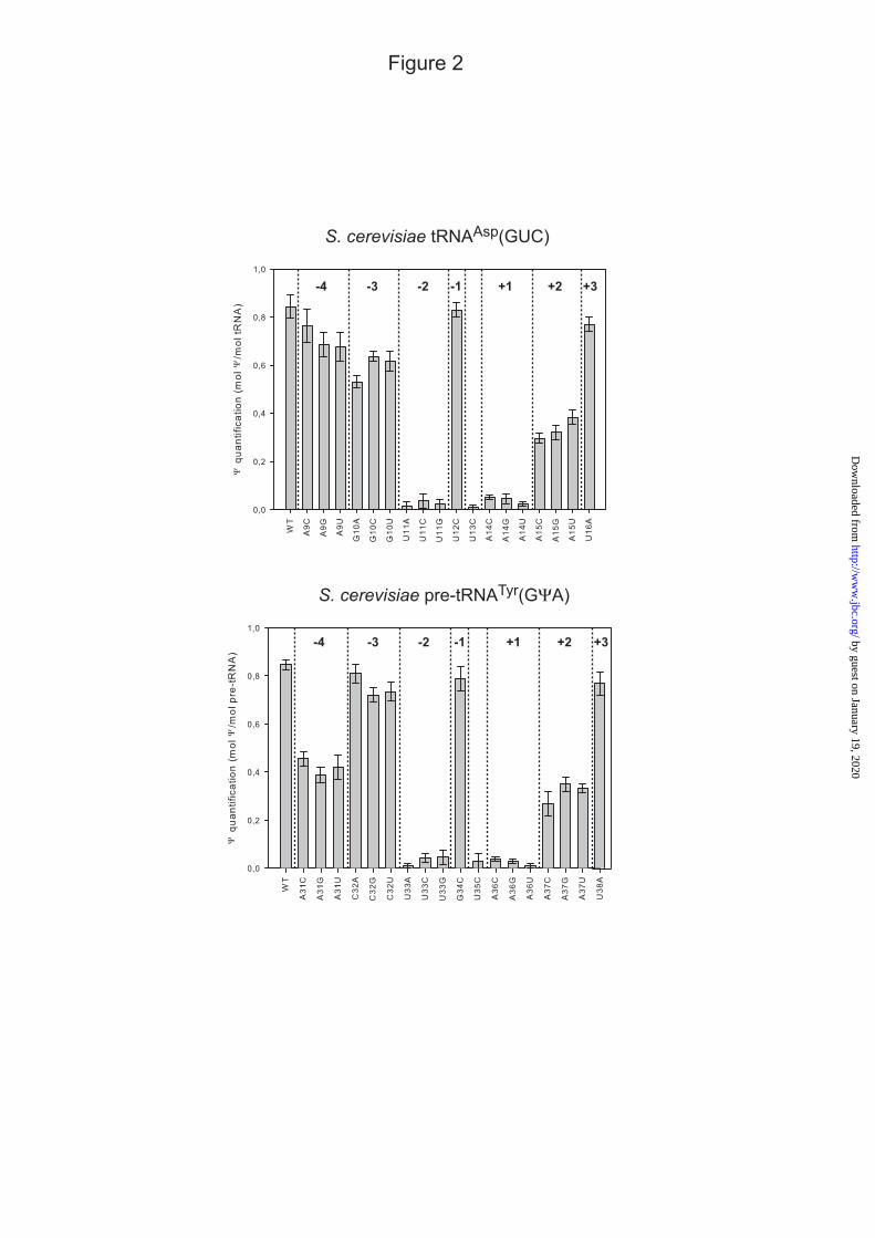
Figure 2
-4 -3 -2 -1 +1 +2 +3
S. cerevisiae tRNAAsp(GUC)
-4 -3 -2 -1 +1 +2 +3
S. cerevisiae pre-tRNATyr(GΨA)
by guest on January 19, 2020http://w
ww
.jbc.org/D
ownloaded from

3 Cp
4 Gp
3 Up
tRNA Ala(IGC)Control
Control tRNA Ala(IGC)C11U
3 Cp4 Gp
3 Up
0.01 ±0.01
0.00 ±0.01
0.04 ±0.02
0.95 ±0.03
3'OH
AG
5'P
UA G
G
C
G
C
U
G C
GG
U
U
G
CG
G
GCG
G C
U
G
U U
C
C
CU
U
C
A
CG
G
CGG
A
UUC GU GCC
A
AG
G
GG
G
UC
C
CC
UU
A G CAU
CC
A
A
U
G
U13
11
Figure 3
S. cerevisiaetRNA Ala (IGC)
S. cerevisiaetRNA Trp (UCA)
mitochondrial
AA
UA A
A
A
A
C
U C
AG
U
U
A
GG A
GUU
U C
U
A
U A
G
U
UC
A
U
U
UC
A
UCG
A
UUG GA AGU
C
AA
U
UC
A
UU
G
AG
UU
U C AAA
CC
A
G
3'OH
5'P
U13
B.
C.
5 Cp
4 Gp 6 Up
Control
0.01 ±0.01 0.77 ±0.02
tRNA Trp(UCA)mitochondrial
9 Ap
UtRNA Ala(IGC) GGGCGUGUGGCG U AGUC GGU AGCGCGCUCCCUUItRNA Arg(1CU) GCUCGCGUGGCG U AAU GGC AACGCGUCUGACUCtRNA Arg(1CU) GCUUGCGUGGCG U AAU GGC AACGCGUCUGACUCtRNA Phe(GAA) GCUUUUAUAGCU U AGU GGU AAAGCGAUAAAUUGtRNA Gly(NCC) AUAGAUAUAAGU U AAUU GGU AAACUGGAUGUCUNtRNA Lys(NUU) GAGAAUAUUGUU U AAU GGU AAAAC-AGUUGUCU
tRNA Arg(ACG) AUAUCUUUAAUU U AAU GGU AAAAUAUUAGAAUAtRNA Arg(NCU) GCUCUCUUAGCU U AAU GGDU AAAGCAUAAUACUNtRNA Thr(UAG) GUAAAUAUAAUU U AAU GGU AAAAUGUAUGUUUAtRNA Trp(!CA) AAGGAUAUAGUU U AAU GGU AAAAC-AGUUGAUUtRNA Ini(CAU) UGCAAUAUGA G U AAUU GGUU AACAUUUUAGGGUC
--
-------
---
---------
------------
---
tRNA Met(CAU) GCUUGUAUAGUU U AAU GGUU AAAACAUUUGUCUNU- -
---
-----
--
----
-
U
15 16 17 18 19 2017a
2020a b
22 23 24 25 26 27 28 29 30 31 32 33 34211 2 3 4 5 6 7 8 9 10 11 12 13 140A.
Conservedsequence +3
Y-3
G/c-2U
-1N
+1A
-4R
+2R
Cytoplasmic
Mitochondrial
0
by guest on January 19, 2020http://w
ww
.jbc.org/D
ownloaded from
3'OH
AA 13
5'P
AA A
A
A
A
C
C
C C
CC
G
U
G
GG
G
GUU
U U
U
U
U U
G
U
GG
C
C
G
GC
G
CCA
A
UUG GC GGU
A
GG
G
GU
G
CC
C
CG
UU
G U CGC
CC
A
GMC3
A.
Figure 4
3'OH
AA
5'P
A A
A
A
A
C
CC
G
U
G
GG
G
GUU
U U
U
UG
U
GG
C
C
GC
GA
U
A
CG
C
GU
G
GC
G
CG
UU
G U CGC
CC
A
G
UU13
B.
0.65 ±0.03
3'OH
AA
5'P
AA A
A
A
A
C
C
C C
CC
G
U
G
GG
G
GUU
U U
U
U
U U
G
U
GG
C
C
G
GC
G
CCA
A
UUG GC GGU
A
AG
A
GU
G
UC
U
CG
UU
G U CGC
CC
A
GMC4
13
3'OH
AA
5'P
AA A
A
AC
C
C CU
G
GG
G
GUU
U U
U U UG
G
CCA
A
UUG GC GGU
A
CG
C
GU
G
GC
G
CG
UU
G U CGC
CC
A
13
0.75 ±0.01
0.81 ±0.01
3'OH
AA
5'P
AA A
A
A
A
C
C
C C
CC
G
U
G
GG
G
GUU
U U
U
U
U U
G
U
GG
C
C
G
GC
G
CCA
A
UUG GC GGU
AC
GG
CC
A
G
G
13
3'OH
AA
5'P
AA
U
A
C
A
C
C
C C
CC
G
U
G
GG
C
GUU
U U
U
U
U U
G
U
GG
C
C
G
GC
G
CCA
A
UUG GC GGU
A
CG
C
GU
G
GC
G
CG
UU
G U CGC
CC
A
G
13
OD-SL
0.73 ±0.02
0.41 ±0.02 0.91 ±0.03
GG
UA
G
GA
GU
A
U
AAU
G GUC
UG
A
3'OH5'P
S. cerevisiaemini tRNAAsp(GUC)
0.28 ±0.02
AC
GU
A
UU
G
UC
G
AA
C
AG
CU
G
AA
u u
u
u
a
ac
c
c
ca
gaa
A
3'OH
5'P
0.32 ±0.02
S. cerevisiaemini pre-tRNATyr (GΨA)
by guest on January 19, 2020http://w
ww
.jbc.org/D
ownloaded from

B.
Figure 5
C.
10 nM
0.81 ±0.02
0.02 ±0.01
1 nM
0.28 ±0.04
0.00 ±0.01
6 Ap
6 Gp 5 Up
5 Cp
4 Ap
5 Gp 3 Up
4 Cp
1 µM
0.86 ±0.03
0.63 ±0.02 0.02 ±0.01
tRNA Tyr U35C
PrecursortRNATyr
MaturetRNATyr
3'OH
AG
5'P
AA G
A
G
G
CC
C C
UC
G
U
G
GG
A
GCC
UC
U
C
U C
U
C
AG
C
C
C
GG
G
GCG
A
UUG GG CG
UA
UU
G
UC
G
AA
C
AG
CU
CC
A
A
AU
UU
G AA
35
A+A / +AA 31
32
4 Ap
5 Gp 2 Up
3 Cp
0.61 ±0.03
tRNATyr+A5 Ap
5 Gp 3 Up
4 Cp
tRNATyr+AA
0.58 ±0.02
6 Ap
5 Gp 3 Up
4 Cp
tRNATyrA38U
0.02 ±0.01
3 Ap
5 Gp 3 Up
4 Cp
+3
UtRNA Tyr(GUA) GGCGCAAGAC G U AAAU UUGA --------------C GA22 23 24 25 26 27 28 29 3031 32 33 34 35 36 37 38 39 40 4142 43 44 45 e
11e13
e15
e14
e16
e12
e17
e1
e2
e3
e4
e5
e27
e26
A.
Conservedsequence +3
Y-3
G/c-2U
-1N
+1A
-4R
+2R
0
S. cerevisiaetRNATyr (GΨA)
by guest on January 19, 2020http://w
ww
.jbc.org/D
ownloaded from
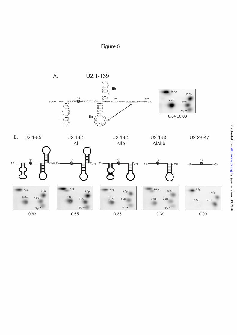
Figure 6
GA
A
U
CU
U
U
UG
C
U UU
GG
CCC
G
UU U
U
AA
AUU
U
UC
GAG
C
AA
AA
G
U
GC
A
C
AC
UG
C
AUCA A U
G
I IIa
IIb
5'p 3'OHUUACC UUUUAAUUUG UUACAAU- -ATG90 139
GACG AAUC
U2:1-139
3510 Cp
6 Gp 10 Up
16 Ap
5'P35
3'OH
U2:1-85
5 Cp
6 Gp 4 Up
7 Ap
0.63
355'P 3'OH
U2:28-47
1 Cp
0 Gp 2 Up
1 Ap
0.00
0.84 ±0.00
5'P35
3'OH
5 Cp
3 Gp 3 Up
7 Ap
0.65
U2:1-85∆I
0.36
5'P35
3'OH
3 Cp
3 Gp 4 Up
6 Ap
U2:1-85∆IIb
5'P
353'OH
3 Cp
3 Gp 3 Up
6 Ap
0.39
U2:1-85∆I∆IIb
B.
A.
by guest on January 19, 2020http://w
ww
.jbc.org/D
ownloaded from

Alan Urban, Isabelle Behm-Ansmant, Christiane Branlant and Yuri Motorin-synthase Pus7pΨmodification by the Saccharomyces cerevisiae RNA:
RNA sequence and 2D structure features required for efficient substrate
published online December 29, 2008J. Biol. Chem.
10.1074/jbc.M807986200Access the most updated version of this article at doi:
Alerts:
When a correction for this article is posted•
When this article is cited•
to choose from all of JBC's e-mail alertsClick here
Supplemental material:
http://www.jbc.org/content/suppl/2008/12/30/M807986200.DC1
by guest on January 19, 2020http://w
ww
.jbc.org/D
ownloaded from