(ricu) pi - clinicaltrials.gov · neurovent page 1 july 14, 2017 title: implementation of neuro...
TRANSCRIPT
NEUROVENT Page 1 July 14, 2017
Title: Implementation of Neuro Lung Protective Ventilation in Patients with Acute Brain Injury (NEUROVENT)
Location: Intermountain Medical Center Neuro Intensive Care Unit (Neuro ICU), Shock Trauma Intensive Care Unit (STICU), and Respiratory Care Intensive Care Unit (RICU)
PI: Colin K. Grissom, MD
Sponsor: Pulmonary and Critical Care Department
Co-Investigators: Don VanBoerum, MD Sarah Majercik, MD Katherine Thomas, MD Michael J. Lanspa, MD MS Ithan Peltan, MD Russ Miller, MD Samuel Brown, MD Lori Carpenter, RRT Dave Collingridge, PhD Brad Isaacson, PhD
Data Analysts: Jason Jacobs, PhD Statistician: Jeff Sorensen, MS Emily Wilson, MS Cerner Physician Consultant: James Sanders
NEUROVENT Page 2 July 14, 2017
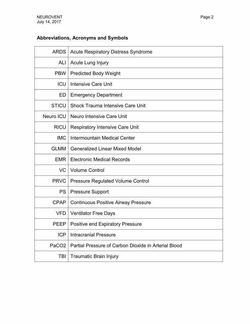
Abbreviations, Acronyms and Symbols
ARDS Acute Respiratory Distress Syndrome
ALI Acute Lung Injury
PBW Predicted Body Weight
ICU Intensive Care Unit
ED Emergency Department
STICU Shock Trauma Intensive Care Unit
Neuro ICU Neuro Intensive Care Unit
RICU Respiratory Intensive Care Unit
IMC Intermountain Medical Center
GLMM Generalized Linear Mixed Model
EMR Electronic Medical Records
VC Volume Control
PRVC Pressure Regulated Volume Control
PS Pressure Support
CPAP Continuous Positive Airway Pressure
VFD Ventilator Free Days
PEEP Positive end Expiratory Pressure
ICP Intracranial Pressure
PaCO2 Partial Pressure of Carbon Dioxide in Arterial Blood
TBI Traumatic Brain Injury
NEUROVENT Page 3 July 14, 2017
Purpose of the Study and Hypotheses: This is a quality improvement study with the purpose of observing and measuring the effects of implementation of a computerized neuro lung protective ventilation protocol, oxygenation protocol, and weaning protocol for mechanically ventilated patients with acute brain injury (TBI, intracerebral hemorrhage, stroke, cerebral edema, or anoxic brain injury) in the new electronic medical record system, iCentra, at IMC in the Neuro ICU, STICU, and RICU. We hypothesize that implementation of a computerized neuro lung protective ventilation protocol, oxygenation protocol, and weaning protocol for patients with acute brain injury (TBI, intracerebral hemorrhage, stroke, cerebral edema, or anoxic brain injury) will achieve a target normal PaCO2 of 35 to 40 mm Hg, decrease initial tidal volumes toward the target 6 ml/kg PBW (range 6 to 8 ml/kg PBW), improve ventilator free days (VFDs), and improve 28-day mortality. The objectives of this study are to: • Measure compliance, percent on target PaCO2 of 35 to 40 mm Hg, and
percent on target tidal volumes with a lung protective tidal volume of 6 ml/kg PBW, after implementation of computerized neuro lung protective ventilation protocol in patients with acute brain injury (TBI, intracerebral hemorrhage, stroke, cerebral edema, or anoxic brain injury).
• Measure compliance with a neuro oxygenation protocol limiting PEEP to 10 cm H2O and a weaning protocol using PS and CPAP spontaneous breathing that are included in the neuro lung protective ventilation protocol.
• Determine if the implementation of a computerized neuro lung protective ventilation protocol, targeting a normal PaCO2 with a 6 ml/kg PBW target tidal volume but allow up to 8 ml/kg PBW tidal volume, will improve outcomes in patients with acute brain injury requiring mechanical ventilation.
• Determine if the implementation of a neuro lung protective ventilation protocol and targeting a normal PaCO2 will improve outcomes in the sub-group of patients with the ARDS and acute brain injury.
Specific Aim #1: Measure compliance with the computerized neuro lung protective ventilation protocol and percent of time each patient has an on target

NEUROVENT Page 4 July 14, 2017
PaCO2 of 35 to 45 mm Hg and a tidal volume of 6 ml/kg PBW after implementation at Intermountain Medical Center (primary outcome).
• Process for Aim #1: Tool utilization and compliance will test the ability to introduce this neuro lung protective ventilation protocol in a new EMR in a controlled clinical environment and whether implementation of the protocol is successful in targeting a normal PaCO2 of 35 to 45 mm Hg and a 6 ml/kg PBW lung protective tidal volumes (allowing up to 8 ml/kg PBW tidal volume to achieve normal PaCO2). To ensure that this is successful, Dr. Grissom will identify local ICU physicians and respiratory care champions to act as a resource for implementation of the protocol in the IMC Neuro ICU, STICU, and RICU.
• Hypothesis 1: Healthcare providers will utilize the computerized neuro lung protective ventilation protocol and will comply with protocol instructions and achieve a target normal PaCO2 and lung protective 6 ml/kg PBW tidal volumes at higher rates post implementation of the protocol.
Specific Aim #2: Determine if the implementation of neuro lung protective ventilation, oxygenation, and weaning protocols with a 6 ml/kg PBW tidal volume and targeting normal PaCO2 in patients with acute brain injury (TBI, intracerebral hemorrhage, stroke, cerebral edema, or anoxic brain injury) who require mechanical ventilation improves ventilator free days (VFDs) to day 28, mortality, and secondary outcomes.
• Process for Aim #2: Standardized management of mechanical ventilation and outcomes will be measured through EMR data. A detailed plan on these metrics has been included (see Research Strategy).
• Hypothesis 2: Deployment of the computerized neuro lung protective ventilation, oxygenation, and weaning protocols in patients with acute brain injury (TBI, intracerebral hemorrhage, stroke, cerebral edema, or anoxic brain injury) will increase VFDs to day 28 and reduce mortality.
Specific Aim #3: Determine if implementation of neuro lung protective ventilation, oxygenation, and weaning protocols with a 6 ml/kg PBW tidal volume ventilation protocol and targeting normal PaCO2 increases VFDs to day 28 in
NEUROVENT Page 5 July 14, 2017
patients with acute brain injury (TBI, intracerebral hemorrhage, stroke, cerebral edema, or anoxic brain injury) and ARDS.
• Process for Aim #3: Standardized management of mechanical ventilation and outcomes for patients with acute brain injury (TBI, intracerebral hemorrhage, stroke, cerebral edema, or anoxic brain injury) and ARDS will be measured through EMR data. A detailed plan on these metrics has been included (see Research Strategy).
• Hypothesis 3: Deployment of the computerized neuro lung protective ventilation, oxygenation, and weaning protocols in patients with acute brain injury (TBI, intracerebral hemorrhage, stroke, cerebral edema, or anoxic brain injury) and ARDS will increase VFDs to day 28 and reduce mortality.
Study Design: This is an observational quality improvement study comparing pre- and post- implementation outcomes of neuro lung protective ventilation, oxygenation, and weaning protocols in the iCentra EMR system at Intermountain Medical Center in the Neuro ICU, STICU, and RICU. Retrospective data and outcomes on mechanical ventilation in patients with acute brain injury (TBI, intracerebral hemorrhage, stroke, cerebral edema, or anoxic brain injury) prior to implementation of the neuro mechanical ventilation protocol will be compared to outcomes post implementation, excluding data during a one-month washout period immediately following protocol implementation. The one-month washout period after implementation will allow physicians and respiratory therapists time to accommodate to the new neuro mechanical ventilation protocols that will be implemented as part of standard care with the iCentra EMR. Post-implementation data will be collected retrospectively from patients in the ICUs and compared with retrospectively queried pre-implementation data. Outcomes will include: use of the protocol by clinicians, compliance with protocol instructions, percent on target normal PaCO2, percent on target low tidal volume ventilation, hospital discharge disposition, 28-day mortality, 90-day mortality, time to first ICU activity, hospital length of stay, ICU length of stay, heath care utilization, quality of life, and costs of care. When the iCentra EMR is implemented at Intermountain Medical Center, clinicians will have the opportunity to use the computerized neuro lung protective ventilation protocols in patients
NEUROVENT Page 6 July 14, 2017
with acute brain injury, or clinicians may choose to order mechanical ventilation settings independently. This is an observational study, designed to measure the frequency with which neuro computerized lung protective ventilation protocols will be ordered, compliance with the instructions of the protocols, and clinical outcomes among patients who are managed with the protocols. Physicians may choose to use the protocols on intubated patients with acute brain injury (TBI, intracerebral hemorrhage, stroke, cerebral edema, or anoxic brain injury) requiring mechanical ventilation or they may choose to order other specific mechanical ventilator settings. Dr. Colin Grissom and Lori Carpenter RRT have already provided education for the lead physicians and respiratory therapists in each ICU at IMC participating in this study of the neuro lung protective ventilation, oxygenation, and weaning protocols in iCentra. Dr. Colin Grissom and Lori Carpenter RRT developed the neuro protective lung ventilation paper protocols for management of ventilation, oxygenation, and weaning in acutely brain injured patients and have used the paper protocols and the computerized protocols in selected patients at IMC who have traumatic brain injury with oversight from the IMC trauma team and Director of Trauma, Dr. Don VanBoerum. In addition, the protocol was reviewed and approved for implementation for patient care by Dr. Katherine Thomas, Medical Director of the Neuro ICU, Dr. Don VanBoerum, Surgical Director of the STICU, and Dr. Sarah Majercik, Director of Trauma Research, as a standard of care for mechanical ventilation of patients with acute brain injury. Selection of the protocols is an encouraged option, but not required, for attending physicians caring patients with acute brain injury requiring mechanical ventilation. Dr. Grissom and Lori Carpenter, RRT, will be available for phone consultation from physicians and respiratory therapists for real-time assistance when utilizing the neuro lung protective mechanical ventilation protocols. Background and Significance: Mechanical ventilation with high tidal volumes may cause mechanical damage to the lung, trigger inflammation, and release cytokines into the systemic circulation.1 This process may cause fever, leukocytosis, new pulmonary infiltrates, prolong duration of mechanical ventilation, and increase mortality. Lung protective ventilation is an approach that limits tidal volume and distending pressure on the alveolus in order to prevent mechanical ventilation induced
NEUROVENT Page 7 July 14, 2017
volutrauma (damage due to high tidal volume), barotrauma (damage due to high pressures), and biotrauma (release of inflammatory mediators due to high tidal volume). The balancing factor for lung protective ventilation in patients with acute brain injury is the goal to maintain a PaCO2 in the normal range to mitigate low PaCO2 causing cerebral vasoconstriction and decreasing delivery of oxygen to acutely injured brain, or high PaCO2 causing cerebral vasodilation and increasing ICP. Lung protective ventilation for patients with the acute respiratory distress syndrome (ARDS) improves outcomes. In a prospective randomized clinical trial performed by the National Institutes of Health, National Heart Lung and Blood Institute (NIH/NHLBI) ARDS Network, ventilation with volume control using a tidal volume of 6 ml/kg as compared to 12 ml/kg predicted body weight (PBW) and targeting a plateau pressure of <30 cm H2O as compared to <50 cm H2O decreased mortality in patients with ARDS.2 Among patients with ARDS, evidence supports that the timing of initiation of low tidal volume ventilation also influences mortality. A retrospective study of patients with ARDS showed that an increase in initial tidal volume of 1 ml/kg above 6 ml/kg PBW in patients with ARDS was associated with a 23% increase in intensive care unit (ICU) mortality risk.3 This finding suggests that initial tidal volume should be strictly set at 6 ml/kg PBW in patients with ARDS. Mounting evidence also indicates that lung protective ventilation in intubated patients without ARDS may decrease the development of ARDS, pulmonary complications, and mortality. A meta-analysis of patients who were intubated and mechanically ventilated, but did not have ARDS, showed that ventilation with a mean tidal volume of 6.5 ml/kg as compared to 10.6 ml/kg PBW resulted in less development of acute lung injury or ARDS, less pulmonary infections, and lower mortality.4 Furthermore, of the 20 studies included in that meta-analysis, 15 set initial tidal volume in the intervention group to ≤6 ml/kg PBW. Higher tidal volumes are an independent predictor for development of acute lung injury (ALI) in patients who did not have ARDS at onset of mechanical ventilation.5 Further evidence of benefit from tidal volume limitation has been supported by a recent patient level data analysis that showed a lower incidence of ARDS and fewer pulmonary complications in patients without ARDS treated with a tidal volume of <7 ml/kg PBW.6 Taken together, these studies indicate that patients with acute respiratory failure requiring mechanical ventilation, but without ARDS, should be
NEUROVENT Page 8 July 14, 2017
supported with volume control ventilation using a tidal volume of no more than 8 ml/kg PBW upon initiation of mechanical ventilation, and may have the best outcomes using an initial tidal volume targeting 6 ml/kg PBW. Patients with acute brain injury who have respiratory failure are also at risk for worsening lung injury and ARDS with high tidal volume ventilation. Respiratory failure requiring mechanical ventilation is common in acutely brain injured patients7 and ARDS occurs in about 25% of patients with acute brain injury and respiratory failure.8 In acutely brain injured patients, ventilation with high tidal volumes is associated with worse outcome9 and development of ARDS8 Lung protective ventilation in patients with acute brain with a low tidal volume strategy, limitation of PEEP, and a weaning protocol improve VFDs.10 Lung protective ventilation and optimal PEEP are recommended for acutely brain injured patients as long as goals for control of ICP are maintained.11 Lung protective ventilation in patients with acute brain injury is commonly applied and reduces risk for development of ARDS.12 The challenge in implementing lung protective ventilation with low tidal volume in patients with acute brain injury is to maintain a normal PaCO2 to mitigate effects of cerebral vasodilation from high PaCO2 that may contribute to increased ICP. The current evidenced based standard mechanical ventilation protocols based on the ARDS Network studies2 for patients with acute respiratory failure with, or without, ARDS, do not control PaCO2, which may result in undesirable high PaCO2 levels in patients with acute brain injury. Lung protective ventilation strategies for patients with acute brain injury, therefore, require a modified approach to target a normal PaCO2 when using lung protective low tidal volume ventilation. A previous before-versus-after implementation quality improvement study of 744 acutely brain injured patients in 20 ICUs in France evaluated a paper protocol prescribing a lung protective low tidal volume ventilation strategy (≤7 ml/kg PBW), moderate PEEP, and criteria for weaning and extubation.10 This study found that implementation of the paper protocol resulted in application of lower tidal volumes and an increase in VFDs. Based on this study, and prior studies showing that lung protective ventilation improves outcome in patients with acute brain injury, our group at Intermountain Healthcare has developed a computerized protocol that targets a normal PaCO2, targets low tidal volume, uses moderate PEEP (≤10 cm H2O), and prescribes parameters to guide weaning and extubation from mechanical ventilation. These computerized neuro
NEUROVENT Page 9 July 14, 2017
ventilation, oxygenation, and weaning protocols will be implemented in the Neuro ICU, STICU, and RICU at Intermountain Medical Center in the iCentra electronic medical record on July 15, 2017 as an option for use by physicians caring for patients with acute brain injury. This computerized neuro lung protective mechanical ventilation protocol will allow standardization of care for patients with acute brain injury and respiratory failure at Intermountain Medical Center.
Research Subjects: Inclusion Criteria
1. Acute brain injury due to non-traumatic causes (stroke, spontaneous intracranial hemorrhage, cerebral edema, anoxic brain injury) or traumatic brain injury.
2. Initiation of mechanical ventilation in the emergency department or intensive care unit at an Intermountain Healthcare hospital
3. Age ≥ 18 years
Exclusion Criteria
1. Transition to comfort care in the emergency department or on the same day of admission to the ICU
2. Death on the same day of admission to the emergency department or ICU
Patient Selection Those to be enrolled must have acute brain injury (TBI, intracerebral hemorrhage, stroke, cerebral edema, or anoxic brain injury) and respiratory failure requiring intubation and initiation of mechanical ventilation. Patients will be divided into two different groups after implementation of the computerized neuro lung protective ventilation protocols (after July 15, 2017 iCentra go live at IMC): patients managed with the computerized neuro lung protective ventilation protocols as ordered by the attending physician and patients managed with physician-specified mechanical ventilation settings. These groups will be used in secondary subgroup analyses among post-implementation patients comparing outcomes of patients on protocol-guided mechanical ventilation with the outcomes of patients on physician-guided mechanical ventilation. Prior experience with implementation of a computerized lung protective ventilation protocol across Intermountain Healthcare suggests that even if relatively low
NEUROVENT Page 10 July 14, 2017
compliance with the computerized lung protective ventilation protocol were to be observed post-implementation, the average set tidal volume would still be expected to decrease, presumably because of education of clinicians and respiratory therapists regarding the benefits of lung protective mechanical ventilation (unpublished data collected as part of the IMPROVENT study, Intermountain IRB # 1050159, an ongoing study at Intermountain Healthcare evaluating the impact of implementation of a lung protective ventilation protocol in iCentra across Intermountain Healthcare Hospitals). Compensation Subjects will not be compensated for participating in this study. Sample Size Retrospectively querying data from 1 Jan 2016 through 31 Dec 2016 at IMC, 364 brain injured patients with complete data were identified in the Intermountain EMR. Accordingly, we estimate that 728 patients (24 months) will be identified and included for analysis. Methods/Procedures: Research Strategy Lung protective ventilation management in patients with acute brain injury (TBI, intracerebral hemorrhage, stroke, cerebral edema, or anoxic brain injury) using the computerized neuro lung protective ventilation protocol (See attachments Neuro Vent Protocol Ventilation, Neuro Vent Protocol Oxygenation, Neuro Vent Protocol Weaning Assessment, and Neuro Vent Protocol Weaning) will be implemented at IMC on July 15, 2017 synchronized with the rollout of iCentra at IMC. This is a quality improvement initiative to introduce a best practice for ventilation, oxygenation, and weaning in mechanically ventilated patients with acute brain injury in the Neuro ICU, STICU, and RICU. We request waiver of informed consent from the Intermountain IRB in order to measure the effect on clinical outcomes and change in practice associated with this implementation. Retrospective data from one year prior to implementation of iCentra on July 15, 2017, will be compared with retrospectively collected data for one year after implementation of the neuro ventilation, oxygenation, and weaning protocols in iCentra starting on August 15, 2017, after a one-month wash-out period. The one month wash-out period from July 15 to August 15, 2017, will allow physicians and respiratory therapists time to acclimate to the new computerized neuro ventilation
NEUROVENT Page 11 July 14, 2017
protocols as well as the new iCentra EMR. Dr. Grissom and Lori Carpenter RRT, will educate physicians, advanced practice clinicians, and respiratory therapists in use of the protocols, and will be available for phone consultation. Co-Investigators Dr. Katherine Thomas, Medical Director of the Neuro ICU, Dr. Don VanBoerum, Surgical Director of the STICU, and Dr. Sarah Majercik, Director of Trauma Research, are all co-investigators on this study and will provide leadership in implementation of the neuro lung protective ventilation protocols. Percent Time on Lung Protective Tidal Volumes Determination of percent time each patient achieves a normal PaCO2 and is on a tidal volume of ≤6.5 ml/kg PBW, ≤7.5 ml/kg PBW, and ≤8.5 ml/kg PBW over the first week of mechanical ventilation will require extraction of data from the EMR. This will require specific data queries to collect information on initial set tidal volume and mode of ventilation from each episode of ventilator charting on each patient and arterial blood gases performed during mechanical ventilation. Jason Jacobs, the lead technical data analyst for pulmonary and critical care research at IMC has extensive experience extracting this data from the legacy EMR and from iCentra for the IMPROVENT study (Intermountain IRB # 1050159). Protocol Compliance As part of the data collection for the ongoing IMPROVENT study (Intermountain IRB # 1050159) Jason Jacobs, data analyst, and James Sanders, Cerner Physician Consultant, have led an effort to establish a data table in iCentra that specifies when a mechanical ventilation protocol is used and stores individual data on different elements of the protocol that allows automated data extraction. This will allow specific data collection on which patients are placed on the computerized Neuro Lung Protective Ventilation Protocols, which specific parts of the protocols were used (ventilation, oxygenation, or weaning), and whether protocol instructions were accepted and implemented, or declined, and the reason for declining a specific protocol instruction. Ventilator Free Days (VFDs) For determination of VFDs to day 28 we will use the same definition for liberation from mechanical ventilation as used in ARDS Network studies2,13 and in the proposed ROSE study from the NIH/NHLBI PETAL Network. Initiation of ventilator free days begins with two ventilator free days once unassisted breathing is present for 48 hours. Unassisted breathing is defined as14:
NEUROVENT Page 12 July 14, 2017
a. Extubated with face mask, nasal prong oxygen, or room air, OR b. T-tube breathing, OR c. Tracheostomy mask breathing, OR d. CPAP ≤ 5 without PS or IMV assistance e. Use of CPAP or BIPAP solely for sleep apnea management f. Use of a high flow oxygen system
Determination of ARDS Determination of ARDS using the Berlin Definition15 requires acute respiratory failure not fully explained by cardiac failure or fluid overload within one week of a known clinical insult, bilateral opacities on chest radiology imaging not fully explained by effusions, lobar/lung collapse, or nodules, and PaO2/FIO2 ≤300 mm Hg with PEEP or CPAP ≥5 cm H2O. We will focus on defining ARDS among those patients with mild, moderate, or severe hypoxemia as defined by a PaO2 to FIO2 ratio ≤ 255 (altitude corrected by multiplying 300 by the ratio of ambient barometric pressure in Salt Lake City to sea level barometric pressure, 300 x 0.85 = 255) and evaluate chest radiographs for bilateral infiltrates in that group. Study Duration 25 months, 7/15/2016 - 8/15/2018 with a one-month peri-implementation wash-out period. Risks The risk of this study is a potential loss of confidentiality, which will be managed as detailed below. Benefits This is a quality improvement study evaluating the benefits of implementation of computerized neuro lung protective ventilation protocols at IMC in the Neuro ICU, STICU, and RICU for patients with acute brain injury (TBI, intracerebral hemorrhage, stroke, cerebral edema, or anoxic brain injury). Application of the protocol is at the discretion of the attending physician for patients with acute brain injury and acute respiratory failure requiring intubation and mechanical ventilation. Based on best practices and the evidenced based medical literature, implementation of the neuro ventilation protocols is expected to improve outcomes.
NEUROVENT Page 13 July 14, 2017
Waiver of Informed Consent This study seeks a waiver of informed consent for these reasons: • The risk of this study is a potential loss of confidentiality. The study involves
no more than minimal risk to the subject, as the study is observational only and the study will not alter the care that enrolled subjects receive. The computerized neuro lung protective mechanical ventilation protocols will be available to clinicians as an order in iCentra, but will not be required.
• The implementation of the neuro lung protective ventilation, oxygenation, and weaning protocols is part of an intent to standardize clinical practice for mechanical ventilation of acutely brain injured patients (TBI, intracerebral hemorrhage, stroke, cerebral edema, or anoxic brain injury) and is supported by the Medical Director of the Neuro ICU, Dr. Katherine Thomas, and the Surgical Director of the STICU, Dr. Don VanBoerum. The implementation of this process of standard of care for mechanical ventilation of patients with acute brain injury is intended as a quality assurance project using evidence based literature. The use of the protocol is not mandated, but is encouraged. The computerized neuro ventilation, oxygenation, and weaning protocols will be implemented regardless of whether this observational study occurs. The investigators on this observational study are using implementation of these computerized neuro lung protective ventilation protocols to formally evaluate compliance with the protocol, effectiveness of the protocol at targeting normal PaCO2 and decreasing tidal volume, and clinical outcomes. The intent of the investigators is to publish our experience with implementation of this protocol in the peer reviewed medical literature, and therefore IRB review is appropriate. Waiver of informed consent is requested because the implementation of the computerized neuro lung protective ventilation protocol is primarily a quality improvement clinical initiative, not a research initiative, and is not mandated, but is left up to the attending physician for each patient.
Protection of Subject Confidentiality PHI will be collected as a part of this study. The PHI will be used only for study purposes and will not be reused or disclosed except as required by law. The information obtained from medical records will be kept separate from clinical records. All digital study records will be kept within the Intermountain Healthcare firewall in a location that is only accessible to authorized members of the study team. All
NEUROVENT Page 14 July 14, 2017
paper study records will be maintained on a floor with secure badge access that only the research team can access. After the study is complete, study identifiers will be removed from the dataset. The data to be collected is detailed below. In particular, the dates to be collected are important for the specific aims of the study. Data Collection: Data Elements Extracted from the EDW for the NEUROVENT Mechanical Ventilation Study • Account Number • EMPI • Admit Date • Discharge Date • Admit Year • Admit ICU • Age • Gender • ICD Diagnosis code • ICD Diagnosis description • ICD Diagnosis long description • ICU Stay • Length of Stay (Days) • Mortality Indicator (Data required for calculation of Acute Physiology Score
and Charlson Comorbidity Index) • Patient Type (inpatient(I) vs outpatient(O)) • Patient Type (Detailed code) • Patient Type Description • Death Location • Discharge Disposition Description • Discharge Reason (same as above) • ED Admit Date • ED Discharge Date • Total Cost • Ventilator Location (First Vent Check Location) • First Date of Ventilator Check
NEUROVENT Page 15 July 14, 2017
• Ventilator Mode • First Date of Ventilator Check • Intubation Date • Intubation Location (Same as Ventilator Location) • All Parameters Included in the Ventilator Checks During the Hospitalization • Room Trace (It is more detailed than the ones above) • In Hospital Mortality • Height • Predicted Body Weight • Difference between admission date and death date • 28 Day Mortality • 60 Day Mortality • 90 Day Mortality • Arterial Blood Gases recorded during hospital stay Data Analysis: Primary Outcomes The primary outcome will be the patient-level proportion of time on mechanical ventilation with a tidal volume ≤ 6.5 ml/kg PBW. Secondary outcomes will include: proportion of time with a target PaCO2 of 35 to 45 mm Hg; protocol compliance; hospital discharge disposition; hospital, 28-day, and 90-day mortality; ventilator-free days to day 28; time to first ICU activity; hospital, ICU length of stay; health care utilization; quality of life (SF-36 or similar); and costs of care. Statistical Analysis Univariable analyses will use Fisher’s exact test and Pearson’s chi-square test for comparing pairs of Bernoulli-distributed variables with and without sparse cells, respectively. Wilcoxon rank-sum test will be used to compare pairs of non-Gaussian, continuous distributions. Bootstrapped Kolomogorov-Smirnov test will be used to compare pairs of distributions of ordinal, discrete data. Multivariable analyses will use linear parametric regression models within the exponential family adjusted for a vector of covariates, having link functions determined by the distribution of the dependent variable. Specifically, Bernoulli-

NEUROVENT Page 16 July 14, 2017
distributed outcomes will use the logit link function, Poisson-distributed outcomes will use the log link function, multinomial outcomes will use the multi-class logit link function, normally distributed outcomes will use the identity link function, and other continuous outcomes (including the primary outcome) whose distributions can be transformed such that they are bounded by zero and one will be treated as quasibinomial dependent variables using the logit link function. The primary outcome (viz., patient-level proportion of time on lung protective ventilation), for example, can be treated as a quasibinomial dependent variable because it can take any value within the unit interval [0,1], and as such can be compared pre- and post-implementation using multivariable quasibinomial logistic regression analysis adjusting for the set of patient-level confounders and effect modifiers, with regression equation taking the form 𝑔𝑔(𝑦𝑦) = 𝛽𝛽0 + 𝛽𝛽1𝑡𝑡 + 𝐗𝐗𝜽𝜽, (1) where 𝑔𝑔(. ) is the link function – quasibinomial logit in the case of the primary analysis – 𝑦𝑦 is the percent time on lung protective ventilation, 𝑡𝑡 is a binary indicator of post-implementation period, and 𝑿𝑿 is a matrix of potential patient-level confounders and effect modifiers. The secondary outcomes will be analyzed using the same linear predictor, but with the appropriate link function as specified above. Power Analysis We empirically estimated the baseline distribution of the primary outcome – viz., patient-level percent time on lung protective ventilation (LPV) – by pulling data from the Intermountain EMR of all brain injured patients seen at IMC in 2016, and computed the patient-level percent time on mechanical ventilation. In so doing, we observed the primary outcome LPV to be distributed such that about 50% of patients had LPV = 0, 25% had LPV = 1, and the LPV distribution of the remaining 25% closely followed a beta distribution with α and β shape parameters having values 0.6 and 0.5, respectively. This information was incorporated into a multidimensional, Monte Carlo simulation-based power analysis of a quasibinomial logistic regression model of the effect of implementation on LPV, assuming total enrollment of 728 patients. The support of the space was determined ex ante in collaboration with the
NEUROVENT Page 17 July 14, 2017
primary investigator in order to ensure the candidate effect sizes were of clinically reasonable magnitude. In so doing, it was found that an odds ratio of 1.5 for the effect of implementation on LPV corresponded to a relative increase in mean LPV of 16% (from baseline mean LPV of 0.429), and a relative increase in complete compliance (LPV=1) of 43.3% (from a baseline rate of complete compliance of 0.248), and would be sufficient to achieve 80% power. Funding: It is anticipated that this project will be funded by the Pulmonary and Critical Care Department at Intermountain Medical Center.
NEUROVENT Page 18 July 14, 2017
References 1. Society AT. Patient Information: Mechanical Ventilation. 5 October 2015. 2. ARDSNetwork. Ventilation with lower tidal volumes as compared with traditional tidal
volumes for acute lung injury and the acute respiratory distress syndrome. The Acute Respiratory Distress Syndrome Network. The New England journal of medicine. 2000;342(18):1301-1308.
3. Needham DM, Yang T, Dinglas VD, et al. Timing of low tidal volume ventilation and intensive care unit mortality in acute respiratory distress syndrome. A prospective cohort study. American journal of respiratory and critical care medicine. 2015;191(2):177-185.
4. Serpa Neto A, Cardoso SO, Manetta JA, et al. Association between use of lung-protective ventilation with lower tidal volumes and clinical outcomes among patients without acute respiratory distress syndrome: a meta-analysis. Jama. 2012;308(16):1651-1659.
5. Gajic O, Dara SI, Mendez JL, et al. Ventilator-associated lung injury in patients without acute lung injury at the onset of mechanical ventilation. Crit Care Med. 2004;32(9):1817-1824.
6. Neto AS, Simonis FD, Barbas CS, et al. Lung-Protective Ventilation With Low Tidal Volumes and the Occurrence of Pulmonary Complications in Patients Without Acute Respiratory Distress Syndrome: A Systematic Review and Individual Patient Data Analysis. Crit Care Med. 2015;43(10):2155-2163.
7. Zygun DA, Kortbeek JB, Fick GH, Laupland KB, Doig CJ. Non-neurologic organ dysfunction in severe traumatic brain injury. Crit Care Med. 2005;33(3):654-660.
8. Elmer J, Hou P, Wilcox SR, et al. Acute respiratory distress syndrome after spontaneous intracerebral hemorrhage*. Crit Care Med. 2013;41(8):1992-2001.
9. Mascia L, Zavala E, Bosma K, et al. High tidal volume is associated with the development of acute lung injury after severe brain injury: an international observational study. Crit Care Med. 2007;35(8):1815-1820.
10. Asehnoune K, Mrozek S, Perrigault PF, et al. A multi-faceted strategy to reduce ventilation-associated mortality in brain-injured patients. The BI-VILI project: a nationwide quality improvement project. Intensive Care Med. 2017;43(7):957-970.
11. Lowe GJ, Ferguson ND. Lung-protective ventilation in neurosurgical patients. Curr Opin Crit Care. 2006;12(1):3-7.
12. Luo XY, Hu YH, Cao XY, et al. Lung-protective Ventilation in Patients with Brain Injury: A Multicenter Cross-sectional Study and Questionnaire Survey in China. Chin Med J (Engl). 2016;129(14):1643-1651.
13. Brower RG, Lanken PN, MacIntyre N, et al. Higher versus lower positive end-expiratory pressures in patients with the acute respiratory distress syndrome. The New England journal of medicine. 2004;351(4):327-336.
14. Angus D MM. Reevaluation Of Systemic Early neuromuscular blockade. NHLBI Petal Network2015.
15. Force ADT, Ranieri VM, Rubenfeld GD, et al. Acute respiratory distress syndrome: the Berlin Definition. Jama. 2012;307(23):2526-2533.
NEUROVENT Page 19 July 14, 2017
APPENDICES Appendix 1. Ventilation Protocol
Appendix 2. Oxygenation Protocol
Appendix 3. Weaning Assessment
Appendix 4. Weaning Protocol
Appendix 5. Tidal Volumes from Predicted Body Weight from Height and Gender
Appendix 6. Rules for iCentra Neuro Ventilator Protocol
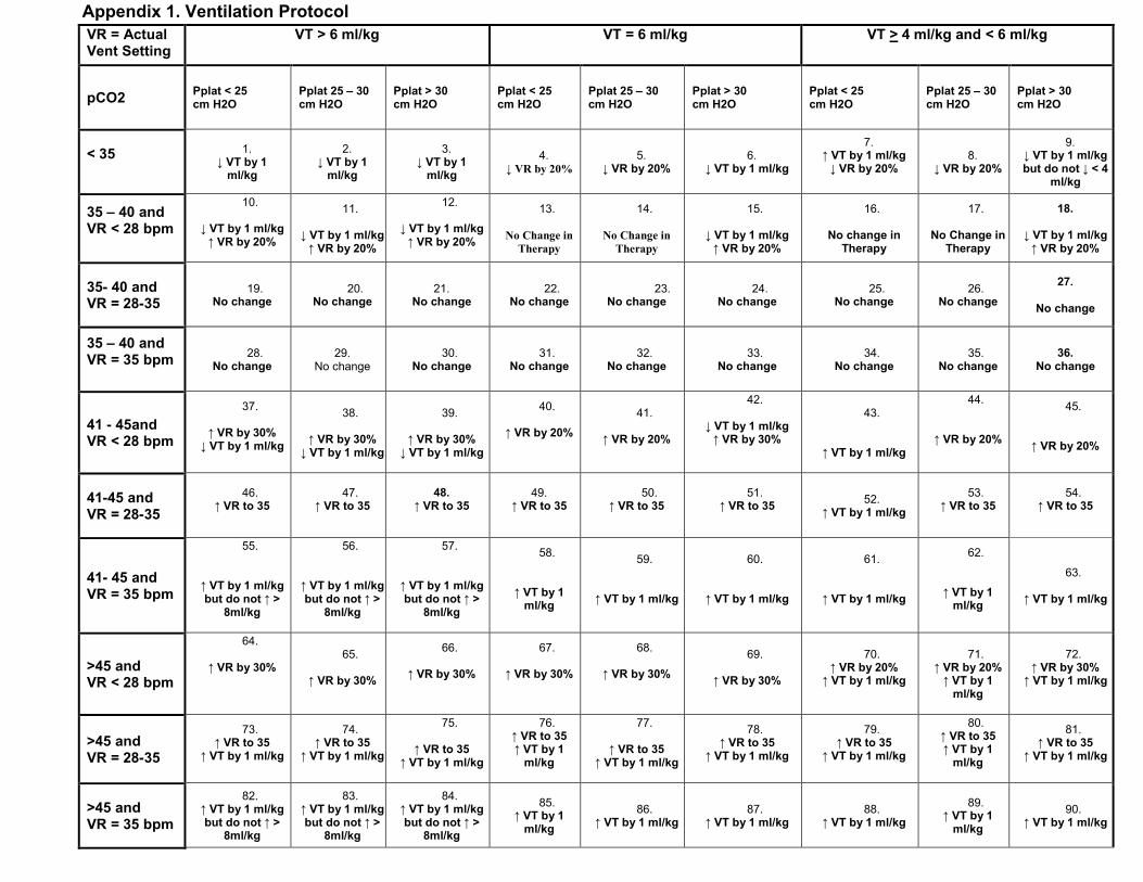
VR = Actual Vent Setting
VT > 6 ml/kg VT = 6 ml/kg VT > 4 ml/kg and < 6 ml/kg
pCO2 Pplat < 25 cm H2O
Pplat 25 – 30 cm H2O
Pplat > 30 cm H2O
Pplat < 25 cm H2O
Pplat 25 – 30 cm H2O
Pplat > 30 cm H2O
Pplat < 25 cm H2O
Pplat 25 – 30 cm H2O
Pplat > 30 cm H2O
< 35 1. ↓ VT by 1
ml/kg
2. ↓ VT by 1
ml/kg
3. ↓ VT by 1
ml/kg
4. ↓ VR by 20%
5. ↓ VR by 20%
6. ↓ VT by 1 ml/kg
7. ↑ VT by 1 ml/kg
↓ VR by 20% 8.
↓ VR by 20%
9. ↓ VT by 1 ml/kg but do not ↓ < 4
ml/kg
35 – 40 and VR < 28 bpm
10.
↓ VT by 1 ml/kg ↑ VR by 20%
11.
↓ VT by 1 ml/kg ↑ VR by 20%
12.
↓ VT by 1 ml/kg ↑ VR by 20%
13.
No Change in Therapy
14.
No Change in Therapy
15.
↓ VT by 1 ml/kg ↑ VR by 20%
16.
No change in Therapy
17.
No Change in Therapy
18.
↓ VT by 1 ml/kg ↑ VR by 20%
35- 40 and VR = 28-35
19. No change
20. No change
21. No change
22. No change
23. No change
24. No change
25. No change
26. No change
27.
No change
35 – 40 and VR = 35 bpm 28.
No change 29.
No change 30.
No change 31.
No change 32.
No change 33.
No change 34.
No change 35.
No change 36.
No change
41 - 45and VR < 28 bpm
37.
↑ VR by 30% ↓ VT by 1 ml/kg
38.
↑ VR by 30% ↓ VT by 1 ml/kg
39.
↑ VR by 30% ↓ VT by 1 ml/kg
40.
↑ VR by 20% 41.
↑ VR by 20%
42.
↓ VT by 1 ml/kg ↑ VR by 30%
43.
↑ VT by 1 ml/kg
44.
↑ VR by 20%
45.
↑ VR by 20%
41-45 and VR = 28-35
46. ↑ VR to 35
47. ↑ VR to 35
48. ↑ VR to 35
49. ↑ VR to 35
50. ↑ VR to 35
51. ↑ VR to 35 52.
↑ VT by 1 ml/kg
53. ↑ VR to 35
54. ↑ VR to 35
41- 45 and VR = 35 bpm
55.
↑ VT by 1 ml/kg but do not ↑ >
8ml/kg
56.
↑ VT by 1 ml/kg but do not ↑ >
8ml/kg
57.
↑ VT by 1 ml/kg but do not ↑ >
8ml/kg
58.
↑ VT by 1 ml/kg
59.
↑ VT by 1 ml/kg
60.
↑ VT by 1 ml/kg
61.
↑ VT by 1 ml/kg
62.
↑ VT by 1 ml/kg
63.
↑ VT by 1 ml/kg
>45 and VR < 28 bpm
64.
↑ VR by 30% 65.
↑ VR by 30%
66.
↑ VR by 30%
67.
↑ VR by 30%
68.
↑ VR by 30% 69.
↑ VR by 30%
70. ↑ VR by 20%
↑ VT by 1 ml/kg
71. ↑ VR by 20%
↑ VT by 1 ml/kg
72. ↑ VR by 30%
↑ VT by 1 ml/kg
>45 and VR = 28-35
73. ↑ VR to 35
↑ VT by 1 ml/kg
74. ↑ VR to 35
↑ VT by 1 ml/kg
75.
↑ VR to 35 ↑ VT by 1 ml/kg
76. ↑ VR to 35 ↑ VT by 1
ml/kg
77.
↑ VR to 35 ↑ VT by 1 ml/kg
78. ↑ VR to 35
↑ VT by 1 ml/kg
79. ↑ VR to 35
↑ VT by 1 ml/kg
80. ↑ VR to 35 ↑ VT by 1
ml/kg
81. ↑ VR to 35
↑ VT by 1 ml/kg
>45 and VR = 35 bpm
82. ↑ VT by 1 ml/kg but do not ↑ >
8ml/kg
83. ↑ VT by 1 ml/kg but do not ↑ >
8ml/kg
84. ↑ VT by 1 ml/kg but do not ↑ >
8ml/kg
85. ↑ VT by 1
ml/kg 86.
↑ VT by 1 ml/kg 87.
↑ VT by 1 ml/kg 88.
↑ VT by 1 ml/kg 89.
↑ VT by 1 ml/kg
90. ↑ VT by 1 ml/kg
Appendix 1. Ventilation Protocol
Page 1 of 2
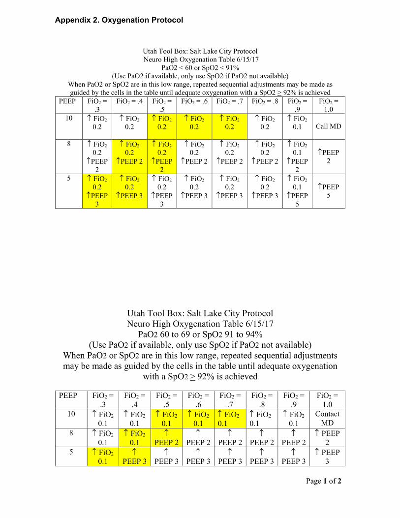
Utah Tool Box: Salt Lake City Protocol Neuro High Oxygenation Table 6/15/17
PaO2 < 60 or SpO2 < 91% (Use PaO2 if available, only use SpO2 if PaO2 not available)
When PaO2 or SpO2 are in this low range, repeated sequential adjustments may be made as guided by the cells in the table until adequate oxygenation with a SpO2 > 92% is achieved
PEEP FiO2 = .3
FiO2 = .4 FiO2 = .5
FiO2 = .6 FiO2 = .7 FiO2 = .8 FiO2 = .9
FiO2 = 1.0
10 ↑ FiO2 0.2
↑ FiO2 0.2
↑ FiO2 0.2
↑ FiO2 0.2
↑ FiO2 0.2
↑ FiO2 0.2
↑ FiO2 0.1 Call MD
8 ↑ FiO2 0.2
↑PEEP 2
↑ FiO2 0.2
↑PEEP 2
↑ FiO2 0.2
↑PEEP 2
↑ FiO2 0.2
↑PEEP 2
↑ FiO2 0.2
↑PEEP 2
↑ FiO2 0.2
↑PEEP 2
↑ FiO2 0.1
↑PEEP 2
↑PEEP 2
5 ↑ FiO2 0.2
↑PEEP 3
↑ FiO2 0.2
↑PEEP 3
↑ FiO2 0.2
↑PEEP 3
↑ FiO2 0.2
↑PEEP 3
↑ FiO2 0.2
↑PEEP 3
↑ FiO2 0.2
↑PEEP 3
↑ FiO2 0.1
↑PEEP 5
↑PEEP 5
Utah Tool Box: Salt Lake City Protocol Neuro High Oxygenation Table 6/15/17
PaO2 60 to 69 or SpO2 91 to 94% (Use PaO2 if available, only use SpO2 if PaO2 not available)
When PaO2 or SpO2 are in this low range, repeated sequential adjustments may be made as guided by the cells in the table until adequate oxygenation
with a SpO2 > 92% is achieved
PEEP FiO2 = .3
FiO2 = .4
FiO2 = .5
FiO2 = .6
FiO2 = .7
FiO2 = .8
FiO2 = .9
FiO2 = 1.0
10 ↑ FiO2 0.1
↑ FiO2 0.1
↑ FiO2 0.1
↑ FiO2 0.1
↑ FiO2 0.1
↑ FiO2 0.1
↑ FiO2 0.1
Contact MD
8 ↑ FiO2 0.1
↑ FiO2 0.1
↑ PEEP 2
↑ PEEP 2
↑ PEEP 2
↑ PEEP 2
↑ PEEP 2
↑ PEEP 2
5 ↑ FiO2 0.1
↑ PEEP 3
↑ PEEP 3
↑ PEEP 3
↑ PEEP 3
↑ PEEP 3
↑ PEEP 3
↑ PEEP 3
Appendix 2. Oxygenation Protocol
Page 2 of 2
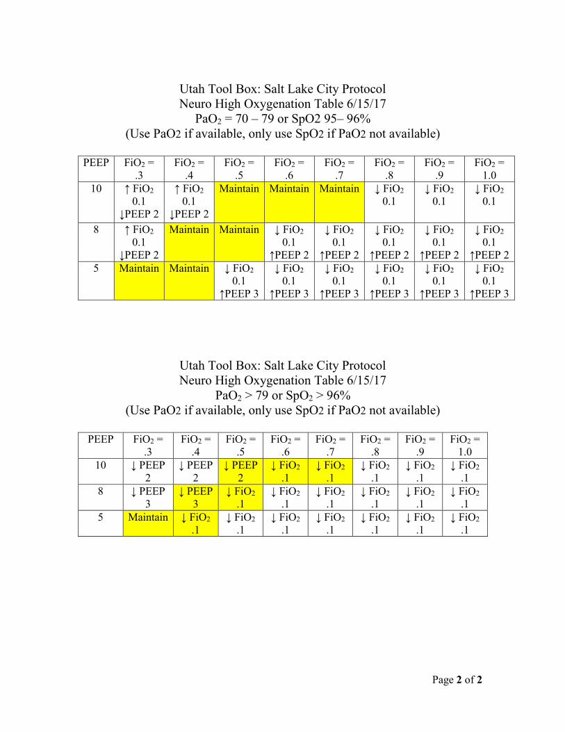
Utah Tool Box: Salt Lake City Protocol Neuro High Oxygenation Table 6/15/17
PaO2 = 70 – 79 or SpO2 95– 96% (Use PaO2 if available, only use SpO2 if PaO2 not available)
PEEP FiO2 = .3
FiO2 = .4
FiO2 = .5
FiO2 = .6
FiO2 = .7
FiO2 = .8
FiO2 = .9
FiO2 = 1.0
10 ↑ FiO2 0.1
↓PEEP 2
↑ FiO2 0.1
↓PEEP 2
Maintain Maintain Maintain ↓ FiO2 0.1
↓ FiO2 0.1
↓ FiO2 0.1
8 ↑ FiO2 0.1
↓PEEP 2
Maintain Maintain ↓ FiO2 0.1
↑PEEP 2
↓ FiO2 0.1
↑PEEP 2
↓ FiO2 0.1
↑PEEP 2
↓ FiO2 0.1
↑PEEP 2
↓ FiO2 0.1
↑PEEP 2 5 Maintain Maintain ↓ FiO2
0.1 ↑PEEP 3
↓ FiO2 0.1
↑PEEP 3
↓ FiO2 0.1
↑PEEP 3
↓ FiO2 0.1
↑PEEP 3
↓ FiO2 0.1
↑PEEP 3
↓ FiO2 0.1
↑PEEP 3
Utah Tool Box: Salt Lake City Protocol Neuro High Oxygenation Table 6/15/17
PaO2 > 79 or SpO2 > 96% (Use PaO2 if available, only use SpO2 if PaO2 not available)
PEEP FiO2 = .3
FiO2 = .4
FiO2 = .5
FiO2 = .6
FiO2 = .7
FiO2 = .8
FiO2 = .9
FiO2 = 1.0
10 ↓ PEEP 2
↓ PEEP 2
↓ PEEP 2
↓ FiO2.1
↓ FiO2.1
↓ FiO2.1
↓ FiO2.1
↓ FiO2.1
8 ↓ PEEP 3
↓ PEEP 3
↓ FiO2.1
↓ FiO2.1
↓ FiO2.1
↓ FiO2.1
↓ FiO2.1
↓ FiO2.1
5 Maintain ↓ FiO2.1
↓ FiO2.1
↓ FiO2.1
↓ FiO2.1
↓ FiO2.1
↓ FiO2.1
↓ FiO2.1
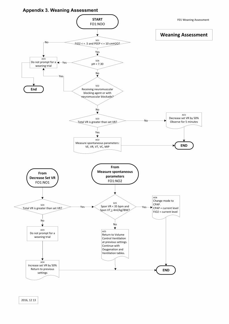
Weaning Assessment
2016, 12 13
STARTFO1:NOO
SO1
FiO2 < = .5 and PEEP < = 10 cmH2O?
AO3
Do not prompt for a weaning trial
SO2 Receiving neuromuscular
blocking agent or with neuromuscular blockade?
SO3
Total VR is greater than set VR?
AO1
Decrease set VR by 50%Observe for 5 minutes
AO2
Measure spontaneous parameters: VE, VR, VT, VC, MIP END
No
Yes
No
Yes
No
SO4
Total VR is greater than set VR?
AO4
Increase set VR by 50%Return to previous
settings
AO3
Do not prompt for a weaning trial
SO5
Spon VR < 35 bpm and Spon VT > 4ml/kg/IBW?
AO5
Return to Volume Control Ventilation at previous settings. Continue with Oxygenation and Ventilation tables.
AO6
Change mode to CPAP. CPAP = current levelFiO2 = current level
END
No
Yes
No
Yes
From Decrease Set VR
FO1:NO1
From Measure spontaneous
parameters FO1:NO2
End
F01 Weaning Assessment
SO6
pH < 7.30
Yes
No
Yes
Appendix 3. Weaning Assessment
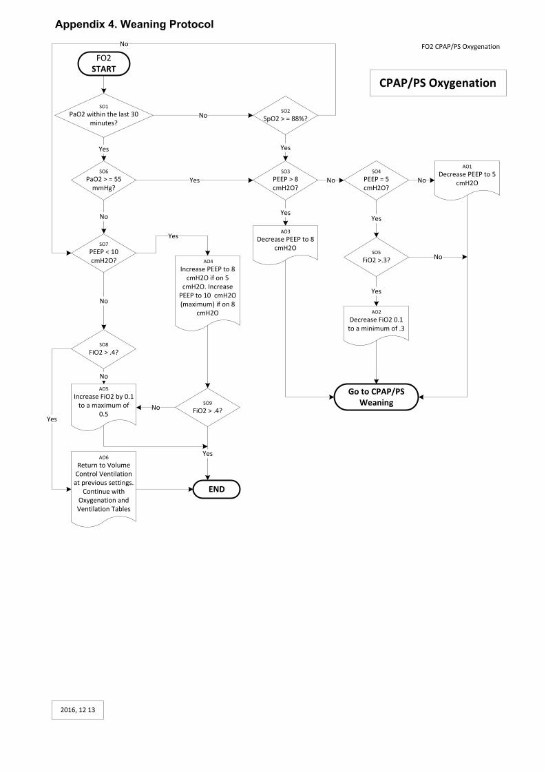
CPAP/PS Oxygenation
2016, 12 13
FO2START
SO1
PaO2 within the last 30 minutes?
SO2
SpO2 > = 88%?
SO6
PaO2 > = 55 mmHg?
SO3
PEEP > 8 cmH2O?
SO4
PEEP = 5 cmH2O?
AO1
Decrease PEEP to 5 cmH2O
SO7
PEEP < 10 cmH2O?
AO3
Decrease PEEP to 8 cmH2O
SO5
FiO2 >.3?
AO2
Decrease FiO2 0.1 to a minimum of .3
SO8
FiO2 > .4?
AO5
Increase FiO2 by 0.1 to a maximum of
0.5
AO6
Return to Volume Control Ventilation at previous settings.
Continue with Oxygenation and Ventilation Tables
Yes
No
No
No
Yes
AO4
Increase PEEP to 8 cmH2O if on 5
cmH2O. Increase PEEP to 10 cmH2O (maximum) if on 8
cmH2O
END
No
No
Yes
No
Yes
Yes
No
Yes
Yes
NoYes
Go to CPAP/PS WeaningSO9
FiO2 > .4?No
Yes
FO2 CPAP/PS Oxygenation
Appendix 4. Weaning Protocol
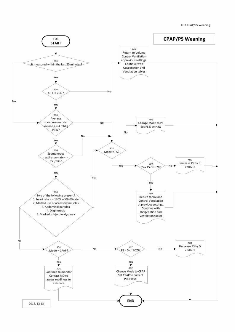
CPAP/PS Weaning
2016, 12 13
FO3
START
END
SO1
pH measured within the last 20 minutes?
SO2
pH > = 7.30?
SO3
Average spontaneous tidal
volume > = 4 ml/kg PBW?
SO4
Spontaneous respiratory rate < =
35 /min?
SO5
Two of the following present?1. heart rate > = 120% of 06:00 rate2. Marked use of accessory muscles
3. Abdominal paradox4. Diaphoresis
5. Marked subjective dyspnea
SO6
Mode = CPAP?
AO1
Continue to monitorContact MD to
assess readiness to extubate
AO2
Change Mode to CPAPSet CPAP to current
PEEP level
SO7
PS = 5 cmH2O?
AO3
Decrease PS by 5 cmH2O
AO7
Return to Volume Control Ventilation at previous settings.
Continue with Oxygenation and Ventilation tables
AO4
Return to Volume Control Ventilation at previous settings.
Continue with Oxygenation and Ventilation tables
SO8
Mode = PS?
SO9
PS = 15 cmH20?
AO6
Increase PS by 5 cmH2O
AO5
Change Mode to PS Set PS 5 cmH2O
Yes
YesNo
No
No
Yes
Yes
Yes
No
No
Yes
Yes
No No
Yes Yes
No
No
FO3 CPAP/PS Weaning
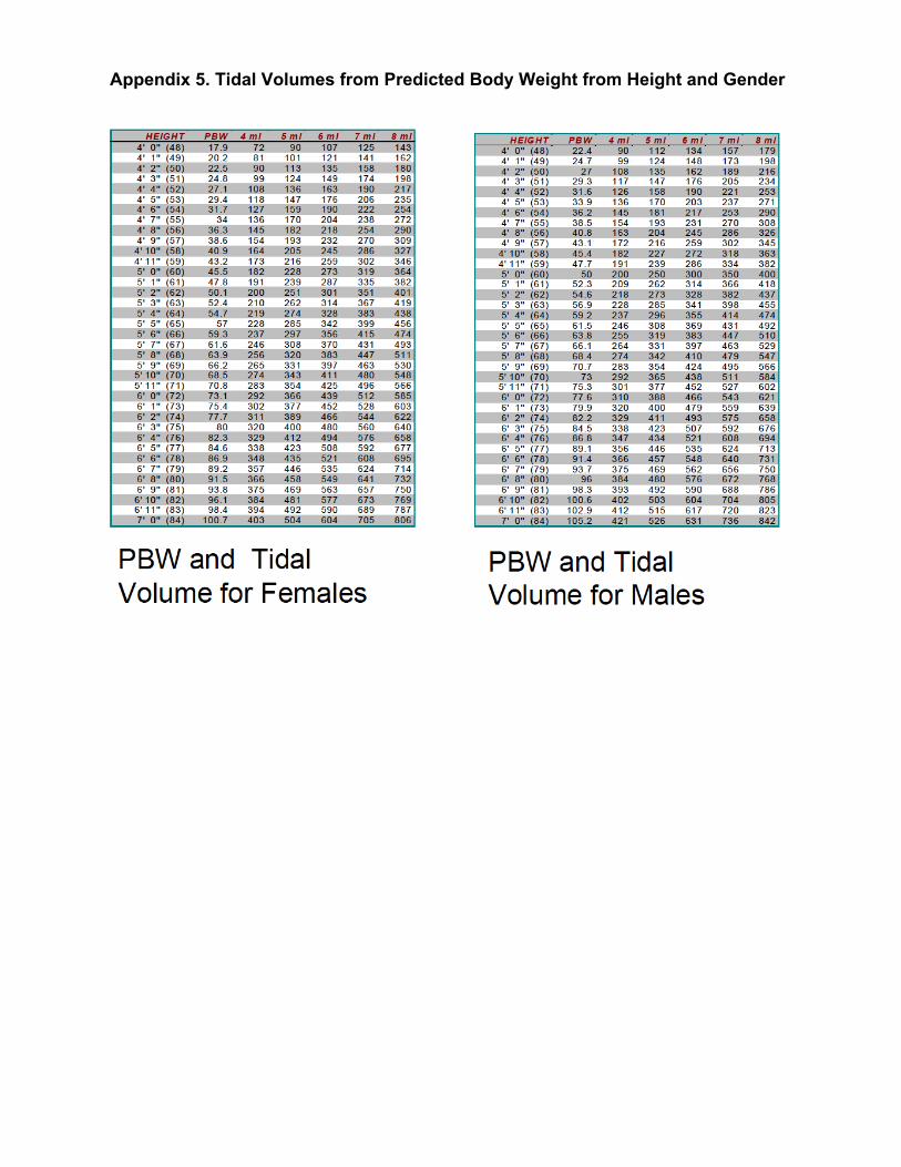
Appendix 5. Tidal Volumes from Predicted Body Weight from Height and Gender
Definitions: BWP: Body weight predicted (formula below)
o Males: PBW (kg) = 50 + 2.3[height (inches) – 60]o Females: PBW (kg = 45.5 + 2.3 [height (inches) – 60]
ABG: arterial blood gas VR: respiratory rate (breaths per minute) VT: tidal volume (milliliters) VE: minute volume (liters per minute) CMV: continuous mandatory ventilation VC: Volume Control PEEP: positive end expiratory pressure (cmH2O) Measured Pplat: actual measured plateau pressure
General: If the PBW does not appear or is too small or too large, check the height and gender
in the computer – they could be entered wrong or not at all. The protocol will not generate new instructions if it has been 2 or more hours since a
complete ventilator assessment has been entered. Always do a ventilator assessment before drawing an ABG. Check for typos. The computer relies on accurate and timely charting. The protocol will run off arterial blood gas or oxygen saturation measured by the
pulse oximeter. Protocol suspensions can be entered proactively or retroactively. Suspend the protocol when the patient will be receiving a procedure, traveling, going
to surgery or hyperbaric. Unsuspend the protocol when back in the unit Enter at the previous settings Enter at the current settings charted (patient may have different needs after the
procedure) The protocols are orders. Deviation from the protocol requires a physician order.
ABG Recommended For: Change in Mode.
ABG Required For: 10% change in VT setting. Change in VR setting if patient is not assisting. Receive ventilation protocol instructions.
Ventilation: The low range for the set ventilatory rate is 6 breaths per minute for all protocols. Ventilation instructions are only given after an ABG.
Appendix 6. Rules for iCentra Neuro Ventilator Protocol
The protocol will set a back up VR if the patient is breathing over the set rate.Backup VR is based on a calculated VE goal.
Set VT at 6ml/kg PWB VE goal = Current VE *(PaCO2/50 * HCO3- /24) Backup set VR = VE goal / set VT Volume control ventilation will be required unless FiO2 < = 0.5 and PEEP < =
10cmH2O, then the patient can be evaluated for pressure support weaning. Tidal Volume (VT) Goal is 6 ml / kg / PBW Measure and record inspiratory plateau pressure (Pplat) with every ventilator
assessment and after changes in VT and PEEP. If Pplat is > 30 cmH2O an ABG is recommended to determine if a VT reduction is
indicated. If unable to measure a Pplat when in PRVC, change the mode to A/C for 2 to 3
minutes. Measure the Pplat. Return the patient to previous mode. Do not increase ventilator rate (VR) above 35 bpm. Do not decrease VT below 4 ml/kg If the patient is not over breathing the set rate do not decrease VT and VR at the
same time.
Oxygenation: The protocol will not decrease PEEP for 6 hours after it has been increased. If PEEP is > 10 cmH2O, do not decrease > 2cmH2O every 2 hours. If the SpO2 or PaO2 fall below the target ranges after a decrease in FiO2 and/or
PEEP and it has been less than 30 minutes, the patient will be returned to theprevious FiO2 and PEEP settings.
Each subsequent repeat of therapy reduction followed by therapy increase will resultin waiting periods (4, 8, and 24 hours).
Night time: Night rests on CMV will start at 22:00 and end at 06:00 when ordered.
Weaning: Weaning may occur 24 hours a day. Weaning may be initiated at any time. Entry criteria for weaning: FiO2 < = .5 Peep < = 10 cmH2O Without neuromuscular blockade Total VR > set VR Weaning assessment will be attempted every 4 hours.