rheumatic heart disease - ksumsc.com work... · a presumptive diagnosis of acute rheumatic fever...
TRANSCRIPT
Rheumatic Heart Disease
Objectives: ● What is ARF And RHD?
● Diagnosis
● Jones Criteria & 2015 revision
● Differential Diagnosis
● Investigations, Management
● Rheumatic Valvular Heart Disease
● Prevention
Team members:Trad Alwakeel + Khalid Aleedan + Yazeed alsuhaibani + Rayan Alqarni
Team leader: Haneen Al Subki
Revised by: Maha AlGhamdi
Designed by: [email protected]
Resources:435 team + Davidson + Recall questions step up to medicine
● Editing file
● Feedback
Color index: IMPORTANT - NOTES - EXTRA - DAVIDSON’S

● Epidemiologic Background:
We are concerned about prevalence and incidence because when we want to diagnose a patient with ARF based on the criteria , we have to know which criteria we should use depending on our country.
Saudi Arabia is classified in moderate risk group.
- Globally RHD is the most common CVD in young people 25 yrs old. - The overall incidence of ARF is 5-51/100000 people, with a mean of 19 per 100000 people. - In children 5-14 yrs old 0.8-5.7 per 1000 children with a median of 1.3 per 1000. - The incidence of RF and the prevalence of RHD has declined substantially in Europe, North America
and other developed nations. This decline has been attributed to improved hygiene, reduced household crowding, and improved medical care. Also due to an improve in socio-economical status and the usage of antibiotics.
- The major burden is currently found in low and middle income countries(India,middle east), and in selected indigenous populations of certain developed countries (Australia and New Zealand).
- A disease of poverty and low socioeconomic status. - In underdeveloped countries RHD is the leading cause of CV death during the first five decades of
life. - In Saudi Arabia: incidence 30 cases/100000 people/yr and prevalence 310/100000 people in 6-15 yrs
age group. - ● Global burden: - Total cases with RHD : 20 Millions - 3 Million have congestive heart failure. - 1 Million require Valve surgery - Annual incidence of RF: 0.5 Million, nearly half develop carditis 1
- Estimated deaths from RHD: 230,000/YR.
1 New cases per year
A. Acute Rheumatic fever: ARF leads to RHD after 10-20 years (latent period) . watch video!
Incidence and pathogenesis
● Acute rheumatic fever usually affects children (most commonly between 5 and 15 years) or young adults .
● The most common cause of acquired heart disease in childhood and adolescence. ● The condition is triggered by an immune-mediated delayed response (manifestations appearing after a
period of 2-4 weeks) to infection with specific strains of group A (beta hemolytic) streptococci, which have antigens that may cross-react with cardiac myosin and sarcolemmal membrane protein. Antibodies produced against the streptococcal antigens cause inflammation in the endocardium, myocardium and pericardium, as well as the joints and skin, but the major effect on health is due to damage to heart valves.
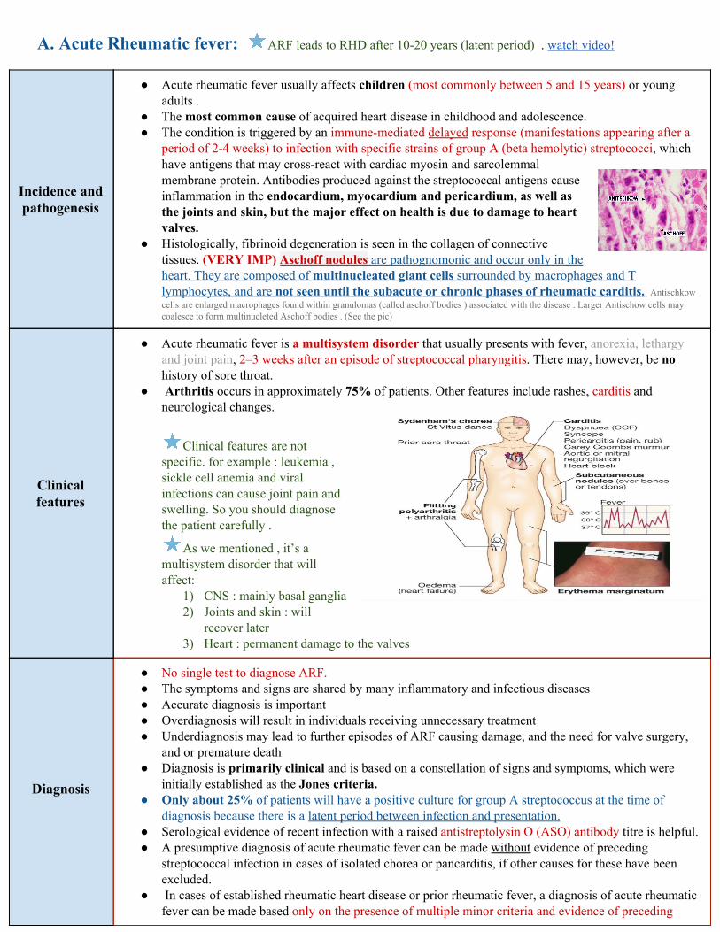
● Histologically, fibrinoid degeneration is seen in the collagen of connective tissues. (VERY IMP) Aschoff nodules are pathognomonic and occur only in the heart. They are composed of multinucleated giant cells surrounded by macrophages and T lymphocytes, and are not seen until the subacute or chronic phases of rheumatic carditis. Antischkow cells are enlarged macrophages found within granulomas (called aschoff bodies ) associated with the disease . Larger Antischow cells may coalesce to form multinucleted Aschoff bodies . (See the pic)
Clinical features
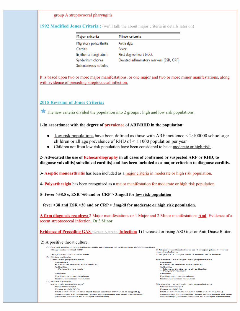
● Acute rheumatic fever is a multisystem disorder that usually presents with fever, anorexia, lethargy and joint pain, 2–3 weeks after an episode of streptococcal pharyngitis. There may, however, be no history of sore throat.
● Arthritis occurs in approximately 75% of patients. Other features include rashes, carditis and neurological changes.
Clinical features are not specific. for example : leukemia , sickle cell anemia and viral infections can cause joint pain and swelling. So you should diagnose the patient carefully .
As we mentioned , it’s a multisystem disorder that will affect:
1) CNS : mainly basal ganglia 2) Joints and skin : will
recover later 3) Heart : permanent damage to the valves
Diagnosis
● No single test to diagnose ARF. ● The symptoms and signs are shared by many inflammatory and infectious diseases ● Accurate diagnosis is important ● Overdiagnosis will result in individuals receiving unnecessary treatment ● Underdiagnosis may lead to further episodes of ARF causing damage, and the need for valve surgery,
and or premature death ● Diagnosis is primarily clinical and is based on a constellation of signs and symptoms, which were
initially established as the Jones criteria. ● Only about 25% of patients will have a positive culture for group A streptococcus at the time of
diagnosis because there is a latent period between infection and presentation. ● Serological evidence of recent infection with a raised antistreptolysin O (ASO) antibody titre is helpful. ● A presumptive diagnosis of acute rheumatic fever can be made without evidence of preceding
streptococcal infection in cases of isolated chorea or pancarditis, if other causes for these have been excluded.
● In cases of established rheumatic heart disease or prior rheumatic fever, a diagnosis of acute rheumatic fever can be made based only on the presence of multiple minor criteria and evidence of preceding
group A streptococcal pharyngitis.
1992 Modified Jones Criteria : (we’ll talk the about major criteria in details later on)
It is based upon two or more major manifestations, or one major and two or more minor manifestations, along with evidence of preceding streptococcal infection.
2015 Revision of Jones Criteria:
The new criteria divided the population into 2 groups : high and low risk populations.
1-In accordance with the degree of prevalence of ARF/RHD in the population:
● low risk populations have been defined as those with ARF incidence < 2:100000 school-age children or all age prevalence of RHD of < 1:1000 population per year
● Children not from low risk population have been considered to be at moderate or high risk.
2- Advocated the use of Echocardiography in all cases of confirmed or suspected ARF or RHD, to diagnose valvulitis( subclinical carditis) and has been included as a major criterion to diagnose carditis.
3- Aseptic monoarthritis has been included as a major criteria in moderate or high risk population.
4- Polyarthralgia has been recognized as a major manifestation for moderate or high risk population
5- Fever >38.5 c, ESR >60 and or CRP > 3mg/dl for low risk population
fever >38 and ESR >30 and or CRP > 3mg/dl for moderate or high risk population.
A firm diagnosis requires: 2 Major manifestations or 1 Major and 2 Minor manifestations And Evidence of a recent streptococcal infection. Or 3 Minor
Evidence of Preceding GAS “Group A strept.”Infection: 1) Increased or rising ASO titer or Anti-Dnase B titer.
2) A positive throat culture.
● Rheumatic Fever Recurrences: - Reliable past history of ARF: 2 major or 1 major and 2 minor or 3 minor manifestations sufficient for
diagnosis - Presence of antecedent (previous) streptococcal infection - When minor manifestations only present exclude other causes.
Jones criteria (major criteria)
1-Carditis:
● A ‘pancarditis’ involves the endocardium, myocardium and pericardium to varying degrees. Its incidence declines with aging, ranging from 90% at 3 years to around 30% in adolescence.
● Occurs in 50-70% of cases ● The only manifestation of ARF that leaves serious permanent damage ● May be subclinical (without murmurs so we should use Echo) ● Cardiomegaly and CHF may occur
2-Arthritis:
● This is the most common major manifestation and occurs early when streptococcal antibody titres are high (present in 35-66% ,Earliest manifestation of ARF).
● An acute painful asymmetric and migratory inflammation (Migrating, “Fleeting”polyarthritis) of the large joints typically affects the knees, ankles, shoulders,elbows. The joints are involved in quick succession and are usually red, swollen and tender.
● Duration short < 1 week. ● Rapid improvement with salicylates. The pain characteristically responds to aspirin; if not, the
diagnosis is in doubt. ● Does not progress to chronic disease.
3-Sydenham Chorea:
● Also known as Saint Vitus's Dance, occur in 10-30%, female predominance. ● This is a late neurological manifestation (extrapyramidal manifestation) that appears at least 3
months after the episode of acute rheumatic fever, when all the other signs may have disappeared (Delayed manifestation of ARF).
● Emotional lability may be the first feature and is typically followed by purposeless, involuntary, choreiform movements of the hands,face, neck, trunk, and limbs.. Speech may be explosive and halting (clumsiness, deterioration of handwriting,emotional lability or grimacing of face).
● Spontaneous recovery usually occurs within a few months. Approximately one-quarter of affected patients will go on to develop chronic rheumatic valve disease..
4-Subcutaneous Nodules:
● Subcutaneous nodules occur in 10% of patients. ● They are small (0.5–2.0 cm), firm non-tender and painless. ● Best felt over extensor surfaces of bone or tendons (Occur over extensor surfaces of
joints, on bony prominences, tendons, spine). ● They typically appear more than 3 weeks after the onset of other manifestations
and therefore help to confirm rather than make the diagnosis ● Short lived: last for few days. ● Associated with severe carditis.
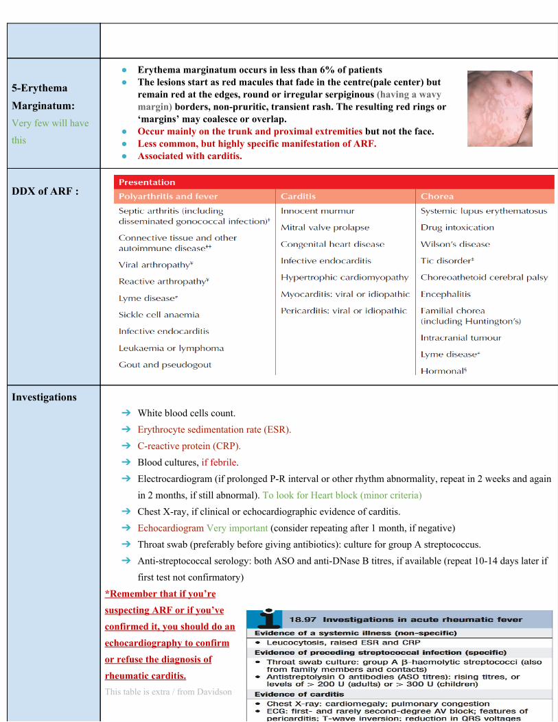
5-Erythema Marginatum: Very few will have
this
● Erythema marginatum occurs in less than 6% of patients ● The lesions start as red macules that fade in the centre(pale center) but
remain red at the edges, round or irregular serpiginous (having a wavy margin) borders, non-pruritic, transient rash. The resulting red rings or ‘margins’ may coalesce or overlap.
● Occur mainly on the trunk and proximal extremities but not the face. ● Less common, but highly specific manifestation of ARF. ● Associated with carditis.
DDX of ARF :
Investigations ➔ White blood cells count. ➔ Erythrocyte sedimentation rate (ESR). ➔ C-reactive protein (CRP). ➔ Blood cultures, if febrile. ➔ Electrocardiogram (if prolonged P-R interval or other rhythm abnormality, repeat in 2 weeks and again
in 2 months, if still abnormal). To look for Heart block (minor criteria) ➔ Chest X-ray, if clinical or echocardiographic evidence of carditis. ➔ Echocardiogram Very important (consider repeating after 1 month, if negative) ➔ Throat swab (preferably before giving antibiotics): culture for group A streptococcus. ➔ Anti-streptococcal serology: both ASO and anti-DNase B titres, if available (repeat 10-14 days later if
first test not confirmatory) *Remember that if you’re suspecting ARF or if you’ve confirmed it, you should do an echocardiography to confirm or refuse the diagnosis of rheumatic carditis. This table is extra / from Davidson
Treatment of ARF
NO specific treatment Now you diagnosed your patient with ARF, what treatment options you can offer him/her?
1. Bed Rest : It’s important, as it lessens joint pain and reduces cardiac workload. The duration should be guided by symptoms, along with temperature, leucocyte count and ESR, and should be continued until these have settled.
2. Salicylates : Like Aspirin, this usually relieves the symptoms of arthritis rapidly and a response within 24 hours helps confirm the diagnosis. We should monitor the patient for toxicity (usually he will have tinnitus and vomiting)
3. Penicillin : Like Procaine Penicillin 4 million units/day x10 days. if the patient is penicillin-allergic, erythromycin or a cephalosporin can be used.
4. Steroids (Prednisolone): 2 mg/kg/day taper over 6 weeks Produces more rapid symptomatic relief than aspirin and is indicated in cases with carditis or severe arthritis (Given when there is severe carditis). There is no evidence that long-term steroids are beneficial.
5. Heart Failure Treatment : Like diuretics and ACEI. If heart failure develops, and does not respond to medical treatment, valve replacement may be necessary and is often associated with a dramatic decline in rheumatic activity. Secondary Prevention of Rheumatic Fever (Prevention of Recurrent Attacks) : شرح للصورة اللي تحت
Primary prevention is important by improving socio-economical status
- Patients are susceptible to further attacks of rheumatic fever if another streptococcal infection occurs, and long-term prophylaxis with penicillin should be given as benzathine penicillin Long acting penicillin (1.2 million U IM monthly), if compliance is in doubt, or oral phenoxymethylpenicillin (250 mg twice daily).
- Sulfadiazine or erythromycin may be used if the patient is allergic to penicillin; sulphonamides prevent infection but are not effective in the eradication of group A streptococci.
- Further attacks of rheumatic fever are unusual after the age of 21, when treatment may be stopped. However, it should be extended if an attack has occurred in the last 5 years, or if the patient lives in an area of high prevalence or has an occupation (e.g. teaching) with high exposure to streptococcal infection.In those with residual heart disease, prophylaxis should continue until 10 years after the last episode or 40 years of age, whichever is later. Long-term antibiotic prophylaxis prevents another attack of acute rheumatic fever but does not protect against infective endocarditis. “Summarised in the picture”
B.Chronic Rheumatic heart disease: (In Adults)
● Chronic valvular heart disease develops in at least half of those affected by rheumatic fever with carditis.
● The mitral valve is affected in more than 70% of cases (usually Stenosis); the aortic valve is the next
most frequently involved (40%), followed by the tricuspid(10%) and then the pulmonary valve(2%).
● Valve disease may be symptomatic during fulminant forms of acute rheumatic fever but may remain
asymptomatic for many years.
● Mitral Stenosis is more common in females (3:1), while males have higher incidence of Aortic
Regurgitation.
Pathology:
● The main pathological process in chronic rheumatic heart disease is progressive fibrosis.
Refresh your memory : The heart sound is basically “LUB” which is S1 - This sound is produced by closure of
Atrioventricular valves- + “DUB” which is S2 - This sound is produced by closure of Aortic and pulmonary valves .
Watch video!
Mitral Stenosis: The most common valvular abnormality in RHD
Notes
The flow of blood from LA to LV is restricted and left atrial pressure rises, leading to pulmonary venous
congestion and breathlessness. There is dilatation and hypertrophy of the LA, and left ventricular lling
becomes more dependent on left atrial contraction. ➔➔ The normal mitral valve area (MVA) = 4-6 cm2 ➔➔ In severe ms <1.5 cm2 ➔➔ High LAP ➔➔ The rise in LAP causes a similar rise in pulmonary capillaries, veins and artery.
Ventricular diastolic pressure will remain normal because it is protected by the Mitral Stenosis !
Clinical Features:
➢ Dyspnea ➢ Fatigue no enough blood bumped to
the body to suply it. ➢ Palpitation ➢ Hemoptysis (10%) due to pulmonary
hypertension so the vessels will rupture.
➢ Hoarseness (Ortner’s syndrome) 2
➢ Dysphagia ➢ Stroke or peripheral embolization
Why ? Left atrium will be enlarged due to stenosis, so the rhythm will be changed from sinus to atrial and the patient usually develops Atrial Fibrillation. Afib will lead to blood clotting that will form a thrombus which travels to the brain causing stroke (and sometimes stroke is the main presentation in a patient with Mitral stenosis).
➢ Cyanosis (Mitral facies , malar 3
flush) ➢ Tapping apex ( S1) 4
➢ Parasternal heave It’s precordial impulse that may be felt in patients with cardiac or respiratory disease.
➢ Diastolic thrill A vibration felt over the heart during ventricular diastole . it may be caused due to mitral valve stenosis .
➢ Accentuated S1 5
➢ Accentuated S2 ➢ Opening snap S2 is followed by an
opening snap , the distance between S2 and the opening snap can give an indication as to the severity of the stenosis . the closer the opening snap follows S2 , the worst the stenosis .
➢ Mid-diastolic rumble
Investigations
Chest X-ray
● Straightening of the left heart border ● Double density ● Kerley B lines These are short parallel lines at the lung periphery
ECG LAE (Left Atrial Enlargement), P Mitrale(left atrial enlargement) , RV dominance , Atrial Fibrillation
Echodoppler . ● Calcification in MV
You can find heart enlargement
2 paralysis of the vocal cords, due to the enlargement of pulmonary artery→ compression of recurrent laryngeal nerve 3 distinctive facial appearance associated with mitralstenosis. Someone with mitral stenosis may present with rosy cheeks, whilst the rest of the face has a bluish tinge due to cyanosis. ... It is due to low cardiac output, and therefore low perfusion of the facial skin, caused by the stenosis. 4 Tapping apex beat is present only in mitral stenosis where the left ventricular size and filling is less . 5 Very obvious
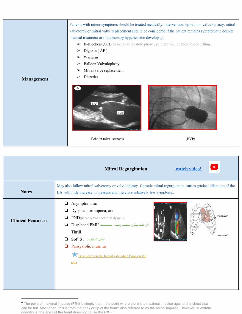
Management
Patients with minor symptoms should be treated medically. Intervention by balloon valvuloplasty, mitral
valvotomy or mitral valve replacement should be considered if the patient remains symptomatic despite
medical treatment or if pulmonary hypertension develops.):
➢ B-Blockers ,CCB to increase diastole phase , so there will be more blood filling.
➢ Digoxin ( AF )
➢ Warfarin
➢ Balloon Valvuloplasty
➢ Mitral valve replacement
➢ Diuretics
Echo in mitral stenosis (BVP)
Mitral Regurgitation watch video!
Notes
May also follow mitral valvotomy or valvuloplasty. Chronic mitral regurgitation causes gradual dilatation of the
LA with little increase in pressure and therefore relatively few symptoms.
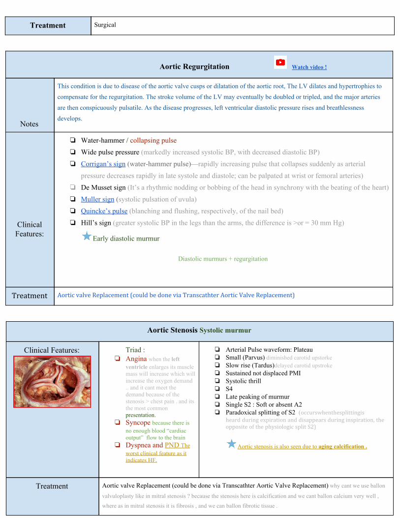
Clinical Features:
❏ Asymptomatic ❏ Dyspnea, orthopnea, and ❏ PND(parxosymal nocturnal dyspnia)
❏ Displaced PMI ألن القلب بیكون متضخم وبیعمل دسبلیسمنت , 6
Thrill ❏ Soft S1 عكس الستینوسز
❏ Pansystolic murmur
Best heard on the lateral side when lying on the
side
6 The point of maximal impulse (PMI) is simply that... the point where there is a maximal impulse against the chest that can be felt. Most often, this is from the apex or tip of the heart: also referred to as the apical impulse. However, in certain conditions, the apex of the heart does not cause the PMI.
Treatment Surgical
Aortic Regurgitation Watch video !
Notes
This condition is due to disease of the aortic valve cusps or dilatation of the aortic root, The LV dilates and hypertrophies to
compensate for the regurgitation. The stroke volume of the LV may eventually be doubled or tripled, and the major arteries
are then conspicuously pulsatile. As the disease progresses, left ventricular diastolic pressure rises and breathlessness
develops.
Clinical Features:
❏ Water-hammer / collapsing pulse ❏ Wide pulse pressure (markedly increased systolic BP, with decreased diastolic BP) ❏ Corrigan’s sign (water-hammer pulse)—rapidly increasing pulse that collapses suddenly as arterial
pressure decreases rapidly in late systole and diastole; can be palpated at wrist or femoral arteries) ❏ De Musset sign (It’s a rhythmic nodding or bobbing of the head in synchrony with the beating of the heart) ❏ Muller sign (systolic pulsation of uvula) ❏ Quincke’s pulse (blanching and flushing, respectively, of the nail bed) ❏ Hill’s sign (greater systolic BP in the legs than the arms, the difference is >or = 30 mm Hg)
Early diastolic murmur
Diastolic murmurs + regurgitation
Treatment Aortic valve Replacement (could be done via Transcathter Aortic Valve Replacement)
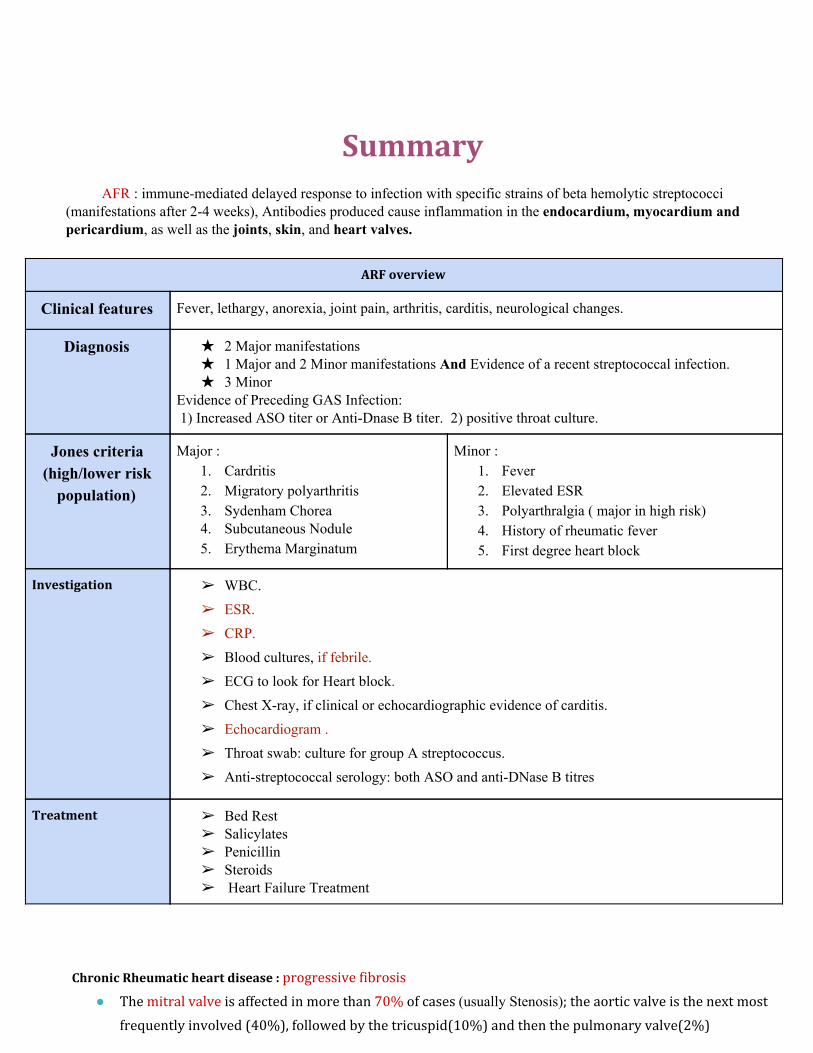
Aortic Stenosis Systolic murmur
Clinical Features:
Triad : ❏ Angina when the left
ventricle enlarges its muscle mass will increase which will increase the oxygen demand .. and it cant meet the demand because of the stenosis > chest pain . and its the most common presentation.
❏ Syncope because there is no enough blood “cardiac output” flow to the brain
❏ Dyspnea and PND The worst clinical feature as it indicates HF.
❏ Arterial Pulse waveform: Plateau ❏ Small (Parvus) diminished carotid upstorke ❏ Slow rise (Tardus)delayed carotid upstroke ❏ Sustained not displaced PMI ❏ Systolic thrill ❏ S4 ❏ Late peaking of murmur ❏ Single S2 : Soft or absent A2 ❏ Paradoxical splitting of S2 (occurswhenthesplittingis
heard during expiration and disappears during inspiration, the opposite of the physiologic split S2)
Aortic stenosis is also seen due to aging calcification .
Treatment Aortic valve Replacement (could be done via Transcathter Aortic Valve Replacement) why cant we use ballon
valvuloplasty like in mitral stenosis ? because the stenosis here is calcification and we cant ballon calcium very well ,
where as in mitral stenosis it is fibrosis , and we can ballon fibrotic tissue .
Summary
AFR : immune-mediated delayed response to infection with specific strains of beta hemolytic streptococci (manifestations after 2-4 weeks), Antibodies produced cause inflammation in the endocardium, myocardium and pericardium, as well as the joints, skin, and heart valves.
ARF overview
Clinical features Fever, lethargy, anorexia, joint pain, arthritis, carditis, neurological changes.
Diagnosis
★ 2 Major manifestations ★ 1 Major and 2 Minor manifestations And Evidence of a recent streptococcal infection. ★ 3 Minor
Evidence of Preceding GAS Infection: 1) Increased ASO titer or Anti-Dnase B titer. 2) positive throat culture.
Jones criteria (high/lower risk
population)
Major : 1. Cardritis 2. Migratory polyarthritis 3. Sydenham Chorea 4. Subcutaneous Nodule 5. Erythema Marginatum
Minor : 1. Fever 2. Elevated ESR 3. Polyarthralgia ( major in high risk) 4. History of rheumatic fever 5. First degree heart block
Investigation ➢ WBC. ➢ ESR. ➢ CRP. ➢ Blood cultures, if febrile. ➢ ECG to look for Heart block. ➢ Chest X-ray, if clinical or echocardiographic evidence of carditis. ➢ Echocardiogram . ➢ Throat swab: culture for group A streptococcus. ➢ Anti-streptococcal serology: both ASO and anti-DNase B titres
Treatment ➢ Bed Rest ➢ Salicylates ➢ Penicillin ➢ Steroids ➢ Heart Failure Treatment
Chronic Rheumatic heart disease : progressive fibrosis
● The mitral valve is affected in more than 70% of cases (usually Stenosis); the aortic valve is the next most
frequently involved (40%), followed by the tricuspid(10%) and then the pulmonary valve(2%)
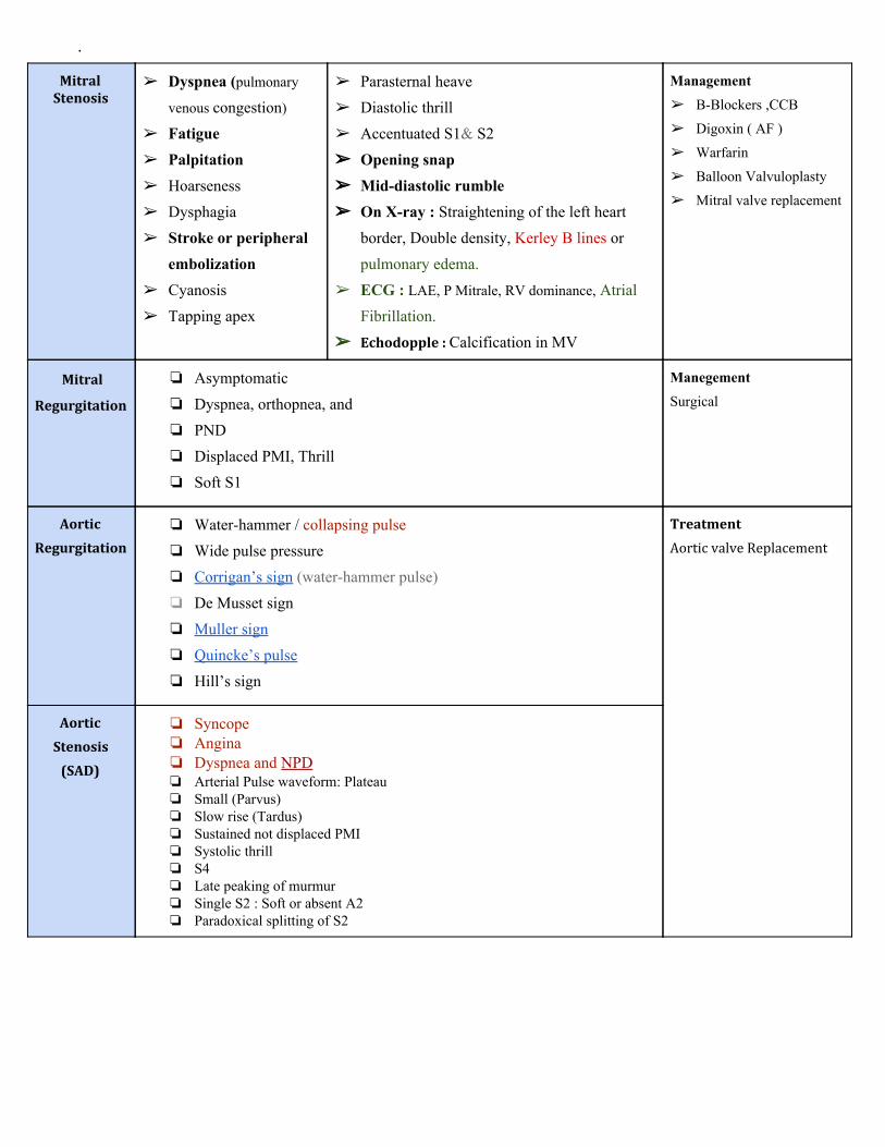
.
Mitral Stenosis
➢ Dyspnea (pulmonary
venous congestion) ➢ Fatigue ➢ Palpitation ➢ Hoarseness ➢ Dysphagia ➢ Stroke or peripheral
embolization ➢ Cyanosis ➢ Tapping apex
➢ Parasternal heave ➢ Diastolic thrill ➢ Accentuated S1& S2 ➢➢ Opening snap ➢➢ Mid-diastolic rumble ➢➢ On X-ray : Straightening of the left heart
border, Double density, Kerley B lines or pulmonary edema.
➢ ECG : LAE, P Mitrale, RV dominance, Atrial Fibrillation.
➢➢ Echodopple : Calcification in MV
Management ➢ B-Blockers ,CCB
➢ Digoxin ( AF )
➢ Warfarin
➢ Balloon Valvuloplasty
➢ Mitral valve replacement
Mitral
Regurgitation
❏ Asymptomatic ❏ Dyspnea, orthopnea, and ❏ PND
❏ Displaced PMI, Thrill ❏ Soft S1
Manegement Surgical
Aortic
Regurgitation
❏ Water-hammer / collapsing pulse ❏ Wide pulse pressure ❏ Corrigan’s sign (water-hammer pulse) ❏ De Musset sign ❏ Muller sign ❏ Quincke’s pulse ❏ Hill’s sign
Treatment
Aortic valve Replacement
Aortic
Stenosis
(SAD)
❏ Syncope ❏ Angina ❏ Dyspnea and NPD ❏ Arterial Pulse waveform: Plateau ❏ Small (Parvus) ❏ Slow rise (Tardus) ❏ Sustained not displaced PMI ❏ Systolic thrill ❏ S4 ❏ Late peaking of murmur ❏ Single S2 : Soft or absent A2 ❏ Paradoxical splitting of S2

Questions
1.Which one of the following Organisms is the most common cause for Rheumatic Heart Disease ? A-group A beta hemolytic streptococcus B-group B alpha hemolytic streptococcus C-staphylococcus aureus D- E.coli 2. Which one of the following group of people are mostly affected by Rheumatic Heart Disease ? A- Elderly from 40-60 B- Teenagers from 14-18 C-Children from 5-15 D-New born babies 3. Which one of the following Valve is mostly affected by Rheumatic Heart Disease ? A- Mitral Valve B- Tricuspid Valve C- Aortic Valve D- Pulmonary Valve 4. Which one of the following diseases is Immunologically Mediated ? A-Hypertension B-DM C-Hyperlipidemia D- Rheumatic Heart Disease 5. Which one of the following serological tests is used to detect recent infection by Group A streptococcus? A-Antistreptolysin O antibodies B-Antinuclear antibodies C-Antimitochondorial antibodies D-Antigliadin antibodies 6. In order to diagnose Rheumatic Heart Disease, which statement is correct ? A-Migratory polyarthritis only B- polyarthralgia + increase ESR only C- Subcutaneous nodules + fever only D- Sydenham chorea + Prolonged PR interval + Fever
7.The only manifestation of ARF that leaves serious permanent damage is ? A-Arthritis B-Subcutaneous nodules C-Carditis D-Erythema Marginatum

8. Which one of the following is seen under the microscope and confirm the diagnosis of ARF ? A-Aschoff Nodules B- Lewi Body C- Caseous Necrosis D- Mallory Body 9. In order to treat patient with Streptococcal pharyngitis , which of the following is the best for him ? A-penicillin B-Ciftriaxone C- Ciprofloxacin D- Gentmycin
10. Which one of the following is the main pathological process in Chronic Rheumatic Heart Disease ? A-Calcification B-Necrosis C-Amyloidosis D- Progressive Fibrosis
11. Which one of the following is Clinical feature for Aortic Regurgitation ? A- Collapsing pulse B- Syncope C-Dyspnea D- Angina
12. Which one of the following is Clinical feature for Mitral Regurgitation ? A-Early diastolic murmur B-Mid systolic click C-Pansystolic murmur D- Systolic Thrill
13. The Radiological feature that is seen on Mitral Stenosis is ? A-Single Density B-Kaerle B line C-Normal left heart boarder D- Coin lesion
14. What is the treatment for Aortic Regurgitation ? A-Warfarin B-Beta Blockers C-Calcium Channel Blockers D-Aortic valve replacement
15.Water-Hammer pulse is feature of which one of the following ? A-Aortic Regurgitation B-Aortic Stenosis C-Mitral Stenosis D-Mitral Regurgitation
16. Syncope , Angina and Dyspnea are characters for which disease ? A- Aortic Regurgitation B-Aortic Stenosis C-Mitral Stenosis D-Mitral Regurgitation
17. Enlargement of the Heart can be seen in which of the following ? A-Aortic Regurgitation B- Aortic Stenosis C-Mitral Stenosis D-Mitral Regurgitation
18. Which one of the following can treat Mitral Stenosis ? A-Digoxin B-Warfarin C-Mitral Valve Replacement D- All the above
19. Which one of the following valve is LESS common affected by Rheumatic Heart Disease? A-Aortic valve B- Mitral valve C-Tricuspid D-Pulmonary
20. Which one of the following is Clinical feature for Mitral Stenosis? A-Opening Snap B-Mid diastolic rumble C- Soft S1 D-A & B Answer key : 1-A 2-C 3-A 4-D 5-A 6-D 7-C 8-A 9-A 10-D 11-A 12-C 13-B 14-D 15-A 16-B 17-B 18-D 19-D 20-D