reviewarticle - cuppingtherapy.org of evidence... · reviewarticle ... acupuncture...
TRANSCRIPT
Hindawi Publishing CorporationEvidence-Based Complementary and Alternative MedicineVolume 2011, Article ID 260510, 6 pagesdoi:10.1155/2011/260510
Review Article
Review of Evidence Suggesting That the Fascia NetworkCould Be the Anatomical Basis for Acupoints and Meridians inthe Human Body
Yu Bai,1 Jun Wang,2 Jin-peng Wu,1 Jing-xing Dai,1 Ou Sha,2 David Tai Wai Yew,3
Lin Yuan,1 and Qiu-ni Liang2
1 Department of Anatomy, Southern Medical University, Guangzhou 510515, China2 Department of Anatomy, School of Medicine of Shenzhen University, Shenzhen 518060, China3 Department of Anatomy, The Chinese University of Hong Kong, Shatin, Hong Kong
Correspondence should be addressed to Lin Yuan, [email protected]
Received 4 August 2010; Revised 4 February 2011; Accepted 28 February 2011
Copyright © 2011 Yu Bai et al. This is an open access article distributed under the Creative Commons Attribution License, whichpermits unrestricted use, distribution, and reproduction in any medium, provided the original work is properly cited.
The anatomical basis for the concept of meridians in traditional Chinese medicine (TCM) has not been resolved. This paperreviews the evidence supporting a relationship between acupuncture points/meridians and fascia. The reviewed evidence supportsthe view that the human body’s fascia network may be the physical substrate represented by the meridians of TCM. Specifically,this hypothesis is supported by anatomical observations of body scan data demonstrating that the fascia network resembles thetheoretical meridian system in salient ways, as well as physiological, histological, and clinical observations. This view represents atheoretical basis and means for applying modern biomedical research to examining TCM principles and therapies, and it favors aholistic approach to diagnosis and treatment.
1. Introduction
The theory of meridians and collaterals, also popularlyknown as channel theory, is a fundamental pillar of tradi-tional Chinese medicine (TCM), particularly in the areasof acupuncture, moxibustion, and massage, as well as oftraditional martial arts such as Tai Chi Chuan [1, 2]. In prin-ciple, meridians (jıngluo in Chinese) are essentially strings ofacupoints, which may be visualized as passageways throughwhich energy flows throughout the body [2, 3]. The meridiansystem is thought to be composed of 12 principal meridians,each of which connects to an organ system and extends to anextremity, and eight collaterals (Figures 1(a), 1(c), and 1(e)).Practitioners of TCM intend that their treatments shouldimprove the flow of energy through the meridian network.
Given the long history of channel theory, which predatesmodern scientific development, and the theory’s intermin-gling with philosophy and ancient metaphysical ideas, rigor-ous scientific and clinical studies are needed to tease out theirtrue, physical nature [2]. Acupuncture is an ancient aspect
of TCM with demonstrated therapeutic effects [2]. Althoughscientific interest in the validity of meridians and acupointshas been growing in the last decade, the basis of the natureand material of acupuncture points and meridians has notbeen resolved. Lo proposed that acupuncture meridians aremade up of polarized molecules [4]. Based on a review of theliterature, Ahn et al. concluded that the available evidence didnot conclusively support the claim that acupuncture pointshad distinct electrical properties [5]. Ma and colleagueshave speculated that some aspect of the perivascular spacemight be the anatomical substrate of meridians [6]. Liet al. reported evidence suggesting that visualized regionalhypodermic migration channels of interstitial fluid consti-tuted the meridians [7]. Other research groups have sinceprovided complementary supportive evidence with variousapproaches for this acupoint simulation-brain activationphenomenon [8–11].
While evidence consistent with the existence of entitiestermed meridians is growing, much work is still needed todelineate their anatomical basis [12]. It has been posited
2 Evidence-Based Complementary and Alternative Medicine
Front Back
A
1234
B 56
78
(a)
B A
(b)
234
5678
A B
1
(c)
A B
(d)
2
34
5
6
78
1
A B
(e)
AB
(f)
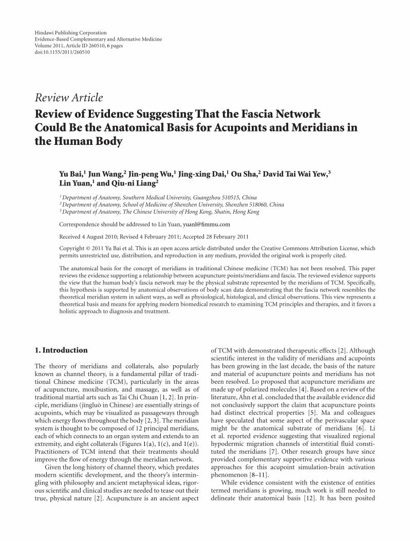
Figure 1: Comparison between acupuncture points (from the anatomical guidelines provided in [19]) and fascia imaging data. The fasciaconnective tissue gathering areas were constructed using MIMICS11.02 software based on the digital datasets of VCH bodies. (a) Locationof the Ren Channel (A) and Du Channel (B): (1) Tanzhong. (2) Juque. (3) Shenque. (4) Qugu. (5) Dazhui. (6) Shendao. (7) Yaoyangguan.(8) Yaoshu. (b) Reconstruction of fascia pathways approximating the Ren Channel (A) and Du Channel (B). (c) Locations of the Tripleenergizer meridian of hand-shaoyang (a) and the Large intestine meridian of hand-yangming (b): (1) Tianjing. (2) Sanyangluo. (3) Yangchi.(4) Guanchong. (5) Quchi. (6) Pianli. (7) Hegu. (8) Shangyang. (d) Reconstruction of fascia pathways in the arm approximating the Tripleenergizer meridian of hand-shaoyang (A) and the Large intestine meridian of hand-yangming (B). (e) Locations of the Kidney meridianof foot-shaoyin (A) and the Gallbladder meridian of foot-shaoyang (B): (1) Yingu. (2) Zhubin. (3) Dazhong. (4) Rangu. (5) Fengshi. (6)Yanglingquan. (7) Xuanzhong. (8) Qiuxu. (f) Reconstruction of fascia pathways in the leg approximating the Kidney meridian of foot-shaoyin (A) and the Gallbladder meridian of foot-shaoyang (B).
that the physical substrate of meridians may include neu-rovascular bundles, neuromuscular attachments, sensorynerve endings, perivascular space and perineurial vessels[6, 13–17]. In particular, Langevin and Yandow proposedthat the anatomical relationship of acupuncture points andmeridians to connective tissue planes [18].
Based on supportive evidence in the literature, thepresent paper provides support for a fascia network hypoth-esis of meridians—that is, the view that the fascia networkmay be the anatomical basis for acupoints and meridiansin the human body. Specifically, we examine whether theevidence supports the ideas that (i) the anatomical basisof meridians is the fascia network that is distributedthroughout the body and (ii) the histological composition ofmeridians is nonspecific connective tissues, including looseconnective tissue and fat tissue. The histological structures
where an acupuncture needle acts are fascia connectivetissue containing nerve endings, capillary vessels, fibroblasts,undifferentiated mesenchymal cells, lymphocytes, and soforth. Acupuncture points are traditionally believed to besites that produce strong reactions when stimulated. Thedistribution of fascia connective tissue throughout the bodyenables acupoints to exist in every part of the body.In our view, the difference between clinically recognizedacupoints and nonacupoints, as well as between mainacupoints and supplementary acupoints, lies principally inthe intensity of biological reactions rather than in the grossstructural components per se. Below, our view that thefascia may be the physical substrate of the meridian systemis evaluated with respect to prior anatomical observationsof body scan data demonstrating that three-dimensional(3D) reconstructions of fascia resemble the theoretical
Evidence-Based Complementary and Alternative Medicine 3
(a) (b) (c) (d)
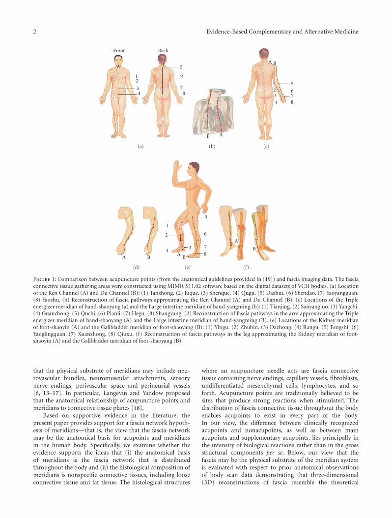
Figure 2: Thick fascia connective tissues in VCH images were marked (green) (a) and their 3 D structures were rendered (b, c). Whenfascia connective tissues of the whole body, including thick and thin fascia tissues, were marked and their 3 D structures were reconstructed,a complete fascia network was observed; (d see reference no. [19]). All of the human organs and tissues were observed to be coated withconnective tissues, and the connective tissues extended into the organs to form septa within the organs.
meridian system in salient ways and relevant physiologicalobservations.
2. Anatomical and Physiological Observations
2.1. Human Body Imaging. The 3D constructions of thehuman body produced by the visible Chinese human (VCH)project, a National Basic Research Program of China, providean intriguing window into human anatomy [19, 20]. Indeed,as illustrated in Figure 1, scan data show that the fascialconnective tissues of the human body approximate conspic-uously the TCM meridian network [19, 20]. Firstly, it is note-worthy that reconstructions of the fascial connective tissuesin the body trunk and limbs show line-like structures whichare similar to those of acupoints and meridians/collaterals[19]. Secondly, these fascial strings form a network of linesthat are close to the virtual meridians in anatomical location[19]. Furthermore, as shown in Figure 2, subsequent 3D fas-cial reconstructive studies involving computed tomographyand magnetic resonance imaging (MRI) of living humanbodies revealed a pattern of line-like structures that appearsimilar in form and distribution to the traditional Chinesemeridians [19, 20]. The VCH and living body imagingstudies together indicate that the anatomy of the fascialnetwork in the human body is consistent with the traditionalview of the meridian network pattern.
2.2. Acupuncture. While Cho et al.’s imaging data suggestinga possible connection between acupuncture and brainactivation discussed above provide evidence that meridiansshould have a physical anatomical substrate, they did notprovide precise information regarding what that substratemay be [21, 22]. Studies of the local acupuncture processitself may shed light on the anatomical substrate. Efficaciousacupuncture is associated with a temporary local sensationof soreness and/or numbness (termed deqi) at the acupointsite [23]. The needle grasp phenomenon has been shown to
occur when a needle physically impacts the connective tissuein the fascia [18, 24]. This observation indicates that theefficacy of acupuncture relies on interaction with the fascia.
2.3. Physiological Observations. Fascia is the soft tissue com-ponent of the connective tissue system that permeates thehuman body. It forms a whole-body continuous 3D matrix ofstructural support [25]. It penetrates and surrounds all of thebody’s vital organs, muscles, bones, and nerve fibers, creatinga unique physiological environment [25]. This network offascial connective tissue is situated to provide ongoing phys-iological support for the body’s metabolically active systemscomposed of specialized cells and tissues [25]. In the view ofTCM, optimal health requires unencumbered flow of energythrough the meridians. Of course, TCM does not specify thephysical nature of such “energy.” If the meridians are fascia,as we posit, then that energy may be nerve signals, flow ofparacrine signaling molecules, electrical signaling throughgap junctions among perineurial cells, distribution ofmechanical forces, or some combination of these processes.
2.4. Fascia Provides Dynamic Connections between and amongMuscles and Bones. Van der Wal used 3D reconstructionstudies to reveal a continuous connective tissue structure thatruns throughout the body allowing for dynamic connectionsbetween the fascia and musculature [26]. Van der Wal’s workwas revolutionary in that it has provided a view of the fasciaas an integrated structure, rather than distinct piecemealstructures associated with particular muscles and/or bones[26]. The integrated nature of the fascia is consistent with itspresently hypothesized role as the meridian network of thebody.
In a study examining the flexor carpi ulnaris in humanpatients undergoing tendon transfer surgery, Smeulders andKreulen found that the intermuscular connections, ratherthan individual tendon-muscle connections, were responsi-ble for most (90%) of the transference of forces between the
4 Evidence-Based Complementary and Alternative Medicine
neighboring muscles [27]. These researchers further foundthat muscle excursion was more limited by intermuscularconnective tissues than by tendon-muscle connections [27].Indeed, Huijing and Baan showed that as much as half of theforce generated by a muscle is transmitted to surroundingconnective tissues [28]. Thus, it appears that the fasciaemediate an active mechanical transference role.
2.5. Responsivity of Cells to Tensional Forces. The utilityof mechanical force activation of neuronal receptors haslong been appreciated for touch and pressure sensationas well as for protection signaling of potential injury dueto hyperextension, hypercompression, or hyperrotation.Indeed integrin receptors expressed in the extracellularmatrix are mechanically coupled to intra-cellular actinfilaments, the contortion of which can initiate biochemicalsignaling resulting in adaptive changes in cell morphology[29]. Hence, mechano-sensation in connective tissues doesnot merely passively relay information to the central nervoussystem, but also directly impacts the properties of the fasciaitself including its fibroblasts and collagen fibers [30–32].Furthermore, Grinnell demonstrated that cells do not adhereindiscriminately to matrix proteins, but rather adhere tospecific matrix fibrils, supporting the notion of an organizedfunction fascial network [33]. Fascia inflammation will beaddressed in the following section, but it is worth mentioninghere that repetitive mechanical strain was experimentallyshown to affect secretion of proinflammatory interleukinsand cell proliferation of human fibroblasts in vitro [34, 35].
2.6. Perineurium and Epineurium Implicated in Pain andInflammation. The fascia tissue surrounding nerve fascicles(the perineurium) and that surrounding the whole nerveand associated vasculature bundles (the epineurium) playimportant roles in pain regulation [36]. Bove and Light con-ducted immunohistochemistry studies revealing peptidergicfine-caliber axons in the epineurium and perineurium thatare consistent with nociceptive function [37]. Subsequentin vitro electrical and chemical nerve stimulation studiesdemonstrated that stimulation of local nociceptive receptorsof the perineurium and/or epineurium can evoke neurogenicinflammation [38]. Such evidence indicates that disruptionof perineural fascial tissues that stimulates perineuralnociceptors can trigger local (neurogenic) inflammation,presumably as a defensive mechanism that functions to helpmaintain the local environment of the nerves [39].
Indeed a convergence of evidence indicates that chroniclow back pain may be emanating from connective tissues,rather than bone, cartilage, or musculature. The magnitudeof low back pain was found not to correlate with magnitudeof disc displacement [40]. Subsequent work has implicatedperispinal ligamentous tissues and lumbar fascia as commonculprits of low back pain [41, 42]. Moreover, Thomas andRobet found corroborating histological evidence indicatingthat low back pain may be due to inflammation in the lumbarfasciae [43]. Based on a convergence of evidence, such as theabove studies among others, Langevin and Sherman havedeveloped a model of chronic low back pain in which thepain is the result of a cycle of protective reduced immobility
leading to fascial remodeling, resulting in inflammationand neural sensitization, which then further restrainmobility and perpetuate the cycle [44]. The phenomenonof neurogenic inflammation triggered by stimulation ofnociceptive receptors in fascial tissues is consistent with thenotion that disruption of fascial physiology can have notableconsequences on human health.
3. Discussion
In this paper, a convergence of evidence from various fieldsrelated to fascial anatomy and physiology were reviewedand considered with respect to the possibility that the fasciamight be the physical substrate referred to as the meridiansystem in TCM. The anatomy of the fascial network inthe human body, as demonstrated through VCH and livingbody imaging studies, is consistent with the traditionalview of the meridian network pattern, and the efficacy ofacupuncture has been shown to rely on interactions withthe fascia. Additionally, it appears that the fasciae mediate anactive mechanical transference role as they provide dynamicconnections between and among the muscles and bones.Moreover, the phenomenon of neurogenic inflammationtriggered by stimulation of nociceptive receptors in fascialtissues is consistent with the notion that disruption of fascialphysiology can have notable consequences on human health.Indeed, it is our view that neurogenic inflammation in fasciaemay constitute a form of disruption of meridian energy flowin TCM.
If the fascia network of the body is indeed the physicalsubstrate of the meridians of TCM, there are important clini-cal and research implications. Specifically, if evidence contin-ues to mount in support of this view, then the fasciae shouldreceive greater attention in both diagnostics and treatment[45]. An important ramification of fascial meridians is thatthis view favors a more holistic approach to medicine, inwhich the body’s interconnections and interactions are con-sidered [45]. Further research resolving the neurophysiologyof perineural receptors and facial architecture should helpinform therapies for chronic pain, spasticity, and perhapsother thus far poorly understood idiopathic conditions.
Considering fascia as the physical substrate of the merid-ians of TCM has fundamental ramifications for biomedicalresearch as well. The meridian view of fascia can pro-vide a theoretical basis and means for applying modernbiomedical research to examining TCM principles andtherapies. Specifically, if true, then contributions of the fasciato ongoing metabolism support and ultimately long-termhealth and longevity should be observable. Also, if true,then the fascia should provide a regenerative resource forthe body, perhaps as a source of stem cells and progenitorcells [46]. The physiological support and progenerative roleof the fascia may emerge early in development; if so, itscontributions during embryonic tissue differentiation shouldreceive greater attention. Perhaps, in the future, more in-depth study of the fascial network could form a newdiscipline, namely, fasciaology.
Evidence-Based Complementary and Alternative Medicine 5
Acknowledgments
The paper was supported by Grants from the National BasicResearch 973 Program (no. 2007CB512705) and the NationalNatural Science Foundation of China (no. 30801464). Theauthors thank Dr. Ann Power Smith of Write Science Rightand Professor Kunmei Ji for editorial revision of this paper.Y. Bai and J. Wang contributed equally to this work.
References
[1] S. J. Birch and R. L. Felt, “The theoretical basis of acupuncture:fundamental concepts and explanatory models,” in Under-standing Acupuncture, pp. 110–113, Churchill Livingstone,New York, NY, USA, 1999.
[2] S. Xutian, J. Zhang, and W. Louise, “New exploration andunderstanding of traditional Chinese medicine,” AmericanJournal of Chinese Medicine, vol. 37, no. 3, pp. 411–426, 2009.
[3] J. A. Sutherland, “Meridian therapy: current research andimplications for critical care,” AACN Clinical Issues, vol. 11,no. 1, pp. 97–104, 2000.
[4] S. Y. Lo, “Meridians in acupuncture and infrared imaging,”Medical Hypotheses, vol. 58, no. 1, pp. 72–76, 2002.
[5] A. C. Ahn, A. P. Colbert, B. J. Anderson et al., “Electricalproperties of acupuncture points and meridians: a systematicreview,” Bioelectromagnetics, vol. 29, no. 4, pp. 245–256, 2008.
[6] W. Ma, H. Tong, W. Xu et al., “Perivascular space: possibleanatomical substrate for the meridian,” Journal of Alternativeand Complementary Medicine, vol. 9, no. 6, pp. 851–859, 2003.
[7] H. Y. Li, J. F. Yang, M. Chen et al., “Visualized regionalhypodermic migration channels of interstitial fluid in humanbeings: are these ancient meridians?” Journal of Alternative andComplementary Medicine, vol. 14, no. 6, pp. 621–628, 2008.
[8] H. Lee, H. J. Park, S. A. Kim et al., “Acupuncture stimulation ofthe vision-related acupoint (Bl-67) increases c-Fos expressionin the visual cortex of binocularly deprived rat pups,” Ameri-can Journal of Chinese Medicine, vol. 30, no. 2-3, pp. 379–385,2002.
[9] C. M. Siedentopf, S. M. Golaszewski, F. M. Mottaghy, C.C. Ruff, S. Felber, and A. Schlager, “Functional magneticresonance imaging detects activation of the visual associationcortex during laser acupuncture of the foot in humans,”Neuroscience Letters, vol. 327, no. 1, pp. 53–56, 2002.
[10] G. Li, R. T. F. Cheung, Q. Y. Ma, and E. S. Yang, “Visual corticalactivations on fMRI upon stimulation of the vision-implicatedacupoints,” NeuroReport, vol. 14, no. 5, pp. 669–673, 2003.
[11] G. Litscher, D. Rachbauer, S. Ropele et al., “Acupuncture usinglaser needles modulates brain function: first evidence fromfunctional transcranial Doppler sonography and functionalmagnetic resonance imaging,” Lasers in Medical Science, vol.19, no. 1, pp. 6–11, 2004.
[12] J. C. Longhurst, “Defining meridians: a modern basis ofunderstanding,” JAMS Journal of Acupuncture and MeridianStudies, vol. 3, no. 2, pp. 67–74, 2010.
[13] J. Bossy, “Morphological data concerning the acupuncturepoints and channel network,” Acupuncture and Electro-Therapeutics Research, vol. 9, no. 2, pp. 79–106, 1984.
[14] H. C. Dung, “Anatomical features contributing to the forma-tion of acupuncture points,” American Journal of Acupuncture,vol. 12, no. 2, pp. 139–143, 1984.
[15] M. Ciszek, J. Szopinski, and V. Skrzypulec, “Investigations ofmorphological structure of acupuncture points and meridi-ans,” Journal of Traditional Chinese Medicine, vol. 5, no. 4, pp.289–292, 1985.
[16] P. H. Hashimoto, “The perineurial vessel: a possible candidatefor the structural basis of the meridian (Jing-Lyo) in Chinesemedicine,” Anatomical Science International, vol. 80, no. 4, pp.177–180, 2005.
[17] K. T. Yung, “A birdcage model for the Chinese meridiansystem: part VI. Meridians as the primary regulatory system,”American Journal of Chinese Medicine, vol. 33, no. 5, pp. 759–766, 2005.
[18] H. M. Langevin and J. A. Yandow, “Relationship of acupunc-ture points and meridians to connective tissue planes,”Anatomical Record, vol. 269, no. 6, pp. 257–265, 2002.
[19] J. Wang, W. R. Dong, C. L. Wang et al., “From meridians andacupoints to self-supervision and control system: a hypothesisof the 10th functional system based on anatomical studiesof digitized virtual human,” Journal of Southern MedicalUniversity, vol. 27, no. 5, pp. 573–579, 2007 (Chinese).
[20] C. L. Wang, L. Yuan, J. Wang, and P. F. Jiao, “Contraststudy on the line course of fascia meridians made by threedimensional reconstruction and classical meridians in humanbody,” Chinese Journal of Anatomy, vol. 30, no. 3, pp. 340–343(Chinese).
[21] Z. H. Cho, S. C. Chung, J. P. Jones et al., “New findings ofthe correlation between acupoints and corresponding braincortices using functional MRI,” Proceedings of the NationalAcademy of Sciences of the United States of America, vol. 95, no.5, pp. 2670–2673, 1998.
[22] Z. H. Cho, S. C. Chung, H. J. Lee, E. K. Wong, and B. I.Min, “Retraction. New findings of the correlation betweenacupoints andcorresponding brain cortices using functionalMRI,” Proceedings of the National Academy of Sciences of theUnited States of America, vol. 103, no. 27, p. 10527, 2006.
[23] K. K. S. Hui, E. E. Nixon, M. G. Vangel et al., “Characterizationof the “deqi” response in acupuncture,” BMC Complementaryand Alternative Medicine, vol. 7, article 33, 2007.
[24] E. E. Konofagou and H. M. Langevin, “Using ultrasound tounderstand acupuncture. Acupuncture needle manipulationand its effect on connective tissue,” IEEE Engineering inMedicine and Biology Magazine, vol. 24, no. 2, pp. 41–46, 2005.
[25] F. Thomas and S. Robet, “Introduction,” in Fascia Research II,Amsterdam Basic Science and Implications for Conventional andComplementary Health Care, p. 2, Elsevier Press, Amsterdam,The Netherland, 2009.
[26] J. van der Wal, “The Architecture of the connective tissue inthe musculoskeletal system—an often overlooked functionalparameter as to proprioception in the locomotor apparatus,”in Fascia Research II, Amsterdam Basic Science and Implicationsfor Conventional and Complementary Health Care, p. 21,Elsevier Press, Amsterdam, The Netherland, 2009.
[27] M. J. C. Smeulders and M. Kreulen, “Myofascial forcetransmission and tendon transfer for patients suffering fromspastic paresis: a review and some new observations,” Journalof Electromyography and Kinesiology, vol. 17, no. 6, pp. 644–656, 2007.
[28] P. A. Huijing and G. C. Baan, “Myofascial force transmission:muscle relative position and length determine agonist andsynergist muscle force,” Journal of Applied Physiology, vol. 94,no. 3, pp. 1092–1107, 2003.
[29] F. Thomas and S. Robet, “Introduction,” in Fascia Research II,Amsterdam Basic Science and Implications for Conventional andComplementary Health Care, p. 4, Elsevier Press, Amsterdam,The Netherlands, 2009.
6 Evidence-Based Complementary and Alternative Medicine
[30] H. M. Langevin, N. A. Bouffard, G. J. Badger, J. C. Iatridis,and A. K. Howe, “Dynamic fibroblast cytoskeletal responseto subcutaneous tissue stretch ex vivo and in vivo,” AmericanJournal of Physiology, vol. 288, no. 3, pp. C747–C756, 2005.
[31] H. M. Langevin, C. J. Cornbrooks, and D. J. Taatjes, “Fibrob-lasts form a body-wide cellular network,” Histochemistry andCell Biology, vol. 122, no. 1, pp. 7–15, 2004.
[32] X. Yu, G. Ding, H. Huang, J. Lin, W. Yao, and R. Zhan, “Roleof collagen fibers in acupuncture analgesia therapy on rats,”Connective Tissue Research, vol. 50, no. 2, pp. 110–120, 2009.
[33] F. Grinnell, “Fibroblast mechanics in three-dimensional colla-gen matrices,” Journal of Bodywork and Movement Therapies,vol. 12, no. 3, pp. 191–193, 2008.
[34] K. R. Meltzer and P. R. Standley, “Modeled repetitive motionstrain and indirect osteopathic manipulative techniques inregulation of human fibroblast proliferation and interleukinsecretion,” Journal of the American Osteopathic Association, vol.107, no. 12, pp. 527–536, 2007.
[35] T. S. Eagan, K. R. Meltzer, and P. R. Standley, “Importance ofstrain direction in regulating human fibroblast proliferationand cytokine secretion: a useful in vitro model for softtissue injury and manual medicine treatments,” Journal ofManipulative and Physiological Therapeutics, vol. 30, no. 8, pp.584–592, 2007.
[36] G. M. Bove, “Epi-perineurial anatomy, innervation, andaxonal nociceptive mechanisms,” Journal of Bodywork andMovement Therapies, vol. 12, no. 3, pp. 185–190, 2008.
[37] G. M. Bove and A. R. Light, “Calcitonin gene-relatedpeptide and peripherin immunoreaetivity in nerve sheaths,”Somatosensory and Motor Research, vol. 12, no. 1, pp. 49–57,1995.
[38] S. K. Sauer, G. M. Bove, B. Averbeck, and P. W. Reeh, “Ratperipheral nerve components release calcitonin gene-relatedpeptide and prostaglandin E in response to noxious stimuli:evidence that nervi nervorum are nociceptors,” Neuroscience,vol. 92, no. 1, pp. 319–325, 1999.
[39] A. R. Light, ““Nocifensor” system re-revisited. Focus on “twotypes of C nociceptor in human skin and their behavior inareas of capaicin-induced secondary hyperalgesia”,” Journal ofNeurophysiology, vol. 91, no. 6, pp. 2401–2403, 2004.
[40] M. C. Jensen, M. N. Brant-Zawadzki, N. Obuchowski, M. T.Modic, D. Malkasian, and J. S. Ross, “Magnetic resonanceimaging of the lumbar spine in people without back pain,” TheNew England Journal of Medicine, vol. 331, no. 2, pp. 69–73,1994.
[41] M. M. Panjabi, “A hypothesis of chronic back pain: ligamentsubfailure injuries lead to muscle control dysfunction,” Euro-pean Spine Journal, vol. 15, no. 5, pp. 668–676, 2006.
[42] R. Schleip, A. Vleeming, F. Lehmann-Horn, and W. Klingler,“Letter to the Editor concerning “A hypothesis of chronicback pain: ligament subfailure injuries lead to muscle controldysfunction” (M. Panjabi),” European Spine Journal, vol. 16,no. 10, pp. 1733–1735, 2007.
[43] F. Thomas and S. Robet, “Introduction,” in Fascia Research II,Amsterdam Basic Science and Implications for Conventional andComplementary Health Care, p. 7, Elsevier Press, Amsterdam,The Netherlands, 2009.
[44] H. M. Langevin and K. J. Sherman, “Pathophysiological modelfor chronic low back pain integrating connective tissue andnervous system mechanisms,” Medical Hypotheses, vol. 68, no.1, pp. 74–80, 2006.
[45] H. M. Langevin, “Connective tissue: a body-wide signalingnetwork?” Medical Hypotheses, vol. 66, no. 6, pp. 1074–1077,2006.
[46] H. Tapp Jr., E. N. Hanley, J. C. Patt, and H. E. Gruber,“Adipose-derived stem cells: characterization and currentapplication in orthopaedic tissue repair,” Experimental Biologyand Medicine, vol. 234, no. 1, pp. 1–9, 2009.

Submit your manuscripts athttp://www.hindawi.com
Stem CellsInternational
Hindawi Publishing Corporationhttp://www.hindawi.com Volume 2014
Hindawi Publishing Corporationhttp://www.hindawi.com Volume 2014
MEDIATORSINFLAMMATION
of
Hindawi Publishing Corporationhttp://www.hindawi.com Volume 2014
Behavioural Neurology
EndocrinologyInternational Journal of
Hindawi Publishing Corporationhttp://www.hindawi.com Volume 2014
Hindawi Publishing Corporationhttp://www.hindawi.com Volume 2014
Disease Markers
Hindawi Publishing Corporationhttp://www.hindawi.com Volume 2014
BioMed Research International
OncologyJournal of
Hindawi Publishing Corporationhttp://www.hindawi.com Volume 2014
Hindawi Publishing Corporationhttp://www.hindawi.com Volume 2014
Oxidative Medicine and Cellular Longevity
Hindawi Publishing Corporationhttp://www.hindawi.com Volume 2014
PPAR Research
The Scientific World JournalHindawi Publishing Corporation http://www.hindawi.com Volume 2014
Immunology ResearchHindawi Publishing Corporationhttp://www.hindawi.com Volume 2014
Journal of
ObesityJournal of
Hindawi Publishing Corporationhttp://www.hindawi.com Volume 2014
Hindawi Publishing Corporationhttp://www.hindawi.com Volume 2014
Computational and Mathematical Methods in Medicine
OphthalmologyJournal of
Hindawi Publishing Corporationhttp://www.hindawi.com Volume 2014
Diabetes ResearchJournal of
Hindawi Publishing Corporationhttp://www.hindawi.com Volume 2014
Hindawi Publishing Corporationhttp://www.hindawi.com Volume 2014
Research and TreatmentAIDS
Hindawi Publishing Corporationhttp://www.hindawi.com Volume 2014
Gastroenterology Research and Practice
Hindawi Publishing Corporationhttp://www.hindawi.com Volume 2014
Parkinson’s Disease
Evidence-Based Complementary and Alternative Medicine
Volume 2014Hindawi Publishing Corporationhttp://www.hindawi.com