review the selective estrogen enzyme modulators in breast ... 04 serms bc... · review the...
TRANSCRIPT
www.bba-direct.com
Biochimica et Biophysica Acta 1654 (2004) 123–143
Review
The selective estrogen enzyme modulators in breast cancer: a review
Jorge R. Pasqualini*
Hormones and Cancer Research Unit, Institut de Puericulture, 26 Boulevard Brune, 75014 Paris, France
Received 21 January 2004; accepted 12 March 2004
Available online 15 April 2004
Abstract
It is well established that increased exposure to estradiol (E2) is an important risk factor for the genesis and evolution of breast tumors,
most of which (approximately 95–97%) in their early stage are estrogen-sensitive. However, two thirds of breast cancers occur during the
postmenopausal period when the ovaries have ceased to be functional. Despite the low levels of circulating estrogens, the tissular
concentrations of these hormones are significantly higher than those found in the plasma or in the area of the breast considered as normal
tissue, suggesting a specific tumoral biosynthesis and accumulation of these hormones. Several factors could be implicated in this process,
including higher uptake of steroids from plasma and local formation of the potent E2 by the breast cancer tissue itself. This information
extends the concept of ‘intracrinology’ where a hormone can have its biological response in the same organ where it is produced. There is
substantial information that mammary cancer tissue contains all the enzymes responsible for the local biosynthesis of E2 from circulating
precursors. Two principal pathways are implicated in the last steps of E2 formation in breast cancer tissues: the ‘aromatase pathway’ which
transforms androgens into estrogens, and the ‘sulfatase pathway’ which converts estrone sulfate (E1S) into E1 by the estrone-sulfatase. The
final step of steroidogenesis is the conversion of the weak E1 to the potent biologically active E2 by the action of a reductive 17h-hydroxysteroid dehydrogenase type 1 activity (17h-HSD-1). Quantitative evaluation indicates that in human breast tumor E1S ‘via sulfatase’
is a much more likely precursor for E2 than is androgens ‘via aromatase’.
Human breast cancer tissue contains all the enzymes (estrone sulfatase, 17h-hydroxysteroid dehydrogenase, aromatase) involved in the
last steps of E2 biosynthesis. This tissue also contains sulfotransferase for the formation of the biologically inactive estrogen sulfates. In
recent years, it was demonstrated that various progestins (promegestone, nomegestrol acetate, medrogestone, dydrogesterone,
norelgestromin), tibolone and its metabolites, as well as other steroidal (e.g. sulfamates) and non-steroidal compounds, are potent sulfatase
inhibitors. Various progestins can also block 17h-hydroxysteroid dehydrogenase activities. In other studies, it was shown that medrogestone,
nomegestrol acetate, promegestone or tibolone can stimulate the sulfotransferase activity for the local production of estrogen sulfates. All
these data, in addition to numerous agents which can block the aromatase action, lead to the new concept of ‘Selective Estrogen Enzyme
Modulators’ (SEEM) which can largely apply to breast cancer tissue. The exploration of various progestins and other active agents in trials
with breast cancer patients, showing an inhibitory effect on sulfatase and 17h-hydroxysteroid dehydrogenase, or a stimulatory effect on
sulfotransferase and consequently on the levels of tissular levels of E2, will provide a new possibility in the treatment of this disease.
D 2004 Elsevier B.V. All rights reserved.
Keywords: Progestin; 17h-Hydroxysteroid dehydrogenase; Estradiol
1. Introduction
In Western countries (Europe, USA, Canada, South
America) breast cancer represents 25–30% of the total
incidence of cancers in women and accounts for 15–18%
of mortality.
0304-419X/$ - see front matter D 2004 Elsevier B.V. All rights reserved.
doi:10.1016/j.bbcan.2004.03.001
* Tel.: +33-1-4542-4121/4539-9109; fax: +33-1-4542-6121.
E-mail address: [email protected] (J.R. Pasqualini).
The risk of a woman developing breast cancer during her
lifetime is 1 in 8 in the United States, 1 in 12 in the
European Community and 1 in 80 in Japan. Two-thirds of
breast cancers are detected in postmenopausal women.
Most breast cancers (about 95%), whether in pre- or
postmenopausal women, are initially hormone-dependent,
where the hormone estradiol plays a crucial role in their
development and progression [1–4]. The hormone and
estrogen receptor (ER) complex can mediate the activation
of proto-oncogenes and oncogenes (e.g. c-fos, c-myc),
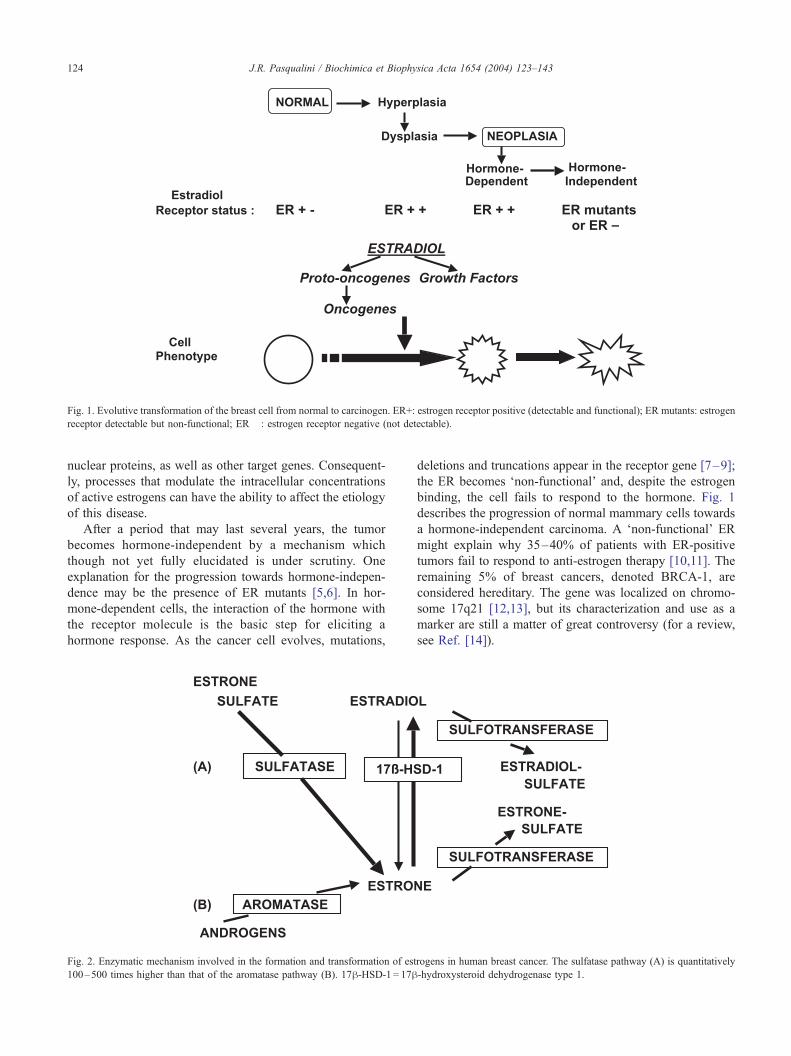
Fig. 1. Evolutive transformation of the breast cell from normal to carcinogen. ER+: estrogen receptor positive (detectable and functional); ER mutants: estrogen
receptor detectable but non-functional; ER� : estrogen receptor negative (not detectable).
J.R. Pasqualini / Biochimica et Biophysica Acta 1654 (2004) 123–143124
nuclear proteins, as well as other target genes. Consequent-
ly, processes that modulate the intracellular concentrations
of active estrogens can have the ability to affect the etiology
of this disease.
After a period that may last several years, the tumor
becomes hormone-independent by a mechanism which
though not yet fully elucidated is under scrutiny. One
explanation for the progression towards hormone-indepen-
dence may be the presence of ER mutants [5,6]. In hor-
mone-dependent cells, the interaction of the hormone with
the receptor molecule is the basic step for eliciting a
hormone response. As the cancer cell evolves, mutations,
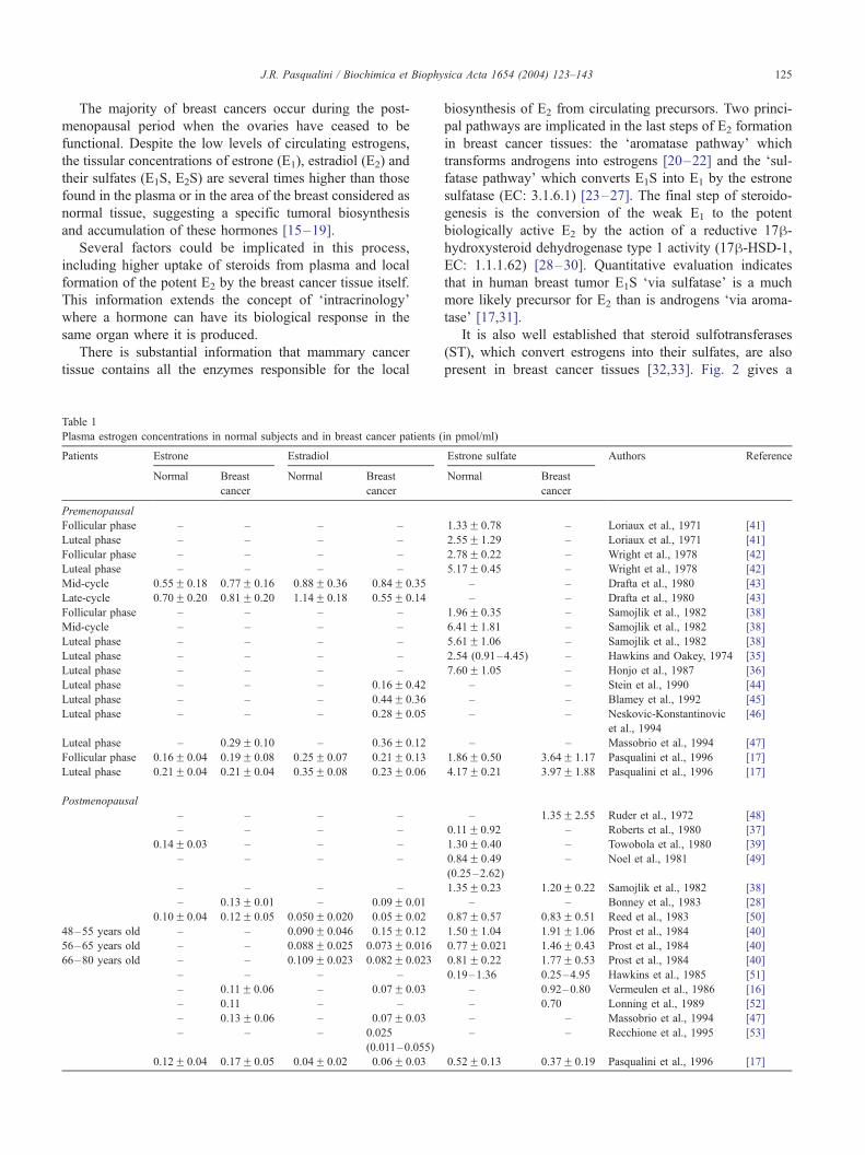
Fig. 2. Enzymatic mechanism involved in the formation and transformation of est
100–500 times higher than that of the aromatase pathway (B). 17h-HSD-1 = 17h
deletions and truncations appear in the receptor gene [7–9];
the ER becomes ‘non-functional’ and, despite the estrogen
binding, the cell fails to respond to the hormone. Fig. 1
describes the progression of normal mammary cells towards
a hormone-independent carcinoma. A ‘non-functional’ ER
might explain why 35–40% of patients with ER-positive
tumors fail to respond to anti-estrogen therapy [10,11]. The
remaining 5% of breast cancers, denoted BRCA-1, are
considered hereditary. The gene was localized on chromo-
some 17q21 [12,13], but its characterization and use as a
marker are still a matter of great controversy (for a review,
see Ref. [14]).
rogens in human breast cancer. The sulfatase pathway (A) is quantitatively
-hydroxysteroid dehydrogenase type 1.
J.R. Pasqualini / Biochimica et Biophysica Acta 1654 (2004) 123–143 125
The majority of breast cancers occur during the post-
menopausal period when the ovaries have ceased to be
functional. Despite the low levels of circulating estrogens,
the tissular concentrations of estrone (E1), estradiol (E2) and
their sulfates (E1S, E2S) are several times higher than those
found in the plasma or in the area of the breast considered as
normal tissue, suggesting a specific tumoral biosynthesis
and accumulation of these hormones [15–19].
Several factors could be implicated in this process,
including higher uptake of steroids from plasma and local
formation of the potent E2 by the breast cancer tissue itself.
This information extends the concept of ‘intracrinology’
where a hormone can have its biological response in the
same organ where it is produced.
There is substantial information that mammary cancer
tissue contains all the enzymes responsible for the local
Table 1
Plasma estrogen concentrations in normal subjects and in breast cancer patients (
Patients Estrone Estradiol
Normal Breast
cancer
Normal Breast
cancer
Premenopausal
Follicular phase – – – –
Luteal phase – – – –
Follicular phase – – – –
Luteal phase – – – –
Mid-cycle 0.55F 0.18 0.77F 0.16 0.88F 0.36 0.84F 0.35
Late-cycle 0.70F 0.20 0.81F 0.20 1.14F 0.18 0.55F 0.14
Follicular phase – – – –
Mid-cycle – – – –
Luteal phase – – – –
Luteal phase – – – –
Luteal phase – – – –
Luteal phase – – – 0.16F 0.42
Luteal phase – – – 0.44F 0.36
Luteal phase – – – 0.28F 0.05
Luteal phase – 0.29F 0.10 – 0.36F 0.12
Follicular phase 0.16F 0.04 0.19F 0.08 0.25F 0.07 0.21F 0.13
Luteal phase 0.21F 0.04 0.21F 0.04 0.35F 0.08 0.23F 0.06
Postmenopausal
– – – –
– – – –
0.14F 0.03 – – –
– – – –
– – – –
– 0.13F 0.01 – 0.09F 0.01
0.10F 0.04 0.12F 0.05 0.050F 0.020 0.05F 0.02
48–55 years old – – 0.090F 0.046 0.15F 0.12
56–65 years old – – 0.088F 0.025 0.073F 0.016
66–80 years old – – 0.109F 0.023 0.082F 0.023
– – – –
– 0.11F 0.06 – 0.07F 0.03
– 0.11 – –
– 0.13F 0.06 – 0.07F 0.03
– – – 0.025
(0.011–0.055)
0.12F 0.04 0.17F 0.05 0.04F 0.02 0.06F 0.03
biosynthesis of E2 from circulating precursors. Two princi-
pal pathways are implicated in the last steps of E2 formation
in breast cancer tissues: the ‘aromatase pathway’ which
transforms androgens into estrogens [20–22] and the ‘sul-
fatase pathway’ which converts E1S into E1 by the estrone
sulfatase (EC: 3.1.6.1) [23–27]. The final step of steroido-
genesis is the conversion of the weak E1 to the potent
biologically active E2 by the action of a reductive 17h-hydroxysteroid dehydrogenase type 1 activity (17h-HSD-1,EC: 1.1.1.62) [28–30]. Quantitative evaluation indicates
that in human breast tumor E1S ‘via sulfatase’ is a much
more likely precursor for E2 than is androgens ‘via aroma-
tase’ [17,31].
It is also well established that steroid sulfotransferases
(ST), which convert estrogens into their sulfates, are also
present in breast cancer tissues [32,33]. Fig. 2 gives a
in pmol/ml)
Estrone sulfate Authors Reference
Normal Breast
cancer
1.33F 0.78 – Loriaux et al., 1971 [41]
2.55F 1.29 – Loriaux et al., 1971 [41]
2.78F 0.22 – Wright et al., 1978 [42]
5.17F 0.45 – Wright et al., 1978 [42]
– – Drafta et al., 1980 [43]
– – Drafta et al., 1980 [43]
1.96F 0.35 – Samojlik et al., 1982 [38]
6.41F1.81 – Samojlik et al., 1982 [38]
5.61F1.06 – Samojlik et al., 1982 [38]
2.54 (0.91–4.45) – Hawkins and Oakey, 1974 [35]
7.60F 1.05 – Honjo et al., 1987 [36]
– – Stein et al., 1990 [44]
– – Blamey et al., 1992 [45]
– – Neskovic-Konstantinovic
et al., 1994
[46]
– – Massobrio et al., 1994 [47]
1.86F 0.50 3.64F 1.17 Pasqualini et al., 1996 [17]
4.17F 0.21 3.97F 1.88 Pasqualini et al., 1996 [17]
– 1.35F 2.55 Ruder et al., 1972 [48]
0.11F 0.92 – Roberts et al., 1980 [37]
1.30F 0.40 – Towobola et al., 1980 [39]
0.84F 0.49
(0.25–2.62)
– Noel et al., 1981 [49]
1.35F 0.23 1.20F 0.22 Samojlik et al., 1982 [38]
– – Bonney et al., 1983 [28]
0.87F 0.57 0.83F 0.51 Reed et al., 1983 [50]
1.50F 1.04 1.91F1.06 Prost et al., 1984 [40]
0.77F 0.021 1.46F 0.43 Prost et al., 1984 [40]
0.81F 0.22 1.77F 0.53 Prost et al., 1984 [40]
0.19–1.36 0.25–4.95 Hawkins et al., 1985 [51]
– 0.92–0.80 Vermeulen et al., 1986 [16]
– 0.70 Lonning et al., 1989 [52]
– – Massobrio et al., 1994 [47]
– – Recchione et al., 1995 [53]
0.52F 0.13 0.37F 0.19 Pasqualini et al., 1996 [17]
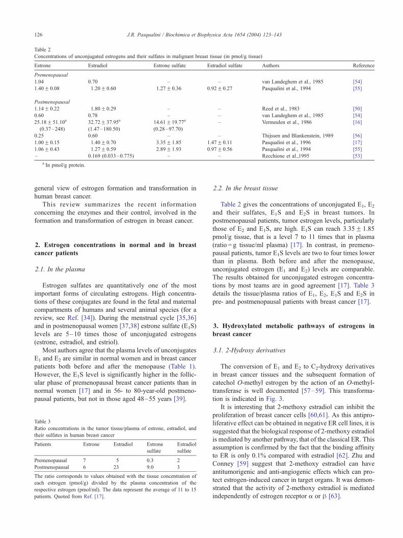
Table 2
Concentrations of unconjugated estrogens and their sulfates in malignant breast tissue (in pmol/g tissue)
Estrone Estradiol Estrone sulfate Estradiol sulfate Authors Reference
Premenopausal
1.04 0.70 – – van Landeghem et al., 1985 [54]
1.40F 0.08 1.20F 0.60 1.27F 0.36 0.92F 0.27 Pasqualini et al., 1994 [55]
Postmenopausal
1.14F 0.22 1.80F 0.29 – – Reed et al., 1983 [50]
0.60 0.78 – – van Landeghem et al., 1985 [54]
25.18F 51.10a
(0.37–248)
32.72F 37.95a
(1.47–180.50)
14.61F19.77a
(0.28–97.70)
– Vermeulen et al., 1986 [16]
0.25 0.60 – – Thijssen and Blankenstein, 1989 [56]
1.00F 0.15 1.40F 0.70 3.35F 1.85 1.47F 0.11 Pasqualini et al., 1996 [17]
1.06F 0.43 1.27F 0.59 2.89F 1.93 0.97F 0.56 Pasqualini et al., 1994 [55]
– 0.169 (0.033–0.775) – – Recchione et al.,1995 [53]
a In pmol/g protein.
J.R. Pasqualini / Biochimica et Biophysica Acta 1654 (2004) 123–143126
general view of estrogen formation and transformation in
human breast cancer.
This review summarizes the recent information
concerning the enzymes and their control, involved in the
formation and transformation of estrogen in breast cancer.
2. Estrogen concentrations in normal and in breast
cancer patients
2.1. In the plasma
Estrogen sulfates are quantitatively one of the most
important forms of circulating estrogens. High concentra-
tions of these conjugates are found in the fetal and maternal
compartments of humans and several animal species (for a
review, see Ref. [34]). During the menstrual cycle [35,36]
and in postmenopausal women [37,38] estrone sulfate (E1S)
levels are 5–10 times those of unconjugated estrogens
(estrone, estradiol, and estriol).
Most authors agree that the plasma levels of unconjugates
E1 and E2 are similar in normal women and in breast cancer
patients both before and after the menopause (Table 1).
However, the E1S level is significantly higher in the follic-
ular phase of premenopausal breast cancer patients than in
normal women [17] and in 56- to 80-year-old postmeno-
pausal patients, but not in those aged 48–55 years [39].
Table 3
Ratio concentrations in the tumor tissue/plasma of estrone, estradiol, and
their sulfates in human breast cancer
Patients Estrone Estradiol Estrone
sulfate
Estradiol
sulfate
Premenopausal 7 5 0.3 2
Postmenopausal 6 23 9.0 3
The ratio corresponds to values obtained with the tissue concentration of
each estrogen (pmol/g) divided by the plasma concentration of the
respective estrogen (pmol/ml). The data represent the average of 11 to 15
patients. Quoted from Ref. [17].
2.2. In the breast tissue
Table 2 gives the concentrations of unconjugated E1, E2
and their sulfates, E1S and E2S in breast tumors. In
postmenopausal patients, tumor estrogen levels, particularly
those of E2 and E1S, are high. E1S can reach 3.35F 1.85
pmol/g tissue, that is a level 7 to 11 times that in plasma
(ratio = g tissue/ml plasma) [17]. In contrast, in premeno-
pausal patients, tumor E1S levels are two to four times lower
than in plasma. Both before and after the menopause,
unconjugated estrogen (E1 and E2) levels are comparable.
The results obtained for unconjugated estrogen concentra-
tions by most teams are in good agreement [17]. Table 3
details the tissue/plasma ratios of E1, E2, E1S and E2S in
pre- and postmenopausal patients with breast cancer [17].
3. Hydroxylated metabolic pathways of estrogens in
breast cancer
3.1. 2-Hydroxy derivatives
The conversion of E1 and E2 to C2-hydroxy derivatives
in breast cancer tissues and the subsequent formation of
catechol O-methyl estrogen by the action of an O-methyl-
transferase is well documented [57–59]. This transforma-
tion is indicated in Fig. 3.
It is interesting that 2-methoxy estradiol can inhibit the
proliferation of breast cancer cells [60,61]. As this antipro-
liferative effect can be obtained in negative ER cell lines, it is
suggested that the biological response of 2-methoxy estradiol
is mediated by another pathway, that of the classical ER. This
assumption is confirmed by the fact that the binding affinity
to ER is only 0.1% compared with estradiol [62]. Zhu and
Conney [59] suggest that 2-methoxy estradiol can have
antitumorigenic and anti-angiogenic effects which can pro-
tect estrogen-induced cancer in target organs. It was demon-
strated that the activity of 2-methoxy estradiol is mediated
independently of estrogen receptor a or h [63].
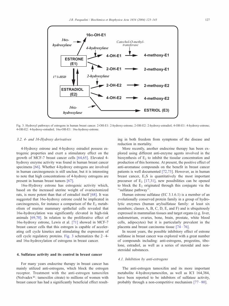
Fig. 3. Hydroxyl pathways of estrogens in human breast cancer. 2-OH-E1: 2-hydroxy-estrone; 2-OH-E2: 2-hydroxy-estradiol; 4-OH-E1: 4-hydroxy-estrone;
J.R. Pasqualini / Biochimica et Biophysica Acta 1654 (2004) 123–143 127
3.2. 4- and 16-Hydroxy derivatives
4-Hydroxy estrone and 4-hydroxy estradiol possess es-
trogenic properties and exert a stimulatory effect on the
growth of MCF-7 breast cancer cells [64,65]. Elevated 4-
hydroxy enzyme activity was found in human breast cancer
specimens [66]. Whether 4-hydroxy estrogens are involved
in human carcinogenesis is still unclear, but it is interesting
to note that high concentrations of 4-hydroxy estrogens are
present in human breast tumors [67].
16a-Hydroxy estrone has estrogenic activity which,
based on the increased uterine weight of ovariectomized
rats, is more potent than that of estradiol itself [68]. It was
suggested that 16a-hydroxy estrone could be implicated in
carcinogenesis, for instance a comparison of the E2 metab-
olism of murine mammary epithelial cells revealed that
16a-hydroxylation was significantly elevated in high-risk
animals [69,70]. In relation to the proliferative effect of
16a-hydroxy estrone, Lewis et al. [71] showed in MCF-7
breast cancer cells that this estrogen is capable of acceler-
ating cell cycle kinetics and stimulating the expression of
cell cycle regulatory proteins. Fig. 3 schematizes the 2–4-
and 16a-hydroxylation of estrogens in breast cancer.
4-OH-E2: 4-hydroxy-estradiol; 16a-OH-E1: 16a-hydroxy-estrone.
4. Sulfatase activity and its control in breast cancer
For many years endocrine therapy in breast cancer has
mainly utilized anti-estrogens, which block the estrogen
receptor. Treatment with the anti-estrogen tamoxifen
(NolvadexR: tamoxifen citrate) to millions of women with
breast cancer has had a significantly beneficial effect result-
ing in both freedom from symptoms of the disease and
reduction in mortality.
More recently, another endocrine therapy has been ex-
plored using different anti-enzyme agents involved in the
biosynthesis of E2 to inhibit the tissular concentration and
production of this hormone. At present, the positive effect of
anti-aromatase compounds on the benefit in breast cancer
patients is well documented [72,73]. However, as in human
breast cancer, E1S is quantitatively the most important
precursor of E2 [17,31], new possibilities can be opened
to block the E2 originated through this conjugate via the
‘‘sulfatase pathway’’.
Human estrone sulfatase (EC 3.1.6.1) is a member of an
evolutionally conserved protein family in a group of hydro-
lytic enzymes (human arylsulfatase family: at least six
members; classes A, B, C, D, E, and F) and is ubiquitously
expressed in mammalian tissues and target organs (e.g. liver,
endometrium, ovaries, bone, brain, prostate, white blood
cells, adipocytes) but it is particularly prevalent in the
placenta and breast carcinoma tissue [74–76].
In recent years, the possible inhibitory effect of estrone
sulfatase in breast cancer was explored with a great number
of compounds including: anti-estrogens, progestins, tibo-
lone, estradiol, as well as a series of steroidal and non-
steroidal substances.
4.1. Inhibition by anti-estrogens
The anti-estrogen tamoxifen and its more important
metabolite 4-hydroxytamoxifen, as well as ICI 164,384,
have been reported to be inhibitors of sulfatase activity,
probably through a non-competitive mechanism [77–80].
iophy
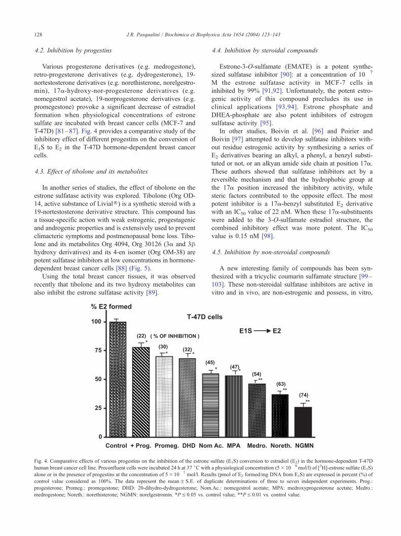
4.2. Inhibition by progestins
Various progesterone derivatives (e.g. medrogestone),
retro-progesterone derivatives (e.g. dydrogesterone), 19-
nortestosterone derivatives (e.g. norethisterone, norelgestro-
min), 17a-hydroxy-nor-progesterone derivatives (e.g.
nomegestrol acetate), 19-norprogesterone derivatives (e.g.
promegestone) provoke a significant decrease of estradiol
formation when physiological concentrations of estrone
sulfate are incubated with breast cancer cells (MCF-7 and
T-47D) [81–87]. Fig. 4 provides a comparative study of the
inhibitory effect of different progestins on the conversion of
E1S to E2 in the T-47D hormone-dependent breast cancer
cells.
4.3. Effect of tibolone and its metabolites
In another series of studies, the effect of tibolone on the
estrone sulfatase activity was explored. Tibolone (Org OD-
14, active substance of LivialR) is a synthetic steroid with a
19-nortestosterone derivative structure. This compound has
a tissue-specific action with weak estrogenic, progestagenic
and androgenic properties and is extensively used to prevent
climacteric symptoms and postmenopausal bone loss. Tibo-
lone and its metabolites Org 4094, Org 30126 (3a and 3hhydroxy derivatives) and its 4-en isomer (Org OM-38) are
potent sulfatase inhibitors at low concentrations in hormone-
dependent breast cancer cells [88] (Fig. 5).
Using the total breast cancer tissues, it was observed
recently that tibolone and its two hydroxy metabolites can
also inhibit the estrone sulfatase activity [89].
J.R. Pasqualini / Biochimica et B128
Fig. 4. Comparative effects of various progestins on the inhibition of the estrone
human breast cancer cell line. Preconfluent cells were incubated 24 h at 37 jC with
alone or in the presence of progestins at the concentration of 5� 10� 7 mol/l. Resu
control value considered as 100%. The data represent the meanF S.E. of dup
progesterone; Promeg.: promegestone; DHD: 20-dihydro-dydrogesterone; Nom
medrogestone; Noreth.: norethisterone; NGMN: norelgestromin. *PV 0.05 vs. co
4.4. Inhibition by steroidal compounds
Estrone-3-O-sulfamate (EMATE) is a potent synthe-
sized sulfatase inhibitor [90]: at a concentration of 10� 7
M the estrone sulfatase activity in MCF-7 cells in
inhibited by 99% [91,92]. Unfortunately, the potent estro-
genic activity of this compound precludes its use in
clinical applications [93,94]. Estrone phosphate and
DHEA-phosphate are also potent inhibitors of estrogen
sulfatase activity [95].
In other studies, Boivin et al. [96] and Poirier and
Boivin [97] attempted to develop sulfatase inhibitors with-
out residue estrogenic activity by synthesizing a series of
E2 derivatives bearing an alkyl, a phenyl, a benzyl substi-
tuted or not, or an alkyan amide side chain at position 17a.
These authors showed that sulfatase inhibitors act by a
reversible mechanism and that the hydrophobic group at
the 17a position increased the inhibitory activity, while
steric factors contributed to the opposite effect. The most
potent inhibitor is a 17a-benzyl substituted E2 derivative
with an IC50 value of 22 nM. When these 17a-substituents
were added to the 3-O-sulfamate estradiol structure, the
combined inhibitory effect was more potent. The IC50
value is 0.15 nM [98].
4.5. Inhibition by non-steroidal compounds
A new interesting family of compounds has been syn-
thesized with a tricyclic coumarin sulfamate structure [99–
103]. These non-steroidal sulfatase inhibitors are active in
vitro and in vivo, are non-estrogenic and possess, in vitro,
sica Acta 1654 (2004) 123–143
sulfate (E1S) conversion to estradiol (E2) in the hormone-dependent T-47D
a physiological concentration (5� 10� 9 mol/l) of [3H]-estrone sulfate (E1S)
lts (pmol of E2 formed/mg DNA from E1S) are expressed in percent (%) of
licate determinations of three to seven independent experiments. Prog.:
.Ac.: nomegestrol acetate; MPA: medroxyprogesterone acetate; Medro.:
ntrol value; **PV 0.01 vs. control value.
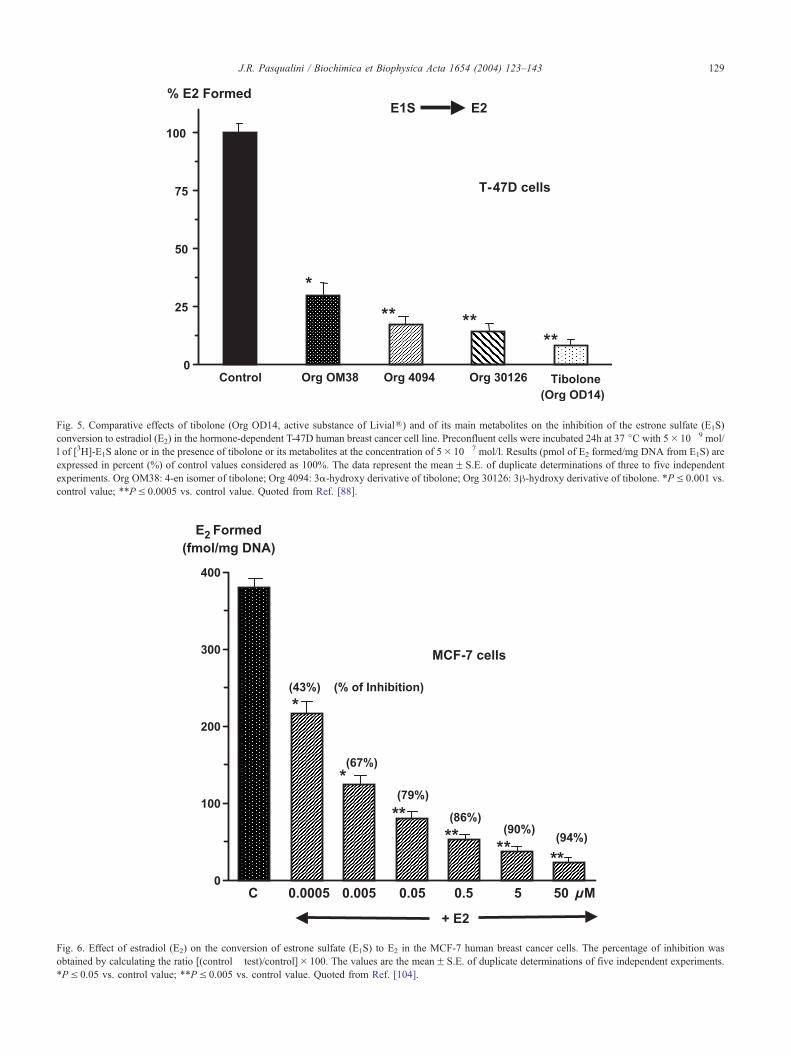
Fig. 5. Comparative effects of tibolone (Org OD14, active substance of LivialR) and of its main metabolites on the inhibition of the estrone sulfate (E1S)
conversion to estradiol (E2) in the hormone-dependent T-47D human breast cancer cell line. Preconfluent cells were incubated 24h at 37 jC with 5� 10� 9 mol/
l of [3H]-E1S alone or in the presence of tibolone or its metabolites at the concentration of 5� 10� 7 mol/l. Results (pmol of E2 formed/mg DNA from E1S) are
expressed in percent (%) of control values considered as 100%. The data represent the meanF S.E. of duplicate determinations of three to five independent
experiments. Org OM38: 4-en isomer of tibolone; Org 4094: 3a-hydroxy derivative of tibolone; Org 30126: 3h-hydroxy derivative of tibolone. *PV 0.001 vs.
control value; **PV 0.0005 vs. control value. Quoted from Ref. [88].
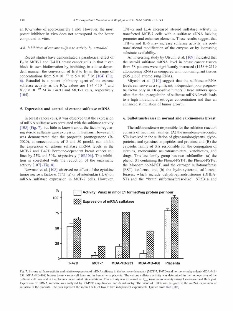
Fig. 6. Effect of estradiol (E2) on the conversion of estrone sulfate (E1S) to E2 in the MCF-7 human breast cancer cells. The percentage of inhibition was
obtained by calculating the ratio [(control� test)/control]� 100. The values are the meanF S.E. of duplicate determinations of five independent experiments.
*PV 0.05 vs. control value; **PV 0.005 vs. control value. Quoted from Ref. [104].
J.R. Pasqualini / Biochimica et Biophysica Acta 1654 (2004) 123–143 129
J.R. Pasqualini / Biochimica et Biophysica Acta 1654 (2004) 123–143130
an IC50 value of approximately 1 nM. However, the most
potent inhibitor in vivo does not correspond to the better
compound in vitro.
4.6. Inhibition of estrone sulfatase activity by estradiol
Recent studies have demonstrated a paradoxical effect of
E2 in MCF-7 and T-47D breast cancer cells in that it can
block its own bioformation by inhibiting, in a dose-depen-
dent manner, the conversion of E1S to E2 in the range of
concentrations from 5� 10� 10 to 5� 10� 5 M [104] (Fig.
6). Estradiol is a potent inhibitory agent of the estrone
sulfatase activity as the IC50 values are 1.84� 10� 9 and
8.77� 10� 10 M in T-47D and MCF-7 cells, respectively
[104].
5. Expression and control of estrone sulfatase mRNA
In breast cancer cells, it was observed that the expression
of mRNA sulfatase was correlated with the sulfatase activity
[105] (Fig. 7), but little is known about the factors regulat-
ing steroid sulfatase gene expression in humans. However, it
was demonstrated that the progestin promegestone (R-
5020), at concentrations of 5 and 50 Amol/l, can inhibit
the expression of estrone sulfatase mRNA levels in the
MCF-7 and T-47D hormone-dependent breast cancer cell
lines by 25% and 50%, respectively [105,106]. This inhibi-
tion is correlated with the reduction of the enzymatic
activity [107] (Fig. 8).
Newman et al. [108] observed no effect of the cytokine
tumor necrosis factor-a (TNF-a) or of interleukin (IL-6) on
mRNA sulfatase expression in MCF-7 cells. However,
Fig. 7. Estrone sulfatase activity and relative expression of mRNA sulfatase in the h
231, MDA-MB-468) human breast cancer cell lines and in human term placenta
different cell lines and in the placenta under initial rate conditions. This activity w
Expression of mRNA sulfatase was analyzed by RT-PCR amplification and den
sulfatase in the placenta. The data represent the meanF S.E. of two to five indep
TNF-a and IL-6 increased steroid sulfatase activity in
transfected MCF-7 cells with a sulfatase cDNA lacking
promoter and enhancer elements. These results suggest that
TNF-a and IL-6 may increase sulfatase activity via post-
translational modification of the enzyme or by increasing
substrate availability.
An interesting study by Utsumi et al. [109] indicated that
the steroid sulfatase mRNA level in breast cancer tissues
from 38 patients were significantly increased (1458F 2119
attomols/mg RNA) as compared with non-malignant tissues
(535F 663 attomols/mg RNA).
Miyoshi et al. [110] suggest that the sulfatase mRNA
levels can serve as a significant, independent poor prognos-
tic factor only in ER-positive tumors. These authors spec-
ulate that the up-regulation of sulfatase mRNA levels leads
to a high intratumoral estrogen concentration and thus an
enhanced stimulation of tumor growth.
6. Sulfotransferases in normal and carcinomous breast
The sulfotransferase responsible for the sulfation reaction
consists of two main families: (A) the membrane-associated
STs involved in the sulfation of glycosaminoglycans, glyco-
proteins, and tyrosines in peptides and proteins, and (B) the
cytosolic family of STs responsible for the conjugation of
steroids, monoamine neurotransmitters, xenobiotics, and
drugs. This last family group has two subfamilies: (a) the
phenol ST containing the Phenol-PST-1, the Phenol-PST-2,
the Monoamine-M-PST, and the estrogen sulfotransferase
(EST) isoforms, and (b) the hydroxysteroid sulfotrans-
ferases, which include dehydroepiandrosterone (DHEA-
ST) and the ‘‘brain sulfotransferase-like’’ ST2B1a and
ormone-dependent (MCF-7, T-47D) and hormone-independent (MDA-MB-
. The estrone sulfatase activity was determined in the homogenates of the
as expressed as Vmax (maximum velocity) using Lineweaver and Burk plot.
sitometry. The value of 100% was assigned to the mRNA expression of
endent experiments. Quoted from Ref. [105].
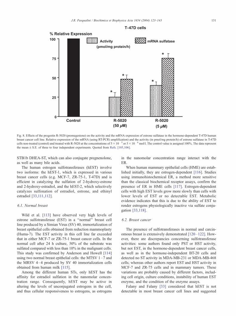
Fig. 8. Effects of the progestin R-5020 (promegestone) on the activity and the mRNA expression of estrone sulfatase in the hormone-dependent T-47D human
breast cancer cell line. Relative expression of the mRNA (using RT-PCR) amplification) and the activity (in pmol/mg protein/h) of estrone sulfatase in T-47D
cells non-treated (control) and treated with R-5020 at the concentrations of 5� 10� 5 or 5� 10� 6 mol/l. The control value is assigned 100%. The data represent
the meanF S.E. of three to four independent experiments. Quoted from Refs. [105,106].
J.R. Pasqualini / Biochimica et Biophysica Acta 1654 (2004) 123–143 131
STB1b DHEA-ST, which can also conjugate pregnenolone,
as well as many bile acids.
The human estrogen sulfotransferases (hEST) involve
two isoforms: the hEST-1, which is expressed in various
breast cancer cells (e.g. MCF-7, ZR-75-1, T-47D) and is
efficient in catalyzing the sulfation of 2-hydroxy-estrone
and 2-hydroxy-estradiol, and the hEST-2, which selectively
catalyzes sulfonation of estradiol, estrone, and ethinyl
estradiol [33,111,112].
6.1. Normal breast
Wild et al. [113] have observed very high levels of
estrone sulfotransferase (EST) in a ‘‘normal’’ breast cell
line produced by a Simian Virus (SV) 40, immortalization of
breast epithelial cells obtained from reduction mammoplasty
(Huma-7). The EST activity in this cell line far exceeded
that in either MCF-7 or ZR-75-1 breast cancer cells. In the
normal cell after 24 h culture, 50% of the substrate was
sulfated compared with less than 10% in the malignant cells.
This study was confirmed by Anderson and Howell [114]
using two normal breast epithelial cells: the MTSV 1–7 and
the MRSV 4–4 produced by SV 40 immortalization cells
obtained from human milk [115].
Among the different human STs, only hEST has the
affinity for estradiol sulfation in the nanomolar concen-
tration range. Consequently, hEST may be active in
altering the levels of unconjugated estrogens in the cell,
and thus cellular responsiveness to estrogens, as estrogens
in the nanomolar concentration range interact with the
ER.
When human mammary epithelial cells (HME) are estab-
lished initially, they are estrogen-dependent [116]. Studies
using immunohistochemical ER, a method more sensitive
than the classical biochemical receptor assays, confirm the
presence of ER in HME cells [117]. Estrogen-dependent
cells with high EST levels grow more slowly than cells with
lower levels of EST or no detectable EST. Metabolic
evidence indicates that this is due to the ability of EST to
render estrogens physiologically inactive via sulfate conju-
gation [33,118].
6.2. Breast cancer
The presence of sulfotransferases in normal and carcin-
omous breast is extensively demonstrated [120–122]. How-
ever, there are discrepancies concerning sulfotransferase
activities: some authors found only PST or HST activity,
but not EST, in the hormone-dependent breast cancer cells,
as well as in the hormone-independent BT-20 cells and
detected no ST activity in MDA-MB-231 or MDA-MB-468
cells; whereas other authors report EST and HST activity in
MCF-7 and ZR-75 cells and in mammary tumors. These
variations are probably caused by different factors, includ-
ing cell origin, culture conditions, instability of human EST
enzyme, and the condition of the enzyme assays.
Falany and Falany [33] considered that hEST is not
detectable in most breast cancer cell lines and suggested
J.R. Pasqualini / Biochimica et Biophysica Acta 1654 (2004) 123–143132
that the sulfoconjugated activity in the cells is mainly due to
the human Phenol-PST, an enzyme that has a higher affinity
with the estrogens at micromolar than at nanomolar con-
centrations. hP-PST has an affinity for estrogen sulfation
about 300-fold lower than that of hEST [119,123].
To explore the difference in EST content between normal
human mammary epithelial and breast cancer cells, and their
correlation with cellular growth, Falany and Falany [33,124]
transformed MCF-7 cells with an EST expression vector,
and observed that after incubation of 20 nM of E2, sulfation
occurs more rapidly with MCF-7 cells transformed with
EST than with the control cells, thereby rendering E2
physiologically inactive. EST/MCF-7 cells require a higher
concentration of E2 to stimulate growth than do control
MCF-7 cells, as EST inactivates E2 via sulfatation, conse-
quently rendering it incapable of binding to the ER and
inhibiting the process of cell growth.
In conclusion, knowledge of the expression and regula-
tion of the different sulfotransferases is of capital impor-
tance in understanding the changes in the normal breast cell
during tumorigenesis, as well as hormonal involvement in
this mechanism.
6.3. Control of sulfotransferase activities in breast cancer
As sulfoconjugates are not biologically active, the con-
trol of the formation of these conjugates in breast cells
represents an important mechanism to modulate the biolog-
ical action of estradiol in this tissue.
Comparative studies on the formation of estrogen sul-
fates after incubation of estrone with the hormone-depen-
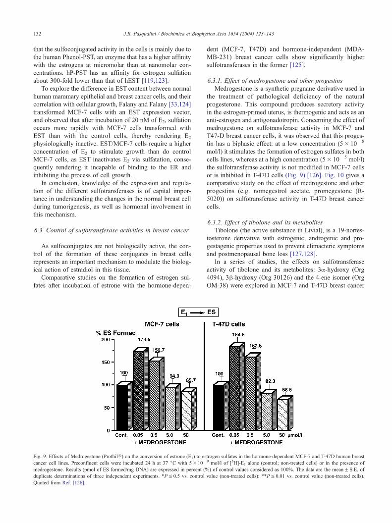
Fig. 9. Effects of Medrogestone (ProthilR) on the conversion of estrone (E1) to es
cancer cell lines. Preconfluent cells were incubated 24 h at 37 jC with 5� 10�
medrogestone. Results (pmol of ES formed/mg DNA) are expressed in percent (
duplicate determinations of three independent experiments. *PV 0.5 vs. control
Quoted from Ref. [126].
dent (MCF-7, T47D) and hormone-independent (MDA-
MB-231) breast cancer cells show significantly higher
sulfotransferases in the former [125].
6.3.1. Effect of medrogestone and other progestins
Medrogestone is a synthetic pregnane derivative used in
the treatment of pathological deficiency of the natural
progesterone. This compound produces secretory activity
in the estrogen-primed uterus, is thermogenic and acts as an
anti-estrogen and antigonadotropin. Concerning the effect of
medrogestone on sulfotransferase activity in MCF-7 and
T47-D breast cancer cells, it was observed that this proges-
tin has a biphasic effect: at a low concentration (5� 10� 8
mol/l) it stimulates the formation of estrogen sulfates in both
cells lines, whereas at a high concentration (5� 10� 5 mol/l)
the sulfotransferase activity is not modified in MCF-7 cells
or is inhibited in T-47D cells (Fig. 9) [126]. Fig. 10 gives a
comparative study on the effect of medrogestone and other
progestins (e.g. nomegestrol acetate, promegestone (R-
5020)) on sulfotransferase activity in T-47D breast cancer
cells.
6.3.2. Effect of tibolone and its metabolites
Tibolone (the active substance in Livial), is a 19-nortes-
tosterone derivative with estrogenic, androgenic and pro-
gestagenic properties used to prevent climacteric symptoms
and postmenopausal bone loss [127,128].
In a series of studies, the effects on sulfotransferase
activity of tibolone and its metabolites: 3a-hydroxy (Org
4094), 3h-hydroxy (Org 30126) and the 4-ene isomer (Org
OM-38) were explored in MCF-7 and T-47D breast cancer
trogen sulfates in the hormone-dependent MCF-7 and T-47D human breast9 mol/l of [3H]-E1 alone (control; non-treated cells) or in the presence of
%) of control values considered as 100%. The data are the meanF S.E. of
value (non-treated cells); **PV 0.01 vs. control value (non-treated cells).
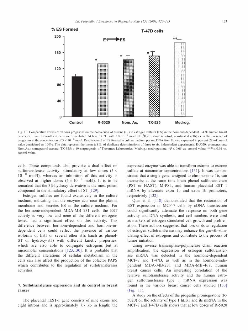
Fig. 10. Comparative effects of various progestins on the conversion of estrone (E1) to estrogen sulfates (ES) in the hormone-dependent T-47D human breast
cancer cell line. Preconfluent cells were incubated 24 h at 37 jC with 5� 10� 9 mol/l of [3H]-E1 alone (control; non-treated cells) or in the presence of
progestins at the concentration of 5� 10� 8 mol/l. Results (pmol of ES formed in culture medium per mg DNA from E1) are expressed in percent (%) of control
value considered as 100%. The data represent the meanF S.E. of duplicate determinations of three to six independent experiments. R-5020: promegestone;
Nom.Ac.: nomegestrol acetate; TX-525: a 19-norprogestin of Theramex Laboratories; Medrog.: medrogestone. *PV 0.05 vs. control value; **PV 0.01 vs.
control value.
J.R. Pasqualini / Biochimica et Biophysica Acta 1654 (2004) 123–143 133
cells. These compounds also provoke a dual effect on
sulfotransferase activity: stimulatory at low doses (5�10� 8 mol/l), whereas an inhibition of this activity is
observed at higher doses (5� 10� 5 mol/l). It is to be
remarked that the 3h-hydroxy derivative is the most potent
compound in the stimulatory effect of ST [129].
Estrogen sulfates are found exclusively in the culture
medium, indicating that the enzyme acts near the plasma
membrane and secretes ES in the culture medium. For
the hormone-independent MDA-MB 231 cells, the EST
activity is very low and none of the different estrogens
tested had a significant effect on this activity. This
difference between hormone-dependent and hormone-in-
dependent cells could reflect the presence of various
isoforms of EST or several other STs (such as phenol-
ST or hydroxy-ST) with different kinetic properties,
which are also able to conjugate estrogens but at
micromolar concentrations [123,130]. It is probable that
the different alterations of cellular metabolism in the
cells can also affect the production of the cofactor PAPS
which contributes to the regulation of sulfotransferases
activities.
7. Sulfotransferase expression and its control in breast
cancer
The placental hEST-1 gene consists of nine exons and
eight introns and is approximately 7.7 kb in length; the
expressed enzyme was able to transform estrone to estrone
sulfate at nanomolar concentrations [131]. It was demon-
strated that a single gene, assigned to chromosome 16, can
transcribe at the same time brain phenol sulfotransferase
(PST or HAST), M-PST, and human placental EST 1
mRNA by alternate exon 1b and exon 1b promoters,
respectively [132].
Qian et al. [118] demonstrated that the restoration of
EST expression in MCF-7 cells by cDNA transfection
could significantly attenuate the response on both gene
activity and DNA synthesis, and cell numbers were used
as markers of estrogen-stimulated cell growth and prolifer-
ation. These authors suggested that loss or downregulation
of estrogen sulfotransferase may enhance the growth-stim-
ulating effect of estrogens and contribute to the process of
tumor initiation.
Using reverse transcriptase-polymerase chain reaction
amplification, the expression of estrogen sulfotransfer-
ase mRNA was detected in the hormone-dependent
MCF-7 and T-47D, as well as in the hormone-inde-
pendent MDA-MB-231 and MDA-MB-468, human
breast cancer cells. An interesting correlation of the
relative sulfotransferase activity and the human estro-
gen sulfotransferase type 1 mRNA expression was
found in the various breast cancer cells studied [133]
(Fig. 11).
A study on the effects of the progestin promegestone (R-
5020) on the activity of type 1 hEST and its mRNA in the
MCF-7 and T-47D cells shows that at low doses of R-5020
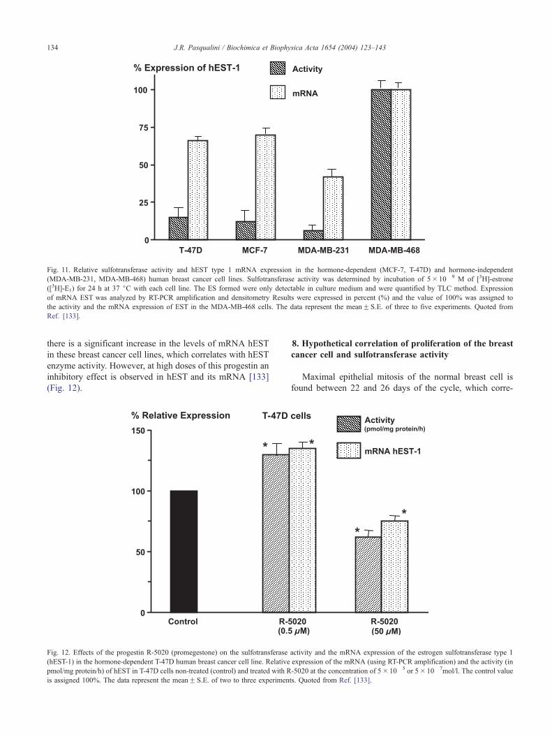
Fig. 11. Relative sulfotransferase activity and hEST type 1 mRNA expression in the hormone-dependent (MCF-7, T-47D) and hormone-independent
(MDA-MB-231, MDA-MB-468) human breast cancer cell lines. Sulfotransferase activity was determined by incubation of 5� 10� 9 M of [3H]-estrone
([3H]-E1) for 24 h at 37 jC with each cell line. The ES formed were only detectable in culture medium and were quantified by TLC method. Expression
of mRNA EST was analyzed by RT-PCR amplification and densitometry Results were expressed in percent (%) and the value of 100% was assigned to
the activity and the mRNA expression of EST in the MDA-MB-468 cells. The data represent the meanF S.E. of three to five experiments. Quoted from
Ref. [133].
J.R. Pasqualini / Biochimica et Biophysica Acta 1654 (2004) 123–143134
there is a significant increase in the levels of mRNA hEST
in these breast cancer cell lines, which correlates with hEST
enzyme activity. However, at high doses of this progestin an
inhibitory effect is observed in hEST and its mRNA [133]
(Fig. 12).
Fig. 12. Effects of the progestin R-5020 (promegestone) on the sulfotransferase
(hEST-1) in the hormone-dependent T-47D human breast cancer cell line. Relative
pmol/mg protein/h) of hEST in T-47D cells non-treated (control) and treated with R
is assigned 100%. The data represent the meanF S.E. of two to three experimen
8. Hypothetical correlation of proliferation of the breast
cancer cell and sulfotransferase activity
Maximal epithelial mitosis of the normal breast cell is
found between 22 and 26 days of the cycle, which corre-
activity and the mRNA expression of the estrogen sulfotransferase type 1
expression of the mRNA (using RT-PCR amplification) and the activity (in
-5020 at the concentration of 5� 10� 5 or 5� 10� 7mol/l. The control value
ts. Quoted from Ref. [133].

Scheme 1. Mechanism of sulfotransferase (ST) activity in normal and breast
cancer cells. In normal breast cancer cells it is suggested that the action of
hEST works at physiological (nanomolar) concentrations of estradiol to
form estradiol sulfate which is biologically inactive. This enzyme is absent
from breast cancer cells where the phenol-ST activity acts only at
micromolar (non-physiological) concentrations.
Scheme 3. Hypothetical effects of medrogestone on human sulfotransferase
(hEST) and proliferation in T-47D and MCF-7 breast cancer cells. As
medrogestone can stimulate hEST in the cancer cell, the effect of estradiol
becomes inactive by the formation of estradiol sulfate and consequently cell
proliferation is inhibited.
J.R. Pasqualini / Biochimica et Biophysica Acta 1654 (2004) 123–143 135
sponds to the high levels of estradiol and progesterone
[134]. During pregnancy, it is suggested that the elevated
values of circulating progesterone are responsible for the
induction of lobular-alveolar development, to prepare the
breast for lactation [135,136]. The data on the effect of
progesterone on breast epithelial proliferation are contradic-
tory. It has been found that progesterone can increase DNA
synthesis in normal breast epithelium in organ culture [137].
Using normal epithelial cells of human breast, it was
demonstrated that the progestin promegestone can decrease
cell proliferation [138,139]. These authors also found that
progestins can inhibit the proliferative effect provoked by
estradiol, whereas McManus and Welsch [140] and Long-
man and Buehring [141] demonstrated no effect.
The proliferative effect of progestins using various iso-
lated breast cancer models: cell lines, organ culture, or
transplantation of breast cancer cells in nude mice, is
contradictory as it was reported that these compound can
either inhibit [142–145], stimulate [146–148], or have no
effect [149].
It was demonstrated that in normal breast cells the
estrogen hEST, which is active at nanomolar concentrations
Scheme 2. Effects of estradiol sulfotransferase (EST) activity on the
proliferation of breast cancer cells. In the normal breast cancer cells, as a
consequence of the hEST activity, the proliferation is inhibited, as estradiol
sulfate (E2S) is biologically inactive. In opposition to breast cancer cells,
hEST activity is very low or inexistent as E2S is not formed and E2 can
stimulate proliferation.
of estradiol, mainly present to form estradiol sulfate (E2S)
and consequently to block the proliferative effect of estra-
diol as E2S, is biologically inactive. However, in the breast
cancer cells the phenol sulfotransferase (P-ST), which is
active at micromolar concentrations of E2 (see Schemes 1
and 2), is present and the hEST is not present [33,117,123].
As the progestins nomegestrol acetate or medrogestone can
stimulate hEST in breast cancer cells, and as these com-
pounds can block the proliferation in breast cancer cells, it is
suggested that the antiproliferative effect of nomegestrol
acetate or medrogestone is correlated with the stimulatory
effect of hEST in the hormone-dependent breast cancer cells
(Scheme 3). More information on the correlation of the
proliferative effect and hEST on breast cancer cells of
various progestins or other substances is needed to verify
this hypothesis.
9. 17B-Hydroxysteroid dehydrogenase and its control in
breast cancer
The last step of biosynthesis of the potent biologically
active estrogen, estradiol, in target tissues is the conversion
of estrone to estradiol by the reductive 17h-HSD 1 (EC
1.1.1.62) activity.
17h-HSD is a widely distributed enzyme in mammalian
tissues, which is implicated in the interconversion of the
inactive 17h-keto- < –> into active 17h-hydroxy in sex
steroid hormones (estrogens and androgens). However,
some types of 17h-HSD may metabolize further substrates
such as bile acids, alcohols, fatty acids and retinols. 17h-HSD belongs to a superfamily of enzymes (to date up to 11
different isoforms are recognized).
9.1. Normal breast
In normal breast tissue, it was observed that the oxidative
17h-HSD activity (E2 to E1) is the preferential direction and
that this activity is more intense during the secretory phase
J.R. Pasqualini / Biochimica et Biophysica Acta 1654 (2004) 123–143136
of the menstrual cycle [150]; 17h-HSD types 1 and 2
mRNAs were both expressed in the glandular epithelium.
In HME cell line, mRNAs for 17h-HSD types 1, 2, and 4
were detected, but only oxidative 17h-HSD activity was
present and it was suggested that this activity is due to 17h-HSD type 2 [151].
Using epithelial cells of normal breast, it was observed
that the progestin promegestone (R-5020) can increase the
17h-HSD activity in the oxidative (E2 to E1) direction; this
stimulatory effect of the progestins depends on preliminary
sensitization by the estrogens [138,152].
9.2. Breast cancer
In breast tumors, in vivo and in vitro studies indicate
that the preferential conversion is the reduction of E1 to
E2. The 17h-HSD type 1 is located in the cytoplasm of
malignant epithelial cells of breast tumors [153]. How-
ever, it was observed that the orientation of the enzymatic
activity (oxidative or reductive) in breast cancer is also
greatly dependent on the local, metabolic or experimental
conditions, including: the nature and concentration of the
cofactors (e.g. NADPH or NADP) and of substrate, pH,
subcellular localization of enzymes. In vitro studies using
human tumor homogenates indicated that the predominant
17h-HSD activity was oxidative rather than reductive
[28]. However, in vivo studies, after isotopic infusion of
estrogens to postmenopausal breast cancer patients, have
shown that the reductive direction is greater than the
oxidative [29].
In hormone-dependent breast cancer cell lines (MCF-7,
T-47D, R-27, ZR-75-1) 17h-HSD type 1 was the predom-
inant reductive isoform, but type 2 and 4 isoforms with
oxidative activities (formation of E1) were also detected
[30,153–155]. It was demonstrated that in intact cells, when
the physiological conditions are more closely protected, the
catalytic activity of each type of 17h-HSD is exclusively
uni-directional, whereas in cell homogenates the bidirec-
tional orientation prevails, but the physiological direction is
favoured [156,157].
In contrast, when breast cancer cells evolve to a hor-
mone-independent status (MDA-MB-231; MDA-MB-436;
Hs-578S) they revert to the oxidative (E2 to E1) 17h-HSDactivity as their preferential enzymatic orientation [30]. This
observation suggests that there is a change in 17h-HSDphenotype in neoplastic cells and that the tumoral process of
the breast is accompanied by a modification of estrogen
metabolism [158].
Fournier et al. [159] have postulated that 17h-HSD might
be a marker for hormone-dependent breast cancer. In more
recent studies, Suzuki et al. [160,161] observed that 17h-HSD type 1 was immunolocalized in carcinoma cells in 68
out of 111 invasive ductal carcinoma cases, while 17h-HSDtype 2, which catalyzes the conversion of E2 to E1, was not
detected in any of these cases. These authors show a
significant correlation between 17h-HSD type 1 and ER
and PR expression, which is in agreement with the data of
Sasano et al. [162] who showed also that 17h-HSD type 2 is
greatly expressed in endometrial carcinoma. Ariga et al.
[163] also found that 17h-HSD type 1 is preferentially
localized in breast tumors and 17h-HSD type 2 in normal
breast, but there is no significant correlation between ER
and 17h-HSD type 1. Recent quantitative real-time PCR
data seem to indicate that 17h-HSD type 1 mRNA expres-
sion levels were significantly higher in postmenopausal than
in premenopausal breast cancer patients [110].
9.3. Control of 17b-hydroxysteroid dehydrogenase activity
in the breast
9.3.1. Control by progestins
Breast tumors from postmenopausal patients receiving
lynestrenol display higher oxidative 17h-HSD activity than
tumors from untreated patients. The activity depends on the
ER or PR status of the tumor [159].
Progestins can induce 17h-HSD type 1 activity with an
increase in both the 1.3 kb mRNA species and enzyme
protein in hormone-dependent T-47D breast cancer cells
[153,164,165]. Org 2058 increases the oxidative direction in
T-47D cells only [153]. Coldham and James [166] showed
that the progestin medroxyprogesterone acetate (MPA)
stimulates the reductive (E1 to E2) activity of MCF-7 cells
when phenol red was excluded from the tissue culture
media. The authors suggested that this could be the way
in which progestins increase cell proliferation in vivo. On
the other hand, Couture et al. [154] observed that in the
treatment of hormone-dependent ZR-75-1 breast cancer
cells with MPA, the oxidative (E2 to E1) direction is
predominant; this effect seems to implicate the androgen
receptor. Other progestins, such as progesterone, levonor-
gestrel, and norethisterone, increase both the oxidative and
reductive 17h-HSD activity in MCF-7 cells [167], whereas
promegestone (R-5020) has no significant effect on the
reductive activity of 17h-HSD [30] but can increase the
oxidative (E2 to E1) activity in T-47D cells [168]. Nome-
gestrol acetate has an inhibitory effect on the 17h-HSDenzyme in T-47D cells (35% and 81% inhibition at
5� 10� 7 and 5� 10� 6 M, respectively) but no significant
effect was found in MCF-7 cells, except at 5� 10� 5 M
[83]. Medrogestone (ProthilR), a synthetic pregnane deriv-
ative of progesterone, significantly decreases the reductive
17h-HSD type 1 activity in MCF-7 and T-47D breast cancer
cells. The inhibitory effect is dose-dependent and is more
intense, even at low doses, in the T-47D cell line than in the
MCF-7 cells; the IC50 values, which correspond to the 50%
inhibition of the conversion of E1 to E2, are 0.45 and
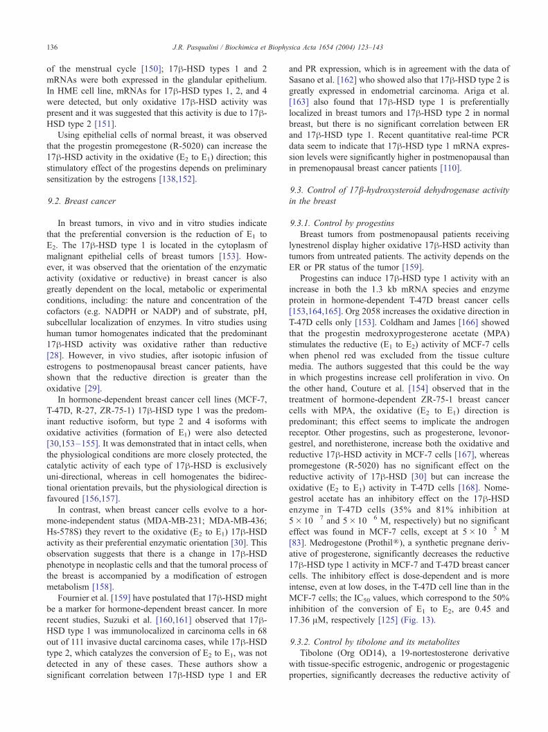
17.36 AM, respectively [125] (Fig. 13).
9.3.2. Control by tibolone and its metabolites
Tibolone (Org OD14), a 19-nortestosterone derivative
with tissue-specific estrogenic, androgenic or progestagenic
properties, significantly decreases the reductive activity of
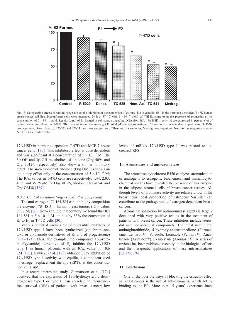
Fig. 13. Comparative effects of various progestins on the inhibition of the conversion of estrone (E1) to estradiol (E2) in the hormone-dependent T-47D human
breast cancer cell line. Preconfluent cells were incubated 24 h at 37 jC with 5� 10� 9 mol/l of [3H]-E1 alone or in the presence of progestins at the
concentration of 5� 10� 7 mol/l. Results (pmol of E2 formed in cell compartment/mg DNA from E1); 17h-HSD-1 activity) are expressed in percent (%) of
control value considered as 100%. The data represent the meanF S.E. of duplicate determinations of three to six independent experiments. R-5020:
promegestone; Dana.: danazol; TX-525 and TX-541 are 19-norprogestins of Theramex Laboratories; Medrog.: medrogestone; Nom.Ac.: nomegestrol acetate.
*PV 0.05 vs. control value.
J.R. Pasqualini / Biochimica et Biophysica Acta 1654 (2004) 123–143 137
17h-HSD in hormone-dependent T-47D and MCF-7 breast
cancer cells [170]. This inhibitory effect is dose-dependent
and was significant at a concentration of 5� 10� 7 M. The
3a-OH and 3h-OH metabolites of tibolone (Org 4094 and
Org 30126, respectively) also show a similar inhibitory
effect. The 4-en isomer of tibolone (Org OM38) shows an
inhibitory effect only at the concentration of 5� 10� 6 M;
The IC50 values in T-47D cells are respectively: 1.44, 2.03,
4.83, and 35.25 AM for Org 30126, tibolone, Org 4094, and
Org OM38 [169].
9.3.3. Control by anti-estrogens and other compounds
The anti-estrogen ICI 164,384 can inhibit by competition
the enzyme 17h-HSD in human breast tumors (IC50 value:
890 AM) [80]. However, in our laboratory we found that ICI
164,384 at 5� 10� 6 M inhibits by 53% the conversion of
E1 to E2 in T-47D cells [30].
Various potential irreversible or reversible inhibitors of
17h-HSD type 1 have been synthesized (e.g. bromoace-
toxy or alkylamide derivatives of E2 and of progesterone)
[171–173]. Thus, for example, the compound 16a-(bro-
moalkylamide) derivative of E2 inhibits the 17h-HSDtype 1 in human placenta with an IC50 value of 10.6
AM [173]. Sawicki et al. [173] obtained 77% inhibition of
17h-HSD type 1 activity with equilin, a component used
in estrogen replacement therapy (ERT), at the concentra-
tion of 1 AM.
In a recent interesting study, Gunnarsson et al. [174]
observed that the expression of 17h-hydroxysteroid dehy-
drogenase type I or type II can correlate to recurrence-
free survival (RFS) of patients with breast cancer; low
levels of mRNA 17h-HSD type II was related to de-
creased RFS.
10. Aromatases and anti-aromatase
The aromatase cytochrome P450 catalyzes aromatization
of androgens to estrogens; biochemical and immunocyto-
chemical studies have revealed the presence of this enzyme
in the adipose stromal cells of breast cancer tissues. Al-
though levels of aromatase activity are relatively low in the
breast, this local production of estrogens ‘on site’ can
contribute to the pathogenesis of estrogen-dependent breast
cancers.
Aromatase inhibition by anti-aromatase agents is largely
developed with very positive results in the treatment of
patients with breast cancer. These inhibitors include steroi-
dal and non-steroidal compounds. The most useful are:
aminoglutethimide, 4-hydroxy-androstenedione (Formes-
tane; LentaronR), Vorosole, Letrozole (FermaraR), Anas-trozole (ArimidexR), Examestane (AromasinR). A series of
reviews has been published recently on the biological effects
and the therapeutic applications of these anti-aromatases
[22,175,176].
11. Conclusions
One of the possible ways of blocking the estradiol effect
in breast cancer is the use of anti-estrogens, which act by
binding to the ER. More than 15 years’ experience have
Fig. 14. The SEEM concept in human hormone-dependent breast cancer
cells. The SEEM can control the enzymatic mechanisms involved in the
formation and transformation of estrogens in breast cancer cells, where the
sulfatase pathway is quantitatively higher than the aromatase. SEEM-I
inhibits the estrone sulfatase; SEEM-II the 17h-HSD-1; SEEM-III the
aromatase activities, and SEEM-IV stimulates the estrone sulfotransferase
activity. It is suggested that E1S is present in the tumor outside the cell and
reaches the cell membrane where it is in contact with the intracellular
estrone sulfatase. ANDR.: androgens; E1: estrone; E2: estradiol; E1S:
estrone sulfate.
J.R. Pasqualini / Biochimica et Biophysica Acta 1654 (2004) 123–143138
shown that breast cancer patients treated with the anti-
estrogen tamoxifen (Nolvadex) have a significantly reduced
risk of recurrence and an increased overall survival. Recent-
ly, tests using a series of new anti-estrogens yielded very
attractive clinical results. However, another way to block
estradiol is by using anti-enzymes (anti-sulfatase, anti-aro-
matase, or anti-17h-hydroxysteroid dehydrogenase (17h-HSD)) which are involved in estradiol biosynthesis in breast
cancer tissues. At present, anti-aromatases are extensively
used in breast cancer treatment with positive benefits.
However, estrone sulfatase is quantitatively the most im-
portant pathway in estradiol bioformation in breast cancer
tissue. Very interesting data were obtained concerning the
inhibitory activity of various progestins (promegestone,
nomegestrol acetate, medrogestone, dydrogesterone, norel-
gestromin), as well as tibolone and its metabolites, on
estrone sulfatase, as well as on 17h-hydroxysteroid dehy-
drogenase, enzymes involved in the other pathway of
estradiol formation in breast cancer cells.
Recent data also show that some progestins (promege-
stone, nomegestrol acetate, medrogestone) as well as tibo-
lone can stimulate sulfotransferase activity in hormone-
dependent breast cancer cells. This is an important point
in the physiopathology of this disease, as it is well known
that estrogen sulfates are biologically inactive.
The fact that estradiol (E2) can block its own bioforma-
tion in the breast cancer cell provides another aspect of this
very complex mechanism in breast cancerization which, in
addition to growth factors, oncogenes, proto-oncogenes and
other factors, needs extensive additional information to be
clarified. The paradoxical effect of E2 could be related to
ERT, a treatment that has been observed to have either no
effect or to slightly increase breast cancer incidence [177]
but significantly decrease mortality [178–182].
For these inhibitory or stimulatory effects on the control
of the enzymes involved in the formation and transforma-
tion of estrogens in breast cancer, we have proposed the
concept of selective estrogen enzyme modulators (SEEM),
which is schematically represented in Fig. 14.
The exploration of various progestins and other substan-
ces in trials with breast cancer patients, showing an inhib-
itory effect on sulfatases and 17h-hydroxysteroid dehy-
drogenase and a stimulatory effect on sulfotransferases
will, in combination with anti-aromatase agents, provide
new possibilities in the treatment of this disease.
Acknowledgements
The author would like to express deep thanks to Dr. G.
Chetrite for help in developing the bibliography and to Ms.
S. MacDonald for assistance in the preparation of this
manuscript.
References
[1] A. Segaloff, Hormones and mammary carcinogenesis, in: W.L.
McGuire (Ed.), Advances in Research and Treatment, Breast Cancer,
vol. 2, Plenum, New York, 1978, pp. 1–22.
[2] M.A. Kirschner, The role of hormones in the development of human
breast cancer, in: W.L. McGuire (Ed.), Advances in Research and
Treatment, Current Topics, Breast Cancer, vol. 3, Plenum, New
York, 1979, pp. 199–226.
[3] M.E. Lippman, R.B. Dickson, S. Bates, C. Knabbe, K. Huff, S.
Swain, M. McManaway, D. Bronzert, A. Kasid, E.P. Gelmann, Auto-
crine and paracrine growth regulation of human breast cancer, Breast
Cancer Res. Treat. 7 (1986) 59–70.
[4] B.E. Henderson, R. Ross, L. Bernstein, Estrogens as a cause of
human cancer: the Richard and Hinda Rosenthal Foundation Award
Lecture, Cancer Res. 48 (1988) 246–253.
[5] S. Raam, N. Robert, C.A. Pappas, H. Tamura, Defective estrogen
receptors in human mammary cancers: their significance in defining
hormone dependence, J. Natl. Cancer Inst. 80 (1988) 756–761.
[6] S.A.W. Fuqua, S.D. Fitzgerald, G.C. Chamness, A.K. Tandon, D.P.
McDonnell, Z. Nawaz, B.W. O’Malley, W.L. McGuire, Variant hu-
man breast tumor estrogen receptor with constitutive transcriptional
activity, Cancer Res. 51 (1991) 105–109.
[7] W.L. McGuire, G.C. Chamness, S.A.W. Fuqua, Abnormal estrogen
receptor in clinical breast cancer, J. Steroid Biochem. Mol. Biol. 43
(1992) 243–247.
[8] S.A.W. Fuqua, G.L. Greene, W.L. McGuire, Inhibition of estrogen
receptor action by a naturally occurring variant in human breast
tumors, Cancer Res. 52 (1992) 483–486.
[9] C.G. Castles, S.A.W. Fuqua, D.M. Klotz, S.M. Hill, Expression of a
constitutively active estrogen receptor variant in the estrogen recep-
tor-negative BT-20 human breast cancer cell line, Cancer Res. 53
(1993) 5934–5939.
[10] D.P. Edwards, G.C. Chamness, W.L. McGuire, Estrogen and pro-
gesterone receptors in breast cancer, Biochim. Biophys. Acta 560
(1979) 457–486.
J.R. Pasqualini / Biochimica et Biophysica Acta 1654 (2004) 123–143 139
[11] S. Litherland, I.M. Jackson, Antiestrogens in the management
of hormone-dependent cancer, Cancer Treat. Rev. 15 (1988)
183–194.
[12] J.M. Hall, M.K. Lee, B. Newman, J.E. Horrow, L.A. Anderson, B.
Huey, M.C. King, Linkage of early-onset familial breast cancer to
chromosome 17q21, Science 250 (1990) 1684–1689.
[13] C.S. Cropp, H.A. Nevanlinna, S. Pyrhonen, U.-H. Stenman, P.
Salmikangas, H. Albertsen, R. White, R. Callahan, Evidence for
involvement of BRCA1 in sporadic breast carcinomas, Cancer Res.
54 (1994) 2548–2551.
[14] B.A. Bove, R.L. Dunbrack Jr., A.K. Godwin, BRCA1, BRCA2, and
hereditary breast cancer, in: J.R. Pasqualini (Ed.), Breast Cancer,
Prognosis, Treatment, and Prevention, Marcel Dekker, New York,
2002, pp. 555–624.
[15] A.A.J. van Landeghem, J. Poortman, M. Nabuurs, J.H.H. Thijssen,
Endogenous concentration and subcellular distribution of estrogens
in normal and malignant human breast tissue, Cancer Res. 45 (1985)
2900–2906.
[16] A. Vermeulen, J.P. Deslypere, R. Paridaens, G. Leclercq, F. Roy, J.C.
Heuson, Aromatase, 17h-hydroxysteroid dehydrogenase and intra-
tissular sex hormone concentrations in cancerous and normal glan-
dular breast tissue in postmenopausal women, Eur. J. Cancer Clin.
Oncol. 22 (1986) 515–525.
[17] J.R. Pasqualini, G. Chetrite, C. Blacker, M.-C. Feinstein, L. Dela-
londe, M. Talbi, C. Maloche, Concentrations of estrone, estradiol,
and estrone sulfate and evaluation of sulfatase and aromatase activ-
ities in pre- and postmenopausal breast cancer, J. Clin. Endocrinol.
Metab. 81 (1996) 1460–1464.
[18] J.R. Pasqualini, J. Cortes-Prieto, G. Chetrite, M. Talbi, A. Ruiz,
Concentrations of estrone, estradiol, and their sulfates and evaluation
of sulfatase and aromatase activities in patients with breast fibroa-
denoma, Int. J. Cancer 70 (1997) 639–643.
[19] R. Clarke, F. Leonessa, J.N. Welch, T.C. Skaar, Cellular and molec-
ular pharmacology of antiestrogen action and resistance, Pharmacol.
Rev. 53 (2001) 1–47.
[20] Y.J. Abul-Hajj, R. Iverson, D.T. Kiang, Aromatization of androgens
by human breast cancer, Steroids 33 (1979) 205–222.
[21] A. Lipton, S.J. Santner, R.J. Santen, H.A. Harvey, P.D. Feil, D.
White-Hershey, M.J. Bartholomew, C.E. Antle, Aromatase activity
in primary and metastatic human breast cancer, Cancer 59 (1987)
779–782.
[22] A.S. Bhatnagar, C. Batzl, A. Hausler, K. Schieweck, M. Lang, P.F.
Trunet, Pharmacology of nonsteroidal aromatase inhibitors, in: J.R.
Katzenellenbogen, B.S. Katzenellenbogen (Eds.), Hormone-Depen-
dent Cancer, Marcel Dekker, New York, 1996, pp. 155–168.
[23] T.L. Dao, C. Hayes, P.R. Libby, Steroid sulfatase activities in human
breast tumors, Proc. Soc. Exp. Biol. Med. 146 (1974) 381–384.
[24] F. Vignon, M. Terqui, B. Westley, D. Derocq, H. Rochefort, Effects
of plasma estrogen sulfates in mammary cancer cells, Endocrinology
106 (1980) 1079–1086.
[25] J.R. Pasqualini, C. Gelly, F. Lecerf, Estrogen sulfates: biological and
ultrastructural responses and metabolism in MCF-7 human breast
cancer cells, Breast Cancer Res. Treat. 8 (1986) 233–240.
[26] J.R. MacIndoe, with the technical assistance of G. Woods, L. Jeffries,
M. Hinkhouse, The hydrolysis of estrone sulfate and dehydroepian-
drosterone sulfate by MCF-7 human breast cancer cells, Endocrinol-
ogy 123 (1988) 1281–1287.
[27] J.R. Pasqualini, C. Gelly, B.-L. Nguyen, C. Vella, Importance of
estrogen sulfates in breast cancer, J. Steroid Biochem. 34 (1989)
155–163.
[28] R.C. Bonney, M.J. Reed, K. Davidson, P.A. Beranek, V.H.T. James,
The relationship between 17h-hydroxysteroid dehydrogenase activ-
ity and oestrogen concentrations in human breast tumours and in
normal breast tissue, Clin. Endocrinol. 19 (1983) 727–739.
[29] J.M. McNeill, M.J. Reed, P.A. Beranek, R.C. Bonney, M.W.
Ghilchik, D.J. Robinson, V.H.T. James, A comparison of the in vivo
uptake and metabolism of 3H-oestrone and 3H-oestradiol by normal
breast and breast tumour tissues in post-menopausal women, Int. J.
Cancer 38 (1986) 193–196.
[30] B.-L. Nguyen, G. Chetrite, J.R. Pasqualini, Transformation of es-
trone and estradiol in hormone-dependent and hormone-independent
human breast cancer cells. Effects of the antiestrogen ICI 164,384,
danazol, and promegestone (R-5020), Breast Cancer Res. Treat. 34
(1995) 139–146.
[31] S.J. Santner, P.D. Feil, R.J. Santen, In situ estrogen production via
the estrone sulfatase pathway in breast tumors: relative importance
versus the aromatase pathway, J. Clin. Endocrinol. Metab. 59 (1984)
29–33.
[32] J.R. Pasqualini, Steroid sulphotransferase activity in human hor-
mone-independent MDA-MB-468 mammary cancer cells, Eur. J.
Cancer 28A (1992) 758–762.
[33] J.L. Falany, C.N. Falany, Expression of cytosolic sulfotransferases in
normal mammary epithelial cells and breast cancer cell lines, Cancer
Res. 56 (1996) 1551–1555.
[34] J.R. Pasqualini, F. Kincl, in: Hormones and the Fetus vol. 1, Perga-
mon, Oxford, 1985, pp. 173–334.
[35] R.A. Hawkins, R.E. Oakey, Estimation of oestrone sulphate, oestra-
diol-17h and oestrone in peripheral plasma: concentrations during
the menstrual cycle and in man, J. Endocrinol. 60 (1974) 1–17.
[36] H. Honjo, J. Kitawaki, M. Itoh, J. Yasuda, K. Iwasaku, M. Urabe, K.
Naitoh, T. Yamamoto, H. Okada, T. Ohkubo, T. Nambara, Serum
and urinary estrone sulfate during the menstrual cycle, measured by
a direct radio-immunoassay, and fate of exogenously injected estrone
sulfate, Horm. Res. 27 (1987) 61–68.
[37] K.D. Roberts, J.G. Rochefort, G. Bleau, A. Chapdelaine, Plasma
estrone sulfate levels in postmenopausal women, Steroids 35
(1980) 179–187.
[38] E. Samojlik, R.J. Santen, T.J. Worgul, Plasma estrone-sulfate assess-
ment of reduced estrogen production during treatment of metastatic
breast carcinoma, Steroids 39 (1982) 497–507.
[39] O.A. Towobolla, R.C. Crilly, R.E. Oakey, Oestrone sulphate in plas-
ma from postmenopausal women and the effects of oestrogen and
androgen therapy, Clin. Endocrinol. (Oxf.) 13 (1980) 461–471.
[40] O. Prost, M.O. Turrel, N. Dahan, C. Craveur, G.L. Adessi, Estrone
and dehydroepiandrosterone sulfatase activities and plasma estrone
sulfate levels in human breast carcinoma, Cancer Res. 44 (1984)
661–664.
[41] D.L. Loriaux, H.J. Ruder, M.B. Lipsett, The measurement of estrone
sulfate in plasma, Steroids 18 (1971) 463–472.
[42] K. Wright, D.C. Collins, P.I. Musey, J.R.K. Preedy, A specific ra-
dioimmunoassay for estrone sulfate in plasma and uring without
hydrolysis, J. Clin. Endocrinol. Metab. 47 (1978) 1092–1098.
[43] D. Drafta, A.E. Schindler, S.M. Milcu, E. Keller, E. Stroe, E.
Horodniceanu, I. Balanescu, Plasma hormones in pre- and postmen-
opausal breast cancer, J. Steroid Biochem. 13 (1980) 793–802.
[44] R.C. Stein, M. Dowsett, A. Hedley, J.-C. Gazet, H.T. Ford, R.C.
Coombes, The clinical and endocrine effects of 4-hydroxyandroste-
nedione alone and in combination with goserelin in premenopausal
women ith advanced breast cancer, Br. J. Cancer 62 (1990) 679–683.
[45] R.W. Blamey, W. Jonat, M. Kaufmann, A.R. Bianco, M. Namer,
Goserelin depot in the treatment of premenopausal advanced breast
cancer, Eur. J. Cancer 28A (1992) 810–814.
[46] Z.B. Neskovic-Konstantinovic, L.B. Vuletic, L.I. Nikolic-Stanojevic,
S.V. Susnjar, S.B. Jelic, M.V. Brankovic-Magic, S.S. Radulovic,
Therapeutic and endocrine effects of DecapeptylR, synthetic LH-
RH agonistic analogue in premenopausal women with metastatic
breast cancer—a pilot phase II study, Oncology 51 (1994) 95–101.
[47] M. Massobrio, M. Migliardi, P. Cassoni, C. Menzaghi, A. Revelli, G.
Cenderelli, Steroid gradients across the cancerous breast: an index of
altered steroid metabolism in breast cancer? J. Steroid Biochem.
Molec. Mol. 51 (1994) 175–181.
[48] H.J. Ruder, L. Loriaux, M.B. Lipsett, Estrone sulfate: production
rate and metabolism in man, J. Clin. Invest. 51 (1972) 1020–1033.
[49] C.T. Noel, M.J. Reed, H.S. Jacobs, V.H.T. James, The plasma con-
J.R. Pasqualini / Biochimica et Biophysica Acta 1654 (2004) 123–143140
centration of oestrone sulphate in postmenopausal women: lack of
diurnal variation, effect of ovariectomy, age and weight, J. Steroid
Biochem. 14 (1981) 1101–1105.
[50] M.J. Reed, R.W. Cheng, C.T. Noel, H.A.F. Dudley, V.H.T. James,
Plasma levels of estrone, estrone sulfate, and estradiol and the per-
centage of unbound estradiol in postmenopausal women with and
without breast disease, Cancer Res. 43 (1983) 3940–3943.
[51] R.A. Hawkins, M.L. Thomson, E. Killen, Oestrone sulphate, adipose
tissue, and breast cancer, Breast Cancer Res. Treat. 6 (1985) 75–87.
[52] P.E. Lonning, D.C. Johannessen, T. Thorsen, Alterations in the pro-
duction rate and the metabolism of oestrone and oestrone sulphate in
breast cancer patients treated with aminoglutethimide, Br. J. Cancer
60 (1989) 107–111.
[53] C. Recchione, E. Venturelli, A. Manzari, A. Cavalleeri, A. Martinetti,
G. Secreto, Testosterone, dihydrotestosterone and oestradiol levels in
postmenopausal breast cancer tissues, J. Steroid Biochem. Mol. Biol.
52 (1995) 541–546.
[54] A.A.J. van Landeghem, J. Poortman, M. Nabuurs, J.H.H. Thijssen,
Endogenous concentration and subcellular distribution of estrogens
in normal and malignant human breast tissue, Cancer Res. 45 (1985)
2900–2906.
[55] J.R. Pasqualini, G. Chetrite, B.-L. Nguyen, C. Blacker, M.-C. Fein-
stein, C. Maloche, M. Talbi, L. Delalonde, Control of estrone sulfa-
tase activity and its expression in human breast cancer, in: M. Motta,
M. Serio (Eds.), Sex Hormones and Antihormones in Endocrine-
Dependent Pathology: Basic and Clinical Aspects, Excerpta Medica
Int. Congr. Series, vol. 1064, 1994, pp. 257–265.
[56] J.H.H. Thijssen, M.A. Blankenstein, Endogenous oestrogens and
androgens in normal and malignant endometrial and mammary tis-
sues, Eur. J. Cancer Clin. Oncol. 25 (1989) 1953–1959.
[57] M. Assicot, G. Contesso, C. Bohuon, Catechol-o-methyltransferase
in human breast cancer, Eur. J. Cancer 13 (1977) 961–966.
[58] Y.J. Abul-Hajj, J.H.H. Thijssen, M.A. Blankenstein, Metabolism of
estradiol by human breast cancer, Eur. J. Cancer Clin. Oncol. 24
(1988) 1171–1178.
[59] B.T. Zhu, A.H. Conney, Is 2-methoxyestradiol an endogenous estro-
gen metabolite that inhibits mammary carcinogenesis, Cancer Res.
58 (1998) 2269–2277.
[60] M.-L. Lottering, M. Haag, J.C. Seegers, Effects of 17h-estradiolmetabolites on cell cycle events in MCF-7 and HeLa cells, Cancer
Res. 52 (1992) 5926–5932.
[61] C. Lippert, H. Seeger, A.O. Mueck, The effect of endogenous estra-
diol metabolites on the proliferation of human breast cancer cells,
Life Sci. 72 (2003) 877–883.
[62] G.R. Merriam, N.J. MacLusky, M.K. Picard, F. Naftolin, Compara-
tive properties of the catechol estrogens: I. Methylation by catechol-
O-methyltransferase and binding to cytosol estrogen receptors, Ste-
roids 36 (1980) 1–11.
[63] N.J. Lakhani, M.A. Sarkar, J. Venitz, W.D. Figg, 2-Methoxyestradiol,
a promising anticancer agent, Pharmacotherapy 23 (2003) 165–172.
[64] N. Schutze, G. Vollmer, I. Tiemann, M. Geiger, R. Knuppen, Cate
cholestrogens are MCF-7 cell estrogen receptor agonists, J. Steroid
Biochem. Mol. Biol. 46 (1993) 781–789.
[65] A.O. Mueck, H. Seeger, T.H. Lippert, Estradiol metabolism and
malignant disease, Maturitas 43 (2002) 1–10.
[66] L.A. Castagnetta, O.M. Granata, F.P. Arcuri, L.M. Polito, F. Rosati,
G.P. Cartini, Gas chromatography/mass spectrometry of catechol
estrogens, Steroids 57 (1992) 437–443.
[67] J.G. Liehr, M.J. Ricci, 4-Hydroxylation of estrogens as marker of
human mammary tumors, Proc. Natl. Acad. Sci. U. S. A. 93 (1996)
3294–3296.
[68] J. Fishman, C. Martucci, Biological properties of 16a-hydroxyes-
trone: implications in estrogen physiology and pathophysiology,
J. Clin. Endocrinol. Metab. 51 (1980) 611–615.
[69] A. Suto, H.L. Bradlow, G.Y. Wong, M.P. Osborne, N.T. Telang,
Persistent estrogen responsiveness of ras oncogene-transformed
mouse mammary epithelial cells, Steroids 57 (1992) 262–268.
[70] N.T. Telang, R. Narayanan, H.L. Bradlow, M.P. Osborne, Coordi-
nated expression of intermediate biomarkers for tumorigenic trans-
formation in ras-transfected mouse mammary epithelial cells, Breast
Cancer Res. Treat. 18 (1991) 155–163.
[71] J.S. Lewis, T.J. Thomas, C.M. Klinge, M.A. Gallo, T. Thomas, Reg-
ulation of cell cycle and cyclins by 16alpha-hydroxyestrone in MCF-
7 breast cancer cells, J. Molec. Endocrinol. 27 (2001) 293–307.
[72] A.M.H. Brodie, L.Y. Wing, M. Dowsett, R.C. Coombes, Aromatase
inhibitors and treatment of breast cancer, J. Steroid Biochem. 24
(1986) 91–97.
[73] P.C. de Jong, J. van de Ven, H.W.R. Nortier, I. Maitimu-Smeele,
T.H. Donker, J.H.H. Thijssen, P.H.T.J. Slee, R.A. Blankenstein, In-
hibition of breast cancer tissue aromatase activity and estrogen con-
centrations by the third-generation aromatase inhibitor Vorozole,
Cancer Res. 57 (1997) 2109–2111.
[74] H. Fujikawa, F. Okura, Y. Kuwano, A. Sekizawa, H. Chiba, K. Saito,
H. Saito, T. Yanaihara, Steroid sulfatase activity in osteoblast cells,
Biochem. Biophys. Res. Commun. 231 (1997) 42–47.
[75] G. Parenti, G. Meroni, A. Ballabio, The sulfatase gene family, Curr.
Opin. Genet. Dev. 7 (1997) 386–391.
[76] P.J. Hughes, L.E. Twist, J. Durham, M.A. Choudhry, M. Drayson, R.
Chandraratna, R.H. Michell, C.J. Kirk, G. Brown, Up-regulation of
steroid sulphatase activity in HL60 promyelocytic cells by reti-
noids and 1alpha,25-dihydroxyvitamin D3, Biochem. J. 355
(2001) 361–371.
[77] J.R. Pasqualini, C. Gelly, Effect of tamoxifen and tamoxifen deriva-
tives on the conversion of estrone-sulfate to estradiol in the MCF-7
andR-27mammary cancer cell lines,Cancer Lett. 40 (1988) 115–121.
[78] J.R. Pasqualini, N. Giambiagi, C. Gelly, G. Chetrite, Antiestrogen
action in mammary cancer and in fetal cells, J. Steroid Biochem.
Mol. Biol. 37 (1990) 343–348.
[79] J.R. Pasqualini, B.-L. Nguyen, Estrone sulfatase activity and effect
of antiestrogens on transformation of estrone sulfate in hormone-
dependent vs. independent human breast cancer cell lines, Breast
Cancer Res. Treat. 18 (1991) 93–98.
[80] S.J. Santner, R.J. Santen, Inhibition of estrone sulfatase and 17h-hydroxysteroid dehydrogenase by antiestrogens, J. Steroid Biochem.
Mol. Biol. 45 (1993) 383–390.
[81] J.R. Pasqualini, B. Schatz, C. Varin, B.-L. Nguyen, Recent data on
estrogen sulfatases and sulfotransferases activities in human breast
cancer, J. Steroid Biochem. Mol. Biol. 41 (1992) 323–329.
[82] J.R. Pasqualini, C. Varin, B.-L. Nguyen, Effect of the progestagen R-
5020 (Promegestone) and of progesterone on the uptake and on the
transformation of estrone sulfate in the MCF-7 and T-47D human
mammary cancer cells: correlation with progesterone receptor levels,
Cancer Lett. 66 (1992) 55–60.
[83] G. Chetrite, J. Paris, J. Botella, J.R. Pasqualini, Effect of nomeges-
trol acetate on estrone-sulfatase and 17h-hydroxysteroid dehydroge-
nase activities in human breast cancer cells, J. Steroid Biochem.
Mol. Biol. 58 (1996) 525–531.
[84] G. Chetrite, C. Ebert, F. Wright, J.-C. Philippe, J.R. Pasqualini,
Control of sulfatase and sulfotransferase activities by medrogestone
in the hormone-dependent MCF-7 and T-47D human breast cancer
cell lines, J. Steroid Biochem. Mol. Biol. 70 (1999) 39–45.
[85] J.R. Pasqualini, P. Caubel, A.J. Friedman, J.-C. Philippe, G. Chetrite,
Norelgestromin as selective estrogen enzyme modulator in human
breast cancer cell lines. Effect on sulfatase activity in comparison to
medroxyprogesterone acetate, J. Steroid Biochem. Mol. Biol. 84
(2003) 193–198.
[86] J.R. Pasqualini, Differential effects of progestins on breast tissue
enzymes, Maturitas 46 (S1) (2003) S7–S16.
[87] J.R. Pasqualini, G. Chetrite, The selective estrogen enzyme modu-
lators (SEEM) in breast cancer, in: J.R. Pasqualini (Ed.), Breast
Cancer: Prognosis, Treatment and Prevention, Marcel Dekker,
New York, 2002, pp. 187–250.
[88] G. Chetrite, H.J. Kloosterboer, J.R. Pasqualini, Effect of tibolone
(Org OD14) and its metabolites on estrone sulphatase activity in
J.R. Pasqualini / Biochimica et Biophysica Acta 1654 (2004) 123–143 141
MCF-7 and T-47D mammary cancer cells, Anticancer Res. 17
(1997) 135–140.
[89] J.R. Pasqualini, H.J. Kloosterboer, G.S. Chetrite, Effect of tibolone
and its two hydroxy metabolites on estrone sulfatase activity in hu-
man breast cancer tissue, Breast Cancer Res. Treat. 76 (Suppl. 1)
(2002) S99(Abst. 371).
[90] K.W. Selcer, P.-K. Li, Estrogenicity, antiestrogenecity and estrone
sulfatase inhibition of estrone-3-amine and estrone-3-thiol, J. Steroid
Biochem. Mol. Biol. 52 (1995) 281–286.
[91] N.M. Howarth, A. Purohit, M.J. Reed, B.V.L. Potter, Estrone sulfa-
mates: potent inhibitors of estrone sulfatase with therapeutic poten-
tial, J. Med. Chem. 37 (1994) 219–221.
[92] A. Purohit, G.J. Williams, C.J. Roberts, B.V.L. Potter, M.J. Reed, In
vivo inhibition of oestrone sulphatase and dehydroepiandrosterone
sulphatase by oestrone-3-O-sulphamate, Int. J. Cancer 63 (1995)
106–111.
[93] W. Elger, S. Schwarz, A. Hedden, G. Reddersen, B. Schneider,
Sulfamates of various estrogens are prodrugs with increased system-
ic and reduced hepatic estrogenicity at oral application, J. Steroid
Biochem. Mol. Biol. 55 (1995) 395–403.
[94] S.D. Valigora, P.-K. Li, G. Dunphy, M. Turner, D.L. Ely, Steroid
sulfatase inhibitor alters blood pressure and steroid profiles in hy-
pertensive rats, J. Steroid Biochem. Mol. Biol. 73 (2000) 113–122.
[95] C.J. Anderson, L.J.H. Lucas, T.S. Widlanski, Molecular recognition
in biological systems: phosphate esters vs. sulfate esters and the
mechanism of action of steroid sulfatase, J. Am. Chem. Soc. 117
(1995) 3889–3890.
[96] R.P. Boivin, V. Luu-The, R. Lachance, F. Labrie, D. Poirier, Struc-
ture–activity relationships of 17alpha-derivates of estradiol as inhib-
itors of steroid-sulfatase, J. Med. Chem. 43 (2000) 4465–4478.
[97] D. Poirier, R.P. Boivin, 17a-alkyl- or 17a-substituted benzyl-17h-estradiols: a new family of estrone-sulfatase inhibitors, Bioorg. Med.
Chem. Lett. 8 (1998) 1891–1896.
[98] L.C. Ciobanu, R.P. Boivin, V. Luu-The, F. Labrie, D. Poirier, Potent
inhibition of steroid sulfatase activity by 3-O-sulfamate 17a-benzyl
(or 4V-tert-butylbenzyl)estra-1,3,5(10)-trienes: combinaison of two
substituents at positions C3 and C17a of estradiol, J. Med. Chem.
42 (1999) 2280–2286.
[99] A. Purohit, K.A. Vernon, A.E.W. Hummelinck, L.W.L. Woo,
H.A.M. Hejaz, B.V.L. Potter, M.J. Reed, The development of a-ring
modified analogues of oestrone-3-O-sulphamate as potent steroid
sulphatase inhibitors with reduce oestrogenecity, J. Steroid Bio-
chem. Mol. Biol. 64 (1998) 269–276.
[100] A. Purohit, L.W.L. Woo, D. Barrow, H.A.M. Hejaz, R.I. Nicholson,
B.V.L. Potter, M.J. Reed, Non-steroidal and steroidal sulfamates:
new drugs for cancer therapy, Mol. Cell. Endocrinol. 171 (2001)
129–135.
[101] B. Malini, A. Purohit, D. Ganeshapillai, L.W.L. Woo, B.V.L. Potter,
M.J. Reed, Inhibition of steroid sulphatase activity by tricyclic
coumarin sulphamates, J. Steroid Biochem. Mol. Biol. 75 (2000)
253–258.
[102] L.W.L. Woo, M. Lightowler, A. Purohit, M.J. Reed, B.V.L. Potter,
Heteroatom-substituted analogues of the active-site directed inhibitor
estra-1,3,5(10)-trien-17-one-3-sulphamate inhibit estrone sulphatase
by a different mechanism, J. Steroid Biochem. Mol. Biol. 57 (1996)
79–88.
[103] L.W.L. Woo, A. Purohit, B. Malini, M.J. Reed, B.V.L. Potter, Potent
active site-directed inhibition of steroid sulphatase by tricyclic cou-
marin-based sulphamates, Chem. Biol. 7 (2000) 773–791.
[104] J.R. Pasqualini, G. Chetrite, Paradoxical effect of estradiol: it can
block its own bioformation in human breast cancer cells, J. Steroid
Biochem. Mol. Biol. 78 (2001) 21–24.
[105] J.R. Pasqualini, C. Maloche, M. Maroni, G. Chetrite, Effect of the
progestagen promegestone (R-5020) on mRNA of the oestrone sul-
phatase in the MCF-7 human mammary cancer cells, Anticancer
Res. 14 (1994) 1589–1593.
[106] J.R. Pasqualini, G. Chetrite, E. Le Nestour, Control and expression
of oestrone sulphatase activities in human breast cancer, J. Endocri-
nol. 150 (1996) S99–S105.
[107] G. Chetrite, C. Varin, L. Delalonde, J.R. Pasqualini, Effect of prom-
egestone, tamoxifen, 4-hydroxytamoxifen and ICI 164,384 on the
oestrone sulphatase activity of human breast cancer cells, Anticancer
Res. 13 (1993) 931–934.
[108] S.P. Newman, A. Purohit, M.W. Ghilchick, B.V.L. Potter, M.J. Reed,
Regulation of steroid sulphatase expression and activity in breast
cancer, J. Steroid Biochem. Mol. Biol. 75 (2000) 259–264.
[109] T. Utsumi, N. Yoshimura, S. Takeuchi, M. Maruta, K. Maeda, N.
Harada, Elevated steroid sulfatase expression in breast cancers,
J. Steroid Biochem. Mol. Biol. 73 (2000) 141–145.
[110] Y. Miyoshi, A. Ando, E. Shiba, T. Taguchi, Y. Tamaki, S. Noguchi,
Involvement of up-regulation of 17beta-hydroxysteroid dehydroge-
nase type 1 in maintenance of intratumoral high estradiol levels in
postmenopausal breast cancers, Int. J. Cancer 94 (2001) 685–689.
[111] I.A. Aksoy, R. Wood, R. Weinshilboum, Human liver estrogen sul-
fotransferase: identification by cDNA cloning and expression, Bio-
chem. Biophys. Res. Commun. 200 (1994) 1621–1629.
[112] F. Faucher, L. Lacoste, I. Dufort, V. Luu-The, High metabolization
of catecholestrogens by type 1 estrogen sulfotransferase (h EST 1),
J. Steroid Biochem. Mol. Biol. 77 (2001) 83–86.
[113] M.J. Wild, P.S. Rudland, D.J. Back, Metabolism of the oral contra-
ceptive steroids ethynylestradiol and norgestimate by normal (Huma
7) and malignant (MCF-7 and ZR 75-1) human breast cells in cul-
ture, J. Steroid Biochem. Mol. Biol. 39 (1991) 535–543.
[114] E. Anderson, A. Howell, Oestrogen sulphotransferases in malignant
and normal human breast tissue, Endocr.-Relat. Cancer 2 (1995)
227–233.
[115] J. Bartek, J. Bartkova, N. Kyprianou, E.-L. Lalani, Z. Staskova, M.
Shearer, S. Chang, J. Taylor-Papadimitriou, Efficient immortalization
of luminal epithelial cells from human mammary gland by introduc-
tion of simian virus 40 large tumour antigen with a recombinant
retrovirus, Proc. Natl. Acad. Sci. U. S. A. 88 (1991) 3520–3524.
[116] E. Leygue, R. Gol-Winkler, A. Gompel, C. Louis-Sylvestre, L.
Soquet, S. Staub, F. Kuttenn, P. Mauvais-Jarvis, Estradiol stimulates
c-myc proto-oncogene expression in normal human breast epithelial
cells in culture, J. Steroid Biochem. Mol. Biol. 52 (1995) 299–305.
[117] C. Malet, A. Gompel, H. Yaneva, H. Cren, N. Fidji, I. Mowszowicz,
F. Kuttenn, P. Mauvais-Jarvis, Estradiol and progesterone receptors
in culture normal breast epithelial cells and fibroblasts: immunocy-
tochemical studies, J. Clin. Endocrinol. Metab. 73 (1991) 8–17.
[118] Y. Qian, C. Deng, W.-C. Song, Expression of estrogen sulfotransfer-
ase in MCF-7 cells by cDNA transfection suppresses the estrogen
response: potential role of the enzyme in regulating estrogen-depen-
dent growth of breast epithelial cells, J. Pharmacol. Exp. Ther. 286
(1998) 555–560.
[119] J.L. Falany, L. Lawing, C.N. Falany, Identification and characteriza-
tion of cytosolic sulfotransferase activities in MCF-7 human breast
carcinoma cells, J. Steroid Biochem. Mol. Biol. 46 (1993) 481–487.
[120] L. Tseng, J. Mazella, L.Y. Lee, M.L. Stone, Estrogen sulfatase and
estrogen sulfotransferase in human primary mammary carcinoma,
J. Steroid Biochem. 19 (1983) 1413–1417.
[121] J.B. Adams, N.S. Phillips, Properties of estrogen and hydroxysteroid
sulphotransferases in human mammary cancer, J. Steroid Biochem.
36 (1990) 695–701.
[122] S. Sharp, J.M. Anderson, M.W.H. Coughtrie, Immunohistochemical
localisation of hydroxysteroid sulphotransferase in human breast
carcinoma tissue: a preliminary study, Eur. J. Cancer 30A (1994)
1654–1658.
[123] C.N. Falany, J. Wheeler, T.S. Oh, J.L. Falany, Steroid sulfation by
expressed human cytosolic sulfotransferases, J. Steroid Biochem.
Mol. Biol. 48 (1994) 369–375.
[124] J.L. Falany, C.N. Falany, Regulation of estrogen activity by sulfation
in human MCF-7 human breast cancer cells, Oncol. Res. 9 (1997)
589–596.
[125] G.S. Chetrite, C. Ebert, F. Wright, J.-C. Philippe, J.R. Pasqualini,
J.R. Pasqualini / Biochimica et Biophysica Acta 1654 (2004) 123–143142
Effect of medrogestone on 17h-hydroxysteroid dehydrogenase ac-
tivity in the hormone-dependent MCF-7 and T-47D human breast
cancer cell lines, J. Steroid Biochem. Mol. Biol. 68 (1999) 51–56.
[126] G.S. Chetrite, C. Ebert, F. Wright, J.-C. Philippe, J.R. Pasqualini,
Control of sulfatase and sulfotransferase activities by medrogestone
in the hormone-dependent MCF-7 and T-47D human breast cancer
cells lines, J. Steroid Biochem. Mol. Biol. 70 (1999) 39–45.
[127] P.M. Kicovic, J. Cortes-Prieto, M. Luisi, F. Franchi, Placebo-con-
trolled cross-over study of the effects of Org OD 14 in menopausal
women, Reproduccion 6 (1982) 81–91.
[128] N.H. Bjarnason, K. Bjarnason, J. Haarbo, C. Christiansen, Tibolone:
prevention of bone loss in late postmenopausal women, J. Clin.
Endocrinol. Metab. 81 (1996) 2422–3219.
[129] G.S. Chetrite, H.J. Kloosterboer, J.-C. Philippe, J.R. Pasqualini, Ef-
fect of Org OD14 (LivialR) and its metabolites on human estrogen
sulphotransferase activity in the hormone-dependent MCF-7 and T-
47D, and the hormone-independent MDA-MB-231, breast cancer
cells lines, Anticancer Res. 19 (1999) 269–276.
[130] C.N. Falany, V. Krasnykh, J.L. Falany, Bacterial expression and
characterization of a cDNA for human liver estrogen sulfotransfer-
ase, J. Steroid Biochem. Mol. Biol. 52 (1995) 529–539.
[131] F. Bernier, G. Leblanc, F. Labrie, V. Luu-The, Structure of human
estrogen and aryl sulfotransferase gene, J. Biol. Chem. 269 (1994)
28200–28205.
[132] V. Luu-The, F. Bernier, I. Dufort, Steroid sulfotransferases, J. Endo-
crinol. 150 (1996) S87–S97.
[133] G. Chetrite, E. Le Nestour, J.R. Pasqualini, Human estrogen sulfo-
transferase (hEST1) activities and its mRNA in various breast cancer
cell lines. Effect of the progestin, promegestone (R-5020), J. Steroid
Biochem. Mol. Biol. 66 (1998) 295–302.
[134] T.A. Longacre, S.A. Bartow, A correlative morphologic study of
human breast and endometrium in the menstrual cycle, Am. J. Surg.
Pathol. 10 (1986) 382–393.
[135] Y.J. Topper, C.S. Freeman, Multiple hormone interactions in the
developmental biology of the mammary gland, Physiol. Rev. 60
(1980) 1049–1106.
[136] J. Russo, I.H. Russo, Mechanisms involved in carcinogenesis of the
breast, in: J.R. Pasqualini (Ed.), Breast Cancer, Prognosis, Treatment,
and Prevention, Marcel Dekker, New York, 2002, pp. 555–624.
[137] I.J. Laidlaw, R.B. Clarke, A. Howell, A.W.M.C. Owen, C.S. Potten,
E. Anderson, The proliferation of normal human breast tissue
implanted into athymic nude mice is stimulated by estrogen but
not progesterone, Endocrinology 136 (1995) 164–171.
[138] A. Gompel, C. Malet, P. Spritzer, J.-P. Lalardrie, F. Kuttenn, P.
Mauvais-Jarvis, Progestin effect on cell proliferation and 17h-hydroxysteroid dehydrogenase activity in normal human breast cells
in culture, J. Clin. Endocrinol. Metab. 63 (1986) 1174–1180.
[139] C. Malet, P. Spritzer, D. Guillaumin, F. Kuttenn, Progesterone effect
on cell growth, ultrastructural aspect and estradiol receptors of nor-
mal human breast epithelial (HBE) cells in culture, J. Steroid Bio-
chem. Mol. Biol. 73 (2000) 171–181.
[140] M.J. McManus, C.W. Welsch, The effect of estrogen, progesterone,
thyroxine, and human placental lactogen on DNA synthesis of hu-
man breast ductal epithelium maintained in athymic nude mice,
Cancer 54 (1984) 1920–1927.
[141] S.M. Longman, G.C. Buehring, Oral contraceptives and breast can-
cer. In vitro effect of contraceptive steroids on human mammary cell
growth, Cancer 59 (1987) 281–287.
[142] F. Vignon, S. Bardon, D. Chalbos, H. Rochefort, Antiestrogenic
effect of R5020, a synthetic progestin in human breast cancer cells
in culture, J. Clin. Endocrinol. Metab. 56 (1983) 1124–1130.
[143] K.B. Horwitz, G.R. Freidenberg, Growth inhibition and increase of
insulin receptors in antiestrogen-resistant T-47Dco human breast
cancer cells by progestins: implication for endocrine therapies, Can-
cer Res. 45 (1985) 167–173.
[144] E.A. Musgrove, C.S. Lee, R.L. Sutherland, Progestins both stimulate
and inhibit breast cancer cell cycle progression while increasing
expression of transforming growth factor alpha, epidermal growth
factor receptor, c-fos and c-myc genes, Mol. Cell. Biol. 11 (1991)
5032–5043.
[145] J. Botella, E. Duranti, I. Duc, A.M. Cognet, R. Delansorne, J. Paris,
Inhibition by nomegestrol acetate and other synthetic progestins on
proliferation and progesterone receptor content of T47-D human
breast cancer cells, J. Steroid Biochem. Mol. Biol. 50 (1994) 41–47.
[146] M.-H. Jeng, C.J. Parker, V.C. Jordan, Estrogenic potential in oral
contraceptives to stimulate human breast cancer cell proliferation,
Cancer Res. 52 (1992) 6539–6546.
[147] W.H. Catherino, V.C. Jordan, Nomegestrol acetate, a clinically
useful 19-norprogesterone derivative which lacks estrogenic activ-
ity, J. Steroid Biochem. Mol. Biol. 55 (1995) 239–246.
[148] E. Kalkhoven, L. Kwakkenbos-Isbrucker, S.W. De Latt, P.T. van der
Saag, B. van den Burg, Synthetic progestins induce proliferaton of
breast tumor cell lines via the progesterone or estrogen receptor,
Mol. Cell. Endocrinol. 102 (1994) 45–52.
[149] R.W. Schatz, A.M. Soto, C. Sonnenschein, Effects of interaction
between estradiol-17h and progesterone on the proliferation of
cloned breast tumor cells (MCF-7 and T-47D), J. Cell. Physiol.
124 (1985) 386–390.
[150] K. Pollow, E. Boquoi, J. Baumann, M. Schmidt-Gollwitzer, B.
Pollow, Comparison of the in vitro conversion of estradiol-17h to
estrone of normal and neoplastic human breast tissue, Mol. Cell.
Endocrinol. 6 (1977) 333–348.
[151] M. Miettinen, M. Mustonen, M. Poutanen, V. Isomaa, M. Wickman,
G. Soderqvist, R. Vihko, P. Vihko, 17h-Hydroxysteroid dehydro-
genases in normal human mammary epithelial cells and breast tissue,
Breast Cancer Res. Treat. 57 (1999) 175–182.
[152] J.F. Prudhomme, C. Malet, A. Gompel, J.P. Lalardie, C. Ochoa, A.
Boue, P. Mauvais-Jarvis, F. Kuttenn, 17h-Hydroxysteroid dehydro-
genase activity in human breast epithelial cells and fibroblast cul-
tures, Endocrinology 114 (1984) 1483–1489.
[153] M. Poutanen, B. Moncharmont, R. Vihko, 17h-Hydroxysteroid de-
hydrogenase gene expression in human breast cancer cells: regula-
tion of expression by a progestin, Cancer Res. 52 (1992) 290–294.
[154] P. Couture, C. Theriault, J. Simard, F. Labrie, Androgen receptor-
mediated stimulation of 17h-hydroxysteroid dehydrogenase activity
by dihydrotestosterone and medroxyprogesterone acetate in ZR-75-1
human breast cancer cells, Endocrinology 132 (1993) 179–185.
[155] M.M. Miettinen, M.V.J. Mustonen, M.H. Poutanen, V. Isomaa, R.K.
Vihko, Human 17h-hydroxysteroid dehydrogenase type 1 and type 2isoenzymes have opposite activities in cultured cells and character-
istic cell- and tissue-specific expression, Biochem. J. 314 (1996)
839–845.
[156] M.M. Miettinen, M.H. Poutanen, R.K. Vihko, Characterization of
estrogen-dependent growth of cultured MCF-7 human breast-cancer
cells expressing 17h-hydroxysteroid-dehydrogenase type 1, Int. J.
Cancer 68 (1996) 600–604.
[157] F. Labrie, V. Luu-The, S.-X. Lin, C. Labrie, J. Simard, R. Breton, A.
Belanger, The key role of 17h-hydroxysteroid dehydrogenases in
sex steroid biology, Steroids 62 (1997) 148–158.
[158] J.R. Pasqualini, G. Chetrite, B.-L. Nguyen, C. Maloche, L. Dela-
londe, M. Talbi, M.-C. Feinstein, C. Blacker, J. Botella, J. Paris,
Estrone sulfate-sulfatase and 17h-hydroxysteroid dehydrogenase ac-
tivities: a hypothesis for their role in the evolution of human breast
cancer from hormone-dependence to hormone-independence, J. Ste-
roid Biochem. Mol. Biol. 53 (1995) 407–412.
[159] S. Fournier, F. Brihmat, J.C. Durand, N. Sterkers, P.M. Martin, F.
Kutten, P. Mauvais-Jarvis, Estradiol 17h-hydroxysteroid dehydro-
genases, a marker of breast cancer hormone dependency, Cancer
Res. 45 (1985) 2895–2899.
[160] T. Suzuki, T. Moriya, N. Ariga, C. Kakeno, M. Kanazawa, H.
Sasano, 17h-Hydroxysteroid dehydrogenase type 1 and type 2 in
human breast carcinoma: a correlation to clinicopathological param-
eters, Br. J. Cancer 82 (2000) 518–523.
[161] T. Suzuki, H. Sasano, S. Andersson, J.I. Masson, 3h-Hydroxysteroid
J.R. Pasqualini / Biochimica et Biophysica Acta 1654 (2004) 123–143 143
dehydrogenase/D5 4-isomerase activity associated with the human
17h-hydroxsteroid dehydrogenase type 2 isoform, J. Clin. Endocri-
nol. Metab. 85 (2000) 3669–3672.
[162] H. Sasano, T. Susuki, J. Takeyama, H. Utsunomiya, K. Ito, N. Ariga,
T. Moriya, 17-Beta-hydroxysteroid dehydrogenase in human breast
and endometrial cancer—a new development in intracrinology, On-
cology 59 (2000) 5–12 (Suppl.).
[163] N. Ariga, T. Moriya, T. Suzuki, M. Kimura, N. Ohuchi, S. Satomi,
H. Sasano, 17h-Hydroxysteroid dehydrogenase type 1 and type 2 in
ductal carcinoma in situ and intraductal proliferative lesions of the
human breast, Anticancer Res. 20 (2000) 1101–1108.
[164] M. Poutanen, V. Isomaa, K. Kainulainen, R. Vihko, Progestin in-
duction of 17h-hydroxsteroid dehydrogenase enzyme protein in the
T-47D human breast-cancer cell line, Int. J. Cancer 46 (1990)
897–901.
[165] H. Peltoketo, V.V. Isomaa, M.H. Poutanen, R. Vihko, Expression
and regulation of 17h-hydroxsteroid dehydrogenase type 1, J. Endo-
crinol. 150 (1996) S21–S30.
[166] N.G. Coldham, V.H.T. James, A possible mechanism for increased
breast cell proliferation by progestins through increased reductive
17h-hydroxysteroid dehydrogenase activity, Int. J. Cancer 45
(1990) 174–178.
[167] E.F. Adams, N.G. Coldham, V.H.T. James, Steroidal regulation of
oestradiol-17h-dehydrogenase activity of the human breast cancer
cell line MCF-7, J. Endocrinol. 118 (1988) 149–154.
[168] C. Malet, A. Vacca, F. Kuttenn, P. Mauvais-Jarvis, 17h-Estradioldehydrogenase (E2DH) activity in T47D cells, J. Steroid Biochem.
Mol. Biol. 39 (1991) 769–775.
[169] G. Chetrite, H.J. Kloosterboer, J.-P. Philippe, J.R. Pasqualini, Effect
of org OD14 (LivialR) and its metabolites on 17h-hydroxysteroiddehydrogenase activity in hormone-dependent MCF-7 and T-47D
breast cancer cells, Anticancer Res. 19 (1999) 261–268.
[170] T.M. Penning, 17h-Hydroxysteroid dehydrogenase: inhibitors and
inhibitor design, Endocr.-Relat. Cancer 3 (1996) 41–56.
[171] M.R. Tremblay, D. Poirier, Overview of a rational approch to design
type I 17h-hydroxysteroid dehydrogenase inhibitors without estro-
genic activity: chemical synthesis and biological evaluation, J. Ste-
roid Biochem. Mol. Biol. 66 (1998) 179–191.
[172] J.D. Pelletier, F. Labrie, D. Poirier, N-butyl, N-methyl, 11[3V,17V((dihydroxy)-1V,3V,5V(10V)-estratrien-16V(-yl]-9(R/S)-bromounde-
canamide: synthesis and 17h-HSD inhibiting, estrogenic and ante-
strogenic activities, Steroids 59 (1994) 536–547.
[173] M.W. Sawicki, M. Erman, T. Puranen, P. Vihko, D. Ghosh, Structure
of the ternary complex of human 17h-hydroxysteroid dehydrogenasetype 1 with 3-hydroxyestra-1,3,5,7-tetraen-17-one (equilin) and
NADP+ , Proc. Natl. Acad. Sci. U. S. A. 96 (1999) 840–845.
[174] C. Gunnarsson, K. Kirschner, E. Hellqvist, O. Stal, Expression of
17h-hydroxysteroid dehydrogenases and correlation to prognosis in
postmenopausal breast cancer patients, Breast Cancer Res. Treat. 82
(Suppl. 1) (2003) S111 (Abst. 459).
[175] A.M.H. Brodie, Aromatase inhibitors and their application to the
treatment of breast cancer, in: J.R. Pasqualini (Ed.), Breast Cancer,
Prognosis, Treatment, and Prevention, Marcel Dekker, New York,
2002, pp. 251–269.
[176] Aromatase 2002, in: A. Brodie, M. Dowsett, N. Harada, P. Lonning,
B. Miller, J.R. Pasqualini, R. Santen, H. Sasano, E. Simpson (Eds.),
Proceedings of the VI International Aromatase Conference, J. Ste-
roid Biochem. Mol. Biol., vol. 86, 2003, pp. 219–507.
[177] D.M. Strickland, R.D. Gambrell, C.A. Butzin, K. Strickland, The
relationship between breast cancer survival and prior postmenopaus-
al estrogen use, Obstet. Gynaecol. 80 (1992) 400–404.
[178] D.B. Willis, E.E. Calle, H.L. Miracle-McMahill, C.W. Heath Jr.,
Estrogen replacement therapy and risk of fatal breast cancer in a
prospective cohort of postmenopausal women in the United States,
Cancer Causes Control 7 (1996) 449–457.
[179] I. Persson, J. Yuen, L. Bergvist, C. Schairer, Cancer incidence and
mortality in women receiving estrogen and estrogen-progestin re-
placement therapy—long-term follow-up of a Swedish cohort, Int.
J. Cancer 67 (1996) 327–332.
[180] P. Bonnier, F. Bessenay, A.J. Sasco, B. Beedassy, C. Lejeune, S.
Romain, C. Charpin, L. Piana, P.M. Martin, Impact of menopausal
hormone-replacement therapy on clinical and laboratory character-
istics of breast cancer, Int. J. Cancer 79 (1998) 278–282.
[181] H. Jernstrom, J. Frenander, M. Ferno, H. Olsson, Hormone replace-
ment therapy before breast cancer diagnosis significantly reduces the
overall death rate compared with never-use among 984 breast cancer
patients, Br. J. Cancer 80 (1999) 1453–1458.
[182] H.P. Schneider, C. Jackisch, Potential benefits of estrogens and pro-
gestogens on breast cancer, Int. J. Fert. Women’s Med. 43 (1998)
278–285.