review proton pump-driven cutaneous chloride … proton pump-driven cutaneous chloride uptake in...
TRANSCRIPT
www.bba-direct.comBiochimica et Biophysica Acta 1618 (2003) 120–132
Review
Proton pump-driven cutaneous chloride uptake in anuran amphibia
Lars Jørn Jensen1, Niels Johannes Willumsen2, Jan Amstrup, Erik Hviid Larsen*
August Krogh Institute, University of Copenhagen, Universitetsparken 13, DK-2100, Copenhagen Ø, Denmark
Received 4 June 2003; accepted 6 July 2003
Abstract
Krogh introduced the concept of active ion uptake across surface epithelia of freshwater animals, and proved independent transports of
Na+ and Cl� in anuran skin and fish gill. He suggested that the fluxes of Na+ and Cl� involve exchanges with ions of similar charge. In the
so-called Krogh model, Cl�/HCO3� and Na+/H+ antiporters are located in the apical membrane of the osmoregulatory epithelium. More recent
studies have shown that H+ excretion in anuran skin is due to a V-ATPase in mitochondria-rich (MR) cells. The pump has been localized by
immunostaining and H+ fluxes estimated by pH-stat titration and mathematical modelling of pH-profiles in the unstirred layer on the external
side of the epithelium. H+ secretion is voltage-dependent, sensitive to carbonic-anhydrase inhibitors, and rheogenic with a charge/ion-flux
ratio of unity. Cl� uptake from freshwater is saturating, voltage independent, and sensitive to DIDS and carbonic-anhydrase inhibitors.
Depending on anuran species and probably on acid/base balance of the animal, apical exit of protons is coupled to an exchange of Cl� with
base (HCO3�) either in the apical membrane (g-type of MR cell) or in the basolateral membrane (a-type MR cell). The g-cell model accounts
for the rheogenic active uptake of Cl� observed in several anuran species. There is indirect evidence also for non-rheogenic active uptake
accomplished by a h-type MR cell with apical base secretion and basolateral proton pumping. Several studies have indicated that the
transport modes of MR cells are regulated via ion- and acid/base balance of the animal, but the signalling mechanisms have not been
investigated. Estimates of energy consumption by the H+-ATPase and the Na+/K+-ATPase indicate that the g-cell accomplishes uptake of
NaCl in normal and diluted freshwater. Under common freshwater conditions with serosa-positive or zero Vt, the K+ conductance of the
basolateral membrane would have to maintain the inward driving force for Na+ uptake across the apical membrane. With the K+ equilibrium
potential across the basolateral membrane estimated to � 105 mV, this would apply to external Na+ concentrations down to 40–120 Amol/l.
NaCl uptake from concentrations down to 10 Amol/l, as observed by Krogh, presupposes that the H+ pump hyperpolarizes the apical
membrane, which would then have to be associated with serosa-negative Vt. In diluted freshwater, exchange of cellular HCO3� with external
Cl� seems to be possible only if the proton pump has the additional function of keeping the external concentration of HCO3� low.
Quantitative considerations also lead to the conclusion that with the above extreme demand, at physiological intracellular pH of 7.2, the
influx of Cl� via the apical antiporter and the passive exit of Cl� via basolateral channels would be possible within a common range of
intracellular Cl� concentrations.
D 2003 Elsevier B.V. All rights reserved.
Keywords: Osmoregulation in freshwater; Krogh model; frog and toad skin; Mitochondria-rich cell; H+-pump V-ATPase; Cl�/HCO3� exchange
1. Introduction electrochemical energy stored in the concentration differ-
Extracellular ion balance in freshwater animals is ener-
gized by the Na+/K+-pump P-ATPase and the H+-pump V-
ATPase in osmoregulatory epithelia of skin, gill, operculum,
gastro-intestinal tract, and kidney. These ion pump ATPases
mediate the conversion of chemical energy stored in ATP to
0005-2736/$ - see front matter D 2003 Elsevier B.V. All rights reserved.
doi:10.1016/j.bbamem.2003.07.002
* Corresponding author. Tel.: +45-3532-1642; fax: +45-3532-1567.
E-mail address: [email protected] (E.H. Larsen).1 Present address: Department of Medical Physiology, Panum Institute,
University of Copenhagen, Blegdamsvej 3, DK-2200 Copenhagen N,
Denmark.2 Present address: Sophion Bioscience A/S, Baltorpvej 154, DK-2750
Ballerup, Denmark.
ence between the extracellular fluid and the external bath for
small diffusible ions like Na+ and Cl�. At steady state, ion
pumping balances the dissipative fluxes via the permeable
renal and extrarenal epithelia. Fairly early it was established
that epithelial Na+ uptake is due to a sodium pump [1]. In
the first model of a transporting epithelium suggested for
frog skin [2], Na+ enters the cell by passive transport
through a Na+-permeable apical plasma membrane and exits
by active transport via the Na+/K+-pump through a K+-
permeable basolateral plasma membrane. In this model the
uptake of Cl� is passive and driven by the transepithelial
electrical potential difference. It accounts for Cl� uptake
from solutions of [NaCl]>10 mmol/l, but fails when applied
L.J. Jensen et al. / Biochimica et Biophysica Acta 1618 (2003) 120–132 121
to Cl� uptake from freshwater both in amphibians and
teleosts [3–5]. In this environment, active uptake of small
diffusible ions and respiratory gas exchange are interdepen-
dent and coupled to regulation of whole body acid/base
balance [6–8] with a proton pump providing additional
energy for transepithelial ion fluxes between freshwater and
body fluids [9–12]. The osmoregulatory epithelia in teleosts
and anuran skin are heterocellular with functions and ion
pathways associated with highly specialized cell types [13–
15]. Recent reviews cover function of individual cell types
[16–19] and genes and isoforms of ion transporters [20].
The evolution, functions, and biochemistry of V-ATPases
have been reviewed in Refs. [21,22].
This review is focused on functions of a proton pump
ATPase in mitochondria-rich (MR) cells of anuran skin
epithelium with a discussion of energy expenditure of
uptake of ions in freshwater.
2. In vivo studies
2.1. Krogh: ‘active chloride uptake’
Metabolically dependent epithelial ion transport was first
demonstrated by Huf [23,24]. In bags of isolated frog skin
exposed on either side to a Ringer’s solution, he demon-
strated transport of Cl� towards the serosal side that could
be abolished by cyanide. The concept of active transport
was analysed by Krogh in a paper from 1937 [25] in which
he raised two fundamental questions that turned out to
become major themes in general and comparative physiol-
ogy. The first one was whether transport of ions across
biological membranes may take place against a prevailing
concentration difference. Krogh studied uptake of ions by
the semi-aquatic frog, Rana esculenta, as he expected that
active ion transport might be disclosed in the surface
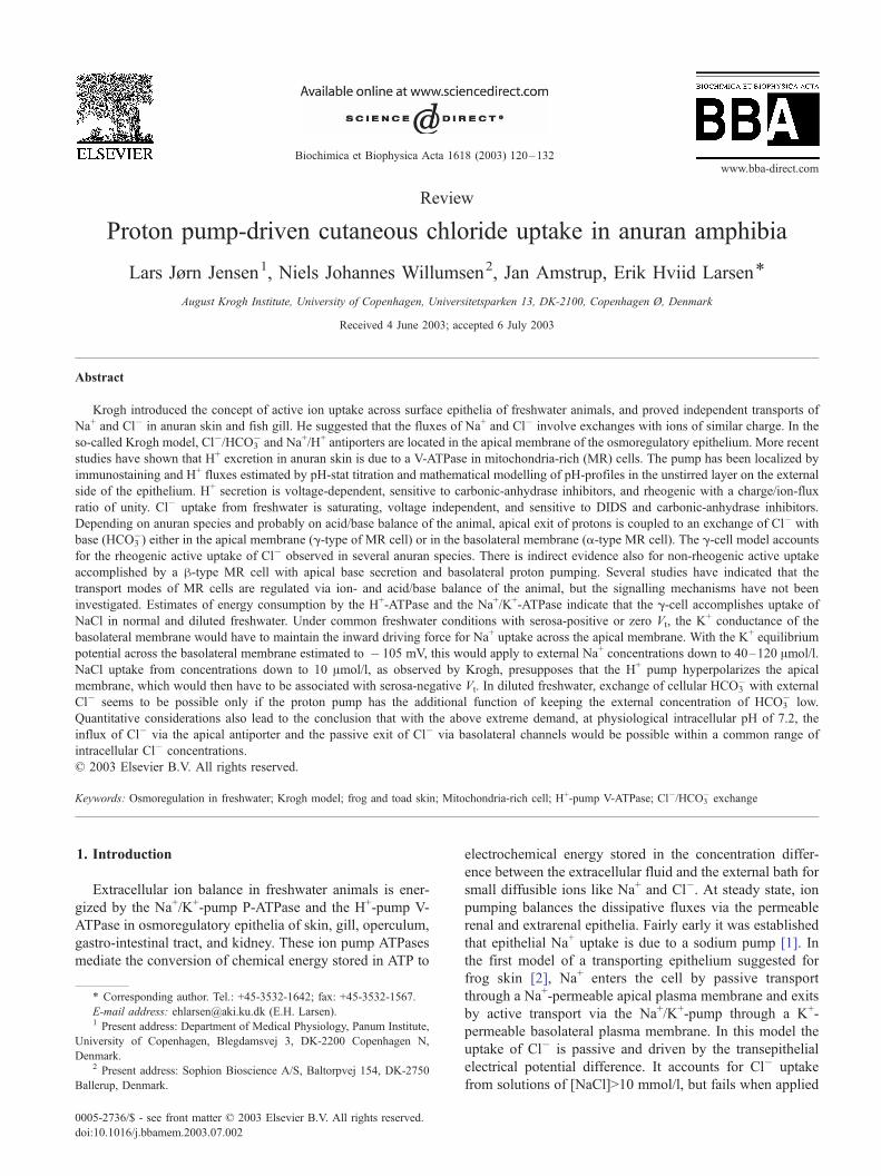
Fig. 1. In experiments with the frog, R. esculenta, August Krogh discovered a pow
box, the frog remains motionless for days with breathing movements stirring the b
resolution of his Cl� titration method corresponds to a bath-[Cl�] of 10 Amol/l. Lef
10 h, the bath was replaced by fresh 1/10 Ringer’s made isotonic by sucrose. The c
water. The frog was kept in running distilled water for 11 days prior to the experim
with [Cl�] of 1/100 of that of Ringer’s solution and Na+ and K+ as cations and gluc
solution that was diluted � 500 as compared to Ringer’s but with glucose added to
the experiment. Data from Table 4 of Ref. [25].
epithelium of a freshwater animal that maintains extracel-
lular ion concentrations above that of the environment. The
other question raised concerned the biological function of
active transport, in casu, the significance of active uptake for
ion balance in freshwater animals. In his ‘Croonean Lecture’
[26] Krogh developed the concept further by including a
comprehensive discussion of the significance of active and
passive mechanisms for maintaining steady-state ion con-
centrations of body cells of eukaryotic organisms. Initially,
the studies focused on Cl�. By titration of relatively large
samples of the bathing solution taken at intervals of several
hours, Krogh discovered a powerful Cl� uptake mechanism
in frog skin as shown in Fig. 1. These results would be
equally consistent with active uptake of Na+, active uptake
of Cl�, or with both ions being submitted to active transport.
For deciding between these possibilities, Krogh applied ion
substitution protocols, and summarized his findings as
follows: (i) Na+ and Cl� are absorbed together from sodium
chloride solutions. (ii) From solutions of potassium chloride,
Cl� and K+ can also be absorbed together, but absorption is
limited to a small amount of the two ions. (iii) Little or no
Ca2 + is absorbed from a CaCl2 solution, but a limited
amount of Cl� is taken up in exchange of HCO3�. From
these finding Krogh concluded, ‘‘that Cl� is the ion always
actively absorbed’’. With respect to the second major
question raised, the biological significance of active trans-
port, it was found that the Cl� mechanism could be
demonstrated only in non-fed animals forced into negative
ion balance by being kept for several weeks in running
distilled water. This treatment induced the active cutaneous
uptake together with a very significant reduction of cutane-
ous and renal loss of Cl� to the surroundings. Krogh
suggested that normally frogs get enough ions from food
and that the cutaneous active uptake of NaCl probably
would be of major significance only during hibernation at
the bottom of ponds where the animal does not feed [25].
erful active Cl� uptake mechanism in anuran skin. Kept in a small celluloid
ath. The cloaca was cannulated for separating voided urine from bath. The
t: Uptake of Cl� from a solution that initially was 1/10 Ringer’s. After about
ontinued uptake of Cl� shows that it is not coupled to an osmotic uptake of
ent. Data from Table 3 of Ref. [25]. Right: Uptake of Cl� from a solution
ose added to isotonicity. After about 7 h, the bath was replaced with a NaCl
isotonicity. The frog was kept in running distilled water for 3 weeks prior to
Table 1
Extracellular concentrations of sodium and chloride of the European toad,
B. bufo, in ion balance (unpublished)
N = 10 Lymph [Na+]
(mmol/l)
Lymph [Cl�]
(mmol/l)
Difference
(mmol/l)
MeanF S.E.M. 105.5F 1.0 85.4F 2.6 20.0F 2.7
The toads were kept in a terrarium with free access to tap water and
mealworms. Fluid samples (40–50 Al) were taken from the dorsal lymph
sac as described in Ref. [34]. Chloride and sodium were measured by
titration (CMT10 Radiometer, Copenhagen, Denmark) and photometry
(FLM3 Radiometer), respectively.
L.J. Jensen et al. / Biochimica et Biophysica Acta 1618 (2003) 120–132122
The analysis was extended to other freshwater animals
and ions. In salt-depleted teleosts, active uptake of Cl� was
observed in gills of bullhead (Ameiurus nebulosus), salmon
(Salmo irideus), roach (Leusiscus rutilus), goldfish (Caras-
ssius auratus), horke (Acerina cernua), perch (Perca fluvia-
tilis), and in two species of sticklebacks (Gasterosteus
aculeatus, G. pungitius). As the only freshwater species
studied by the above technique, eels (Anguilla vulgaris)
did not show active uptake of Cl� [27]. In a more detailed
investigation of goldfish and mitten crab (Eriocheir sinen-
sis), Krogh found independent and selective active transports
of cations and anions to be developed in the gills and he also
discovered excretion of NH4+ across the gill epithelium [28].
In Krogh’s terminology, active transport of an ion was
demonstrated if the ion is transported against a concentration
gradient. In the above experiments with the frog, where
active transport of Cl� was demonstrated, only the bath-
concentration of this ion was measured, so it ‘‘was provi-
sionally assumed that cations were attracted electrostatical-
ly’’ (quoted from Ref. [28]). In this study [28] the hypothesis
was tested in experiments in which also the Na+ concentra-
tion was measured. It was now directly shown that Na+ and
Cl� are taken up together from millimolar solutions and
found that the rate of uptake of the two ions could vary
considerably. A major conclusion of this paper was that the
two ions, Na+ and Cl�, are submitted to active transport by
independent mechanisms. Krogh did not consider the possi-
ble existence of an electrical potential difference across
plasma membranes or epithelia, but he was aware of the
large electrostatic forces involved in separating a charged ion
from its environment. So he assumed that tight coupling of
ions moving across the epithelium would be a necessity for
electroneutral charge transfer in epithelial cells. In one of his
summary statements, he therefore writes: ‘‘The transport is
supposed to involve an exchange with an ion of the same
sign’’. And further: ‘‘The CO2 produced by metabolism and
excreted through the skin or gills is probably sufficient to
serve in exchange for Cl� absorbed’’ [28]. In subsequent
investigations, these considerations have been referred to as
the ‘Krogh model’ of ion uptake from freshwater with an
epithelial Cl�/HCO3� exchange mechanism and an epithelial
Na+/H+ exchange mechanism, respectively.
2.2. Flux-ratio analysis confirmed active transport of Cl�
While the above investigations demonstrated ion uptake
by frogs from very diluted solutions, in a subsequent study
of the mechanism of cutaneous Cl� transport, the trans-
epithelial electrical potential difference was also taken into
account. With radioisotopes as tracers, Jørgensen et al. [3]
showed that the fluxes of Cl� fulfill the strict criterion for
active transport as defined by Ussing’s flux-ratio equation
for electrodiffusion across a composite membrane [29].
While in animals exposed to 3 mmol/l NaCl the potential
difference varied between 50 and 120 mV (inside of the skin
positive), uptake of Cl� from a 3 mmol/l KCl solution could
take place at a potential difference of reversed polarity, e.g.,
in the range of � 20 to � 50 mV. It was shown that the
sodium and the chloride uptake mechanisms could be
stimulated separately.
2.3. Quantitative studies of chloride/base- and sodium/
proton exchange
The considerations of Krogh and Jørgensen et al. dis-
cussed above were applied and further developed by Garcia
Romeu et al. [30] in a study of ion exchange across the skin
of the South American frog Calyptocephalella gayi. With
salt-depleted animals, they obtained results confirming up-
take of Na+ and Cl� in a near 1:1 proportion from dilute
concentrations of NaCl ( < 2 mmol/l). In experiments where
frogs individually were selectively depleted from Cl�, or
Na+, they confirmed that the skin has capacity for steady-
state net uptake of Cl� during a long period of time without
a concomitant uptake of Na+, and vice versa. A major result
of this [30] and subsequent studies with other amphibian
species [4,31,32] was the demonstration of a near 1:1
transcutaneous exchange of external Na+ for H+ and a near
1:1 transcutaneous exchange of external Cl� for base,
probably HCO3�. They were both depressed by the carbonic
anhydrase inhibitor diamox [32]. It was hypothesized that at
low external concentrations, the exchange of Cl� with
HCO3�, and Na+ with H+, respectively, is obligatory and
suggested that in experiments in which both Na+ and Cl�
are taken up at about similar rates, elimination of protons
and bicarbonate ions would be masked by them forming
H2O and CO2 in the external bath. In concordance with
results of a previous investigation [33] no evidence was
obtained for NH4+ excretion by frog skin [30,32].
2.4. Energy expenditure of NaCl uptake from freshwater
Extracellular concentrations of Cl� and Na+ of anurans
are fairly low as compared to those of other vertebrates
(Table 1). The [HCO3�]/pCO2 system is the major buffer of
extracellular pH and both on land and in freshwater at 20 jCis plasma-pH maintained at about 7.8. With pulmonary gas
exchange, [HCO3�] is about 22 mmol/l (B. marinus, Ref.
[35]), which is compatible with the estimated difference of
[Na+]lymph and [Cl�]lymph of Bufo bufo (20.0F 2.7 mmol/l,
L.J. Jensen et al. / Biochimica et Biophysica Acta 1618 (2003) 120–132 123
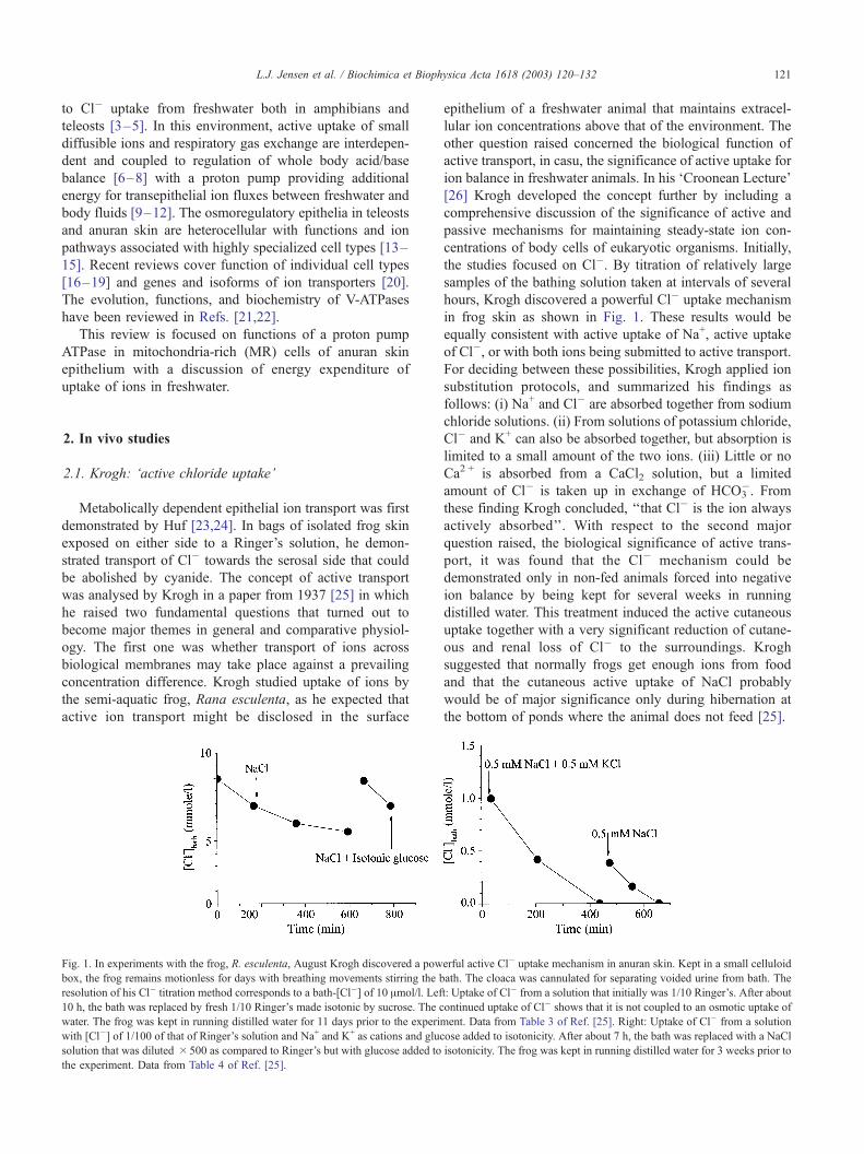
Table 1). With no access to food, during the first week in
running distilled water, toads experience a decrease of
extracellular [Cl�], on average for seven animals, from
87.0F 1.1 (control) to 74.4F 1.0 mmol/l (Fig. 2). During
the following 2 weeks, the decrease was relatively smaller
(day 21, [Cl�]lymph = 70.1F1.9 mmol/l). All of the seven
animals lost body mass (Fig. 2). For the purpose of
illustration, if this loss is assumed associated with the
reduced extracellular water volume, with an initial extracel-
lular space of 30% of the body mass [36] the extracellular
Cl� pool would have been reduced from 2611F 33 to
1414F 124 Amol/100 g body weight, corresponding to a
loss of 46% of the initial steady-state pool. Even if this is an
overestimation, it is indicated that the above mentioned in
vivo studies have been carried out with animals that had
experienced a fairly large perturbation of their ion balance.
Toads recover fully from this treatment when transferred to
a terrarium with free access to mealworms and a pool of tap
water.
The expenditure of metabolic energy associated with
active uptake of Na+ and Cl� must exceed the sum of the
two ions’ transcutaneous electrochemical potential differ-
ence, which is given by:
DlNa þ DlCl ¼ RT lnðNaÞiðNaÞo
þ FVt þ RT lnðC1ÞiðC1Þo
� FVt
¼ RT lnf Nai ½Na�if Nao ½Na�o
þ RT lnf Cli ½Cl�if Clo ½Cl�o
ð1Þ
Here, parentheses indicate activity, brackets concentra-
tion, f activity coefficient, and Vt the transcutaneous electri-
Fig. 2. Body mass and concentration of Cl� in samples from the dorsal
lymph sac of N= 7 toads (B. bufo) transferred to running distilled water at
time zero. Prior to the experiments, the animals had free access to
mealworms and tap water. Paired t-test with {null-hypothesis}: Body mass,
{Day0–Day7V 0} P< 0.001; {Day7–Day14V 0} P < 0.001; {Day14–
Day21V 0} P>0.1; {Day0–Day21V 0} P< 0.01; chloride concentration,
{Day0–Day7V 0} P < 0.001; {Day7–Day14V 0} P>0.5; {Day14–
Day21V 0} P>0.5; {Day0–Day21V 0} P < 0.001. (Unpublished).
cal potential difference. Krogh observed that salt-depleted
frogs could take up Cl� from the bath with a NaCl concen-
tration as low as about 0.01 mmol/l, which was the resolution
of his method. With the ratio of activity coefficients
of plasma and external bath being f 0.76, [Cl�]i = 70
mmol/l (Fig. 2), and [Na+]i = 90 mmol/l, from Eq. (1) at 20
jC the limiting thermodynamic work would have been,
DlNa +DlCl = 42 kJ/mol NaCl transported. In Ussing and
coworker’s studies of NaCl transport by frog skin it was
shown that the net inward flux of chloride in the isolated
preparation exposed to an external [Cl�]>10 mmol/l is
driven by the transepithelial electrical potential difference
generated by the sodium pump [37]. For uptake of Na+
energized by the pump with a stoichiometry 3Na+:2K+:
1ATP, the Cl� uptake driven by Vt, and a DGATP of about
� 60 kJ/mol, the useful work of the sodium pump is less than
20 kJ/mol NaCl transported (i.e., <�DGATP/3). Thus, we
have to assume that in Krogh’s experiments another pump, in
addition to the sodium pump, was in operation. The above-
mentioned efflux of protons would indicate that a proton exit
mechanism provided the required additional coupling to
cellular energy metabolism. An apical Na+/H+ exchange
mechanism can account for the kinetic relationship between
sodium uptake and proton excretion. However, according to
this hypothesis discussed in Ref. [5], it is still the sodium
pump that is doing the entire work. From this point of view,
the epithelial H+-pump discovered in Steinmetz’ studies of
turtle urinary bladder [38,39], which was concluded to be
present also in the skin of several anuran species [40–43],
would be a more likely candidate for supplying metabolic
energy to the cutaneous uptake of NaCl in freshwater. In
what follows is a discussion of the function of the proton
excretion mechanism in anuran skin, which has been hy-
pothesized to energize apical Na+ uptake [43] and apical
active uptake of Cl� [44].
3. In vitro studies
3.1. Interdependence of proton secretion and sodium uptake
Already in the early studies, it was observed that
isolated frog skin has capacity for acidifying the external
solution [45–47]. Acidification was found to take place at
transepithelial equilibrium, in the absence of external CO2,
being sensitive to carbonic anhydrase- and metabolic
inhibitors as well as being depressed in the absence of
O2 [40,41]. It was concluded that proton secretion is active
and depends on metabolically produced CO2. It is note-
worthy that species differences were observed in regards to
dependence on external anions [42]. Thus, with prepara-
tions of Rana ridibunda, the efflux of H+ was independent
of whether Cl� or SO42� was present in the external bath.
In contrast, the H+ efflux in skins of B. bufo could not be
detected if external SO42� was replaced by Cl�. Also
another difference was noted between the two anuran
L.J. Jensen et al. / Biochimica et Bioph124
species. Whreas proton secretion by short-circuited frog
skin was unaffected by Na+ uptake perturbation (amiloride
inhibition, antidiuretic hormone stimulation, external Na+/
Mg+ substitutions), with a similar protocol perturbations of
the active Na+ influx in toad skin resulted in parallel
changes of proton secretion [42]. Furthermore, it was noted
that the stoichiometric relationship between the H+ efflux
and Na+ influx that was described at low external NaCl�
concentrations vanishes at high external [NaCl]. Thus, at
an external [NaCl] = 2 mmol/l, diamox inhibited both of
these fluxes [43], whereas the proton efflux, only, was
inhibited by diamox if the skin was exposed to Ringer’s
solution on the outside [41,43]. From these findings and
further studies on the kinetics of Na+-uptake, Ehrenfeld
and Garcia-Romeu suggested that two uptake mechanisms
prevail. The first one would be dominating at concentra-
tions above 2 mmol/l and is not coupled to an efflux of
protons. The other one would be dominating at low
external NaCl concentrations and is coupled to an oblig-
atory exchange with metabolically produced H+ [48]. More
recently, Harvey and Ehrenfeld [49] suggested that the two
pathways for Na+ entrance are located in principal cells
and MR cells, respectively. Like principal cells, also MR
cells express amiloride blockable Na+ channels in the
apical membrane [50–52], although not all cells may
exhibit this feature [52]. Thus, they hypothesized [11,49]
that at low concentrations a rheogenic apical proton pump
by hyperpolarizing the apical membrane forces the flux of
Na+ from the external bath into the MR cells. Such a
mechanism would be operating independently of the ex-
ternal anion, and, in agreement with their experimental
findings, a 1:1 Na+/H+ stoichiometry would be expected if
Na+ were the only ion that is transported across the
epithelium. In a subsequent paper, Harvey [11] suggested
that HCO3� exits across the basolateral membrane in
exchange for serosal Cl�, a model similar to the A-type
( = a-type) intercalated cells of vertebrate distal renal
epithelia [53–56]. This interpretation would be consistent
with proton efflux being independent of the external anion
(Cl� or SO42�) as observed in frog skin [41] as well as the
observed exit of base towards the serosal bath [57]. By
immunofluorescence microscopy, polarized expression of
the targeted epitope of a V-ATPase was localised to the
apical region of the MR cells in the skin epithelium [58].
Table 2
The active flux of Cl� in anuran skin is associated with an outward current that
Species ICl=�FJ Cl�net
(AA/cm2)
INa+=FJ Na+net
(AA/cm2)
Leptodactyllus ocellatus �19F 3 76F 12
B. bufo �5F 1 30F 2
B. arenariuma �6.9F 1.7 0
Steady-state unidirectional fluxes of Cl� and Na+ were measured in short-circui
estimate of the associated electrical current (second column).a The active Na+ flux was blocked by amiloride in the solution bathing the apica
direction and became outward (negative).
3.2. Interdependence of proton secretion and chloride
uptake
The active inward chloride flux can be studied in the
isolated skin, and in some species is the flux of a
magnitude that compares with the active sodium flux.
The advantage of using isolated preparations is that the
active component is well defined and can be measured
accurately. Table 2 lists Cl� fluxes obtained under trans-
epithelial thermodynamic equilibrium conditions in which
the net flux is a measure of the active component. The
indicated agreement between the observed transepithelial
electric current and the sum of ion currents calculated from
the isotope tracer fluxes is a strong argument in favour of a
rheogenic inwardly directed active transport of chloride.
The above chloride current as well as the component of the
chloride flux that exhibits saturation kinetics at low exter-
nal concentrations are sensitive to carbonic anhydrase
inhibitors [60,62–65]. As mentioned above, the proton
pump is also depressed by application of inhibitors of
carbonic anhydrase, whose activity in the skin seems to be
confined to MR cells [66]. As a testable working hypoth-
esis, it was suggested, therefore, that a primary function of
the proton pump in toad skin epithelium would be to
energize the transepithelial active uptake of Cl� [13,44].
With the proton pump and the Cl�/HCO3� exchanger in the
apical membrane of MR cells, this hypothesis would also
account for the active Cl� influx being rheogenic.
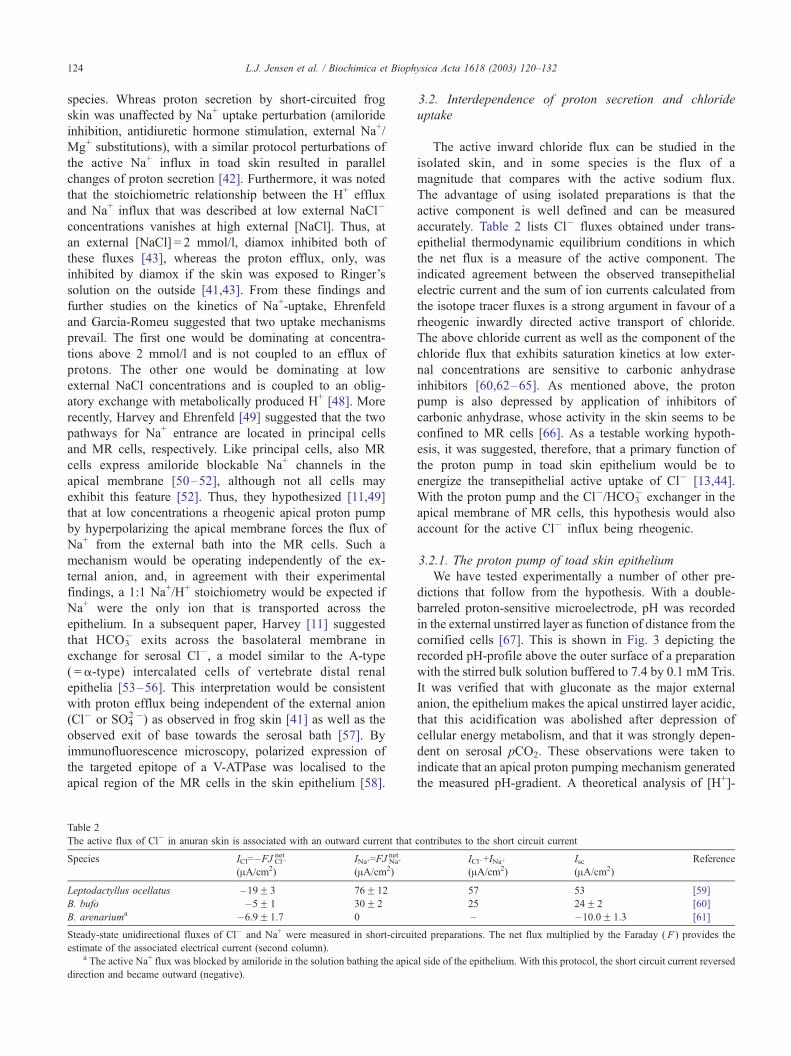
3.2.1. The proton pump of toad skin epithelium
We have tested experimentally a number of other pre-
dictions that follow from the hypothesis. With a double-
barreled proton-sensitive microelectrode, pH was recorded
in the external unstirred layer as function of distance from the
cornified cells [67]. This is shown in Fig. 3 depicting the
recorded pH-profile above the outer surface of a preparation
with the stirred bulk solution buffered to 7.4 by 0.1 mM Tris.
It was verified that with gluconate as the major external
anion, the epithelium makes the apical unstirred layer acidic,
that this acidification was abolished after depression of
cellular energy metabolism, and that it was strongly depen-
dent on serosal pCO2. These observations were taken to
indicate that an apical proton pumping mechanism generated
the measured pH-gradient. A theoretical analysis of [H+]-
ysica Acta 1618 (2003) 120–132
contributes to the short circuit current
ICl�+INa+
(AA/cm2)
Isc(AA/cm2)
Reference
57 53 [59]
25 24F 2 [60]
– �10.0F 1.3 [61]
ted preparations. The net flux multiplied by the Faraday ( F ) provides the
l side of the epithelium. With this protocol, the short circuit current reversed
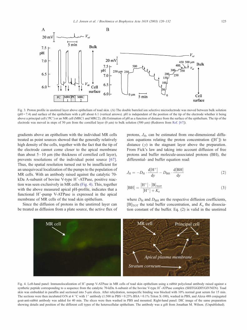
Fig. 3. Proton profile in unstirred layer above epithelium of toad skin. (A) The double barreled ion selective microelectrode was moved between bulk solution
(pH= 7.4) and surface of the epithelium with a pH about 6.1 (vertical arrows). pH is independent of the position of the tip of the electrode whether it being
above a principal cell (‘PC’) or an MR cell (MRC1 and MRC2). (B) Estimation of pH as a function of distance from the surface of the epithelium. The tip of the
electrode was moved in steps of 50 Am from the cornified layer (0 Am) to bulk solution (500 Am) (Redrawn from Ref. [67]).
L.J. Jensen et al. / Biochimica et Biophysica Acta 1618 (2003) 120–132 125
gradients above an epithelium with the individual MR cells
treated as point sources showed that the generally relatively
high density of the cells, together with the fact that the tip of
the electrode cannot come closer to the apical membrane
than about 5–10 Am (the thickness of cornified cell layer),
prevents resolutions of the individual point source [67].
Thus, the spatial resolution turned out to be insufficient for
an unequivocal localization of the pumps to the population of
MR cells. With an antibody raised against the catalytic 70-
kDa A-subunit of bovine V-type H+-ATPase, positive reac-
tion was seen exclusively in MR cells (Fig. 4). This, together
with the above measured apical pH-profile, indicates that a
functional H+-pump V-ATPase is expressed in the apical
membrane of MR cells of the toad skin epithelium.
Since the diffusion of protons in the unstirred layer can
be treated as diffusion from a plate source, the active flux of
Fig. 4. Left-hand panel: Immunolocalization of H+-pump V-ATPase in MR cells
synthetic peptide corresponding to a sequence from the catalytic 70-kDa A-subun
skin was embedded in paraffin and sectioned into 5-Am slices. After rehydration,
The sections were then incubated O/N at 4 jC with 1j antibody (1:500 in PBS+ 0.
goat-anti-rabbit antibody was added for 40 min. The slices were then washed in
showing details and position of the different cell types of the heterocellular epith
protons, JH, can be estimated from one-dimensional diffu-
sion equations relating the proton concentration ([H+]) to
distance ( y) in the stagnant layer above the preparation.
From Fick’s law and taking into account diffusion of free
protons and buffer molecule-associated protons (BH), the
differential- and buffer equation read:
JH ¼ �DH d½Hþ�
dy� DBH d½BH�
dyð2Þ
½BH� ¼ ½Hþ� ½B�TOT½Hþ� þ Ka
ð3Þ
where DH and DBH are the respective diffusion coefficients,
[B]TOT the total buffer concentration, and Ka the dissocia-
tion constant of the buffer. Eq. (2) is valid in the unstirred
of toad skin epithelium using a rabbit polyclonal antibody raised against a
it of the bovine V-type H+-ATPase complex (SHITGGDIYGIVNEN). Toad
nonspecific binding was blocked with 10% normal goat serum for 15 min.
25% BSA+0.1% Triton X-100), washed in PBS, and Alexa 488 conjugated
PBS and mounted. Right-hand panel: DIC image of the same preparation
elium. The antibody was a gift from Jonathan M. Wilson. (Unpublished).
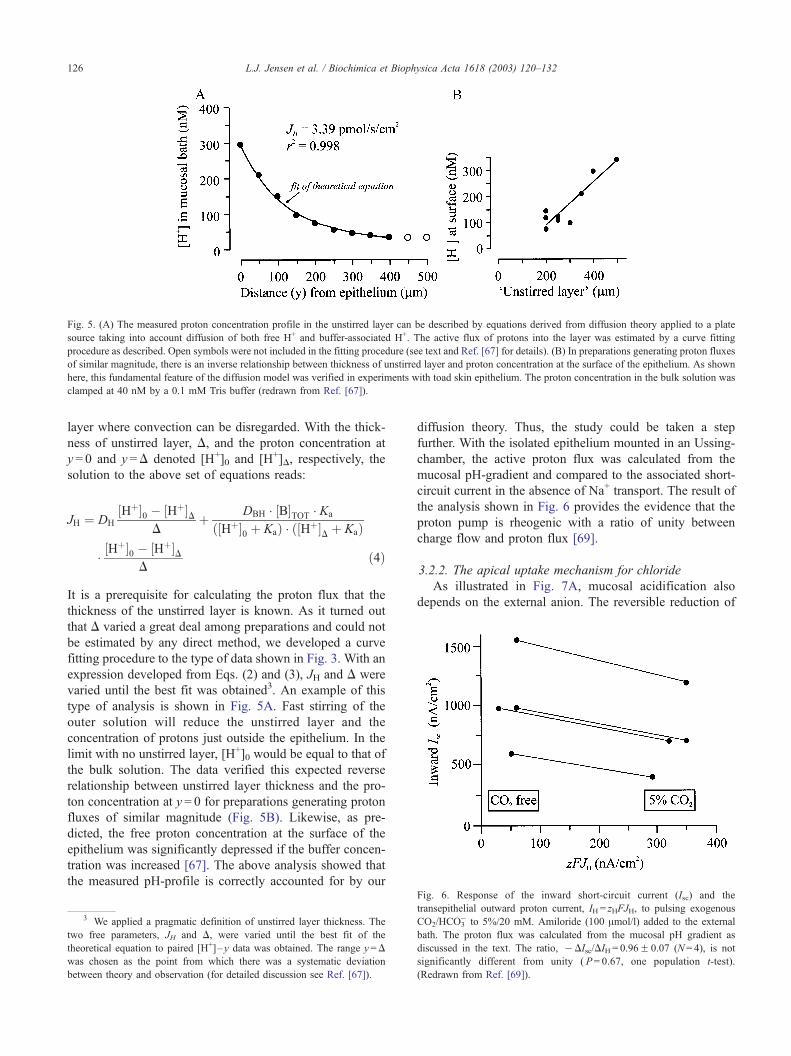
Fig. 5. (A) The measured proton concentration profile in the unstirred layer can be described by equations derived from diffusion theory applied to a plate
source taking into account diffusion of both free H+ and buffer-associated H+. The active flux of protons into the layer was estimated by a curve fitting
procedure as described. Open symbols were not included in the fitting procedure (see text and Ref. [67] for details). (B) In preparations generating proton fluxes
of similar magnitude, there is an inverse relationship between thickness of unstirred layer and proton concentration at the surface of the epithelium. As shown
here, this fundamental feature of the diffusion model was verified in experiments with toad skin epithelium. The proton concentration in the bulk solution was
clamped at 40 nM by a 0.1 mM Tris buffer (redrawn from Ref. [67]).
L.J. Jensen et al. / Biochimica et Biophysica Acta 1618 (2003) 120–132126
layer where convection can be disregarded. With the thick-
ness of unstirred layer, D, and the proton concentration at
y = 0 and y =D denoted [H+]0 and [H+]D, respectively, the
solution to the above set of equations reads:
JH ¼ DH
½Hþ�0 � ½Hþ�DD
þ DBH ½B�TOT Ka
ð½Hþ�0 þ KaÞ ð½Hþ�D þ KaÞ
½Hþ�0 � ½Hþ�D
Dð4Þ
It is a prerequisite for calculating the proton flux that the
thickness of the unstirred layer is known. As it turned out
that D varied a great deal among preparations and could not
be estimated by any direct method, we developed a curve
fitting procedure to the type of data shown in Fig. 3. With an
expression developed from Eqs. (2) and (3), JH and D were
varied until the best fit was obtained3. An example of this
type of analysis is shown in Fig. 5A. Fast stirring of the
outer solution will reduce the unstirred layer and the
concentration of protons just outside the epithelium. In the
limit with no unstirred layer, [H+]0 would be equal to that of
the bulk solution. The data verified this expected reverse
relationship between unstirred layer thickness and the pro-
ton concentration at y = 0 for preparations generating proton
fluxes of similar magnitude (Fig. 5B). Likewise, as pre-
dicted, the free proton concentration at the surface of the
epithelium was significantly depressed if the buffer concen-
tration was increased [67]. The above analysis showed that
the measured pH-profile is correctly accounted for by our
3 We applied a pragmatic definition of unstirred layer thickness. The
two free parameters, JH and D, were varied until the best fit of the
theoretical equation to paired [H+]–y data was obtained. The range y=D
was chosen as the point from which there was a systematic deviation
between theory and observation (for detailed discussion see Ref. [67]).
diffusion theory. Thus, the study could be taken a step
further. With the isolated epithelium mounted in an Ussing-
chamber, the active proton flux was calculated from the
mucosal pH-gradient and compared to the associated short-
circuit current in the absence of Na+ transport. The result of
the analysis shown in Fig. 6 provides the evidence that the
proton pump is rheogenic with a ratio of unity between
charge flow and proton flux [69].
3.2.2. The apical uptake mechanism for chloride
As illustrated in Fig. 7A, mucosal acidification also
depends on the external anion. The reversible reduction of
Fig. 6. Response of the inward short-circuit current (Isc) and the
transepithelial outward proton current, IH = zHFJH, to pulsing exogenous
CO2/HCO3� to 5%/20 mM. Amiloride (100 Amol/l) added to the external
bath. The proton flux was calculated from the mucosal pH gradient as
discussed in the text. The ratio, �DIsc/DIH = 0.96F 0.07 (N= 4), is not
significantly different from unity ( P= 0.67, one population t-test).
(Redrawn from Ref. [69]).
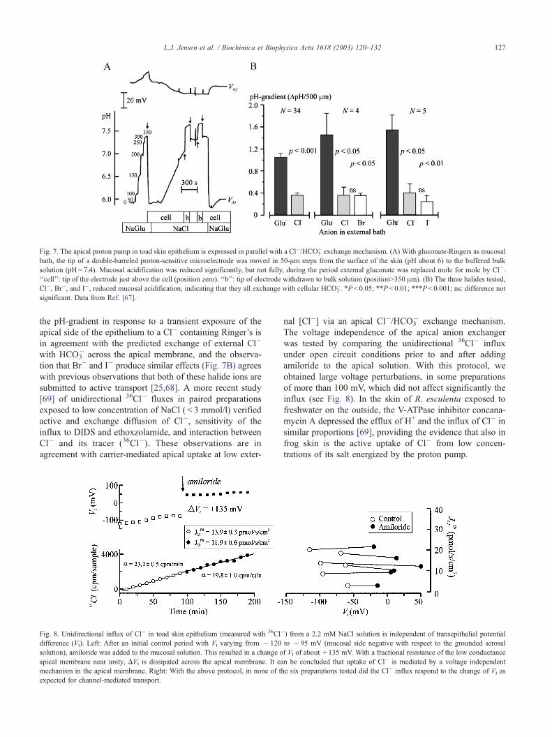
Fig. 7. The apical proton pump in toad skin epithelium is expressed in parallel with a Cl�/HCO3� exchange mechanism. (A) With gluconate-Ringers as mucosal
bath, the tip of a double-barreled proton-sensitive microelectrode was moved in 50-Am steps from the surface of the skin (pH about 6) to the buffered bulk
solution (pH= 7.4). Mucosal acidification was reduced significantly, but not fully, during the period external gluconate was replaced mole for mole by Cl�.
‘‘cell’’: tip of the electrode just above the cell (position zero). ‘‘b’’: tip of electrode withdrawn to bulk solution (position>350 Am). (B) The three halides tested,
Cl�, Br�, and I�, reduced mucosal acidification, indicating that they all exchange with cellular HCO3�. *P < 0.05; **P< 0.01; ***P< 0.001; ns: difference not
significant. Data from Ref. [67].
L.J. Jensen et al. / Biochimica et Biophysica Acta 1618 (2003) 120–132 127
the pH-gradient in response to a transient exposure of the
apical side of the epithelium to a Cl� containing Ringer’s is
in agreement with the predicted exchange of external Cl�
with HCO3� across the apical membrane, and the observa-
tion that Br� and I� produce similar effects (Fig. 7B) agrees
with previous observations that both of these halide ions are
submitted to active transport [25,68]. A more recent study
[69] of unidirectional 36Cl� fluxes in paired preparations
exposed to low concentration of NaCl ( < 3 mmol/l) verified
active and exchange diffusion of Cl�, sensitivity of the
influx to DIDS and ethoxzolamide, and interaction between
Cl� and its tracer (36Cl�). These observations are in
agreement with carrier-mediated apical uptake at low exter-
Fig. 8. Unidirectional influx of Cl� in toad skin epithelium (measured with 36Cl�
difference (Vt). Left: After an initial control period with Vt varying from � 120
solution), amiloride was added to the mucosal solution. This resulted in a change o
apical membrane near unity, DVt is dissipated across the apical membrane. It ca
mechanism in the apical membrane. Right: With the above protocol, in none of th
expected for channel-mediated transport.
nal [Cl�] via an apical Cl�/HCO3� exchange mechanism.
The voltage independence of the apical anion exchanger
was tested by comparing the unidirectional 36Cl� influx
under open circuit conditions prior to and after adding
amiloride to the apical solution. With this protocol, we
obtained large voltage perturbations, in some preparations
of more than 100 mV, which did not affect significantly the
influx (see Fig. 8). In the skin of R. esculenta exposed to
freshwater on the outside, the V-ATPase inhibitor concana-
mycin A depressed the efflux of H+ and the influx of Cl� in
similar proportions [69], providing the evidence that also in
frog skin is the active uptake of Cl� from low concen-
trations of its salt energized by the proton pump.
) from a 2.2 mM NaCl solution is independent of transepithelial potential
to � 95 mV (mucosal side negative with respect to the grounded serosal
f Vt of about + 135 mV. With a fractional resistance of the low conductance
n be concluded that uptake of Cl� is mediated by a voltage independent
e six preparations tested did the Cl� influx respond to the change of Vt as
L.J. Jensen et al. / Biochimica et Biophysica Acta 1618 (2003) 120–132128
However clear-cut the results of the testing of the model
may seem, we also obtained results that are not in full
agreement with the above interpretations. In 11 out of a total
of 45 preparations tested with the protocol of Fig. 7A,
mucosal acidification did not respond as shown in this
figure [67]. This finding would indicate heterogeneity in
the polarization of the acid–base secretory mechanisms
among toads (see Section 4.1). In a series of experiments
with frog skin and toad skin producing fairly large proton
fluxes, concanamycin Awas without effect [69]. This would
indicate that a type of proton-pump ATPase, other than the
V-ATPase, might also be expressed in anuran skin epithe-
lium. Further studies are needed to characterize putative
concanamycin A insensitive H+-fluxes.
4. Hypotheses and models
4.1. MR cells of anuran skin
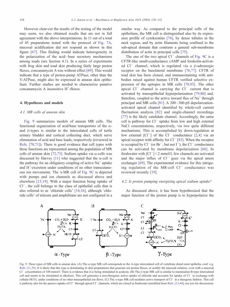
Fig. 9 summarizes models of anuran MR cells. The
functional organization of acid/base transporters of the a-
and h-types is similar to the intercalated cells of turtle
urinary bladder and cortical collecting duct, which serve
elimination of acid and base loads, respectively (reviewed in
Refs. [70,71]). There is good evidence that cell types with
these functions are represented among the population of MR
cells of anuran skin [72,73]. Sodium uptake via a-cells was
discussed by Harvey [11] who suggested that the a-cell is
the pathway for an obligatory coupling of active Na+ uptake
and H+-excretion under conditions of no other transcutane-
ous ion movements. The g-MR cell of Fig. 9C is depicted
with pumps and ion channels as discussed above and
elsewhere [13,19]. With a major function being uptake of
Cl�, the cell belongs to the class of epithelial cells that is
also referred to as ‘chloride cells’ [14,16], although ‘chlo-
ride cells’ of teleosts and amphibians are not configured in a
Fig. 9. Three types of MR cells in anuran skin. (A) The a-type MR cell correspond
Refs. [11,39]. It is likely that this type is dominating in skin preparations that gene
Cl� concentration of 100 mmol/l. There is evidence that it is being stimulated in ac
cell and seems to be stimulated in alkalosis. This cell generates a non-rheogenic
cellular HCO3� under conditions of no other transepithelial ion flows. (C) The g-typ
is pathway also for the passive uptake of Cl� through apical Cl� channels, which ar
similar way. As compared to the principal cells of the
epithelium, the MR cell is distinguished also by its expres-
sion profile of cytokeratins [74], by dense tubules in the
neck region, and by actin filaments being confined to the
sub-apical domain that contrasts a general sub-membrane
distribution of actin in principal cells [75].
The one of the two apical Cl� channels of Fig. 9C is a
CFTR-like small-conductance cAMP and forskolin-activat-
ed Cl� channel, which is regulated via a h-adrenergicreceptor on the basolateral membrane [76,77]. CFTR of
toad skin has been cloned, and immunostaining with anti-
bodies raised against human CFTR verified selective ex-
pression of the epitopes in MR cells [78,93]. The other
apical Cl� channel is carrying the Cl� current that is
activated by transepithelial hyperpolarization [79,80] and,
therefore, coupled to the active inward flux of Na+ through
principal and MR cells [81]. A 200–300-pS depolarization-
activated apical channel identified by whole-cell current
fluctuation analysis [82] and single-channel recordings
[77] is the likely candidate channel. Accordingly, the same
cell is pathway for Cl� uptake from low and high external
NaCl concentrations, respectively, via two quite different
mechanisms. This is accomplished by down-regulation at
low external [Cl�] of the Cl� conductance [2,4] via an
apical receptor with affinity for Cl� [83]. When the receptor
is occupied by Cl� (or Br�, but not I�), the Cl� conductance
can be activated by membrane depolarization [68]. In
freshwater with [Cl�] < 2 mmol/l, few channels are activated
and the major influx of Cl� goes via the apical anion
exchanger [69]. The experimental evidence for this intrigu-
ing regulation of the MR-cell Cl� conductance was
reviewed recently [19].
4.2. Is proton pumping energizing apical sodium uptake?
As discussed above, it has been hypothesized that the
major function of the proton pump is to hyperpolarize the
s to the A-type intercalated cell of vertebrate distal renal epithelia, conf. e.g.
rate net proton fluxes, or acidify the mucosal solution, even with a mucosal
idosis. (B) The h-type MR cell is similar to mammalian B-type intercalated
active uptake of chloride and accounts for uptake of Cl� in exchange with
e MR cell mediates active transport of Cl� in a rheogenic fashion. This cell
e closed in freshwater (modified from Refs. [13,44]; see text for discussion).
4 In the calculation, a solubility coefficient of CO2 = 0.06 mmol/mm
Hg and a pK= 6.3 were used together with a pCO2 = 9.8 mm Hg (20 jC).
L.J. Jensen et al. / Biochimica et Biophysica Acta 1618 (2003) 120–132 129
apical membrane for generating a sufficient driving force for
uptake of Na+ from freshwater. It is unlikely that proton
pumping is driving the cell potential to a value that is
numerically larger than the K+-equilibrium potential across
the basolateral membrane. Otherwise, potassium ions flow
from the interstitial fluid into the cell both via basolateral K+
channels and Na+/K+-pumps. In a skin with dominating
sodium current, the inside of the epithelium would be
relatively positive ([3], Fig. 4). Thus, the apical membrane
potential is at most � 105 mV (EK of the inner membrane
[12], with Vt = 0 mV), and with an intracellular [Na+] of,
e.g., 3–10 mmol/l, the passive flux of Na+ would reverse at
an external concentration of 35–118 Amol/l (20 jC, f cellNa /
f oNa = 0.76). This indicates that even at an external Na+
concentration that is smaller than in most lakes and creeks,
a relatively large K+ conductance of the basolateral mem-
brane is sufficient for establishing the driving force for the
net flux of Na+ from the environment into the cell.
The above example also illustrates that uptake of Na+ and
Cl� from 10 Amol/l of the salt (Fig. 1) can be accomplished
only if proton pumping reverses the transepithelial potential
difference as has been observed in vivo [3] and in the
isolated skin [69]. Due to the small concentrations of Na+ in
cell and freshwater, even with large apical sodium perme-
ability, the conductance of the apical membrane would be
very small as compared to that of the basolateral membrane.
Thus, within the range observed, � 20 mVVVtV� 50 mV
(serosal side of the skin negative), and a fractional resistance
of the apical membrane close to unity, the apical membrane
potential would be between, e.g., � 120 and � 145 mV,
which probably is a sufficient electrical driving force for
generating a net influx of Na+ through the apical channels at
10 Amol/l external [Na+]. In connection with this hypothe-
sis, it is interesting that in zebrafish (Danio rerio) bafilo-
mycin (1 Amol/l) depressed Na+ uptake at an environmental
[Na+] of 35 Amol/l, but was without effect on the Na+
uptake from a [Na+] of 1.5 mmol/l [92].
4.3. Proton pumping and chloride/bicarbonate exchange
Uptake of both ions from 10 Amol/l NaCl solution was
calculated to cost 42 kJ/mol of NaCl transported (Section
2.4). With the energy input of the sodium pump of 20 kJ/mol,
that of the proton pump would have to be larger than 22 kJ/
mol. With a stoichiometry of the V-ATPase of 2 H+ pumped
per ATP hydrolyzed [84] and DGATP=� 60 kJ/mol (as
above), the useful work of the proton pump is 30 kJ/mol
of Cl� transported. In this estimate, we assume that all of the
HCO3� formed leaves the MR cell through the apical Cl�/
HCO3� exchange mechanism. Thus, from the point of view
of energy conservation, our g-MR cell model would accom-
plish the uptake of NaCl from a 10 Amol/l NaCl solution.
The coupling of Cl� influx and HCO3� efflux in the apical
membrane requires that cell-to-bath concentration gradient
of HCO3� be larger than that of Cl�. A quantitative evalu-
ation is a fairly complicated matter and cannot, as yet, lead to
a definitive conclusion, but is useful for elucidating the
nature of the problem. The cellular [HCO3�] depends on
pCO2 in the epithelial cell, which would have to be lower
than that of arterial pCO2 and larger than that of bath-pCO2.
With an intracellular pH of f 7.2 [85,86] and pulmonary
gas exchange, arterial pCO2 is f 10 mm Hg [35]. Then,
from the Henderson–Hasselbalch equation [HCO3�]cellV 4.5
mmol/l.4 With cutaneous gas exchange arterial pCO2 is
smaller, e.g., pCO2 = 3–5 mm Hg (teleost fish [87,88]).
Accordingly, at a pHcell = 7.2, as an upper-limit estimate
we obtain, [HCO3�]cellV 2.3 mmol/l. With these estimates
and [Cl�]out = 10 Amol/l, uptake of Cl� would be possible if
[Cl�]cell is less than 4.5 and 2.3 mmol/l for the two types of
breathing, respectively, and providing the acid secretion
maintains [HCO3�]out < 10 Amol/l in the external unstirred
layer. With Ringer’s solution on the outside [Cl�]cell is
30–45 mM [50,52,77]. Since this reflects a passive
distribution [50,77], with freshwater outside, the concen-
tration of Cl� in the MR cell would probably be much
smaller. The exit across the basolateral membrane through
anion selective channels [89] requires that intracellular
Cl� be above equilibrium across this membrane. With a
membrane potential close to � 105 mV (EK, conf. above)
and [Cl�]extc 70 mmol/l (Fig. 2), this would be fulfilled
for [Cl�]cell>1 mmol/l. This is interesting because it
shows that with extreme demands on the uptake mecha-
nism, influx of Cl� via the apical exchange mechanism
and passive exit of Cl� across the basolateral membrane
would be possible within a common range of intracellular
Cl� concentrations. The conclusion is that as a prerequi-
site for the g-cell model (Fig. 9C) to be compatible with
Krogh’s observations (Fig. 1), the apical proton pump would
have to have the additional function of maintaining the
bicarbonate concentration low in the fluid layer outside the
skin.
There is another problem that also would have to be
considered. Frogs that were selectively depleted of Cl� took
up Cl� in exchange for HCO3� apparently with no other
transcutaneous ion movements [30]. This would indicate
non-rheogenic active uptake of Cl�, which is incompatible
with the g-type MR-cell of Fig. 9C, where a positive charge
moves in the outward direction across the apical and baso-
lateral membrane for each chloride ion entering the animal
from the bath. Cells configured according to the h-type MR-
cell of Fig. 9B accomplishes this type of anion uptake,
which results in acidification of body fluids. Tadpoles that
experience acidosis in response to external hypercapnia
compensate by mobilizing a labile pool of CaCO3 [86]. It
is an interesting problem whether the predicted acidification
of body fluids in frogs with Cl� uptake via h-MR cells in a
similar way is being compensated by mobilizing buffer base
from the paraventral and subcutaneous lime sacs that are
developed in the metamorphosed anuran.
L.J. Jensen et al. / Biochimica et Biophysica Acta 1618 (2003) 120–132130
5. Concluding remarks
Considering the thermodynamic work with conservative
assumptions, the g-type model of the MR cells (Fig. 9C)
seems to account for the findings Krogh published in 1937
[25]. In the most extreme situation of low external [NaCl],
hyperpolarization of the apical membrane by proton pump-
ing is required for inward transport of Na+ across the
apical membrane. There is no evidence of a similar
demand on the proton pump under common freshwater
conditions. A large number of recent experimental obser-
vations are also fully compatible with this model, which
accounts for passive as well as active uptake of Cl� across
anuran skin.
Krogh observed that the ‘leak’ permeability of the skin is
down-regulated while the active mechanism is up-regulated
in animals forced into negative Cl� balance [25]. The
passive mechanisms are submitted to fast and slow regu-
lations that involve cellular signalling and proliferation and
growth in number of MR cells [90,91]. Undoubtedly,
difficulties in reproducing observations on anuran skin
function in freshwater are due to a gap in our knowledge
about the regulation of the active Cl� mechanism, the
capacity of which varies between species and with acclima-
tion of the animals in study. This point was emphasized
recently also in a study comparing individuals of zebrafish
(D. rerio) acclimated to soft water and hard water, respec-
tively. It was found that the first group up-regulated the
maximum flux of Cl� as well the uptake affinity for external
Cl� [92]. Thus, the plasticity of osmoregulatory mecha-
nisms in freshwater may also involve regulation of affinity
of transporters via cellular co-factors or shift between
different membrane transporters for the same ion. Added
to this, there is good evidence that anuran acid- and base-
secreting mechanisms serve three functions, i.e., restoration
of an acid load, restoration of a base load, and whole body
Cl� balance. It is unknown how these specialized functions
are regulated. In Fig. 9, tentatively three different cell types
are depicted for each of these functions. This is in analogy
with mammalian kidney in which the a-cell and the h-cellhave been discovered and studied in details. Studies on
anurans acid/base balance are too few to decide whether
similar heterocellular differentiations characterize the an-
uran skin epithelium or whether a single cell type shifts
functional polarity according to acid/base balance.
Acknowledgements
Comments on the manuscript by Dr. Frank Bo Jensen,
University of Southern Denmark, are much appreciated. We
also wish to thank Dr. Martin Grosell for discussions during
his stay at the August Krogh Institute, and Dr. Jonathan M.
Wilson, University of British Columbia, for the generous
gift of bovine V-ATPase antibody. Our experimental work
referred to in this review was supported by grants from the
Danish Natural Science Research Council, the Carlsberg
Foundation, the Novo-Nordisk Foundation, and the Alfred
Benzon Foundation.
References
[1] H.H. Ussing, K. Zerahn, Active transport of sodium as the source of
electric current in the short-circuited isolated frog skin, Acta Physiol.
Scand. 23 (1951) 110–127.
[2] V. Koefoed-Johnsen, H.H. Ussing, The nature of the frog skin poten-
tial, Acta Physiol. Scand. 42 (1958) 298–308.
[3] C.B. Jørgensen, H. Levi, K. Zerahn, On active uptake of sodium and
chloride ions in anurans, Acta Physiol. Scand. 30 (1954) 178–190.
[4] L.B. Kirschner, The study of NaCl in aquatic animals, Am. Zool. 10
(1970) 365–376.
[5] R. Motais, F. Garcia-Romeu, Transport mechanisms in the teleostean
gill and amphibian skin, Annu. Rev. Physiol. 34 (1972) 141–176.
[6] D.G. McDonald, Y. Tang, R.G. Boutilier, Acid and ion transfer across
the gills of fish: mechanisms and regulation, Can. J. Zool. 67 (1989)
3046–3054.
[7] G.G. Goss, S.F. Perry, J.N. Fry, P. Laurent, Gill morphology and acid
base regulation in freshwater fish, Comp. Biochem. Physiol. 119A
(1998) 107–115.
[8] M. Busk, F.B. Jensen, T. Wang, Effects of feeding on metabolism, gas
transport, and acid–base status in the bullfrog Rana catesbeiana, Am.
J. Physiol. 278 (2000) R185–R195.
[9] H. Lin, D.J. Randal, Evidence for the presence of an electrogenic
proton pump on the trout gill epithelium, J. Exp. Biol. 161 (1991)
119–134.
[10] H. Lin, D.C. Pfeiffer, A.W. Vogl, J. Pan, D.J. Randall, Immunoloc-
alization of H+ ATPase in the gill epithelia of rainbow trout, J. Exp.
Biol. 195 (1994) 16–183.
[11] B.J. Harvey, Energization of sodium absorption by the H+-ATPase
pump in mitochondria-rich cells of frog skin, J. Exp. Biol. 172
(1992) 289–309.
[12] E.H. Larsen, N.J. Willumsen, B.C. Christoffersen, Role of proton
pump of mitochondria-rich cells for active transport of chloride ions
in toad skin epithelium, J. Physiol. 450 (1992) 203–216.
[13] E.H. Larsen, Chloride transport by high-resistance heterocellular epi-
thelia, Physiol. Rev. 71 (1991) 235–283.
[14] S.F. Perry, The chloride cell: structure and function in the gills of
freshwater fishes, Annu. Rev. Physiol. 59 (1997) 325–347.
[15] J.M. Wilson, P. Laurent, B.L. Tufts, D.J. Benos, M. Donowitz, A.W.
Vogl, D.J. Randal, NaCl uptake by the branchial epithelium in fresh-
water teleost fish: an immunological approach to ion-transport protein
localization, J. Exp. Biol. 203 (2000) 2279–2296.
[16] W.S. Marshall, Na+, Cl�, Ca2 + and Zn+ transport by fish gills: retro-
spective review and prospective synthesis, J. Exp. Zool. 293 (2002)
264–283.
[17] W.S. Marshall, T.D. Singer, Cystic fibrosis transmembrane conduc-
tance regulator in teleost fish, Biochim. Biophys. Acta 1566 (2002)
16–27.
[18] C.M. Wood, S.P. Kelly, B. Zhou, M. Fletcher, M. O’Donnell, B.
Eletti, P. Part, Cultured gill epithelia as models for freshwater fish
gill, Biochim. Biophys. Acta 1566 (2002) 72–83.
[19] N.J. Willumsen, J. Amstrup, N. Møbjerg, A. Jespersen, P. Kristensen,
E.H. Larsen, Mitochondria-rich cells as experimental model in studies
of epithelial chloride channels, Biochim. Biophys. Acta 1566 (2002)
28–43.
[20] C.P. Cutler, G. Cramb, Molecular physiology of osmoregulation in
eels and teleosts: the role of transport isoforms and gene duplication,
Comp. Biochem. Physiol. 130A (2001) 551–564.
[21] S.L. Gluck, D.M. Underhill, M. Iyori, L.S. Holliday, T.Y. Kostromi-
nova, B.S. Lee, Physiology and biochemistry of the kidney vacuolar
H+-ATPase, Annu. Rev. Physiol. 58 (1996) 427–445.
L.J. Jensen et al. / Biochimica et Biophysica Acta 1618 (2003) 120–132 131
[22] N. Nelson, W.R. Harvey, Vacuolar and plasma membrane proton-
adenosinetriphosphatases, Physiol. Rev. 79 (1999) 361–385.
[23] E.G. Huf, Versuche uber den Zusammenhang zwischen Stoffwechel,
Potentialbildung und Funktion des Froschhaut, Pfluegers Arch. 235
(1935) 655–673.
[24] E.G. Huf, Uber activen Wasser-und Saltztransport durch die Frosch-
haut, Pflugers Arch. 237 (1936) 143–166.
[25] A. Krogh, Osmotic regulation in the frog (R. esculenta) by active
absorption of chloride ions, Skand. Arch. Physiol. 76 (1937) 60–74.
[26] A. Krogh, The active and passive exchange of inorganic ions through
the surface of living cells and through living membranes generally,
Proc. Royal Soc., B 133 (1946) 140–200.
[27] A. Krogh, Osmotic regulation in fresh water fishes by active absorp-
tion of chloride ions, Z. vgl. Physiol. 24 (1937) 656–666.
[28] A. Krogh, The active absorption of ions in some freshwater animals,
Z. vgl. Physiol. 25 (1938) 335–350.
[29] H.H. Ussing, The distinction by means of tracers between active
transport and diffusion, Acta Physiol. Scand. 19 (1949) 43–56.
[30] F. Garcia Romeu, A. Salibian, S. Pezzani-Hernandez, The nature of in
vivo sodium and chloride uptake mechanism through the epithelium
of the Chilean frog Calyptocephalella gayi (Dum. et Bibr., 1841).
Exchanges of hydrogen against sodium and of bicarbonate against
chloride, J. Gen. Physiol. 53 (1969) 816–835.
[31] L.B. Kirschner, L. Greenwald, T.H. Kerstetter, Effect of amiloride on
sodium transport across body surfaces of freshwater animals, Am. J.
Physiol. 224 (1973) 832–837.
[32] F. Garcia-Romeu, J. Ehrenfeld, In vivo Na+ and Cl� independent
transport across the skin of Rana esculenta, Am. J. Physiol. 228
(1975) 839–844.
[33] F. Garcia-Romeu, A. Salibian, Sodium uptake and ammonia excretion
through the in vivo skin of the South American frog Leptodactylus
ocellatus (L. 1758), Life Sci. 7 (1968) 465.
[34] S.D. Hillyard, E.H. Larsen, Lymph osmolality and rehydration from
NaCl solutions by toads, Bufo marinus, J. Comp. Physiol., B. 171
(2001) 283–292.
[35] R.G. Boutilier, D.J. Randal, G. Shelton, D.P. Toews, Acid–base re-
lationships in the blood of the toad, Bufo marinus: I. The effects of
environmental CO2, J. Exp. Biol. 82 (1979) 331–334.
[36] C.B. Jørgensen, K. Brems, P. Geckler, Volume and osmotic regulation
in the toad Bufo bufo bufo (L.) at low temperature, with special
reference to amphibian hibernation, in: C.B. Jørgensen, E. Skadhauge
(Eds.), Proc. Alfred Benzon Symposium XI, Osmotic and Volume
Regulation, Munksgaard, Copenhagen, 1978, pp. 62–79.
[37] V. Koefoed-Johnsen, H. Levi, H.H. Ussing, The mode of passage of
chloride ions through the isolated frog skin, Acta Physiol. Scand. 25
(1952) 150–163.
[38] P.R. Steinmetz, Characteristics of hydrogen ion transport in urinary
bladder of water turtle, J. Clin. Invest. 46 (1967) 1531–1540.
[39] P.R. Steinmetz, Cellular mechanism of urinary acidification, Physiol.
Rev. 54 (1974) 890–956.
[40] M.G. Emilio, M.M. Machado, H.P. Menano, The production of a
hydrogen ion gradient across the isolated frog skin. Quantitative as-
pects and the effect of acetazolamide, Biochim. Biophys. Acta 203
(1970) 394–409.
[41] T. Machen, D. Erlij, Some features of hydrogen (ion) secretion by the
frog skin, Biochim. Biophys. Acta 406 (1975) 120–130.
[42] M.G. Emilio, H.P. Menano, The excretion of hydrogen ion by the
isolated amphibian skin: effects of antidiuretic hormone and amilor-
ide, Biochim. Biophys. Acta 382 (1975) 344–352.
[43] J. Ehrenfeld, F. Garcia-Romeu, Active hydrogen excretion and so-
dium absorption through isolated frog skin, Am. J. Physiol. 233
(1977) F46–F54.
[44] E.H. Larsen, NaCl transport by amphibian skin, in: R. Greger (Ed.),
NaCl Transport in Epithelia, Adv. Comp. Environ. Physiology, vol. 1,
Springer-Verlag, Berlin, 1988, pp. 189–248.
[45] H.H. Ussing, The active transport through the isolated frog skin in the
light of tracer studies, Acta Physiol. Scand., (1949) 1–37.
[46] E.G. Huf, J. Parish, C. Weatherford, Active salt and water uptake by
isolated frog skin, Am. J. Physiol. 164 (1951) 137–142.
[47] W.R. Fleming, On the role of hydrogen ion and potassium ion in the
active transport of sodium across the isolated frog skin, J. Cell. Comp.
Physiol. 49 (1957) 129–152.
[48] J. Ehrenfeld, F. Garcia-Romeu, Kinetics of ionic transport across frog
skin: two concentration dependent processes, J. Membr. Biol. 56
(1980) 139–147.
[49] B.J. Harvey, J. Ehrenfeld, Epithelial pH and ion transport regulation
by proton pumps and exchangers, Proton Passage Across Cell Mem-
branes, Ciba Foundation Symposium 139, Wileym, Chichester, 1988,
pp. 139–164.
[50] E.H. Larsen, H.H. Ussing, K.R. Spring, Ion transport by mitochon-
dria-rich cells in toad skin, J. Membr. Biol. 99 (1987) 25–40.
[51] B.J. Harvey, E.H. Larsen, Sodium and chloride currents in single
mitochondria-rich cells of amphibian skin, J. Physiol. 459 (1993)
241 (abstract).
[52] R. Rick, Intracellular ion concentrations in the isolated frog skin
epithelium: evidence for different types of mitochondria-rich cells,
J. Membr. Biol. 127 (1992) 227–236.
[53] D.L. Stetson, P.R. Steinmetz, a- and h-types of carbonic anhydrase-
rich cells in turtle bladder, Am. J. Physiol. 249 (1985) F553–F565.
[54] P.R. Steinmetz, Cellular mechanisms of urinary acidification, Am. J.
Physiol. 251 (1986) F173–F187.
[55] D. Brown, S. Hirsch, S. Gluck, Localization of proton-pumping
ATPase in rat kidney, J. Clin. Invest. 82 (1988) 2114–2126.
[56] D. Brown, S. Breton, Mitochondria-rich, proton-secreting epithelial
cells, J. Exp. Biol. 199 (1996) 2345–2358.
[57] E. Duranti, J. Ehrenfeld, B.J. Harvey, Acid secretion through the Rana
esculenta skin: involvement of anion-exchange mechanism at the
basolateral membrane, J. Physiol. 378 (1986) 195–211.
[58] U. Klein, M. Timme, W. Zeiske, J. Ehrenfeld, The H+ pump in frog
skin (Rana esculenta): identification and localization of a V-ATPase,
J. Membr. Biol. 157 (1997) 117–126.
[59] J.A. Zadunaisky, O.A. Candia, D.J. Chiarandini, The origin of short-
circuit current in the isolated skin of the South American frog Lep-
todactyllus ocellatus, J. Gen. Physiol. 47 (1963) 393–402.
[60] K. Bruus, P. Kristensen, E.H. Larsen, Pathways for chloride and sodium
transport across toad skin, Acta Physiol. Scand. 97 (1976) 31–47.
[61] D.M. Berman, O.M. Soria, A. Coviello, Reversed short-circuit current
across isolated skin of the toad Bufo arenarium, Pflugers Arch. 409
(1987) 616–619.
[62] D. Erlij, Salt transport across isolated frog skin, Philos. Trans. R. Soc.,
B 262 (1971) 153–161.
[63] P. Kristensen, Chloride transport across frog skin, Acta Physiol.
Scand. 84 (1972) 338–346.
[64] F. Garcia-Romeu, J. Ehrenfeld, Chloride transport through the non-
short-circuited isolated skin of Rana esculenta, Am. J. Physiol. 228
(1975) 845–849.
[65] J. Ehrenfeld, F. Garcia-Romeu, Coupling between chloride absorption
and base excretion in isolated skin of Rana esculenta, Am. J. Physiol.
235 (1978) F33–F39.
[66] S. Rosen, N.J. Friedley, Carbonic anhydrase activity in Rana pipiens
skin: biochemical and histochemical analysis, Histochemistry 36
(1973) 1–4.
[67] L.J. Jensen, J.N. Sørensen, E.H. Larsen, N.J. Willumsen, Proton pump
activity of mitochondria-rich cells. The interpretation of external pro-
ton-concentration gradients, J. Gen. Physiol. 109 (1997) 73–91.
[68] A.F. Harck, E.H. Larsen, Concentration dependence of halide fluxes
and selectivity of the anion pathway in toad skin, Acta Physiol.
Scand. 128 (1986) 289–304.
[69] L.J. Jensen, N.J. Willumsen, E.H. Larsen, Proton pump activity is
required for active uptake of chloride in isolated amphibian skin ex-
posed to freshwater, J. Comp. Physiol., B. 172 (2002) 503–511.
[70] S.L. Gluck, M. Iyori, L.S. Holliday, T.Y. Kostrominova, B.S. Lee,
Distal urinary acidification from Homer Smith to the present, Kidney
Int. 49 (1996) 1160–1664.
L.J. Jensen et al. / Biochimica et Biophysica Acta 1618 (2003) 120–132132
[71] S.L. Gluck, B.S. Lee, S.-P. Wang, D. Underhill, J. Nemoto, L.S.
Holliday, Plasma membrane V-ATPase in proton-secreting cells of
the mammalian kidney and osteoclast, Acta Physiol. Scand. 163
(Suppl. 643) (1998) 203–212.
[72] J.C. Vanatta, L.W. Frazier, The epithelium of Rana pipiens excretes
H+ and NH4+ in acidosis and HCO3
� in alkalosis, Comp. Biochem.
Physiol. 68A (1981) 511–513.
[73] R.D. Page, L.W. Frazier, Morphological changes in the skin of Rana
pipiens in response to metabolic acidosis, Proc. Soc. Exp. Biol. Med.
184 (1987) 416–422.
[74] I. Spies, Immunolocation of mitochondria-rich cells in epidermis of
the common toad, Bufo bufo (L.), Comp. Biochem. Physiol. 118B
(1997) 285–291.
[75] N.J. Willumsen, J.W. Mills, Distribution of cytoskeletal proteins in
toad skin epithelium, FASEB J. 12 (1998) A729.
[76] N.J. Willumsen, L. Vestergaard, E.H. Larsen, Cyclic-AMP and h-agonist activated chloride conductance of a toad skin epithelium,
J. Physiol. 449 (1992) 641–653.
[77] J.B. Sørensen, E.H. Larsen, Heterogeneity of chloride channels in the
apical membrane if isolated mitochondria-rich cells from toad skin,
J. Gen. Physiol. 108 (1996) 421–433.
[78] J. Amstrup, J. Frøslev, N.J. Willumsen, N. Møbjerg, A. Jespersen,
E.H. Larsen, Expression of cystic fibrosis transmembrane conduc-
tance regulator in the skin of the toad, Bufo bufo-possible role for
Cl� transport across the heterocellular epithelium, Comp. Biochem.
Physiol., Part A 130 (2001) 539–550.
[79] E.H. Larsen, P. Kristensen, S. Nedergaard, N.J. Willumsen, Role of
mitochondria-rich cells for passive chloride transport–with a discus-
sion of Ussing’s contributions to our understanding of shunt pathways
in epithelia, J. Membr. Biol. 184 (2001) 247–254.
[80] W. Nagel, P. Somiesky, U. Katz, The route of passive movement
across amphibian skin: localization and regulatory mechanisms, Bio-
chim. Biophys. Acta 1566 (2002) 44–54.
[81] E.H. Larsen, B.E. Rasmussen, A mathematical model of amphibian
skin epithelium with two types of transporting cellular units, Pflugers
Arch. 401 (1984) S50–S58.
[82] E.H. Larsen, B.J. Harvey, Chloride currents of single mitochondria-
rich cells of toad skin epithelium, J. Physiol. 478.1 (1994) 7–15.
[83] P. Kristensen, Chloride transport in frog skin, in: J.A. Zadunaisky
(Ed.), Transport Mechanisms in Epithelia, Academic Press, London,
New York, 1982, pp. 310–332.
[84] H. Kibak, L. Taiz, T. Starke, P. Bernasconi, J.P. Gogarten, Evolution
of structure and function of V-ATPases, J. Bioenerg. Biomembranes
24 (1992) 415–424.
[85] B.J. Harvey, S.R. Thomas, J. Ehrenfeld, Intracellular pH controls cell
membrane Na+ and K+ conductances and transport in frog skin epi-
thelium, J. Gen. Physiol. 92 (1988) 767–791.
[86] M. Busk, E.H. Larsen, F.B. Jensen, Acid–base regulation in tadpoles
of Rana catesbeiana exposed to environmental hypercapnia, J. Exp.
Biol. 200 (1997) 2507–2512.
[87] F.B. Jensen, R.E. Weber, Kinetics of the acclimational responses of
tench to combined hypoxia and hypercapnia, J. Comp. Physiol. 156B
(1985) 205–211.
[88] M. Nikinmaa, F.B. Jensen, Blood oxygen transport and acid–base
status of stressed trout (Salmo gairdnerii): pre- and postbranchial
values in winter fish, Comp. Biochem. Physiol. 84A (1986) 391–396.
[89] N.J. Willumsen, E.H. Larsen, Identification of anion-selective chan-
nels in the basolateral membrane of mitochondria-rich cells, J.
Membr. Biol. 157 (1997) 255–269.
[90] P.E. Budtz, B.C. Christoffersen, J.S. Johansen, I. Spies, N.J. Will-
umsen, Tissue kinetics, ion transport, and recruitment of mitochon-
dria-rich cells in the skin of the toad (Bufo bufo) in response to
exposure to distilled water, Cell Tissue Res. 280 (1994) 65–75.
[91] U. Katz, S. Gabay, Dynamics and density of mitochondria-rich cells
in toad skin epithelium, Biol. Cell 85 (1995) 185–190.
[92] A.M.Z. Boisen, J. Amstrup, I. Novak, M. Grosell, Transport in soft
water and hard water acclimated zebrafish (Danio rerio), Comp. Bio-
chem. Physiol. 134A (2003) S83.
[93] E.H. Larsen, J. Amstrup, N.J. Willumsen, h-Adrenergic receptors
couple to CFTR chloride channels of intercalated mitochondria-rich
cells in the heterocellular toad skin epithelium. Biochim Biophys Acta
(this issue).