review article the emerging role of complement lectin...
TRANSCRIPT
Hindawi Publishing CorporationThe Scientific World JournalVolume 2013, Article ID 675898, 12 pageshttp://dx.doi.org/10.1155/2013/675898
Review ArticleThe Emerging Role of Complement Lectin Pathway inTrypanosomatids: Molecular Bases in Activation, GeneticDeficiencies, Susceptibility to Infection, and ComplementSystem-Based Therapeutics
Ingrid Evans-Osses,1 Iara de Messias-Reason,2 and Marcel I. Ramirez1
1 Laboratorio de Biologia Molecular de Parasitas e Vetores, Instituto Oswaldo Cruz, FIOCRUZ, 21040-900 Rio de Janeiro, Brazil2 Laboratorio de Imunopatologia, Departamento de Patologia Medicina, Universidade Federal do Parana,Curitiba, PR, Brazil
Correspondence should be addressed to Marcel I. Ramirez; [email protected]
Received 7 December 2012; Accepted 1 January 2013
Academic Editors: S. Amaral Goncalves da Silva and P. Grellier
Copyright © 2013 Ingrid Evans-Osses et al. This is an open access article distributed under the Creative Commons AttributionLicense, which permits unrestricted use, distribution, and reproduction in any medium, provided the original work is properlycited.
The innate immune system is evolutionary and ancient and is the pivotal line of the host defense system to protect against invadingpathogens and abnormal self-derived components. Cellular and molecular components are involved in recognition and effectormechanisms for a successful innate immune response. The complement lectin pathway (CLP) was discovered in 1990. Thesenew components at the complement world are very efficient. Mannan-binding lectin (MBL) and ficolin not only recognize manymolecular patterns of pathogens rapidly to activate complement but also display several strategies to evade innate immunity. Manystudies have shown a relation between the deficit of complement factors and susceptibility to infection. The recently discoveredCLP was shown to be important in host defense against protozoan microbes. Although the recognition of pathogen-associatedmolecular patterns by MBL and Ficolins reveal efficient complement activations, an increase in deficiency of complement factorsand diversity of parasite strategies of immune evasion demonstrate the unsuccessful effort to control the infection. In the presentpaper, we will discuss basic aspects of complement activation, the structure of the lectin pathway components, genetic deficiencyof complement factors, and new therapeutic opportunities to target the complement system to control infection.
1. Introduction
The innate immune system represents the first line of defenseagainst microbial infections. The defense is a nonspecificresponse to infection and comprehends three importantprocesses, namely, pathogen identification and activation ofhumoral and cellular responses, pathogen destruction andclearance, and activation of the adaptive immune system.The complement system is one of the main players ofthe innate immune system, and its activation results inpathogen opsonisation, production of anaphylotoxins (whichrecruit cells to the site of infection), phagocytosis, andlysis.
Nowadays, the importance of the complement lectinpathway (CLP) has received attention mainly in bacterialinfections. Knowledge on the role of the lectin pathway inparasitic infections is scarce. Considering the long-standingevolutionary interplay between parasitic infections andinnate defense mechanisms, a better understanding ofthe complement system in counter parasitic infections isnecessary. In this paper, we will highlight the importantaspects of the current knowledge on CLP activationand resistance by protozoan parasites, particularly thetrypanosomatids. We will discuss the molecular mechanismsof lectin pathway activation, mechanisms of immune evasionby pathogens, genetic association studies on susceptibility
2 The Scientific World Journal
of infection, and the advances in complement-basedtherapeutics to control infection.
2. Complement Lectin Pathway (CLP)Activation
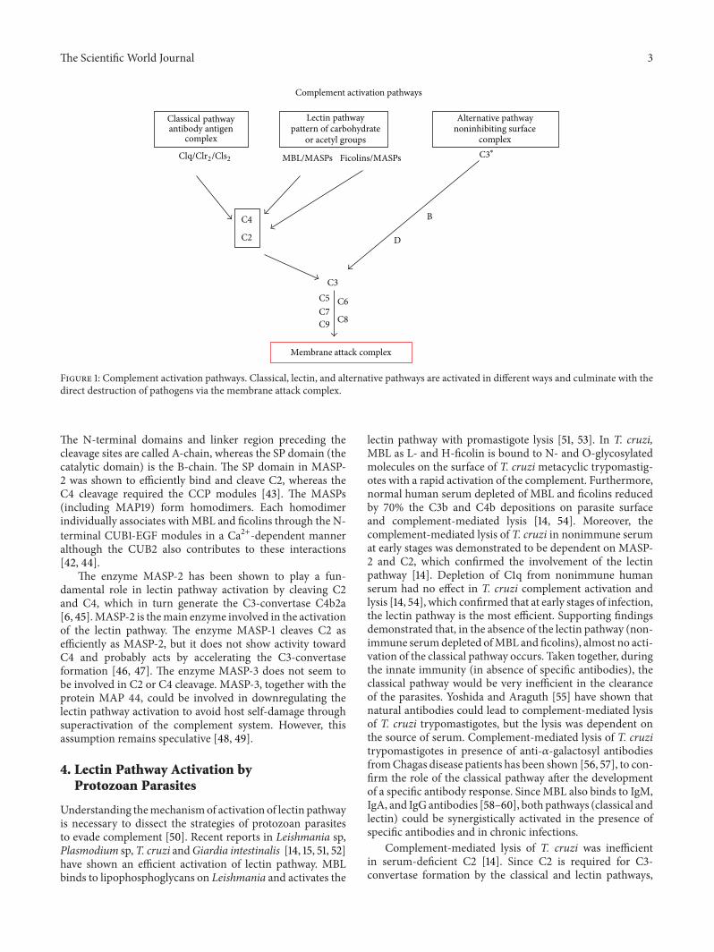
The lectin pathway is initiated when MBL (mannan-bindinglectin) or ficolins (L-H-M) bind to patterns of carbohydrates[1–3] or acetyl groups on the surface of protozoan, virus,fungi, or bacteria [4]. These patterns are found in threepairs of protease serine complexes, namely, mannan-bindinglectin-associated serine protease (MASPs) such as MASP-1,MASP-2, MASP-3, MAP-19, andMAP-44.When recognitionmolecules bind to a pattern, the MASPs are activated [5–7]. MASP (MASP-1 and -2) cleaves to the components C2and C4. The C4b fragment (product of C4 cleavage) bindsto the pathogen surface and associates with C2a to form theC3-convertase (C4b2a, similar to the C3-convertase of theclassical pathway) (see Figure 1). Once C3 cleaves, the C3bfragment can bind to the pathogen surface to activate thealternative pathway (Figure 1), or it can bind to the C4b2a(classical or lectin pathway C3-convertase) to form the C5-convertase (C4b2a3b). C3b can also bind to the alternativepathway C3-convertase, C3bBb, and form the C5-convertaseC3bBb3b. The C5-convertase cleaves C5 into C5a and C5b.C5b then binds to the pathogen surface to form an anchor,together with C6, C7, and C8, to support the formation of themembrane attack complex (MAC)with several C9molecules.
3. Components of the Lectin Pathway:Structural and Functional Considerations
3.1. MBL. MBL has a bouquet-like structure which is similarto the classical pathway initiator protein C1q [8, 9]. MBLis 32 kDa protein that forms oligomeric structures rangingfrom dimers to hexamers [8]. The protein is characterisedby a lectin (or carbohydrate-recognition domain (CRD)),a hydrophobic neck region, a collagenous region, and acysteine-rich N-terminal region [8, 10, 11]. Weis et al. [8]demonstrated that each domain binds to a Ca2+ ion andcoordinates the interactionwith the 3- and 4-hydroxyl groupsof specific sugars, such as GlcNAc, mannose, N-acetyl-mannosamine, fucose, and glucose. MBL binds to severalmicroorganisms including bacteria, viruses, and protozoaparasites such as Trypanosoma cruzi, Leishmania sp., andPlasmodium sp. [12–15].
3.2. Ficolins. Three members of the ficolin family of proteinshave been described in humans, such as L-ficolin (or ficolin-2), H-ficolin (or ficolin-3 or Hakata antigen), and M-ficolin(or ficolin-1) [16–19]. Although previous works [3, 6, 19,20] found the L- and H-ficolins in the human plasma inthe form of soluble proteins, the presence in serum of M-ficolin was only demonstrated recently [21]. This ficolin isnot hepatically synthesized and is able to have a similarcomplement activation. Ficolin proteins are composed of ashort N-terminal region with one or two cysteine residues,followed by a collagen-like domain, a short link region, and
a subsequent fibrinogen-like domain [1]. Ficolin proteinsform trimeric subunits through the binding of a collagen-likedomain [1, 22]. These subunits in turn assemble into activeoligomers through the binding of four subunits via disulfidebridges at the N-terminal regions [1, 23]. Ficolins recognizeacetylated carbohydrates through the C-terminal fibrinogen-like domain [1, 2, 24]. They bind mainly to the terminalGlcNAc residues, which are widely present on a variety ofpathogens but not in human cells [1, 3, 6, 25, 26].
L-ficolin is an oligomeric protein consisting of 35 kDasubunits [22, 27]. Similar to MBL and C1q, the overallstructure of L-ficolin resembles a “bouquet.” The protein isa tetramer consisting of 4 triple helices formed by 12 sub-units [22]. Although the protein binds mainly to acetylatedcarbohydrates [2, 26], it can also recognize other acetylatedmolecules, such as lipoteichoic acid on the surface of Gram-positive bacteria, peptidoglycan, surface lipopolysaccharideof Gram-negative bacteria, 𝛽-1,3-glucans of fungi, envelopeglycoconjugate of viruses, and glycosylated proteins on thesurface of T. cruzi [1, 14, 28–31].
H-ficolin is a protein of 34-kDa and thought to formoligomers of different sizes [3, 27, 32, 33]. H-ficolin hasbeen shown to bind to N-acetylgalactosamine (GalNAc) andfucose, but not to mannose and lactose [1, 3]. Althoughthe protein binds to GlcNAc, its affinity seems to be veryweak compared with that of L-ficolin [34]. Structural studiesdemonstrated that the fibrinogen-like domain of H-ficolincould not be cocrystallized with many acetylated compoundstested including GlcNAc [1]. H-ficolin has been shown tobind to lipopolysaccharides and recognizes surface exposedcarbohydrates on pathogens such as Salmonella typhimuriumand T. cruzi [6, 14, 35].
M-ficolin was initially reported to be expressed on thesurface of peripheral blood monocytes and promonocyticU937 cells [36]. More recently, the protein was reportedto be present in human plasma in significant amounts [25,37]. M-ficolin binds to acetylated carbohydrates, such asGlcNAc and GalNAc [36, 38, 39], and only the human ficolinrecognizes sialic acid [34]. In addition, M-ficolin has alsobeen shown to bind to C-reactive protein (CRP) and to fibrin[24, 40].The protein also recognizes Streptococcus sp. bacteriaand the protozoa parasite T. cruzi [14, 41]. M-ficolin canindependently activate the lectin pathway.
3.3. MASPs. MBL-associated serine proteases (MASPs) aresoluble serine proteases present in human serum. MASP-1, MASP-2, and MASP-3 are synthesised as proenzymeswith apparent molecular weights of 90 kDa, 74 kDa, and94 kDa, respectively. They are composed of an N-terminalCUB (Complement C1r/C1s, Uefg, Bmp1) domain (CUB1),followed by a Ca2+-binding type epidermal growth factor-(EGF-) like domain, a second CUB domain (CUB2), twocomplement control protein modules (CCP1 and CCP2), ashort linker, and a chymotrypsin-like serine protease (SP)domain [42]. Binding of the MBL-MASP or ficolin-MASPcomplexes to their ligands autoactivates the MASPs by pro-moting proteolytic cleavage of arginine-isoleucine residueswithin the linker region, resulting in two polypeptides heldtogether by disulfide bound and activation of the enzymes.
The Scientific World Journal 3
Complement activation pathways
Classical pathwayantibody antigen
complex
Lectin pathwaypattern of carbohydrate
or acetyl groups
Alternative pathwaynoninhibiting surface
complex
Membrane attack complex
MBL/MASPs Ficolins/MASPs C3∗
B
D
C3C5
C9C7
C6
C8
C4
C2
Clq/Clr2/Cls2
Figure 1: Complement activation pathways. Classical, lectin, and alternative pathways are activated in different ways and culminate with thedirect destruction of pathogens via the membrane attack complex.
The N-terminal domains and linker region preceding thecleavage sites are called A-chain, whereas the SP domain (thecatalytic domain) is the B-chain. The SP domain in MASP-2 was shown to efficiently bind and cleave C2, whereas theC4 cleavage required the CCP modules [43]. The MASPs(including MAP19) form homodimers. Each homodimerindividually associates with MBL and ficolins through the N-terminal CUB1-EGF modules in a Ca2+-dependent manneralthough the CUB2 also contributes to these interactions[42, 44].
The enzyme MASP-2 has been shown to play a fun-damental role in lectin pathway activation by cleaving C2and C4, which in turn generate the C3-convertase C4b2a[6, 45].MASP-2 is themain enzyme involved in the activationof the lectin pathway. The enzyme MASP-1 cleaves C2 asefficiently as MASP-2, but it does not show activity towardC4 and probably acts by accelerating the C3-convertaseformation [46, 47]. The enzyme MASP-3 does not seem tobe involved in C2 or C4 cleavage. MASP-3, together with theprotein MAP 44, could be involved in downregulating thelectin pathway activation to avoid host self-damage throughsuperactivation of the complement system. However, thisassumption remains speculative [48, 49].
4. Lectin Pathway Activation byProtozoan Parasites
Understanding themechanismof activation of lectin pathwayis necessary to dissect the strategies of protozoan parasitesto evade complement [50]. Recent reports in Leishmania sp,Plasmodium sp,T. cruzi andGiardia intestinalis [14, 15, 51, 52]have shown an efficient activation of lectin pathway. MBLbinds to lipophosphoglycans on Leishmania and activates the
lectin pathway with promastigote lysis [51, 53]. In T. cruzi,MBL as L- and H-ficolin is bound to N- and O-glycosylatedmolecules on the surface of T. cruzimetacyclic trypomastig-otes with a rapid activation of the complement. Furthermore,normal human serum depleted of MBL and ficolins reducedby 70% the C3b and C4b depositions on parasite surfaceand complement-mediated lysis [14, 54]. Moreover, thecomplement-mediated lysis of T. cruzi in nonimmune serumat early stages was demonstrated to be dependent on MASP-2 and C2, which confirmed the involvement of the lectinpathway [14]. Depletion of C1q from nonimmune humanserum had no effect in T. cruzi complement activation andlysis [14, 54], which confirmed that at early stages of infection,the lectin pathway is the most efficient. Supporting findingsdemonstrated that, in the absence of the lectin pathway (non-immune serumdepleted ofMBL and ficolins), almost no acti-vation of the classical pathway occurs. Taken together, duringthe innate immunity (in absence of specific antibodies), theclassical pathway would be very inefficient in the clearanceof the parasites. Yoshida and Araguth [55] have shown thatnatural antibodies could lead to complement-mediated lysisof T. cruzi trypomastigotes, but the lysis was dependent onthe source of serum. Complement-mediated lysis of T. cruzitrypomastigotes in presence of anti-𝛼-galactosyl antibodiesfromChagas disease patients has been shown [56, 57], to con-firm the role of the classical pathway after the developmentof a specific antibody response. SinceMBL also binds to IgM,IgA, and IgG antibodies [58–60], both pathways (classical andlectin) could be synergistically activated in the presence ofspecific antibodies and in chronic infections.
Complement-mediated lysis of T. cruzi was inefficientin serum-deficient C2 [14]. Since C2 is required for C3-convertase formation by the classical and lectin pathways,
4 The Scientific World Journal
but not for the alternative pathway, the alternative pathwayis inefficiently activated by T. cruzi [14]. The kinetics ofcomplement-mediated lysis with serum treated with EGTA(and MgCl
2) to chelate Ca2+ (classical and lectin pathways
are not functional under these conditions) confirmed thatthe alternative pathway is slowly activated by T. cruzi andLeishmania sp. [54, 61, 62]. Alternative pathway seems tohave less importance at the early activation by T. cruzitrypomastigotes because of the poor binding of factor b tothe parasite surface [63]. Other authors [62, 64] also showeda slow activation of the complement system in Leishmaniaspp., using serum-deficient C2. This result indicates that thealternative pathway activation can be delayed in the absenceof the classical and lectin pathways. Moreover, the activationof lectin pathway results in C3-convertase formation, whichin turn cleaves C3 into C3b to trigger the alternative pathway.This synergistic activation and the rapid deposition of C4 andC2 factors convert the lectin pathway in the most vital time[14, 54, 61].
Besides trypanosomatids, a variety of microorganismshave been shown to activate the lectin pathway. For example,Plasmodium sp. activates the lectin pathway [15]. MBL andficolins have also been shown to recognize Giardia sp.,resulting in CLP activation [52]. Human serum depletedof MBL and L-ficolins that failed to destroy the parasitesdemonstrated that the lectin pathway is involved to controlGiardia infections [52]. Several other pathogens includingbacteria and viruses were also shown to activate the lectinpathway [50, 65]. Altogether, the importance of the lectinpathway in pathogen recognition at initial stages of infectionis demonstrated.
5. Mechanisms of Complement Evasion byProtozoan Parasites
Several molecules involved in complement evasion have beendescribed in different pathogens and have been recentlysummarized in a review by [66]. Briefly, we can reinforcethat themainmechanism to complement evasion is to inhibitthe progression of the complement cascade to prevent theC3-convertase formation. T. cruzi uses different moleculesto block the complement cascade. Complement C2 receptorinhibitor trispanning (CRIT) is a surface molecule expressedat the metacyclic trypomastigotes stage (infectious state) andinhibits the C3-convertase [14, 61]. GP160, also called com-plement regulatory protein (CRP), is a molecule expressedin T. cruzi trypomastigotes that binds to C3b and C4b anddissociates the classical and alternative pathways of C3-convertase [67, 68].
Overexpression of the gene CRP andCRIT inT. cruzi epi-mastigote stage (the insect stage is sensitive to complement-mediated lysis) conferred complement resistance to theparasites, respectively [61]. Some authors [69] demonstratedthe complement inhibition before C3-convertase formation.Calreticulin (CRT) present at the surface of trypomastigotesis able to bind C1q and MBL to alter the classic andlectin pathways [70–72]. Similarly, an 87 to 93 kDa pro-tein identified on the surface of T. cruzi trypomastigotes,
called trypomastigotes-decay accelerating factor (T-DAF),was shown to share cDNA similarity to human DAF andinhibits parasite lysis [73]. Anothermolecule shown to inhibitthe C3-convertase formation in T. cruzi was the gp58/68[74]. Purified gp58/68 inhibited the formation of cell-boundand fluid-phase alternative pathway C3-convertase in a dose-dependent fashion. Different from DAF, gp58/68 was unableto dissociate the C3-convertase. However, the inhibition ofthe C3-convertase seems to be dependent on its associationwith factor B (rather than with C3b) [74]. Gp58/68 providestrypomastigotes with an additional potential mechanism toescape complement lysis by the alternative pathway.
In other trypanosomatids such as Leishmania sp., thesurface glycoprotein, also known as major surface protease,was shown to be the major C3b acceptor [75]. C3b bindingto GP63 results in its conversion to iC3b (inactive C3b) toprevent the C3-convertase formation on the parasite surface[76]. Furthermore, surface deposited iC3b is recognizedby the complement receptor 3 (also known as MAC-1),resulting in parasite phagocytosis by macrophages [76],in which the parasites can multiply and further develop.Deletion of the gene coding for the Golgi GDP-mannosetransporter LPG-2, required for the synthesis of surfacelipophosphoglycan, resulted in parasites highly susceptible tocomplement-mediated lysis [77]. LPG-2 null L. major wasincapable to establish macrophage infection and presenteddiminished mice infection. L. major releases the MAC (C5b-9) deposited on its surface during complement activation[78], to demonstrate that the parasite combines differentevasion mechanism to the complement system.
For the African trypanosome T. brucei, evasion of thecomplement system is dependent on the expression of a singlevariant surface glycoprotein (VSGs) that forms a coat on theparasite surface [79, 80].This parasite has a repertoire ofmorethan 1000 genes (and pseudogenes) coding for VSGs, whichare anchored to the parasite surface by glycosylphosphatidyli-nositol. Once the host develops a specific antibody responseagainst the surface VSG, a switch in gene expression occurs,resulting in a different surface coat, which allows the parasiteto escape the host antibody response [81]. Furthermore, theremoval of immune complexes deposited on the parasitesurface has also been shown to be dependent on VSGs [79,80]. Host immunoglobulins (Ig) form immune complexeswith VSG on the cell surface. They are rapidly removedby a hydrodynamic force generated by the parasite motilitythat results in the transfer of the immune complexes to theposterior cell pole of the cell, in which they are endocytosed.The backward movement of immune complexes on the cellsurface seems to require the forward parasite motility and tobe independent of endocytosis and actin function [80].
Complement receptors capable of inhibiting complementactivity have been shown in Schistosoma sp.A 97 kDa proteincalled paramyosin (also known as schistosoma complementinhibitory protein-1) was reported to bind to the complementcomponents C8 and C9 and inhibit C9 polymerization [82,83]. The protein CRIT (aforementioned to inhibit classicaland lectin pathway activation in T. cruzi and humans) wasfirst identified in a cDNA library of S. haematobium [84,85]. This protein was shown to be expressed on the surface
The Scientific World Journal 5
tegumental plasmamembrane and tegumental surface pits ofadult schistosomes [85]. The protein binds to C2 and inhibitsC3-convertase formation [86]. Schistosomeswere also shownto acquire the host protein DAF from erythrocytes [87].Schistosomes containing surface-bound DAF were able toevade complement-mediated lysis.
To summarize, protozoan parasites display several mech-anisms to evade the complement system. The parasite needsto survive to produce the infection. Protozoa invade the hostcell rapidly to be protected intracellularly. This strategy isused by some strains of T. cruzi. Other way to block thecomplement pathway is expressing receptor for complementon the parasite surface [50]. A broad strategy should be theexpression of a layer at the surface. This strategy seems tobe used by Leishmania with LPG and in T. brucei with VSGand the antigenic variation. Finally, the remotion of immunecomplex fromparasite surface seems to be important to evadethe complement and phagocytosis by opsonisation. Sheddingwas shown in Leishmania and in T. cruzi [88]. In addition, T.brucei would remove complement factors and antibodies toevade the innate immunity.
6. Deficiencies of the Lectin Pathway andSusceptibility to Parasitic Infections: Studiesin Human Populations
6.1. MBL. MBL may contribute to the control of parasitemiaduring malaria infections. MBL genotype and phenotypewere assessed in a prospective matched-control study ofGabonese children with malaria [89]. The 100 patients withsevere malaria both had significantly more frequently lowMBL levels, defined as <200 ng/mL (0.35 versus 0.19, 𝑃 =0.02) and a higher frequency of mutant alleles at codons54 and 57 (0.45 versus 0.31, 𝑃 = 0.04) compared to 100children with mild malaria. Matching was performed forsex, age, and provenance. These findings were confirmedin other study where the MB12 C missense mutation wasfound in 35% of healthy controls and in 42% of infectedbut asymptomatic children, in contrast to 46% of childrenwith severe malaria (𝑃 = 0.007) [15]. The populationattributable fraction of severe malaria cases to MBL2∗Cheterozygosity was estimated to be 17%. Similar findings wereobtained in a recent study to assess MBL2 haplotypes in262 Gabonese malaria patients, where haplotypes associatedwith low MBL levels were significantly more often found inchildren with severe malaria [90]. Interestingly, the authorsmeasured a combination of cytokines at admission andshowed that plasma MBL levels at admission correlatednegatively with proinflammatory cytokine/chemokines. Bycontrast, a different study investigating several SNPs inGabonese children with asymptomatic malaria did not finda significant association with MBL2 variants [91]. Garredet al. investigated MBL variant alleles in 551 children fromGhana in relation to Plasmodium falciparum.Nodifference inMBL genotype frequency was observed between infected andnoninfected children in their study. However, they observedsignificantly higher parasite counts and lower blood glucose
levels in patients who were homozygous for MBL variantalleles [92].
These data indicate that MBLmay act as disease modifier,but the precise mechanisms need to be elucidated. Althoughmost of the cases reported in bacterial infections have beenrelated with an MBL deficiency, the case of intracellularprotozoan seems to be different. In Leishmania [76], theinfection tomacrophages could be increased by opsonisation.Moreover, Santos et al. [93], in a case-control study, reinforcethe concept to show that an epidemical Brazilian popula-tion with visceral leishmaniasis contains high MBL serumlevels. Analyzing the genetic background of the population,mutant alleles with low MBL production were associatedwith protected patients. A recent study [94] showed thatin the same region a population with high-producing MBLgenotypes were associated with an increased risk of severevisceral leishmaniasis.
These findings were reproduced by a recent study in anAzerbaijan population in which alleles associated with highMBL levels were found more frequently in patients withvisceral leishmaniasis compared to healthy controls (𝑃 =0.03) [95]. Other examples of protozoa and in particularof Trypanosomatids family indicate contradictory resultscompared with the MBL/MASP-2 serum levels in Chagasdisease patients. High levels of MBL were associated withsevere cardiomyopathy, probably because of the proinflam-matory activities ofMBL [96]. However, another recent study[97] showed a higher presence of low-producing MASP-2 genotypes in patients with cardiomyopathy in chronicChagas disease.We have included a table with the deficienciesassociated with complement factors (Table 1).
6.2. Ficolins. Contrary to the large number of studies thatinvestigates MBL geno- and phenotype in different clinicalsettings, the role of the closely related ficolins remainslargely unknown. M-ficolin, the only lectin pathway memberproduced by leukocytes and not by hepatocytes, was onlyrecently discovered to be present in significant amounts inhuman serum [21, 98]. M-ficolin serum concentrations mayreflect phagocyte activation in the course of infection [41], butsubstantial research is necessary to understand this patternrecognition receptor fully.
H-ficolin deficiency is extremely rare. Two reports [99,100] described the occurrence of the deficiency in a patientwith repeated infections and in a neonate with necrotizingenterocolitis, respectively. Moreover, increased infectionswith gram + have been associated with neonates with lowerH-ficolin serum levels [101].
This year, some clinical data have been associated withL-ficolin deficiency and parasite infections. Ouf et al. [102]showed that ficolin-2 (l-ficolin) levels and FCN2 geneticpolymorphism could be important as a susceptibility factorin schistosomosis. In Leishmaniosis [103], some evidenceindicated a haplotype of ficolin-2 with susceptibility factor.
A later study that assessed ficolin SNPs in a largerpopulation-based cohort did not find a correlation withrecurrent respiratory infections [104]. Recently, Kilpatrick etal. showed lower L-ficolin concentrations in patients with
6 The Scientific World Journal
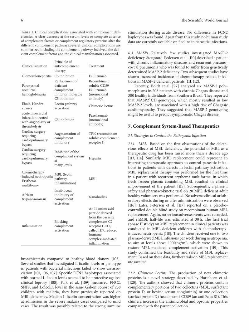
Table 1: Clinical complications associated with complement defi-ciencies. A clear decrease at the serum levels or complete absenceof complement factors or complement regulatory proteins alter thedifferent complement pathways.Several clinical complications aresummarized including the complement pathway involved, the defi-cient complement factor and the clinical manifestation associated.
Clinical situationPrinciple ofanticomplementtherapy
Treatment
Glomerulonephritis C5 inhibition Eculizumab
Paroxysmalnocturnalhemoglobinuria
Replacement ofdeficientcomplementinhibitor moleculeC5 inhibition
Recombinantsoluble CD59Eculizumab(monoclonalantibody)
Ebola, Hendraviruses
Lectin pathwayactivation Chimeric lectins
acute myocardialinfarction treatedwith angioplasty orthrombolysis
C5 inhibitionPexelizumab(monoclonalantibody)
Cardiac surgeryrequiringcardiopulmonarybypass
Augmentation ofcomplementinhibitory
TP10 (recombinantsoluble complementreceptor 1)
Cardiac surgeryrequiringcardiopulmonarybypass
Inhibition of thecomplement systematmany levels
Heparin
Chemotherapyinduced neutropeniaErythemamultiforme
MBL (lectinpathway,inflammation)
MBL
Africantrypanosomiasis
Inhibit coatinhibitory ofcomplementactivation
Nanobodies
InflammationBlockingcomplementactivation
An 11 amino acidpeptide derivedfrom the parasitecomplement C2receptor CRIT,called H17, reducedimmunecomplex-mediatedinflammation
bronchiectasis compared to healthy blood donors [105].Several studies that investigated L-ficolin levels or genotypein patients with bacterial infections failed to show an asso-ciation [101, 106, 107]. Specific FCN2 haplotypes associatedwith normal L-ficolin levels seemed to be protective againstclinical leprosy [108]. Faik et al. [109] measured FNC2,SNPs, and L-ficolin level in the same Gabon cohort of 238children with malaria, they have previously reported onMBL deficiency. Median L-ficolin concentration was higherat admission in the severe malaria cases compared to mildcases. The result was possibly related to the strong immune
stimulation during acute disease. No difference in FCN2haplotypes was found. Apart from this study, no human studydata are currently available on ficolins in parasitic infections.
6.3. MASPs. Relatively few studies investigated MASP-2deficiency. Stengaard-Pedersen et al. [110] described a patientwith chronic inflammatory diseases and recurrent pneumo-coccal pneumonia who was found to suffer from geneticallydeterminedMASP-2 deficiency. Two subsequent studies haveshown increased incidence of chemotherapy-related infec-tions in MASP-2 deficient patients [111, 112].
Recently, Boldt et al. [97] analyzed six MASP-2 poly-morphisms in 208 patients with chronic Chagas disease and300 healthy individuals from Southern Brazil. They reportedthat MASP2∗CD genotypes, which mostly resulted in lowMASP-2 levels, are associated with a high risk of Chagasiccardiomyopathy. They suggested that MASP-2 genotypingmight be useful to predict symptomatic Chagas disease.
7. Complement System-Based Therapeutics
7.1. Strategies to Control the Pathogenic Infection
7.1.1. MBL. Based on the first observations of the delete-rious effects of MBL deficiency, the potential of MBL as atherapeutic drug has been raised more than a decade ago[113, 114]. Similarly, MBL replacement could represent aninteresting therapeutic approach to control parasitic infec-tions in patients with defects in lectin pathway activation.MBL replacement therapy was performed for the first timein a patient with recurrent erythema multiforme, in whichfresh frozen plasma containing MBL resulted in clinicalimprovement of the patient [115]. Subsequently, a phase Isafety and pharmacokinetic trial on 20 MBL deficient adulthealthy volunteers was performed. No adverse clinical or lab-oratory effects during or after administration were observed[116]. Later, Petersen et al. [117] reported on a placebo-controlled double blind study on recombinant human MBLreplacement. Again, no serious adverse events were recorded,and rhMBL half-life was estimated at 30 h. The first trial(phase II study) on MBL replacement in clinical patients wasconducted in MBL deficient children with chemotherapy-induced neutropenia [118]. The children received one to twoplasma-derivedMBL infusions per week during neutropenia,to aim at levels above 1000 ng/mL, which were shown torestore MBL-mediated complement activation [119]. Thisstudy confirmed the feasibility and safety of MBL replace-ment. Based on these data, further trials onMBL replacementare awaited.
7.1.2. Chimeric Lectins. The production of new chimericproteins is a novel strategy described by Hartshorn et al.[120]. The authors showed that chimeric proteins containcomplementary portions of two collectins (MBL, surfactantprotein D, or bovine serum conglutinin) or one collection(surfact protein D) fused to anti-CD89 (an anti Fc-𝛼RI).Thischimera increases the antimicrobial and opsonic propertiescompared with the parent collection
The Scientific World Journal 7
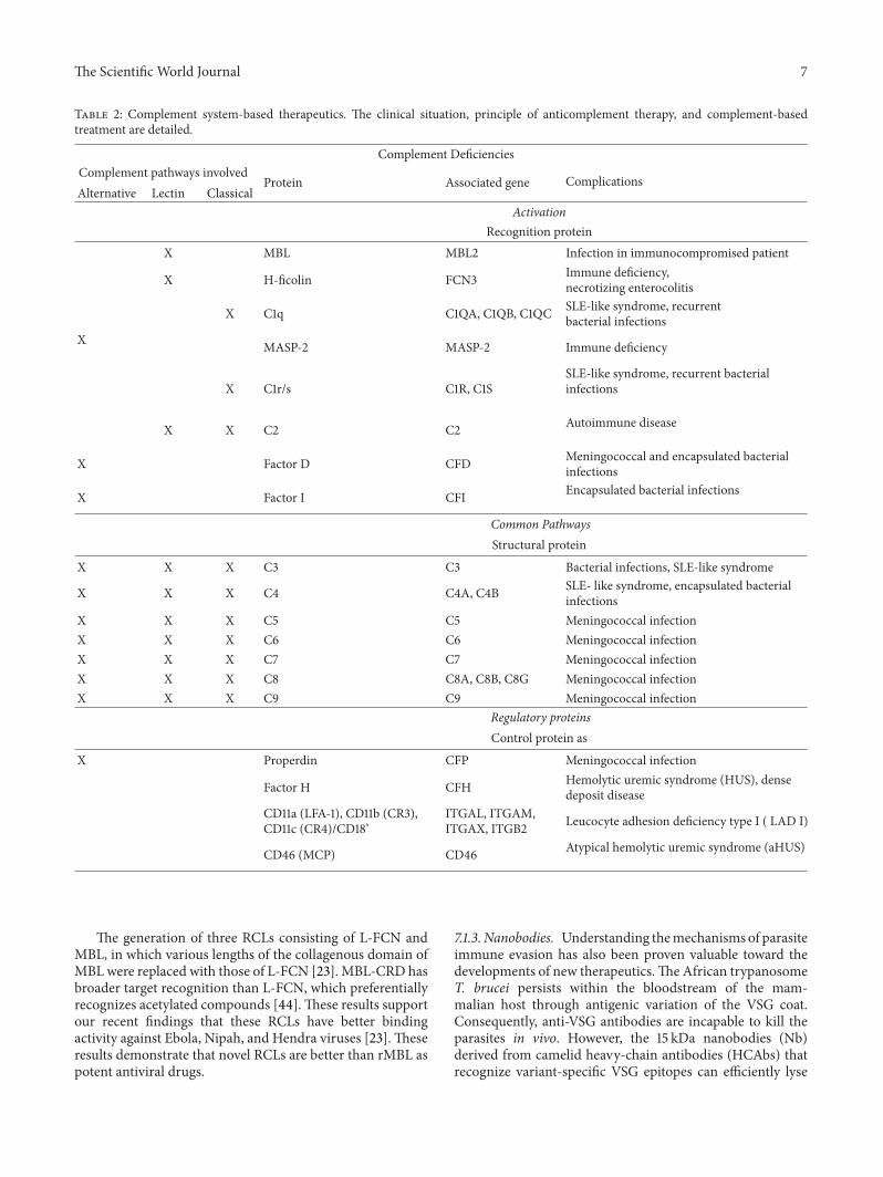
Table 2: Complement system-based therapeutics. The clinical situation, principle of anticomplement therapy, and complement-basedtreatment are detailed.
Complement DeficienciesComplement pathways involved
Protein Associated gene ComplicationsAlternative Lectin Classical
ActivationRecognition protein
X MBL MBL2 Infection in immunocompromised patient
X H-ficolin FCN3 Immune deficiency,necrotizing enterocolitis
X C1q C1QA, C1QB, C1QC SLE-like syndrome, recurrentbacterial infections
X MASP-2 MASP-2 Immune deficiency
X C1r/s C1R, C1SSLE-like syndrome, recurrent bacterialinfections
X X C2 C2 Autoimmune disease
X Factor D CFD Meningococcal and encapsulated bacterialinfections
X Factor I CFI Encapsulated bacterial infections
Common PathwaysStructural protein
X X X C3 C3 Bacterial infections, SLE-like syndrome
X X X C4 C4A, C4B SLE- like syndrome, encapsulated bacterialinfections
X X X C5 C5 Meningococcal infectionX X X C6 C6 Meningococcal infectionX X X C7 C7 Meningococcal infectionX X X C8 C8A, C8B, C8G Meningococcal infectionX X X C9 C9 Meningococcal infection
Regulatory proteinsControl protein as
X Properdin CFP Meningococcal infection
Factor H CFH Hemolytic uremic syndrome (HUS), densedeposit disease
CD11a (LFA-1), CD11b (CR3),CD11c (CR4)/CD18’
ITGAL, ITGAM,ITGAX, ITGB2 Leucocyte adhesion deficiency type I ( LAD I)
CD46 (MCP) CD46 Atypical hemolytic uremic syndrome (aHUS)
The generation of three RCLs consisting of L-FCN andMBL, in which various lengths of the collagenous domain ofMBL were replaced with those of L-FCN [23]. MBL-CRD hasbroader target recognition than L-FCN, which preferentiallyrecognizes acetylated compounds [44].These results supportour recent findings that these RCLs have better bindingactivity against Ebola, Nipah, and Hendra viruses [23].Theseresults demonstrate that novel RCLs are better than rMBL aspotent antiviral drugs.
7.1.3. Nanobodies. Understanding themechanisms of parasiteimmune evasion has also been proven valuable toward thedevelopments of new therapeutics.The African trypanosomeT. brucei persists within the bloodstream of the mam-malian host through antigenic variation of the VSG coat.Consequently, anti-VSG antibodies are incapable to kill theparasites in vivo. However, the 15 kDa nanobodies (Nb)derived from camelid heavy-chain antibodies (HCAbs) thatrecognize variant-specific VSG epitopes can efficiently lyse
8 The Scientific World Journal
trypanosomes both in vitro and in vivo [121]. Nanobodies-mediated lysis of trypanosomes results in the enlargementof the parasite flagellar pocket, blockade of endocytosis, andsevere metabolic perturbations culminating in cell death.The generation of low molecular weight VSG-specific try-panolytic nanobodies offers a new opportunity to developnovel trypanosomiasis therapeutics.
7.1.4. Peptides. Parasite-derived complement receptors havealso been proposed to control complement-mediated hostdamage. An 11 amino acid peptide derived from the parasitecomplement C2 receptor CRIT, called H17, was shown toreduce immune complex-mediated inflammation (dermalreversed passive Arthus reaction) inmice, in vivo [122]. Uponthe intradermal injection of CRIT-H17, a 41% reduction inoedema and haemorrhage, a 72% reduction in neutrophilinflux, and a reduced C3 deposition were observed. Further-more, administration of intravenousH17 at a 1mg/kg dose ledto reduced inflammation by 31%, to demonstrate that CRIT-H17 is a potential therapeutic agent against complement-mediated inflammatory tissue destruction [122].
7.2. Strategies Used in Other Clinical States. The idea touse complement inhibition as therapy for various diseasesand conditions, such as organ transplantation, ischemia-reperfusion injury, coronary artery disease, myocardialinfarction, stroke, cancer, immunosuppression, paroxys-mal nocturnal hematuria, glomerulonephritis, rheumatoidarthritis, and acute respiratory distress syndrome, wasreviewed recently [123].
Most of the principles of anticomplement therapy arebased on the C5 inhibition [124], the replacement of deficientcomplement inhibitor molecule CD 59 [125], and the aug-ment of complement inhibitory molecules [126].
We have summarized all the strategies from Sections 7.1and 7.2 in Table 2.
8. Conclusion
Understanding pathogen and host interaction is a key aspectin the development of new therapeutics against infectiousdiseases. Although many things remain to be investigatedon the molecular basis of parasitic infection, the currentknowledge in complement system highlights the importanceof the lectin pathway as a key mediator of host defenseagainst parasitic infections and its potential for a therapeutictarget in the control of infection. Some reports have pointedout the importance of the activation of CLP in protozoa, toshow that MBL and ficolins require seconds to activate thecomplement cascade. Probably, the MBL and ficolins presentin large quantities in normal human serum guarantee anefficient pathway activation of lectins. However, two aspectsare surprising: the extreme diversity of evasion mechanismsdeployed by the parasites to produce infection and the highfrequency of genetic deficiencies in MBL and complementfactors. Although complement-based therapies are promisingin some specific clinical states [123], they require a morecomprehensive understanding on the characteristics of acti-vation and resistance to each model in infectious diseases
to formulate a treatment. For example, for Neisseria spp.and Leishmania spp. [76], the complement factors might beopsonising. Providing input to the parasitic invasion intoeukaryotic cells is contradictory to the MBL-ficolin chimerasthat block virus entry to eukaryotic cells [125]. More detailedknowledge on early complement activation, development ofspecific inhibitors, and trials on human population should benext steps to block the pathogen invasion to avoid infection.Therapies to compensate factor deficiencies are welcomewhen the case is studied carefully.
The complement system could be the central point tocontrol the protozoa, and new chemotherapeutic alternativesshould improve the current situation.
Acknowledgments
The authors are very grateful to Programa de ParasitologiaBasica/CAPES. I. Evans-Osses is a recipient of CAPES Fel-lowship and M. I. Ramirez is a CNPq fellow.
References
[1] V. Garlatti, N. Belloy, L. Martin et al., “Structural insightsinto the innate immune recognition specificities of L- and H-ficolins,” EMBO Journal, vol. 26, no. 2, pp. 623–633, 2007.
[2] A. Krarup, D. A. Mitchell, and R. B. Sim, “Recognition ofacetylated oligosaccharides by human L-ficolin,” ImmunologyLetters, vol. 118, no. 2, pp. 152–156, 2008.
[3] M. Matsushita, M. Kuraya, N. Hamasaki, M. Tsujimura, H.Shiraki, and T. Fujita, “Activation of the lectin complementpathway byH-ficolin (Hakata antigen),” Journal of Immunology,vol. 168, no. 7, pp. 3502–3506, 2002.
[4] V. L. Runza, W. Schwaeble, and D. N. Mannel, “Ficolins: novelpattern recognition molecules of the innate immune response,”Immunobiology, vol. 213, no. 3-4, pp. 297–306, 2008.
[5] T. Vorup-Jensen, S. V. Petersen, A. G. Hansen et al., “Dis-tinct pathways of mannan-binding lectin (MBL)- and C1-complex auatoactivation revealed by reconstitution of MBLwith recombinant MBL- associated serine protease-2,” Journalof Immunology, vol. 165, no. 4, pp. 2093–2100, 2000.
[6] M. Matsushita, Y. Endo, and T. Fujita, “Cutting edge:complement-activating complex of ficolin and mannose-binding lectin-associated serine protease,” Journal ofImmunology, vol. 164, no. 5, pp. 2281–2284, 2000.
[7] C. M. Stover, S. Thiel, M. Thelen et al., “Two constituents ofthe initiation complex of the mannan-binding lectin activationpathway of complement are encoded by a single structuralgene,” Journal of Immunology, vol. 162, no. 6, pp. 3481–3490,1999.
[8] W. I. Weis, K. Drickamer, and W. A. Hendrickson, “Structureof a C-type mannose-binding protein complexed with anoligosaccharide,” Nature, vol. 360, no. 6400, pp. 127–134, 1992.
[9] M. Wong and R. B. Sim, “Comparison of the complement sys-tem protein complexes formed by Clq and MBL,” BiochemicalSociety Transactions, vol. 25, no. 1, p. 41, 1997.
[10] M. S. Quesenberry and K. Drickamer, “Determination of theminimum carbohydrate-recognition domain in two C-typeanimal lectins,” Glycobiology, vol. 1, no. 6, pp. 615–621, 1991.
[11] M. S. Quesenberry and K. Drickamer, “Role of conserved andnonconserved residues in the Ca2+-dependent carbohydrate-recognition domain of a rat mannose-binding protein. Analysis
The Scientific World Journal 9
by random cassette mutagenesis,” Journal of Biological Chem-istry, vol. 267, no. 15, pp. 10831–10841, 1992.
[12] T. Kawai, T. Kase, Y. Suzuki et al., “Anti-influenza A virusactivities of mannan-binding lectins and bovine conglutinin,”Journal of VeterinaryMedical Science, vol. 69, no. 2, pp. 221–224,2007.
[13] D. L. Jack,G.A. Jarvis, C. L. Booth,M.W.Turner, andN. J. Klein,“Mannose-binding lectin accelerates complement activationand increases serum killing of Neisseria meningitidis serogroupC,” Journal of Infectious Diseases, vol. 184, no. 7, pp. 836–845,2001.
[14] I. D. S. Cestari, A. Krarup, R. B. Sim, J. M. Inal, and M.I. Ramirez, “Role of early lectin pathway activation in thecomplement-mediated killing ofTrypanosoma cruzi,”MolecularImmunology, vol. 47, no. 2-3, pp. 426–437, 2009.
[15] V. Holmberg, F. Schuster, E. Dietz et al., “Mannose-bindinglectin variant associated with severe malaria in young Africanchildren,” Microbes and Infection, vol. 10, no. 4, pp. 342–348,2008.
[16] J. Lu, Y. Le, O. L. Kon, J. Chan, and S. H. Lee, “Biosynthesisof human ficolin, and Escherichia coli-binding protein, bymonocytes: comparison with the synthesis of two macrophage-specific proteins, C1q and the mannose receptor,” Immunology,vol. 89, no. 2, pp. 289–294, 1996.
[17] S. Harumiya, K. Takeda, T. Sugiura et al., “Characterization officolins as novel elastin-binding proteins andmolecular cloningof human ficolin-1,” Journal of Biochemistry, vol. 120, no. 4, pp.745–751, 1996.
[18] Y. Endo, Y. Sato, M. Matsushita, and T. Fujita, “Cloning andcharacterization of the human lectin P35 gene and its relatedgene,” Genomics, vol. 36, no. 3, pp. 515–521, 1996.
[19] M. Matsushita, Y. Endo, S. Taira et al., “A novel humanserum lectin with collagen- and fibrinogen-like domains thatfunctions as an opsonin,” Journal of Biological Chemistry, vol.271, no. 5, pp. 2448–2454, 1996.
[20] Y. Le, S. H. Lee, O. L. Kon, and J. Lu, “Human L-ficolin: plasmalevels, sugar specificity, and assignment of its lectin activity tothe fibrinogen-like (FBG) domain,” FEBS Letters, vol. 425, no.2, pp. 367–370, 1998.
[21] T. Wittenborn, S. Thiel, L. Jensen, H. J. Nielsen, and J. C.Jensenius, “Characteristics and biological variations of M-ficolin, a pattern recognition molecule, in plasma,” Journal ofInnate Immunity, vol. 2, no. 2, pp. 167–180, 2010.
[22] T. Hummelshoj, N. M. Thielens, H. O. Madsen, G. J. Arlaud,R. B. Sim, and P. Garred, “Molecular organization of humanFicolin-2,” Molecular Immunology, vol. 44, no. 4, pp. 401–411,2007.
[23] T. Ohashi and H. P. Erickson, “The disulfide bonding pattern inficolin multimers,” Journal of Biological Chemistry, vol. 279, no.8, pp. 6534–6539, 2004.
[24] M. Tanio, S. Kondo, S. Sugio, and T. Kohno, “Trivalent recog-nition unit of innate immunity system: crystal structure oftrimeric human M-ficolin fibrinogen-like domain,” Journal ofBiological Chemistry, vol. 282, no. 6, pp. 3889–3895, 2007.
[25] Y. Liu, Y. Endo, D. Iwaki et al., “Human M-ficolin is a secretoryprotein that activates the lectin complement pathway,” Journalof Immunology, vol. 175, no. 5, pp. 3150–3156, 2005.
[26] A. Krarup, S. Thiel, A. Hansen, T. Fujita, and J. C. Jensenius,“L-ficolin is a pattern recognition molecule specific for acetylgroups,” Journal of Biological Chemistry, vol. 279, no. 46, pp.47513–47519, 2004.
[27] T. Ohashi and H. P. Erickson, “Oligomeric structure and tissuedistribution of ficolins frommouse, pig and human,”Archives ofBiochemistry and Biophysics, vol. 360, no. 2, pp. 223–232, 1998.
[28] N. J. Lynch, S. Roscher, T. Hartung et al., “L-ficolin specificallybinds to lipoteichoic acid, a cell wall constituent of Gram-positive bacteria, and activates the lectin pathway of comple-ment,” Journal of Immunology, vol. 172, no. 2, pp. 1198–1202,2004.
[29] A. M. Nahid and S. Sugii, “Binding of porcine ficolin-𝛼 tolipopolysaccharides from Gram-negative bacteria and lipotei-choic acids from Gram-positive bacteria,” Developmental andComparative Immunology, vol. 30, no. 3, pp. 335–343, 2006.
[30] J. Liu,M. A.M. Ali, Y. Shi et al., “Specifically binding of L-ficolinto N-glycans of HCV envelope glycoproteins E1 and E2 leadsto complement activation,”Cellular andMolecular Immunology,vol. 6, no. 4, pp. 235–244, 2009.
[31] Y. G. Ma, M. Y. Cho, M. Zhao et al., “Human mannose-bindinglectin and L-ficolin function as specific pattern recognitionproteins in the lectin activation pathway of complement,”Journal of Biological Chemistry, vol. 279, no. 24, pp. 25307–25312,2004.
[32] M. Kuraya, M. Matsushita, Y. Endo, S. Thiel, and T. Fujita,“Expression of H-ficolin/Hakata antigen, mannose-bindinglectin-associated serine protease (MASP)-1 and MASP-3 byhuman glioma cell line T98G,” International Immunology, vol.15, no. 1, pp. 109–117, 2003.
[33] T. Ohashi and H. P. Erickson, “Two oligomeric forms of plasmaficolin have differential lectin activity,” Journal of BiologicalChemistry, vol. 272, no. 22, pp. 14220–14226, 1997.
[34] E.Gout, V.Garlatti, D. F. Smith et al., “Carbohydrate recognitionproperties of human ficolins: glycan array screening reveals thesialic acid binding specificity of M-ficolin,” Journal of BiologicalChemistry, vol. 285, no. 9, pp. 6612–6622, 2010.
[35] A. Swierzko, J. Lukasiewicz, M. Cedzynski et al., “New func-tional ligands for ficolin-3 among lipopolysaccharides ofHafniaalvei,” Glycobiology, vol. 22, no. 2, pp. 267–280, 2012.
[36] C. Teh, Y. Le, S. H. Lee, and J. Lu, “M-ficolin is expressed onmonocytes and is a lectin binding to N-acetyl-D-glucosamineand mediates monocyte adhesion and phagocytosis ofEscherichia coli,” Immunology, vol. 101, no. 2, pp. 225–232, 2000.
[37] C. Honore, S. Rørvig, L. Munthe-Fog et al., “The innatepattern recognition molecule Ficolin-1 is secreted by mono-cytes/macrophages and is circulating in human plasma,”Molec-ular Immunology, vol. 45, no. 10, pp. 2782–2789, 2008.
[38] V. Garlatti, L. Martin, E. Gout et al., “Structural basis forinnate immune sensing by M-ficolin and its control by apH-dependent conformational switch,” Journal of BiologicalChemistry, vol. 282, no. 49, pp. 35814–35820, 2007.
[39] P. D. Frederiksen, S.Thiel, C. B. Larsen, and J. C. Jensenius, “M-ficolin, an innate immune defence molecule, binds patterns ofacetyl groups and activates complement,” Scandinavian Journalof Immunology, vol. 62, no. 5, pp. 462–473, 2005.
[40] M. Tanio, K. Wakamatsu, and T. Kohno, “Binding site of C-reactive protein on M-ficolin,” Molecular Immunology, vol. 47,no. 2-3, pp. 215–221, 2009.
[41] T. R. Kjaer, A. G. Hansen, U. B. S. Sørensen, O. Nielsen, S.Thiel,and J. C. Jensenius, “Investigations on the pattern recognitionmolecule M-ficolin: quantitative aspects of bacterial bindingand leukocyte association,” Journal of Leukocyte Biology, vol. 90,no. 3, pp. 425–437, 2011.
10 The Scientific World Journal
[42] H. Feinberg, J. C. M. Uitdehaag, J. M. Davies, R. Wallis, K.Drickamer, andW. I.Weis, “Crystal structure of the CUB1-EGF-CUB2 region of mannose-binding protein associated serineprotease-2,” EMBO Journal, vol. 22, no. 10, pp. 2348–2359, 2003.
[43] V. Rossi, I. Bally, N. M. Thielens, A. F. Esser, and G. J. Arlaud,“Baculovirus-mediated expression of truncated modular frag-ments from the catalytic region of human complement serineprotease C1s. Evidence for the involvement of both complementcontrol protein modules in the recognition of the C4 proteinsubstrate,” Journal of Biological Chemistry, vol. 273, no. 2, pp.1232–1239, 1998.
[44] R. Wallis and R. B. Dodd, “Interaction of mannose-bindingprotein with associated serine proteases. Effects of naturallyoccurring mutations,” Journal of Biological Chemistry, vol. 275,no. 40, pp. 30962–30969, 2000.
[45] S. Thiel, T. Vorup-Jensen, C. M. Stover et al., “A second serineprotease associated with mannan-binding lectin that activatescomplement,” Nature, vol. 386, no. 6624, pp. 506–510, 1997.
[46] J. S. Presanis, K. Hajela, G. Ambrus, P. Gal, and R. B. Sim,“Differential substrate and inhibitor profiles for humanMASP-1and MASP-2,” Molecular Immunology, vol. 40, no. 13, pp. 921–929, 2004.
[47] G. Ambrus, P. Gal, M. Kojima et al., “Natural substrates andinhibitors ofmannan-binding lectin-associated serine protease-1 and -2: a study on recombinant catalytic fragments,” Journal ofImmunology, vol. 170, no. 3, pp. 1374–1382, 2003.
[48] M. Takahashi, D. Iwaki, K. Kanno et al., “Mannose-bindinglectin (MBL)-associated serine protease (MASP)-1 contributesto activation of the lectin complement pathway,” Journal ofImmunology, vol. 180, no. 9, pp. 6132–6138, 2008.
[49] M. O. Skjoedt, T. Hummelshoj, Y. Palarasah et al., “A novelmannose-binding lectin/ficolin-associated protein is highlyexpressed in heart and skeletal muscle tissues and inhibitscomplement activation,” Journal of Biological Chemistry, vol.285, no. 11, pp. 8234–8243, 2010.
[50] J. D. Lambris, D. Ricklin, and B. V. Geisbrecht, “Complementevasion by human pathogens,” Nature Reviews Microbiology,vol. 6, no. 2, pp. 132–142, 2008.
[51] A. R. Ambrosio and I. J. T. De Messias-Reason, “Leishmania(Viannia) braziliensis: interaction of mannose-binding lectinwith surface glycoconjugates and complement activation. Anantibody-independent defence mechanism,” Parasite Immunol-ogy, vol. 27, no. 9, pp. 333–340, 2005.
[52] I. Evans-Osses, E. A. Ansa-Addo, J. M. Inal, and M. I. Ramirez,“Involvement of lectin pathway activation in the complementkilling of Giardia intestinalis,” Biochemical and BiophysicalResearch Communications, vol. 395, no. 3, pp. 382–386, 2010.
[53] P. J. Green, T. Feizi, M. S. Stoll, S. Thiel, A. Prescott, and M. J.McConville, “Recognition of the major cell surface glycocon-jugates of Leishmania parasites by the human serum mannan-binding protein,” Molecular and Biochemical Parasitology, vol.66, no. 2, pp. 319–328, 1994.
[54] I. Cestari and M. I. Ramirez, “Inefficient complement systemclearance of Trypanosoma cruzi metacyclic trypomastigotesenables resistant strains to invade eukaryotic cells,” PloS One,vol. 5, no. 3, Article ID e9721, 2010.
[55] N. Yoshida and M. F. Araguth, “Trypanolytic activity andantibodies to metacyclic trypomastigotes of Trypanosoma cruziin non-Chagasic human sera,” Parasite Immunology, vol. 9, no.3, pp. 389–393, 1987.
[56] I. C. Almeida, M. A. J. Ferguson, S. Schenkman, andL. R. Travassos, “Lytic anti-𝛼-galactosyl antibodies from
patients with chronic Chagas’ disease recognize novel O-linkedoligosaccharides on mucin-like glycosyl-phosphatidylinositol-anchored glycoproteins of Trypanosoma cruzi,” BiochemicalJournal, vol. 304, no. 3, pp. 793–802, 1994.
[57] I. C. Almeida, S. R. Milani, P. A. J. Gorin, and L. R. Travassos,“Complement-mediated lysis of Trypanosoma cruzi trypo-mastigotes by human anti-𝛼-galactosyl antibodies,” Journal ofImmunology, vol. 146, no. 7, pp. 2394–2400, 1991.
[58] J. N. Arnold, M. R. Wormald, D. M. Suter et al., “Human serumIgM glycosylation: identification of glycoforms that can bindto Mannan-binding lectin,” Journal of Biological Chemistry, vol.280, no. 32, pp. 29080–29087, 2005.
[59] R. Malhotra, M. R. Wormald, P. M. Rudd, P. B. Fischer, R. A.Dwek, and R. B. Sim, “Glycosylation changes of IgG associatedwith rheumatoid arthritis can activate complement via themannose-binding protein,” Nature Medicine, vol. 1, no. 3, pp.237–243, 1995.
[60] A. Roos, L. H. Bouwman, D. J. Van Gijlswijk-Janssen, M.C. Faber-Krol, G. L. Stahl, and M. R. Daha, “Human IgAactivates the complement system via the Mannan-Bindinglectin pathway,” Journal of Immunology, vol. 167, no. 5, pp. 2861–2868, 2001.
[61] I. D. S. Cestari, I. Evans-Osses, J. C. Freitas, J. M. Inal, andM. I. Ramirez, “Complement C2 receptor inhibitor trispanningconfers an increased ability to resist complement-mediated lysisin Trypanosoma cruzi,” Journal of Infectious Diseases, vol. 198,no. 9, pp. 1276–1283, 2008.
[62] M. Domınguez, I. Moreno, M. Lopez-Trascasa, and A. Torano,“Complement interaction with trypanosomatid promastigotesin normal human serum,” Journal of ExperimentalMedicine, vol.195, no. 4, pp. 451–459, 2002.
[63] K. Joiner, A. Sher, T. Gaither, and C. Hammer, “Evasion ofalternative complement pathway by Trypanosoma cruzi resultsfrom inefficient binding of factor B,” Proceedings of the NationalAcademy of Sciences of the United States of America, vol. 83, no.17, pp. 6593–6597, 1986.
[64] I. Moreno, R. Molina, A. Torano, E. Laurin, E. Garcıa, and M.Domınguez, “Comparative real-time kinetic analysis of humancomplement killing of Leishmania infantum promastigotesderived from axenic culture or from Phlebotomus perniciosus,”Microbes and Infection, vol. 9, no. 14-15, pp. 1574–1580, 2007.
[65] K. A. Stoermer and T. E. Morrison, “Complement and viralpathogenesis,” Virology, vol. 411, no. 2, pp. 362–373, 2011.
[66] I. Cestari, E. Ansa-Addo, P. Deolindo, J. M. Inal, and M. I.Ramirez, “Trypanosoma cruzi immune evasion mediated byhost cell-derived microvesicles,” Journal of Immunology, vol.188, no. 4, pp. 1942–1952, 2012.
[67] K. A. Norris, “Stable transfection of Trypanosoma cruzi epi-mastigotes with the trypomastigote-specific complement regu-latory protein cDNA confers complement resistance,” Infectionand Immunity, vol. 66, no. 6, pp. 2460–2465, 1998.
[68] K. A. Norris, G. Harth, and M. So, “Purification of a Try-panosoma cruzi membrane glycoprotein which elicits lyticantibodies,” Infection and Immunity, vol. 57, no. 8, pp. 2372–2377, 1989.
[69] V. Ferreira, M. C. Molina, C. Valck et al., “Role of calreticulinfrom parasites in its interaction with vertebrate hosts,”Molecu-lar Immunology, vol. 40, no. 17, pp. 1279–1291, 2004.
[70] V. Ferreira, C. Valck, G. Sanchez et al., “The classical activationpathway of the human complement system is specificallyinhibited by calreticulin from Trypanosoma cruzi,” Journal ofImmunology, vol. 172, no. 5, pp. 3042–3050, 2004.
The Scientific World Journal 11
[71] G. Ramırez, C. Valck, M. C. Molina et al., “Trypanosoma cruzicalreticulin: a novel virulence factor that binds complement C1on the parasite surface and promotes infectivity,” Immunobiol-ogy, vol. 216, no. 1-2, pp. 265–273, 2011.
[72] C. Valck, G. Ramırez, N. Lopez et al., “Molecular mechanismsinvolved in the inactivation of the first component of humancomplement by Trypanosoma cruzi calreticulin,” MolecularImmunology, vol. 47, no. 7-8, pp. 1516–1521, 2010.
[73] D. V. Tambourgi, T. L. Kipnis, W. D. Da Silva et al., “A partialcDNA clone of trypomastigote decay-accelerating factor (T-DAF), a developmentally regulated complement inhibitor ofTrypanosoma cruzi, has genetic and functional similarities tothe human complement inhibitor DAF,” Infection and Immu-nity, vol. 61, no. 9, pp. 3656–3663, 1993.
[74] E. Fischer, M. A. Ouaissi, P. Velge, J. Cornette, and M. D.Kazatchkine, “Gp 58/68, a parasite component that contributesto the escape of the trypomastigote form of T. cruzi from dam-age by the human alternative complement pathway,” Immunol-ogy, vol. 65, no. 2, pp. 299–303, 1988.
[75] D. G. Russell, “The macrophage-attachment glycoprotein gp63is the predominant C3-acceptor site on Leishmania mexicanapromastigotes,” European Journal of Biochemistry, vol. 164, no.1, pp. 213–221, 1987.
[76] A. Brittingham, C. J. Morrison,W. R. McMaster, B. S. McGwire,K. P. Chang, and D. M.Mosser, “Role of the Leishmania surfaceprotease gp63 in complement fixation, cell adhesion, and resis-tance to complement-mediated lysis,” Journal of Immunology,vol. 155, no. 6, pp. 3102–3111, 1995.
[77] G. F. Spath, L. Epstein, B. Leader et al., “Lipophosphoglycanis a virulence factor distinct from related glycoconjugates inthe protozoan parasite Leishmania major,” Proceedings of theNational Academy of Sciences of the United States of America,vol. 97, no. 16, pp. 9258–9263, 2000.
[78] S. M. Puentes, R. P. Da Silva, D. L. Sacks, C. H. Hammer, andK. A. Joiner, “Serum resistance of metacyclic stage Leishmaniamajor promastigotes is due to release of C5b-9,” Journal ofImmunology, vol. 145, no. 12, pp. 4311–4316, 1990.
[79] D. C. W. Russo, D. J. L. Williams, and D. J. Grab, “Mechanismsfor the elimination of potentially lytic complement-fixing vari-able surface glycoprotein antibody-complexes in Trypanosomabrucei,” Parasitology Research, vol. 80, no. 6, pp. 487–492, 1994.
[80] M. Engstler, T. Pfohl, S. Herminghaus et al., “Hydrodynamicflow-mediated protein sorting on the cell surface of Try-panosomes,” Cell, vol. 131, no. 3, pp. 505–515, 2007.
[81] G. Rudenko, “African trypanosomes: the genome and adapta-tions for immune evasion,” Essays in Biochemistry, vol. 51, pp.47–62, 2011.
[82] J. Deng, D. Gold, P. T. LoVerde, and Z. Fishelson, “Inhibitionof the complement membrane attack complex by Schistosomamansoni paramyosin,” Infection and Immunity, vol. 71, no. 11, pp.6402–6410, 2003.
[83] J. Deng, D. Gold, P. T. LoVerde, and Z. Fishelson, “Mappingof the complement C9 binding domain in paramyosin of theblood fluke Schistosoma mansoni,” International Journal forParasitology, vol. 37, no. 1, pp. 67–75, 2007.
[84] J. M. Inal and R. B. Sim, “A Schistosoma protein, Sh-TOR, is anovel inhibitor of complement which binds human C2,” FEBSLetters, vol. 470, no. 2, pp. 131–134, 2000.
[85] J. M. Inal, “Schistosoma TOR (trispanning orphan receptor),a novel, antigenic surface receptor of the blood-dwelling,Schistosoma parasite,” Biochimica et Biophysica Acta, vol. 1445,no. 3, pp. 283–298, 1999.
[86] J. M. Inal and J. A. Schifferli, “Complement C2 receptorinhibitor trispanning and the 𝛽-chain of C4 share a binding sitefor complement C2,” Journal of Immunology, vol. 168, no. 10, pp.5213–5221, 2002.
[87] M. F. M. Horta and F. J. Ramalho-Pinto, “Role of humandecay-accelerating factor in the evasion of Schistosomamansonifrom the complement-mediated killing in vitro,” Journal ofExperimental Medicine, vol. 174, no. 6, pp. 1399–1406, 1991.
[88] U. Frevert, S. Schenkman, and V. Nussenzweig, “Stage-specificexpression and intracellular shedding of the cell surface trans-sialidase ofTrypanosoma cruzi,” Infection and Immunity, vol. 60,no. 6, pp. 2349–2360, 1992.
[89] A. J. F. Luty, J. F. J. Kun, and P. G. Kremsner, “Mannose-bindinglectin plasma levels and gene polymorphisms in Plasmodiumfalciparum malaria,” Journal of Infectious Diseases, vol. 178, no.4, pp. 1221–1224, 1998.
[90] A. B. W. Boldt, A. Luty, M. P. Grobusch et al., “Association ofa new mannose-binding lectin variant with severe malaria inGabonese children,” Genes and Immunity, vol. 7, no. 5, pp. 393–400, 2006.
[91] L. E. Mombo, C. Y. Lu, S. Ossari et al., “Mannose-binding lectinalleles in sub-Saharan Africans and relation with susceptibilityto infections,” Genes and Immunity, vol. 4, no. 5, pp. 362–367,2003.
[92] P. Garred, F. Larsen, H. O. Madsen, and C. Koch, “Mannose-binding lectin deficiency—revisited,” Molecular Immunology,vol. 40, no. 2–4, pp. 73–84, 2003.
[93] I. K. F. De Miranda Santos, C. H. N. Costa, H. Krieger etal., “Mannan-binding lectin enhances susceptibility to visceralleishmaniasis,” Infection and Immunity, vol. 69, no. 8, pp. 5212–5215, 2001.
[94] D. P. Alonso, A. F. B. Ferreira, P. E. M. Ribolla et al., “Genotypesof themannan-binding lectin gene and susceptibility to visceralleishmaniasis and clinical complications,” Journal of InfectiousDiseases, vol. 195, no. 8, pp. 1212–1217, 2007.
[95] M. Asgharzadeh, A.Mazloumi, H. S. Kafil, and A. Ghazanchaei,“Mannose-binding lectin gene and promoter polymorphism invisceral leishmaniasis caused byLeishmania infantum,”PakistanJournal of Biological Sciences, vol. 10, no. 11, pp. 1850–1854, 2007.
[96] P. R. Luz, M. I. Miyazaki, N. C. Neto, R. M. Nisihara, and I.J. Messias-Reason, “High levels of mannose-binding lectin areassociated with the risk of severe cardiomyopathy in chronicChagas Disease,” International Journal of Cardiology, vol. 143,no. 3, pp. 448–450, 2010.
[97] A. B. W. Boldt, P. R. Luz, and I. J. T. Messias-Reason, “MASP2haplotypes are associated with high risk of cardiomyopathy inchronic Chagas disease,”Clinical Immunology, vol. 140, no. 1, pp.63–70, 2011.
[98] S. Sallenbach, S. Thiel, C. Aebi et al., “Serum concentrations oflectin-pathway components in healthy neonates, children andadults: mannan-binding lectin (MBL), M-, L-, and H-ficolin,and MBL-associated serine protease-2 (MASP-2),” PediatricAllergy and Immunology, vol. 22, no. 4, pp. 424–430, 2011.
[99] L. Munthe-Fog, T. Hummelshøj, C. Honore, H. O. Madsen,H. Permin, and P. Garred, “Immunodeficiency associated withFCN3 mutation and ficolin-3 deficiency,” New England Journalof Medicine, vol. 360, no. 25, pp. 2637–2644, 2009.
[100] L. J. Schlapbach, S. Thiel, U. Kessler, R. A. Ammann, C.Aebi, and J. C. Jensenius, “Congenital H-ficolin deficiency inpremature infants with severe necrotising enterocolitis,” Gut,vol. 60, no. 10, pp. 1438–1439, 2011.
12 The Scientific World Journal
[101] L. J. Schlapbach, M. Mattmann, S. Thiel et al., “Differential roleof the lectin pathway of complement activation in susceptibilityto neonatal sepsis,” Clinical Infectious Diseases, vol. 51, no. 2, pp.153–162, 2010.
[102] E. A. Ouf, O. Ojurongbe, A. A. Akindele et al., “Ficolin-2 levelsand FCN2 genetic polymorphisms as a susceptibility factor inschistosomiasis,”The Journal of Infectious Diseases, vol. 206, no.4, pp. 562–570, 2012.
[103] A. Assaf, T. van Hoang, I. Faik et al., “Genetic evidenceof functional ficolin-2 haplotype as susceptibility factor incutaneous leishmaniasis,” PLoS ONE, vol. 7, no. 3, Article IDe34113, 2012.
[104] J. M. Ruskamp, M. O. Hoekstra, D. S. Postma et al., “Exploringthe role of polymorphisms in ficolin genes in respiratory tractinfections in children,” Clinical and Experimental Immunology,vol. 155, no. 3, pp. 433–440, 2009.
[105] D. C. Kilpatrick, J. D. Chalmers, S. L. MacDonald et al.,“Stable bronchiectasis is associated with low serum L-ficolinconcentrations,” Clinical Respiratory Journal, vol. 3, no. 1, pp.29–33, 2009.
[106] S. J. Chapman, F. O. Vannberg, C. C. Khor et al., “Functionalpolymorphisms in the FCN2 gene are not associated withinvasive pneumococcal disease,” Molecular Immunology, vol.44, no. 12, pp. 3267–3270, 2007.
[107] D. C. Kilpatrick, L. A. Mclintock, E. K. Allan et al., “No strongrelationship between mannan binding lectin or plasma ficolinsand chemotherapy-related infections,” Clinical and Experimen-tal Immunology, vol. 134, no. 2, pp. 279–284, 2003.
[108] I. De Messias-Reason, P. G. Kremsner, and J. F. J. Kun, “Func-tional haplotypes that produce normal ficolin-2 levels protectagainst clinical leprosy,” Journal of Infectious Diseases, vol. 199,no. 6, pp. 801–804, 2009.
[109] I. Faik, S. I. Oyedeji, Z. Idris et al., “Ficolin-2 levels and geneticpolymorphisms of FCN2 in malaria,”Human Immunology, vol.72, no. 1, pp. 74–79, 2011.
[110] K. Stengaard-Pedersen, S. Thiel, M. Gadjeva et al., “Inheriteddeficiency of mannan-binding lectin-associated serine protease2,”NewEngland Journal ofMedicine, vol. 349, no. 6, pp. 554–560,2003.
[111] L. J. Schlapbach, C. Aebi, M. Otth, K. Leibundgut, A. Hirt,and R. A. Ammann, “Deficiency of mannose-binding lectin-associated serine protease-2 associated with increased risk offever and neutropenia in pediatric cancer patients,” PediatricInfectious Disease Journal, vol. 26, no. 11, pp. 989–994, 2007.
[112] M. Granell, A. Urbano-Ispizua, B. Suarez et al., “Mannan-binding lectin pathway deficiencies and invasive fungal infec-tions following allogeneic stem cell transplantation,” Experi-mental Hematology, vol. 34, no. 10, pp. 1435–1441, 2006.
[113] O. Neth, I. Hann, M. W. Turner, and N. J. Klein, “Deficiencyof mannose-binding lectin and burden of infection in childrenwith malignancy: a prospective study,”The Lancet, vol. 358, no.9282, pp. 614–618, 2001.
[114] J. A. Summerfield, “Clinical potential of mannose-bindinglectin-replacement therapy,” Biochemical Society Transactions,vol. 31, no. 4, pp. 770–773, 2003.
[115] H.Valdimarsson, “Infusion of plasma-derivedmannan-bindinglectin (MBL) into MBL-deficient humans,” Biochemical SocietyTransactions, vol. 31, no. 4, pp. 768–769, 2003.
[116] H. Valdimarsson, T. Vikingsdottir, P. Bang et al., “Humanplasma-derived mannose-binding lectin: a phase I safety andpharmacokinetic study,” Scandinavian Journal of Immunology,vol. 59, no. 1, pp. 97–102, 2004.
[117] K. A. Petersen, F. Matthiesen, T. Agger et al., “Phase I safety,tolerability, and pharmacokinetic study of recombinant humanmannan-binding lectin,” Journal of Clinical Immunology, vol.26, no. 5, pp. 465–475, 2006.
[118] N. Brouwer, F. N. J. Frakking, M. D. Van De Wetering etal., “Mannose-Binding Lectin (MBL) substitution: recovery ofopsonic function in vivo lags behindMBL serum levels,” Journalof Immunology, vol. 183, no. 5, pp. 3496–3504, 2009.
[119] F. N. J. Frakking, N. Brouwer, M. D. van de Weteringet al., “Safety and pharmacokinetics of plasma-derivedmannose-binding lectin (MBL) substitution in children withchemotherapy-induced neutropaenia,” European Journal ofCancer, vol. 45, no. 4, pp. 505–512, 2009.
[120] K. L. Hartshorn, K. N. Sastry, D. Chang, M. R. White, and E.C. Crouch, “Enhanced anti-influenza activity of a surfactantprotein D and serum conglutinin fusion protein,” AmericanJournal of Physiology, vol. 278, no. 1, pp. L90–L98, 2000.
[121] B. Stijlemans, G. Caljon, S. K. A. Natesan et al., “High affinitynanobodies against the trypanosome brucei VSG are potenttrypanolytic agents that block endocytosis,” PLoS Pathogens,vol. 7, no. 6, Article ID e1002072, 2011.
[122] J. M. Inal, B. Schneider, M. Armanini, and J. A. Schifferli, “Apeptide derived from the parasite receptor, complement C2receptor inhibitor trispanning, suppresses immune complex-mediated inflammation in mice,” Journal of Immunology, vol.170, no. 8, pp. 4310–4317, 2003.
[123] P. A. Kulkarni and V. Afshar-Kharghan, “Anticomplementtherapy,” Biologics, vol. 2, no. 4, pp. 671–685, 2008.
[124] T. C. Thomas, S. A. Rollins, R. P. Rother et al., “Inhibitionof complement activity by humanized anti-C5 antibody andsingle-chain Fv.,” Molecular Immunology, vol. 33, no. 17-18, pp.1389–1401, 1996.
[125] D. Spitzer, J. Unsinger, M. Bessler, and J. P. Atkinson, “ScFv-mediated in vivo targeting of DAF to erythrocytes inhibits lysisby complement,”Molecular Immunology, vol. 40, no. 13, pp. 911–919, 2004.
[126] Y. Banz, O.M. Hess, S. C. Robson et al., “Attenuation ofmyocar-dial reperfusion injury in pigs by Mirococept, a membrane-targeted complement inhibitor derived from human CR1,”Cardiovascular Research, vol. 76, no. 3, pp. 482–493, 2007.

Submit your manuscripts athttp://www.hindawi.com
Hindawi Publishing Corporationhttp://www.hindawi.com Volume 2014
Anatomy Research International
PeptidesInternational Journal of
Hindawi Publishing Corporationhttp://www.hindawi.com Volume 2014
Hindawi Publishing Corporation http://www.hindawi.com
International Journal of
Volume 2014
Zoology
Hindawi Publishing Corporationhttp://www.hindawi.com Volume 2014
Molecular Biology International
GenomicsInternational Journal of
Hindawi Publishing Corporationhttp://www.hindawi.com Volume 2014
The Scientific World JournalHindawi Publishing Corporation http://www.hindawi.com Volume 2014
Hindawi Publishing Corporationhttp://www.hindawi.com Volume 2014
BioinformaticsAdvances in
Marine BiologyJournal of
Hindawi Publishing Corporationhttp://www.hindawi.com Volume 2014
Hindawi Publishing Corporationhttp://www.hindawi.com Volume 2014
Signal TransductionJournal of
Hindawi Publishing Corporationhttp://www.hindawi.com Volume 2014
BioMed Research International
Evolutionary BiologyInternational Journal of
Hindawi Publishing Corporationhttp://www.hindawi.com Volume 2014
Hindawi Publishing Corporationhttp://www.hindawi.com Volume 2014
Biochemistry Research International
ArchaeaHindawi Publishing Corporationhttp://www.hindawi.com Volume 2014
Hindawi Publishing Corporationhttp://www.hindawi.com Volume 2014
Genetics Research International
Hindawi Publishing Corporationhttp://www.hindawi.com Volume 2014
Advances in
Virolog y
Hindawi Publishing Corporationhttp://www.hindawi.com
Nucleic AcidsJournal of
Volume 2014
Stem CellsInternational
Hindawi Publishing Corporationhttp://www.hindawi.com Volume 2014
Hindawi Publishing Corporationhttp://www.hindawi.com Volume 2014
Enzyme Research
Hindawi Publishing Corporationhttp://www.hindawi.com Volume 2014
International Journal of
Microbiology