review article - downloads.hindawi.comdownloads.hindawi.com/journals/sarcoma/2012/849456.pdf ·...
TRANSCRIPT

Hindawi Publishing CorporationSarcomaVolume 2012, Article ID 849456, 16 pagesdoi:10.1155/2012/849456
Review Article
The Molecular Biology of Soft-Tissue Sarcomas and CurrentTrends in Therapy
Jorge Quesada1, 2 and Robert Amato1
1 Division of Oncology, Department of Internal Medicine, University of Texas Health Science Center at Houston (Medical School),Houston, TX 77030, USA
2 Memorial Hermann Cancer Center, The University of Texas, 6410 Fannin Street, Suite 830, Houston, TX 77030, USA
Correspondence should be addressed to Jorge Quesada, [email protected]
Received 29 November 2011; Revised 23 February 2012; Accepted 24 February 2012
Academic Editor: Alessandro Gronchi
Copyright © 2012 J. Quesada and R. Amato. This is an open access article distributed under the Creative Commons AttributionLicense, which permits unrestricted use, distribution, and reproduction in any medium, provided the original work is properlycited.
Basic research in sarcoma models has been fundamental in the discovery of scientific milestones leading to a better understandingof the molecular biology of cancer. Yet, clinical research in sarcoma has lagged behind other cancers because of the multiple clinicaland pathological entities that characterize sarcomas and their rarity. Sarcomas encompass a very heterogeneous group of tumorswith diverse pathological and clinical overlapping characteristics. Molecular testing has been fundamental in the identificationand better definition of more specific entities among this vast array of malignancies. A group of sarcomas are distinguished byspecific molecular aberrations such as somatic mutations, intergene deletions, gene amplifications, reciprocal translocations, andcomplex karyotypes. These and other discoveries have led to a better understanding of the growth signals and the molecularpathways involved in the development of these tumors. These findings are leading to treatment strategies currently under intenseinvestigation. Disruption of the growth signals is being targeted with antagonistic antibodies, tyrosine kinase inhibitors, andinhibitors of several downstream molecules in diverse molecular pathways. Preliminary clinical trials, supported by solid basicresearch and strong preclinical evidence, promises a new era in the clinical management of these broad spectrum of malignanttumors.
1. Introduction
Remarkable gains in the understanding of cancer biologyhave been attained in the past two decades. Novel method-ologies and laboratory techniques have allowed moleculardissection of cancer cells leading to a more precise portrait oftumorigenesis. The biological and molecular characteristicsof transformed cells that produce and sustain malignantgrowth have been organized in a coherent and compre-hensive manner by Hanahan and Weinberg [1]. Sustainedgrowth, evasion of growth suppressors, resistance to death,induction of angiogenesis, and the ability to invade andspread are fundamental tumor characteristics, all of whichhave underlying molecular correlates that scientists arebeginning to unravel and understand. More recently, otherenabling characteristics have supplemented these initial
concepts, namely, avoidance of immune destruction, tumor-promoting inflammation, deregulation of cellular energypathways, and genomic instability [1].
Although these principles are applicable to all malignan-cies, individual classes of tumors and even individual patientsdiffer in the particular specificities of the complex processof malignant growth. Further, this is a multistep dynamicprocess subject to change and adaptation over the course ofthe disease, from premalignant lesions to metastatic spread.
Sarcoma research has lagged behind other cancersbecause of the rarity of these tumors and the multiple clinicaland pathological entities that compose these malignancies.Yet, research work with sarcomas has been central inelucidating many of the modern concepts of cancer biologyincluding the molecular signals driving tumor growth andpermanence. Even though few advances in the treatment of

2 Sarcoma
these highly resistant tumors have occurred, some milestoneshave been carved based on clinical and bench research insarcoma. This paper highlights some of the most outstandingwork in the past, current knowledge of the molecular biologyof sarcomas and the challenges to control or cure these rare,heterogeneous malignancies.
1.1. The Rous Sarcoma Virus. The seminal paper by Rousdescribing the ability to transfer an avian spindle cellsarcoma from one Plymouth Rock hen to another waslargely dismissed as irrelevant to human disease [2]. Rousdescribed experiments that eventually led to our modernunderstanding of the genesis of cancer. Later, Rous publishedwork describing the transmission of sarcoma using tumorcell-free extracts indicating that a biological agent on thefiltrate could cause tumor growth and could be propagatedthrough subsequent passages. These observations opened thefield of tumor virology and would link sarcoma researchintimately to the field of cancer research.
The agent responsible for this unprecedented discoverywas a retrovirus (Rous sarcoma virus (RSV)). A cadre ofresearchers began to unravel the mystery of this finding overthe years. Remarkably, it took more than 50 years from theinitial report for the world to realize the magnitude of thediscovery when the existence of a genetic sequence in the RVScapable of inducing transformation, the src gene was dis-covered. The src-encoded tyrosine kinase (TK) was the firstevidence of TK activity involved in malignant transformation[3], and it was the first to demonstrate that activation occursby phosphorylation of the aminoacid tyrosine in host cellproteins. These enzymes have been shown to be essential forthe malignant transformation of cells by oncogenic signals[4]. Steadily the functional relationship between oncogenicprotein activity and receptor signaling began to emerge intoa cohesive model that occupies much of the current researchin carcinogenesis and biology of cancer cells. The eventualdevelopment of targeted therapies, interfering with thesemolecular pathways saw the dawn of a modern approach tocancer therapy.
The viral etiology of cancer in vertebrates evolvedfrom these observations [5]. Viral particles were describedin multiple vertebrate species and culminated with theidentification of the first retroviral oncogene named “v-src”and its cellular homologue (c-src). It was realized that theviral oncogenes derived from functional cellular genes (pro-tooncogenes) capable of inducing malignant transformationupon activation [6]. Subsequent years saw the identificationof a number oncogenes and tumor-suppressor genes thatwere altered in human cancer. The concept of clonalevolution and a molecular model of multistep tumorigenesisevolved from these discoveries [7].
1.2. RAS and Human Oncogenes. As the concept that cancerwas a disease of altered genes gained momentum, researchwith other animal sarcoma-inducing viruses led to furtherremarkable discoveries. The viruses that stand out were therat sarcoma virus from the 1960s. The ras genes from theKirsten sarcoma virus (KRAS), the Harvey sarcoma virus
(HRAS), and later NRAS were shown to encode highlyrelated proteins (H-Ras, N-Ras, K-Ras4A, and K-Ras4B) [8].Later on, the cellular homologous of KRAS was found inhuman cancer cells (1982). A single aminoacid mutation wasproven to allow the constitutive activation of the oncogene.Thus, the concept of cellular oncogenes capable of inducinghuman cancer was confirmed by the cloning of c-RAS thefirst human oncogene. The Ras proteins were later foundto be GTPases involved in cellular signal transduction forcell growth and division. Activating mutations in Ras havebeen described in 20–25% of all human tumors and muchhigher in some specific tumor types [9]. During the 1990sthe role of KRAS in tumorigenesis was established and themolecular signaling pathways involved in this process beganto be unraveled [10].
Enormous scientific gains in the understanding ofhuman cancer derived from the study of these viral agents.These discoveries, derived from the study of sarcomas, willremain engrained in the annals of history of cancer.
1.3. Coley’s Vaccine. The first systematic study of immun-otherapy for the treatment of malignant tumors began in1891 by William Coley. Following the clinical observationof a dramatic response of one young patient to an injectionof streptococcal organisms Coley treated, amidst much crit-icism and controversy, hundreds of patients with soft-tissueand bone sarcomas over a span of four decades [11]. Laterhe would use heat-killed streptococcal organism and Bacillusprodigiosus (Serratia marcescens), a concoction that becameknown as Coley’s Toxin. By 1920 his work would become ofill repute. The Bone Sarcoma Registry (the first establishedcancer registry) declared lack of sufficient evidence of thetherapeutic benefit claimed by Coley. Regardless of thelonglasting debate surrounding his work, Coley’s researchwould set the foundations on cancer immunotherapy. Thescientific interest on this approach will rapidly grow asthe biology of the immune system was progressively betterunderstood. We could now assume that the effects of theColey’s toxins were mediated by activation of the immunesystem, with ensuing cellular activation and production ofpowerful cytokines (e.g., interleukins, interferons, tumornecrosis factor, etc.), some of which would become therapeu-tic tools in contemporary cancer immunotherapy.
1.4. Sarcomas and Interferon. The discovery of interferonwas dismissed as a laboratory curiosity until Gresser showedthat human leukocytes produced substantial amounts ofthese cytokines and later described an antiproliferativeeffect on cultured cells and animals [12]. Immunomod-ulating properties were also ascribed to interferon (IFN)and triggered enormous interest to investigate IFN as apotential anticancer agent, amidst the growing evidenceof the existence of cancer-inducing transforming viruses.Strander pioneered the initial clinical use of IFN in humancancer. His group showed in vitro evidence of the activityof IFN in osteosarcoma (OS) cells and in human sarcomatumors transplanted into mice which led to studies using IFNtreatment in OS after surgery [13]. At a decade of followup,

Sarcoma 3
Table 1: HHV-8 Genes and role in Kaposi Sarcoma.
Latent genes Effects Other properties
V-cyclin (ORF 72)
Constitutively activates CDK6Interferes with cyclin-dependent cell cycle arrestBypasses inhibitory controllers of cell cycleregulation (p16, p21, and p27)
Phosphorylates pRb;Releases E2FActivates S-phase genes
v-FLIP (ORF 71)Competes with proapoptotic signals mediatedvia FAS-FADD
Oncogenic
LANA (ORF 73)Blocks tumor suppressor genes p53Competes with E2F transcription factor forbinding to pRb
Controls genes triggering the switch to lytic phaseOncogenic
Kaposin (K12)Upregulates PORX1; induces reprogramming ofvascular endothelial cells
Oncogenic
K10.1 (LANA-2)Blocks IFN- and IRF-mediated transcriptionalactivation; binds p53?
Lytic genes Effects Other properties
vIL-6 (ORF K2)
Activates gp130 independently of IL-6RActivates NFκB; constitutively activates GPCR.Activates JAK1 and STAT 1/3 pathwaysInduces transcription of VEGF and MMP-9
Autocrine and paracrine loopsOncogenic
vGPCR (ORF 74) Constitutively activate GPCRs; binds IL-8OncogenicAngiogenic
vMIP-I and vMIP-II (ORF K4)CCR-3 and 4 agonistBroad spectrum cytokine receptor antagonist.
Entry coreceptor for HIV-1Angiogenic
vBcl2 (ORF 16)Antiapoptotic activityInhibits bax
Oncogenic
sarcoma-specific survival was 38% for the patients treatedbefore 1985 and 63% for the group treated with highertotal IFN dose afterwards. Because of the uncontrolled studydesign, there is no way to confirm a direct clinical benefitfrom IFN therapy in this pioneering study. Yet, continuousinterest remains in exploring the potential benefit of IFNin OS, and a prospective randomized trial was launchedby the European and American Osteosarcoma Study Group(EURAMOS 1) using pegylated IFN. We know now that IFNis an endogenous antiangiogenic regulator. IFN-α mediatescellular signaling via the JAK/STAT cascade with complexinteraction with other signaling pathways including MAPK,PI3K, mTOR, and IFN-induced apoptosis via activation ofBak and Bax and the mitochondrial pathway. All of thesemolecular cascades are seemingly necessary in generating anIFN response [14, 15].
1.5. Kaposi’s Sarcoma: A Model of Viral Carcinogenesis.Kaposi’s sarcoma (KS) was originally described in 1872and for decades remained as an uncommon low-grade,slow-growing tumor affecting predominantly patients ofJewish and Mediterranean origin (“Classsic” KS). In the1950s, an endemic type of KS was identified in equatorialAfrica regions with some patients exhibiting rapid andaggressive course and high mortality. Epidemiological datafrom this endemic form raised suspicion of a viral etiologyin association with increased relevance of oncogenic virusesin other malignancies [16].
Renewed efforts to identify the cause of KS came as aresult of the high incidence of KS in patients with AIDS.
Epidemiological data led to the proposal that a sexuallytransmitted agent was responsible for KS [17]. Distinct DNAfragments of a herpes virus from KS were first isolated in1994 [18]. The new virus was found to be a gamma herpesvirus and was designated HHV-8. HHV-8 DNA is foundin over 90% of specimens from classic KS, endemic KS,posttransplant KS, and epidemic KS.
Expression of multiple cellular-derived oncogenes hasbeen identified by genetic dissection of the HHV-8 duringits latent and lytic phases [19, 20]. KS lesions containpredominantly HHV-8 in latent phase. In these cells, HHV-8 expresses a limited number of “latent” genes (Table 1).The lytic phase of HHV-8 is more closely implicated in thedevelopment of KS and its ability to spread from lymphoidtissue to endothelial cells. The switch to the lytic phase resultsin the expression of a number of virally encoded cellularhomologues (Table 1). The gene expression during the lyticphase is initiated by the HHV-8 replication transactivationactivator gene (ORF 50; RTA), which is under regulatorycontrol by LANA-1. RTA activates DNA and protein synthesisresulting in replication and assembly of new infective viralparticles [19]. Lastly, the role of HIV virus in AIDS-relatedKS has been studied and suggests a synergistic effect withHHV-8 in the genesis of the tumor in these patients. The HIVTat protein has been shown to compete with heparin sulfateproteoglycans, which bind β-fibroblast growth factor (β-FGF), increasing free levels of this angiogenic factor. KS hasbeen described as an angiogenic neoplasm, and the geneticprofile of the spindle cells, a major cellular component of KSlesions, closely resembles lymphatic endothelium [21]. These

4 Sarcoma
endothelial cells are responsive to angiogenic growth factors.Second, HIV Tat activates HHV-8 increasing expression ofseveral early viral genes (Table 2).
IFN was shown to produce antitumor effects in AIDSpatients with KS [22, 23] leading to the FDA approval of IFNfor treatment of AIDS-associated KS in 1988. However, it wasthe control of HIV replication with highly antiviral antiretro-viral therapies (HAART) combinations that resulted in asignificant decline in the incidence of KS in AIDS patients.Whether this was the result of suppression of HIV-Tat (orother HIV genes) or the partially restoration of immunity,this epidemiological shift reinforces the participatory role ofHIV in the tumorigenesis of KS.
KS stands out as one bona fide example of viral carcino-genesis in man. Although many factors are required for themalignant transformation to occur, HHV-8 is the primarytrigger of a cascade of molecular events that lead to thedevelopment of this unique type of sarcoma.
1.6. Gastrointestinal Stromal Tumors (GISTs). The term“gastrointestinal stromal tumors” (GIST) refers to mes-enchymal tumors of the gastrointestinal tract with featuresof neurogenic or myogenic differentiation but neither ofneurogenic (S100-protein negative) or of smooth muscleorigin (absence of myofilaments). Somatic mutations ofc-KIT are involved in the genesis of these tumors. Thetransforming viral gene, Hardy-Zuckerman 4 feline sarcomavirus (v-KIT; HZ4-FeSV), was discovered in 1983 in felinesarcomas [24]. The c-KIT gene is the cellular homologue ofa v-KIT that encodes a TK growth factor receptor (CD 117)and has been associated with growth and differentiation ofimmature cells [25]. The CD117 receptor is expressed bythe interstitial cells of Cajal (ICC) and by neoplastic cellsmost notably by GIST [26]. From these observations, it wasconcluded that the ICC was most likely the cell of originof GIST. The binding of the ligand Steel factor (SLF), orstem-cell factor to CD117 results in activation of KIT TKand its downstream substrates, which serve as effectors ofsignal transduction. Mutations of c-KIT resulted in gain offunction of the KIT TK and constitutive activation of itsmolecular downstream pathways, predominantly the PI3Kpathway [27, 28].
Approximately 75–85% of GIST patients express acti-vating mutations of KIT of which exon 11 is the mostcommon (57–70%), followed by exon 9 (5–18%) and rarelyexon 13 and 17 domains (<2%). Only 5% of patientsexhibit mutations in PDGFR and some patients exhibitneither KIT nor PDGFR (10–15%) [29]. Work with a c-KIT expressing human myeloid leukemia cell line provedthat STI 571 (imatinib mesylate) was able to block c-KITautophosphorylation [30]. Shortly after, a human GISTcell line expressing an active KIT mutation was completelyinhibited by imatinib [31]. These findings translated into thefirst report on the clinical efficacy of imatinib in a singlepatient with GIST [32] which was followed by an auspiciousphase II trial [33] and a fast track FDA approval. Thus, GISTbecame the first solid tumor, where therapeutic interferenceof a mutated TK mutation showed clear benefit.
Table 2: HIV and Kaposi Sarcoma. HHV-8 genes activated by HIV-Tat.
HHV-8 genes Effects Other properties
vGPCR (ORF 74)
Constitutively activatesGPCRPromotes synthesis ofVEGF
OncogenicAngiogenic
vBcI2 (ORF 16)Antiapoptotic activityInhibits bax
Oncogenic
vIRF-1 (ORF K9)
Interferon regulatoryfactor homologActivates the c-myconcogene
AntagonizesIFN-mediatedantiviralimmunity
Later work demonstrated that the specific location andnature of the activating mutation on KIT influences theclinical behavior of GIST. Most nonresponsive patients andalmost all patients with early relapses were shown to carryan exon 9 KIT, PDGFR mutations or a wild type. AlthoughKIT exon 11 missense mutations are found predominantly inlower-grade, favorable-outcome, GIST mutations involvingdeletion or duplication of multiple aminoacids in exon 11have been related to poorer outcome [34]. Imatinib resis-tance has been related to survival pathways that circumventKIT blockade linked to secondary mutations in KIT exons 13,14, 17, or 18 which typically develop in tumors with primaryexon 11 mutations leading to late progression in 50 to 70%of the patients [35].
Sunitinib, a multifunctional TK inhibitor, producesresponses in patients with relapsing or resistant to imatinib[29]. In vitro inhibition of PI3K pathway in imatinibresistant cells arrests cell growth and induces apoptosis[35], suggesting that this may be a mechanism of action ofsunitinib in imatinib-resistant patients. These observationsare important as they relate to the fact that even specifictargeted molecular therapies are not infallible or permanentand extensive further work lies ahead.
2. Molecular Biology of Sarcomas
The molecular findings in KS and GIST paved the wayto an intensive search for molecular targets with a clini-cal translational potential. Bone and soft-tissue sarcomasencompass a very heterogeneous group of tumors withdiverse pathological and clinical overlapping characteristics.Histomorphology distinguishes pleomorphic and nonpleo-morphic tumors and spindle cell, epitheloid and smallblue round cell tumors. With immunohistochemistry sometumors can be identified with epithelial or mesothelial lineof differentiation. The advent of molecular testing has furtherassisted in the identification of more specific entities amongthis vast array of malignancies.
At a molecular level, a group of sarcomas are distin-guished by specific molecular aberrations including somaticmutations, intergene deletions, gene amplifications, andreciprocal translocations. Others carry complex karyotypes

Sarcoma 5
Table 3: Reciprocal translocations in sarcomas.
Tumor EWS translocations Fused genes Incidence (%)
Ewing sarcoma/PNET
t(11; 22)(q24; q12)t(21; 22)(q22; q12)t(7; 22)(p24; q12)
t(17; 22)(q12; q12)t(2; 22)(q33; q12)
EWS-Fli1EWS-ERGEWS-ETV1EWS-E1AFEWS-FEV
8510
RareRareRare
Desmoplastic small round cell tumors t(11; 22)(q13; q12) EWS-WT1 75
Myxoid liposarcoma t(12; 22)(q13; q12) EWS-CHOP 5
Clear cell sarcoma t(12; 22)(q13; q12) EWS-ATF1 >90
Myxoid liposarcoma t(12; 16)(q13; q11) FUS-CHOP 95
Extraskeletal myxoid chondrosarcomat(9; 22)(q22; q11)t(9; 17)(q22; q11)
EWS-CHNRBP56-CHN
7520
Tumor Non-EWS Translocations Fused genes Incidence (%)
Synovial sarcomat(X; 22)(p11.23; q11)t(X; 18)(p11.21; q11)
SYT-SSX1SYT-SSX2
6535
Alveolar Rhabdomyosarcomat(2; 23)(q35; q14)t(1; 13)(q36; q14)
PAX3-FKHRPAX7-FKHR
7510
Congenital fibrosarcoma t(12; 15)(q13; q25) ETV6-NRTK3 Unknown
Alveolar sort part sarcoma t(X; 17)(p11; q25) ASPSCR1-TFE3 99
and nonspecific genetic alterations. The majority of high-grade sarcomas with complex karyotypes have a high fre-quency of protein 53 (p53) and retinoblastoma protein (pRb)mutations as well as impairments in DNA repair and severechromosomal instability, but no specific genetic alterations[36–40].
These molecular aberrations have undoubtedly improvedthe diagnostic capabilities to confirm the nature of manySTS. However, the predictive and prognostic significance ofthese anomalies is hampered by the lack of standardizationof methodologies and the small sampling amongst many ofthe several subtypes of sarcomas. Early reports that associatemolecular abnormalities in ES and SS with prediction ofoutcome have been disputed in larger studies [41, 42].
2.1. Somatic Mutations. The gain-of-function KIT andPDGFR gene mutations in GIST are the most notableexamples of this type of genetic abnormality in soft-tissuesarcomas.
2.2. Intergene Deletions. Rhabdoid tumors, which are highlyaggressive and carry a poor prognosis, show partial or com-plete loss of the hSNF5/INI1 gene (chromosome 22q11.2).This gene is a core member of the SWI/SNF chromatinremodeling complex, and its loss leads to cell cycle progres-sion. This abnormality is also present in 50% of malignantperipheral nerve sheath tumors and epithelioid sarcomas[37].
2.3. Amplifications. Amplifications of genomic regions arenot specific for a given sarcoma subtype, but amplificationof the murine double minute gene (MDM2) and the cyclin-dependent kinase 4 (CDK4) at chromosome 12q13–15 are
highly characteristic in dedifferentiated liposarcomas (LPS)[40].
2.4. Reciprocal Translocations. Numerous translocationshave been described in the past two decades that define15–20% of sarcomas (Table 3). The first translocationidentified in patients with Ewing sarcoma (ES) betweenthe EWSR1 gene on chromosome 22q12 and FLI1 (Friendleukemia virus integration 1), a member of the ETS genefamily of transcription factors on chromosome 11q24, wasreported in 1992 [41]. This remains the most frequentabnormality, present in 85% of the patients. Other lessfrequent translocations have been described with othermembers of the ETS gene family. These translocationsdefined the ES family of tumors including the peripheralprimitive neuroectodermal tumors (PNET) and became acritical diagnostic tool and a window to the pathogenesis ofthese and other sarcomas. The translocated genes give originto a chimeric fusion gene encoding an aberrant transcriptionfactor which alters several cell signaling pathways affectingproliferation and apoptosis leading to invasiveness andmetastasis [43].
Several fusion proteins have been described resultingfrom translocations that involve the EWS family of genesand the ETS family in various types of sarcomas. ES includesfusion genes between EWS and FLI1 (85%) and ERG (10%)and rare cases involving ETV1, ETV4, FEV, and other ETSgenes. Other gene products with a high homology to EWScan replace EWS in different fusions including CHN (orNOR1) in extraskeletal myxoid chondrosarcomas and CHOP(or CEBPE) in a proportion of myxoid LPS [38–40].
A second group of fusion genes are formed by non-EWS translocations and have been described in varioustypes of sarcomas notably synovial sarcoma (SS), alveolar,rhabdomyosarcoma (ARMS), and myxoid LPS (Table 3).

6 Sarcoma
Table 4: The IGF System in sarcoma pathogenesis.
Tumor Abnormal genes Involved ligand Involved receptor Other properties
Ewing sarcoma/PNET EWS-FLI1 IGF-1 IGF-1R Downregulates IGFBP-3
Synovial sarcoma SYT-SSX1/SYT-SSX2 IGF-2 IGF-1R
Alveolar rhabdomyosarcoma PAX3-FKHR/PAX7-FKHR IGF-2 IGF-1R Autocrine loop
Desmoplastic small round cell tumors EWS-WT1 ? IGF-1R
Congenital fibrosarcoma ETV6-NTRK3 IGF-2 IGF-1R
Embryonal rhabdomyosarcoma LOH 11p15.5 pften IGF-2 IGF-1R Autocrine loop
Leiomyosarcoma Complex karyotypes IGF-2 IGF-1R PI3K highly activated
Osteosarcoma Complex karyotypes IGF-1, IGF-2 IGF-1R
Kaposi sarcoma Complex karyotypes ? IGF-1R, IR-A
GIST Lacking KIT, PDHFR mutations IGF-1, IGF-2 IGF-1R Poorer Prognosis
These translocations have a tight specificity for a partic-ular cell type. Gene fusions seem to only occur in susceptiblecells at a given stage of development. Those cells survivingthe abnormal translocation will eventually suffer malignanttransformation. Accordingly, these fusion genes and theirtranscriptional targets may define the tissue lineage wherethe tumor originates, the phenotype of the transformed cells,and perhaps the clinical course of each subtype of sarcoma.ES is a striking example of lineage specificity [44].
The fusion genes are believed to promote carcinogenesisvia stimulation or suppression of other genes involved incell proliferation (i.e., upregulation of PDGF-C, CCDN1,and c-MYC), evasion of growth inhibition (via downreg-ulation of CDK inhibitors and TGF-β receptor), escapefrom senescence by upregulation of hTERT, escape fromapoptosis by repression of IGFBP-3 promoter, induction ofangiogenesis by increasing VEGF and promoting invasionand metastases via matrix metalloproteinases (MMPs). Somefusion genes perform as autocrine stimulation loops. In der-matofibrosarcoma protuberans (DFSP), the growth factorplatelet-derived growth factor B (PDGFB) is constitutivelyactivated by the collagen, type I, alpha 1 (COL1A1) promoterin chromosome 17. In congenital fibrosarcoma the ETV6-NTRK3 fusion product (t(12; 15)(p13; q25)) results inconstitutive activation of Ras/MAPK mitogenic pathway andPI3K/Akt pathway-mediated cell survival via the insulin-likegrowth factor 1 receptor [45, 46].
Overlapping of these groups is exemplified by clearcell sarcoma (CCS) and alveolar soft part sarcoma (ASPS)which are biologically linked by a common mechanismthat upregulates expression of c-Met and tumorigenesis.These seemingly unrelated tumors dysregulate the MiTfamily of transcription factors and are referred as “MiTtumors” because of the involvement of the microphthalmia-associated transcription factor (MIFT) in their pathogenesis.In CSS the t(12; 22)(q13; q12) translocation fuses theEWS-ATF1 and constitutively activates AFT turning onseveral genes including MIFT [47]. ASPS is characterizedby a non-EWS translocation between the ASPL locus onchromosome 17 and the TFE3 locus on the X chromosome(der(17)t(X; 17)(p11q25)) [48]. MITF, TFE3, TFEB, andTFEC comprise a family of transcription factors that share a
highly homologous DNA binding and dimerization domain.These proteins bind identical DNA elements, suggesting thatthey may activate common downstream targets [49].
2.5. The Insulin-Like Growth Factor System (IGF). The roleof this pathway has been recognized as one of the majorsignaling pathways in the tumorigenesis of several sarcomas.The IGF consists of 3 main ligands: IGF-I, IGF-II, and insulinwhich bind to four types of membrane receptors, namely,IGF-1R, igf-2R, the insulin receptor (IR), and hybrid recep-tors. IGF-1R and Insulin receptors are 84% homologous andinteract with each other to form heterodimers with highaffinity to their ligands. In addition, there are IGF circulatingbinding proteins (IGFBP) that modulate the availability ofthe free ligands to the receptors. The concentration of theligands and the binding proteins are under the influenceof growth hormone and liver synthesis. The binding ofligand and receptor activates intracellular signaling cascadesvia the PI3K and MAPK pathways. Abnormalities amongor along these numerous molecules have been described indifferent human malignancies and particularly in sarcomas[50, 51].
Several sarcoma fusion genes and other molecularanomalies have been shown to upregulate ligand (IGF-1 or IGF-2) or receptor (IGF-1R) expression and, in ES,to downregulation of IGFBP-3 (Table 4) The IGF system,particularly the IGF-1R, has been validated both in vitroand in experimental animal models as a central participantin the induction of malignant transformation in ES, RMS,leiomyosarcoma (LMS), and other sarcomas. Furthermore,preliminary clinical observations have related markers of theIGF system to tumor behavior and prognosis [51].
2.6. The PI3K Pathway. Heightened expression of IGF-1Rhas been related to loss of transcriptional repressors (p53)or increase in transcriptional activators in various humancancers. Upregulation of IGF-1R results in sustained activa-tion of downstream PI3K and MAPK pathways (Figure 1).The carcinogenic potential of abnormal or unregulatedPI3K/AKT or RAS/MAPK pathways’ signaling is related totheir regulatory role in cell cycle progression, cell growth
Sarcoma 7
IRS PI3K PDK1
PIP2 PIP3
eIF-4EeIF-48rpS6
MYC CKD1
Cap-dependenttranslation
Top-dependenttranslation
BAD
Bcl2
Survival
AKTmTOR-2
PTEN
GSK3
mTOR-1
S6K1 4EBP1
TSC1 TSC2
Shc
RAF
MEK
MAPK
GRB2
SOS
Proliferation
Growth factors
Receptors
Rheb
The PI3K/AKT, RAF/MAPK, and MTORpathways
RAS
ELK1
P
P
P
P
PP
PP
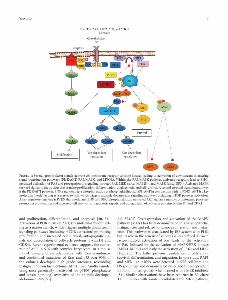
Figure 1: Several growth factor signals activate cell membrane receptor tyrosine kinases leading to activation of downstream interactingsignal transduction pathways (PI3K/AKT, RAF/MAPK, and MTOR). Within the RAF/MAPK pathway, activated receptors lead to SHC-mediated activation of RAS and propagation of signalling through RAF, MEK (a.k.a. MAP2K), and MAPK (a.k.a. ERK). Activated MAPKforward signals to the nucleus that regulate proliferation, differentiation, angiogenesis, and cell survival. A second essential signalling pathwayis the PI3K/AKT pathway. PI3K catalyzes triple phosphorylation of phosphatidylinositol (PI) AKT in conjunction with its PDK1. AKT is a keymolecular “node” acting as a master switch, which triggers multiple downstream signaling pathways including mTOR pathway activation.A key regulatory enzyme is PTEN that modulates PI3K and SHC phosphorylation. Activated AKT signals a number of mitogenic processespromoting proliferation and increased cell survival, antiapoptotic signals, and upregulation of cell-cycle proteins (cyclin D1 and CDK4).
and proliferation, differentiation, and apoptosis [50, 51].Activation of PI3K turns on AKT, key molecular “node” act-ing as a master switch, which triggers multiple downstreamsignaling pathways (including mTOR activation) promotingproliferation and increased cell survival, antiapoptotic sig-nals and upregulation of cell-cycle proteins (cyclin D1 andCDK4). Recent experimental evidence supports the centralrole of AKT in STS with complex karyotypes. In a mousemodel using and an adenovirus with Cre-recombinaseand conditional mutations of Kras and p53 over 90% ofthe animals developed high grade sarcomas, resemblingmalignant fibrous histiocytoma (MFH) [52]. Another modelusing mice genetically inactivated for pTEN (phosphataseand tensin homolog) over 80% of the animals developedabdominal LMS [53].
2.7. MAPK. Overexpression and activation of the MAPKpathway (MKK) has been demonstrated in several epithelialmalignancies and related to tumor proliferation and metas-tases. This pathway is coactivated by IRS system with PI3Kbut its role in the genesis of sarcoma is less defined. Growthfactor-induced activation of Ras leads to the activationof Raf, followed by the activation of MAPK/ERK kinases(MEK1-MEK2) and lastly the activation of ERK1 and ERK2(Figure 1). The latter proteins regulate cell proliferation,survival, differentiation, and migration. In one study, RAF1and MEK 1/2 mRNA were detected in STS cell lines andOS specimens and demonstrated dose- and time-dependentinhibition of cell growth when treated with a MEK inhibitor[54]. Similar observations have been reported in SS whereTK inhibition with sorafenib inhibited the MKK pathway,

8 Sarcoma
downregulated cyclin D1 and pRb levels, caused G1 arrest,and induced apoptosis [55]. In a xenograft model, MKK sig-naling was necessary for tumor growth and vascularizationand treatment with anthrax lethal toxin (LeTx) producedextensive tumor death and antiangiogenic effects. LeTxcontains lethal factor (LF), a zinc metalloprotease that cleavesand inactivates several MKK proteins, suggesting that MKKhad a predominantly proangiogenic effect in this model [56].
2.8. The Role of mTOR Pathway. Activation of PI3K-AKTis a convergence point of activation of growth factorreceptors driving the growth of various sarcomas (Figure 1).Intrinsic activation of the mTOR pathway in sarcomas is theresult of abnormal signaling of these pathways [57]. Othermechanisms explaining over activation of mTOR involve theloss of regulatory inhibitory gene activity of the tuberoussclerosis complex (TSC) proteins or PTEN inactivationby methylation of the promoter, gene mutation, or allelicdeletions such as those reported in perivascular epithelioidcell tumors (PEComas). Loss of LKB1 protein also leads tohyper activation of mTOR signaling in a manner similar tothe loss of PTEN [58–60].
2.9. Angiogenesis. Angiogenesis is regulated by equilibriumbetween proangiogenic (i.e., VEGF, FGF, EGF, PDGF, HIF)and antiangiogenic factors (i.e., angiostatin, IFN, throm-bospondin, and interleukins 1, 4, 12, 18, and 21) which areproduced by both the malignant cells and the microenviron-ment including endothelial cells, fibroblasts, and immunecells. Vascular endothelial growth factor pathway plays adominant role in the pathogenesis and biology of STS.VEGF is overexpressed in 25% of tumors, and high VEGFis associated with an increased risk of metastases andpoorer prognosis. Strong VEGF expression is often presentin tumors rich in vasculature and epithelioid featuressuch as epithelioid sarcoma, KS, and ASPS. Serum VEGFlevels have been strongly correlated with tumor grade andmass and usually poorly differentiated tumors. STS withhigh VEGF expression are associated with resistance tochemotherapy [61, 62]. As with IGF, the VEGF downstreampathway involves PI3K and MAPK indicative of intermingleof cell membrane signals. Distinct from these pathwaysis the VEGF-induced activation of PGLG1 which exertsproangiogenic effects via protein kinase C (PKC) [61].
Other proangiogenic factors are upregulated in STSincluding PDGFR, MMP-2, and Notch-1 and Notch-4, basicFGF and angiopoietin-2 with certain variability according totype and grade of the sarcoma. For instance, fibrosarcomaand LMS showed the highest bFGF levels [63]. Somechromosomal translocations and their fusion proteins canact as transcription factors for promoters of the VEGF geneas is the case of the highly vascular ASPS. Similarly theactivation of mTOR may promote angiogenesis via control ofthe hypoxia inducible factor (HIF)-1α and mTOR inhibitionresults in an antiangiogenic effect [58].
2.10. Telomeres. Cell senescence has been considered ahallmark of cancer and has been related to chromosomal
telomere maintenance mechanisms currently under scrutinyin several malignancies. In a LPS model, using high-resolution DNA mapping array, high level of genome insta-bility and genetic amplifications were identified. In contrastto most stem cells and other cancer cells, that use reversetranscriptase telomerase for telomere maintenance, sarcomacells activate the alternative lengthening of telomeres (ALT)mechanism as often as telomerase. ALT positive LPS havehigher genetic instability and a worse prognosis than non-ALT tumors [64]. A genetic change, the deletion of chromo-some 1q32.2-q44, is seemingly specific to the activation ofALT mechanism in this model [65].
2.11. Hedgehog (Hh) Pathway. Activation of Hh pathwayresults in stimulation of a wide range of prosurvival tran-scription factor genes. Abnormal activation of Hh pathwayhas been implicated in the genesis of various cancersparticularly basal cell carcinoma, medulloblastomas, andRMS. Activation of Hh is manifested by the expressionof several proteins (Ptch1, Gli1, Gli3, and Myf5). Thesemarkers are expressed by embryonal RMS and fusion gene-negative alveolar RMS, whereas FOXO1-PAX3/PAX7 positiveARMS are not [64]. Preliminary observations have shownaugmented Hh signaling in cancer stem cells as well as instromal nonmalignant cells surrounding malignant tumorsand may constitute another cofactor in the genesis ofsarcomas and other cancers [66].
2.12. Cancer Stem Cells and Mesenchymal Cells in Sarcoma.A growing body of evidence suggests the existence ofcancer stem cells (CSCs), pluripotential stem cells that canperpetuate the generation or renewal of tumor forming cellsin solid tumors [67]. The first evidence of cancer stem cellsin sarcoma was reported in 2009. Using surgically resectedES primary tumors, a population of CD133 positive cellsfulfilling in vitro and in vivo criteria of CSC was identified.These criteria included the capacity to generate and sustaintumor growth in a xenograft model and to differentiate,in vitro, along adipogenic, osteogenic, and chondrogeniclineages [68]. These data are supportive of the mesenchymalstem cell (MSC) origin of ES. Thus, ES-initiating cells seemto conserve the properties of their putative cell precursors.In this model, the expression of the EWS-FLI-1 fusionprotein in MSC cells was sufficient to develop ES-like tumors.Lastly, the CD133+ CSC studied showed significantly higherexpression of OCT4 (octamer-binding transcription factor4) and NANOG genes known to be critically involved in self-renewal of embryonic stem cells [68].
While testing other sarcoma-inducing fusion genes,only FUS-CHOP-expressing transfected cells were found togenerate tumors resembling human myxoid LPS. Togetherwith the findings in ES, these data raise the intriguing pos-sibility that ES and MLS may originate from closely relatedmesenchymal cells, but at different anatomical locations.Perhaps the microenvironment (bone in ES, soft tissue inMLS) contributes to the line of differentiation that the MSCfollow according to the adaptability of the transformed cellto survive in a given tissue environment [69].
Sarcoma 9
2.13. Mesenchymal-Epithelial Transition in Sarcoma. Anoth-er puzzling and intriguing feature of certain sarcomas is theapparent spontaneous transition from a mesenchymal tumorto an epithelial enriched tumor (mesenchymal-epithelialtransition (MET)). There is growing evidence supportingthe role of epithelial-to-mesenchymal transition (EMT)during carcinoma progression and metastases linked to theconsequences in cell morphology, cell-to-cell adhesion, cellmotility, and plasticity to migrate and growth in the extra-cellular matrix. The mesenchymal to epithelial transition(MET) in sarcoma progression is considerably less wellstudied. Triggering of MET has been shown to be induced byc-met proto-oncogene, a TK receptor for HGF/SF. Increasedexpression of this protein leads to epithelial differentiation.Epigenetic regulation of DNA methylation has also beenrelated to the MET [70].
This phenomenon is particularly notable in SS.Monophasic SS is entirely composed of spindle cellswith or without solid epithelial areas, whereas the biphasicSS contains a lining of epithelial cells amongst the spindlecells [71]. At a molecular level, the SYT-SSX1 fusionoccurs five times more frequently in biphasic SS exhibitingMET in comparison to SYT-SSX2 in monophasic SS.These data indicates a possible role of SYS-SSX1 andSYT-SSX2 in the MET phenomenon by interacting withtranscription receptors (snail or slug, resp.) interferingwith the E-cadherin gene and triggering the MET epithelialdifferentiation program in the affected SS cells [72].
Similar observations have been described in a chon-drosarcoma model, where upregulation of 4 distinct epithe-lial markers and the downregulation of snail were found. Lossof DNA methylation was demonstrated in maspin and 14-3-3σ genes leading to increased expression of these 2 epithelial-specific genes during chondrosarcoma genesis. These resultssupport the relationship between MET and an epigeneticswitch in chondrosarcoma [73].
2.14. MicroRNA in Sarcomas. The discovery of microRNA(miRNA) has been one further step into the molecularuniverse of gene regulation and cell biology and consequentlyinto the biology of cancer cells and oncogenesis. There isan intense search for miRNA profiles in diverse humancancers to better understand the role of these minuteRNA molecules in cancer control and to explore potentialtherapeutic implications [74].
Preliminary work in sarcomas has disclosed uniquemiRNA expression signatures according to the histologicaltypes seemingly reflecting the cell lineage and differentiationstatus of the tumors [75]. Hierarchal clustering of 87miRNAs disclosed four main groups, whereby almost allSS, RMS, LMS, and GIST were grouped distinctively. InGIST, miR-221 and miR-222 had low expression, suggestingthat the decreased suppressive activity of these miRNAsallows increased translation of KIT. The miRNAs that play amajor role in myogenesis, miR-1, miR-133A, and miR-133B,were overrepresented in LMS, whereas miR-335, involved inskeletal muscle differentiation, was present in ARMS. In SS,miR-143 whose target is ERK5 (MAPK7), was expressed at
very low levels suggesting that this miRNA may be involvedin the expression of the SYT-SSX1 oncoprotein [76]. Othershave linked the expression of miR-200 to mesenchymal-epithelial differentiation often reported in SS [77].
In a recent study exploring the genesis of CSC phenotypein ES, Riggi and collaborators [78] published evidence thatrepression of miRNA-145 and expression of the EWS-FLI-1fusion gene were both necessary to induce transformation.They showed evidence that their common target may be theSOX2 gene. This gene is known to code for transcriptionfactors required for the development of pluripotent stemcells. These observations provided valuable insight into themechanisms, whereby a single oncogene (EWS-FLI1) canreprogram cells towards CSC.
3. Therapeutic Implications
Targeting the multiple molecular pathways and mechanismssummarized above is one of the areas of intense basic andclinical research. The objective is to find means to modifytumor behavior and to find long awaited clinical therapeuticoptions for these patients. Search for antagonistic antibodies,TK inhibitors, and inhibitors of downstream molecules of thePI3K, MAPK, and mTOR paths are at the forefront of theseefforts (Figure 2). The preliminary clinical trials have notyet crystallized into therapeutic breakthroughs despite solidpreclinical evidence, suggesting a broad range of favorablebiological effects from the inhibition of these pathways.
3.1. IGF-1R Antibodies. Preclinical data proves that effectiveblockade of IGF-1 and IGFII ligands to the IGF-1R is feasible[79]. Monoclonal antibodies against the IGF-1R have beenthe favored approach to date. Phase I and II studies of IGF1Rantagonists figitumumab, cixutumumab, AMG479, R1507,and SCH 717454 either alone or in combination with otheragents, are currently under clinical investigation for patientswith sarcomas. Table 5 summarizes the results of reportedphase I/II studies with anti-IGF-R1 agent [79–83].
Further, IGF-1R has been implicated in chemotherapyresistance from in vitro work with malignant cells. In anES tumor model, combination of ADW742 with imatinib,vincristine, and doxorubicin induced a significant reductionof tumor cell growth, mainly by increasing apoptosis [84].Similarly, NVP-AEW541 led to cytotoxicity and inducedapoptosis in imatinib resistant or wildtype GIST [85].Finally, there is experimental evidence that bidirectionalcrosstalk between the IGF system and the erbB family ofreceptors may confer means of escape or resistance to targettherapy of these receptor pathways [51]. Thus, the useof combined therapies aiming to block several pathwayssimultaneously is being actively investigated in varioustumors.
3.2. TKs Inhibitors and Antiangiogenesis. As described above,TKs account for a large number of defective signalingpathways in sarcomas. Based on the success of imatinib inGIST, TKs inhibitors are of major interest in other sarcomas.Sunitinib has been shown to be effective in imatinib-resistant

10 Sarcoma
SCF HGF VEGF
PP
PP
PP
PP
PP
PP
PP
PP
PP
PP
FGFR
FGFIGF1-2
IGF-1R c-KIT C-MET VEFG-A
Ligandregulation
Receptorblocking
Tyrosinekinaseinhibitors
Downstreamsignal inhibitors PI3K/MAPK/MTOR
Molecular biology of sarcomasThe PI3K/AKT, MAPK, and MTOR pathways
Targeted therapy strategies
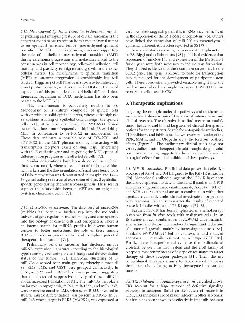
Figure 2: Several ligands (IGF1/2, SCF, HGF, VEGF, and FGF) activate cell membrane receptor tyrosine kinases (IGF1R, C-KIT, C-MET,VEGFR-A, and FGFR) triggering shared interacting signal transduction pathways (PI3K/AKT, RAF/MAPK, and MTOR). The availabilityof ligands via paracrine secretion (IGF1), autocrine loops (VEGF), or ligand binding (IGFBPs) can modulate activation of these pathways.Molecular anomalies of the receptors or the downstream signals lead to constitutive activation or dysregulation of these signals. Multiple cellprocesses including proliferation, differentiation, angiogenesis, and survival are promoted as a result of the activation of these main pathwaysin sarcomas and other neoplasms. Actionable targets are listed that may interfere with the abnormal signalling and could result in beneficialbiologic and clinical effects.
Table 5: Current Anti-IGF treatment of sarcomas.
Monoclonal antibodies Human trialsDisease control rate(CR, PR, and SD)
Comments
Figitumumab CP-751, 871 ES/STS 10/28 (35%) Anti-IGF-IR
Cixutumumab STS 22/37 (59%)Liposarcomas; block hybridreceptors
Robatumumab SCH 717454 Preclinical OS, RMS Anti-IGF-IR
Ganitumab AMG479 ES 2/15 (13%)
RI 507 ES 18/125 (14.4%) No SD included
TK inhibitors Status Target disease
NVP-AEW541 Preclinical ES/STS/GIST Synergy with chemoRx
NVP-ADW 742 Preclinical ES Synergy with imatinib
BMS-536924 Preclinical STS ATP-competitive IGF-IR
Others Status Target disease
Nordihydroguaiaretic acid (NDGA) Preclinical STS Disrupts IGF-1R; blocks HER-2
GISTs [86]. Patients with chordomas, desmoids, and DFSPtumors reportedly responded to imatinib in small trials andisolated case reports, likely via inhibition of PDGFR [87].
Initial attempts intended to block signaling pathwayswith other available agents known to interfere with eithercellular receptors or TKs have not met with evidence of majoror consistent clinical benefit. Phase II studies with EGFRinhibitors (gefitinib or erlotinib) showed no clinical activity
in SS or malignant nerve-sheath tumors. Use of trastuzumabin ES and OS, alone or in combination with chemotherapy,has had no therapeutic benefit. Phase II studies withsunitinib in non-GIST sarcomas have only shown occasionalobjective responses, but disease stabilization over 12 weekswas noted in several histologies [87]. Sorafenib, but notsunitinib, was reported to produced a 14% PR rate and amedian OS of 14.3 months in angiosarcomas [88]. Some

Sarcoma 11
Table 6: Current clinical trials with tyrosine kinase inhibitors in sarcomas.
Agent Human trials Clinical benefit rate (CR, PR, and SD) Targets: notes
Imatinib GIST 85% (5% CR; 45% PR) KIT. FDA Approved
Sunitinib GIST 65% (7% PR)VEGFR-1, VEGFR-2, VEGFR-3, PDGFR a/β, KIT.FDA Approved
Sorafenib Angiosarcoma, GIST 14% VEGFR-2, VEGFR-3, PDGFR, c-RAS, b-RAF, KIT
PazopanibPaletteSTS
PFS = 20 versus 7 weeks (P = 0.0001) VEGFR-1, VEGFR-2, VEGFR-3, PDGFR-a/β, KIT
BrivanibSTSASCO 2011
30% overall3 PR in angiosarcomas
FGF and VEGF(FGF-positive did better)
CediranibSTSASCO 2011
78% (43%, PR)Alveolar soft part sarcoma
VEGFR-1, VEGFR-2, and VEGFR-3
TivantinibSTSASCO 2011
80% (5%, PR)Clear cell sarcoma, ASPS
c-Met
Axitinib STS, angiosarcoma N/A VEGFR-1, VEGFR-2, and VEGFR-3
Table 7: Current clinical trials with rapalogs in sarcomas.
Agent Human trials Clinical benefit rate (CR, PR, and SD) Notes
TemsirolimusGISTASCO 2010
GIST = 4/15 (27%)STS = 2/15 (13%)
Refractory GIST
TemsirolimusPhase II STSMayo Clinic
5%
Sirolimus Kaposi sarcoma 15/15 (100%)Posttransplant KSDermal lesions
Ridaforolimus Phase II-STS 61/212 (29%; 2% PR) IV formulation
Ridaforolimus SUCEED trial PFS: HR = 0.69 (P < 0.0001) (22.4 versus 14.7 weeks) Maintenance after ChemoRX
sarcoma subtypes with a predominant vascular connectivetissue component such as angiosarcomas, intimal sarcomas,and hemangiopericytomas may arise from endothelial cells,suggesting that proangiogenic proteins may be relevantto their growth and potential treatment targets. Severalmultitargeted receptor TK inhibitors with antiangiogeniceffects are currently on clinical trials. Table 6 shows publishedor reported data on various new multitargeted TKs [89–93].
ABT-510, a peptide that mimics the antiangiogenicactivity of thrombospondin-1, was tested in a phase II trial inpatients with advanced STS. Approximately 50% of patientsachieved SD, with 1 objective response. Perifosine an AKTinhibitor has been tested in phase I and phase II trialsin patients with advanced STS with inconsistent results.No objective responses were observed but 27% of patientsexperienced SD in one study with a 5% PR rate and 45%of patients experienced SD for >4 months in a retrospectiveevaluation [94].
3.3. mTOR Inhibitors. The clinical impact of analogs ofrapamycin (rapalogs) in the management of renal cellcarcinoma has confirmed the antitumor potential of mTORpathway interference [95]. The participation of mTOR in thegenesis of sarcoma is related to the primordial role of theIGF system in these tumors. Thus, mTOR inhibitors were anatural choice to test clinically in sarcomas.
Rapamycin exert its biological effects by forming acomplex with FKBP12 which binds to the FK-rapamycin
binding domain of mTOR inhibiting the function ofmTORC1-mediated signal pathway and resulting in a directantiangiogenic effect [58]. A number of preclinical studieswith various rapalogs paved the way to subsequent humantrials. These have included in vitro and in vivo observationsin mouse xenograft models using sirolimus, temsirolimus,everolimus, and ridaforolimus. Of relevance, temsirolimusinhibits HIF-1α translation and interferes with VEGF proteinexpression in RMS demonstrating suppressed tumor growthvia anti-angiogenesis [96]. Other models have suggestedthat sarcomas associated with PTEN loss or inactivationmay be particularly susceptible to the therapeutic effectsof mTOR inhibitors [58, 59]. Rapalogs have been shownto be less effective or ineffective in the presence of KRasmutations or overexpression of Bcl2, whereas tumors withcyclin D1 expression and “angiogenesis addiction” are moresusceptible [97].
Phase I/II clinical trials with several rapalogs and com-binations aim to determine efficacy in sarcoma patients.Ongoing studies include testing efficacy of sirolimus in KS,temsirolimus, and valproic acid in OS and STS; or combina-tions with vinorelbine for uterine sarcoma, liposomal dox-orubicin or irinotecan for recurrent or refractory sarcomas[96, 97]. Table 7 summarizes data on early phase studies withrapalogs. Ridaforolimus is a nonprodrug rapalog that hasshown promising clinical activity in STS [98, 99]. Despitedisputed clinical impact (the difference in median PFS wasonly 3 weeks), this drug is awaiting approval in 23 countriesincluding the FDA in the US based on these studies.

12 Sarcoma
3.4. Combinatorial Studies. A number of early clinicalstudies explored combination of agents intended to blockredundant or cross-talking molecular pathways or to cir-cumvent chemotherapy resistance. These include everolimuswith imatinib, phase II study in imatinib-resistant GIST;everolimus and figitumumab; temsirolimus with cixutu-mumab in ES (65% of patients had tumor reduction >20%)ridaforolimus and doxorubicin; everolimus and pegylatedliposomal doxorubicin, mTOR inhibitors and hormonother-apy, and many others will follow [96–99]. There is also agrowing interest in the use of other agents including mela-tonin, metformin, celecoxib, statins, and others which havebeen shown to modulate molecular signals in the pathwaysinvolved in tumor growth. Melatonin has been shown to havea broad range of activities resulting in an oncostatic effectin vitro and in vivo including suppression of tumorigenesisin methylcholanthracene-induced fibrosarcomas in mice[100]. In an ES in vitro model, melatonin has been shownto induce apoptosis and synergism when combined withchemotherapeutic agents [101]. Metformin reduces insulinlevel by decreasing insulin resistance a favorable effect oncancer cells dependent on the IGF system. A direct inhibitoryeffect of metformin on cancer cell growth has also beenreported and has been associated to a regulatory role in theAMP-activated protein kinase (AMPK) and mTOR pathways[102].
4. Conclusions
A wealth of information is accumulating at a rapid pacethat, undoubtedly, will continue to contribute to the furtherunderstanding of the molecular biology of sarcomas. Thereare, however, enormous challenges ahead, particularly inthe clinical translation of these discoveries. Despite thesuccess noted in KS and the success of imatinib treatmentin GIST, only modest gains have been attained in ourattempts to alter or modulate growth of tumor in mostother sarcomas. It is clear that the rarity of this vast numberof pathological entities complicates the clinical applicationof the newly developing agents. But the existence of fairlyspecific genetic abnormalities, mainly specific translocationsand a generation of discernible fusion genes in varioussarcomas make research in these tumors an attractive moldfor advancing studies of solid tumors.
Finding key molecular “nodes” or pathways that are com-mon amongst these tumors will help to further understandthe pathogenesis of sarcomas and may help improve currenttherapeutic choices. Yet, the redundancy of these molecularcascades, the loopholes and the fact that tumors may findadaptive escape routes necessitate incessant search for deeperunderstanding of the signaling pathways in the context ofall the hallmarks of cancer discussed above. The conceptof system biology and the development of computationaland mathematical models are areas that will facilitate theorganization of thoughts directing future research strategies.
Clinically, biologic effects are largely cytostatic andtransient which indicate the need to devise new strategies toreach long-term clinical control and potential cure of some of
these entities. Newer clinical trials are beginning to modifythe chemotherapy-oriented criteria of response and criteriain the designing of clinical studies [103]. Emphasis is beingplaced in the selection of patient populations to avoid thepitfalls of unselected trials that have often missed the targetin the past. There is a clear trend to consider progression-freesurvival (PFS) as a more relevant endpoint when assessingthe antitumor effects of molecular targeted therapies. It hasbeen suggested that first-line treatments should achieve PFSrates of >30% at 6 months for the results to be consideredclinically meaningful in phase II trials in STS [86].
Assessment of “metabolic” response by FDG-PET isbeing examined as a better mean of assessing antitumoractivity of targeted therapies as opposed to traditionalRECIST-defined responses. Support for this is coming inmany fields including sarcoma trials [104]. FDG-PET hasbeen shown superior to CT in predicting early response andmonitoring of response and progression to imatinib therapyin GIST and other STS [105, 106]. Furthermore, FDG-PETmay also have applicability as an early pharmacodynamicmarker of molecular targeted therapies [106].
Although the concept of “personalized” therapy has beenoverestimated there is clearly a need to continue searchingfor uniqueness among subgroups of patients, particularlyin sarcomas, this most heterogeneous collection of diseases.Cardinal to these efforts is the search for molecular markersthat identify patients most likely to benefit from a givenintervention as well as to monitor the biological effectsof the treatment or to identify the optimal dose of theseagents. It is necessary to dismiss the chemotherapy-drivenconcept of maximum tolerated dose and find instead theoptimal biologic dose of the agents being tested. So farthe identification of clinically relevant molecular markershas been daunting but progress in the area is expectedas methodologies to examine efficiently multitude putative“markers of interest” are developed.
These interventions are not strictly tumor-specific andelicit cellular and clinical toxicity and may hamper ourenthusiasm to some extent as we have already witnessed inother fields including RCC, melanoma, colon, breast, andother cancers. The identification of more precise targets andrefinement of the specificity of targeted agents is requiredto make progress in this field. These targets should bethose that are essential for the malignant cells to subsist, aconcept that has been termed “oncogene addiction.” But evenwell-characterized mutations can have different molecularisoforms that could alter the specificity and durability ofthe binding to the therapeutic agent or render the agentless efficacious. Identification of precise “pockets” withinthe abnormal targeted molecule may also improve thetherapeutic index of future targeted therapies.
Enormous clinical challenges lie ahead, but a new worldof possibilities is opening. Combination strategies are veryattractive because of the multitude of potential targets andbecause of the different properties and mechanisms of actionof drugs in our growing arsenal. However, the combinationof some of these agents has already shown additive toxicitieswithout an additional antitumor effect. Antagonism has beenobserved in some combinations that seemed logical and

Sarcoma 13
promising and, although observed in preclinical models, weare yet to prove synergism when combining these agents.Combination therapies should not be limited to molecular-targeted drugs as we can judiciously combine these withchemotherapeutic agents. Furthermore, these combinationstrategies must include efforts to modulate the microenvi-ronment of the tumor and the immune system to attain a fulland comprehensive approach to the control of the malignantgrowth in sarcomas and other cancers.
Acknowledgments
The authors wish to express their gratitude to Dr. RobertBrown for reviewing the paper and Ms. Marika Stepankiwfor her editorial input.
References
[1] D. Hanahan and R. A. Weinberg, “Hallmarks of cancer: thenext generation,” Cell, vol. 144, no. 5, pp. 646–674, 2011.
[2] P. Rous, “A sarcoma of the fowl transmissible by an agentseparable from the tumor cells,” The Journal of ExperimentalMedicine, vol. 13, pp. 397–411, 1911.
[3] H. Rubin, “The early history of tumor virology: Rous, RIF,and RAV,” Proceedings of the National Academy of Sciencesof the United States of America, vol. 108, no. 35, pp. 14389–14396, 2011.
[4] J. S. Butel, “Viral carcinogenesis: revelation of molecularmechanisms and etiology of human disease,” Carcinogenesis,vol. 21, no. 3, pp. 405–426, 2000.
[5] R. J. Huebner and G. J. Todaro, “Oncogenes of rna tumorviruses as determinants of cancer,” Proceedings of the NationalAcademy of Sciences of the United States of America, vol. 64,no. 3, pp. 1087–1094, 1969.
[6] D. Stehelin, H. E. Varmus, J. M. Bishop, and P. K. Vogt, “DNArelated to the transforming gene(s) of avian sarcoma virusesis present in normal avian DNA,” Nature, vol. 260, no. 5547,pp. 170–173, 1976.
[7] E. R. Fearon and B. Vogelstein, “A genetic model forcolorectal tumorigenesis,” Cell, vol. 61, no. 5, pp. 759–767,1990.
[8] M. Malumbres and M. Barbacid, “RAS oncogenes: the first30 years,” Nature Reviews Cancer, vol. 3, no. 6, pp. 459–465,2003.
[9] D. S. Goodsell, “The molecular perspective: the RAS onco-gene,” Oncologist, vol. 4, no. 3, pp. 263–264, 1999.
[10] J. Downward, “Targeting ras signalling pathways in cancertherapy,” Nature Reviews Cancer, vol. 3, no. 1, pp. 11–22,2003.
[11] J. Bickels, Y. Kollender, O. Merinsky, and I. Meller, “Coley’stoxin: historical perspective,” Israel Medical Association Jour-nal, vol. 4, no. 6, pp. 471–472, 2002.
[12] I. Gresser, “Antitumor effects of interferon,” Advances inCancer Research, vol. 16, pp. 97–140, 1973.
[13] H. Strander, “Interferons and osteosarcoma,” Cytokine andGrowth Factor Reviews, vol. 18, no. 5-6, pp. 373–380, 2007.
[14] L. C. Platanias, “Mechanisms of type-I- and type-II-interferon-mediated signalling,” Nature Reviews Immunol-ogy, vol. 5, no. 5, pp. 375–386, 2005.
[15] T. Panaretakis, L. Hjortsberg, K. P. Tamm, A. C. Bjorklund,B. Joseph, and D. Grander, “Interferon α induces nucleus-independent apoptosis by activating extracellular signal-regulated kinase 1/2 and c-Jun NH2-terminal kinase down-stream of phosphatidylinositol 3-kinase and mammaliantarget of rapamycin,” Molecular Biology of the Cell, vol. 19,no. 1, pp. 41–50, 2008.
[16] M. S. R. Hutt, “Historical introduction, burkitt’s lymphoma,nasopharyngeal carcinoma and Kaposi’s sarcoma,” Transac-tions of the Royal Society of Tropical Medicine and Hygiene,vol. 75, no. 6, pp. 761–765, 1981.
[17] V. Beral, T. A. Peterman, R. L. Berkelman, and H. W. Jaffe,“Kaposi’s sarcoma among persons with AIDS: a sexuallytransmitted infection?” The Lancet, vol. 335, no. 8682, pp.123–128, 1990.
[18] P. S. Moore and Y. Chang, “Detection of herpesvirus-like dnasequences in Kaposi’s sarcoma in patients with and thosewithout HIV infection,” New England Journal of Medicine,vol. 332, no. 18, pp. 1181–1185, 1995.
[19] J. T. West and C. Wood, “The role of Kaposi’s sarcoma-associated herpesvirus/human herpesvirus-8 regulator oftranscription activation (RTA) in control of gene expression,”Oncogene, vol. 22, no. 33, pp. 5150–5163, 2003.
[20] R. G. Jenner, K. Maillard, N. Cattini et al., “Kaposi’s sarcoma-associated herpesvirus-infected primary effusion lymphomahas a plasma cell gene expression profile,” Proceedings of theNational Academy of Sciences of the United States of America,vol. 100, no. 18, pp. 10399–10404, 2003.
[21] H. W. Wang, M. W. B. Trotter, D. Lagos et al., “Kaposisarcoma herpesvirus-induced cellular reprogramming con-tributes to the lymphatic endothelial gene expression inKaposi sarcoma,” Nature Genetics, vol. 36, no. 7, pp. 687–693,2004.
[22] S. E. Krown, F. X. Real, and S. Cunningham Rundles,“Preliminary observations on the effect of recombinantleukocyte a interferon in homosexual men with Kaposi’ssarcoma,” New England Journal of Medicine, vol. 308, no. 18,pp. 1071–1076, 1983.
[23] A. Rios, P. W. A. Mansell, G. R. Newell et al., “Treatmentof acquired immunodeficiency syndrome-related Kaposi’ssarcoma with lymphoblastoid interferon,” Journal of ClinicalOncology, vol. 3, no. 4, pp. 506–512, 1985.
[24] P. Besmer, W. D. Hardy Jr., E. E. Zuckerman et al., “Thehardy-zuckerman 2-FeSV, a new feline retrovirus with onco-gene homology to abelson-MuLV,” Nature, vol. 303, no. 5920,pp. 825–828, 1983.
[25] Y. Yarden, W. J. Kuang, T. Yang-Feng et al., “Human proto-oncogene c-KIT: a new cell surface receptor tyrosine kinasefor an unidentified ligand,” EMBO Journal, vol. 6, no. 11, pp.3341–3351, 1987.
[26] K. Sircar, B. R. Hewlett, J. D. Huizinga, K. Chorneyko, I.Berezin, and R. H. Riddell, “Interstitial cells of cajal asprecursors of gastrointestinal stromal tumors,” AmericanJournal of Surgical Pathology, vol. 23, no. 4, pp. 377–389,1999.
[27] S. Hirota, K. Isozaki, Y. Moriyama et al., “Gain-of-functionmutations of c-KIT in human gastrointestinal stromaltumors,” Science, vol. 279, no. 5350, pp. 577–580, 1998.
[28] M. C. Heinrich, B. P. Rubin, B. J. Longley, and J. A. Fletcher,“Biology and genetic aspects of gastrointestinal stromaltumors: KIT activation and cytogenetic alterations,” HumanPathology, vol. 33, no. 5, pp. 484–495, 2002.

14 Sarcoma
[29] P. Reichardt, “Optimal use of targeted agents for advancedgastrointestinal stromal tumours,” Oncology, vol. 78, no. 2,pp. 130–140, 2010.
[30] M. C. Heinrich, D. J. Griffith, B. J. Druker, C. L. Wait,K. A. Ott, and A. J. Zigler, “Inhibition of c-KIT receptortyrosine kinase activity by STI 571 a selective tyrosine kinaseinhibitor,” Blood, vol. 96, no. 3, pp. 925–932, 2000.
[31] D. A. Tuveson, N. A. Willis, T. Jacks et al., “STI571inactivation of the gastrointestinal stromal tumor c-KIToncoprotein: biological and clinical implications,” Oncogene,vol. 20, no. 36, pp. 5054–5058, 2001.
[32] H. Joensuu, P. J. Roberts, M. Sarlomo-Rikala et al., “Effectof the tyrosine kinase inhibitor STI571 in a patient witha metastatic gastrointestinal stromal tumor,” New EnglandJournal of Medicine, vol. 344, no. 14, pp. 1052–1056, 2001.
[33] G. D. Demetri, M. Von Mehren, C. D. Blanke et al., “Efficacyand safety of imatinib mesylate in advanced gastrointestinalstromal tumors,” New England Journal of Medicine, vol. 347,no. 7, pp. 472–480, 2002.
[34] S. Singer, B. P. Rubin, M. L. Lux et al., “Prognostic value ofkit mutation type, mitotic activity, and histologic subtype ingastrointestinal stromal tumors,” Journal of Clinical Oncol-ogy, vol. 20, no. 18, pp. 3898–3905, 2002.
[35] S. Bauer, A. Duensing, G. D. Demetri, and J. A. Fletcher,“KIT oncogenic signaling mechanisms in imatinib-resistantgastrointestinal stromal tumor: PI3-kinase/AKT is a crucialsurvival pathway,” Oncogene, vol. 26, no. 54, pp. 7560–7568,2007.
[36] E. Alava, “Molecular pathology in sarcomas,” Clinical andTranslational Oncology, vol. 9, no. 3, pp. 130–144, 2007.
[37] J. V. M. G. Bovee and P. C. W. Hogendoorn, “Molecularpathology of sarcomas: concepts and clinical implications,”Virchows Archiv, vol. 456, no. 2, pp. 193–199, 2010.
[38] Y. Oda and M. Tsuneyoshi, “Recent advances in the molec-ular pathology of soft tissue sarcoma: implications fordiagnosis, patient prognosis, and molecular target therapy inthe future,” Cancer Science, vol. 100, no. 2, pp. 200–208, 2009.
[39] L. J. Helman and P. Meltzer, “Mechanisms of sarcomadevelopment,” Nature Reviews Cancer, vol. 3, no. 9, pp. 685–694, 2003.
[40] F. Mertens, I. Panagopoulos, and N. Mandahl, “Genomiccharacteristics of soft tissue sarcomas,” Virchows Archiv, vol.456, no. 2, pp. 129–139, 2010.
[41] M. C. Le Deley, O. Delattre, K. L. Schaefer et al.,“Impact of EWS-ETS fusion type on disease progressionin ewing’s sarcoma/peripheral primitive neuroectodermaltumor: prospective results from the cooperative Euro-E.W.I.N.G. 99 trial,” Journal of Clinical Oncology, vol. 28, no.12, pp. 1982–1988, 2010.
[42] L. Guillou, J. Benhattar, F. Bonichon et al., “Histologic grade,but not SYT-SSX fusion type, is an important prognosticfactor in patients with synovial sarcoma: a multicenter,retrospective analysis,” Journal of Clinical Oncology, vol. 22,no. 20, pp. 4040–4050, 2004.
[43] O. Delattre, J. Zucman, B. Plougastel et al., “Gene fusionwith an ETS DNA-binding domain caused by chromosometranslocation in human tumours,” Nature, vol. 359, no. 6391,pp. 162–165, 1992.
[44] F. G. Barr, “Translocations, cancer and the puzzle of speci-ficity,” Nature Genetics, vol. 19, no. 2, pp. 121–124, 1998.
[45] S. R. Knezevich, D. E. McFadden, W. Tao, J. F. Lim, andP. H. B. Sorensen, “A novel ETV6-NTRK3 gene fusion incongenital fibrosarcoma,” Nature Genetics, vol. 18, no. 2, pp.184–187, 1998.
[46] C. L. Lannon, M. J. Martin, C. E. Tognon, W. Jin, S. J.Kim, and P. H. B. Sorensen, “A highly conserved NTRK3 c-terminal sequence in the ETV6-NTRK3 oncoprotein bindsthe phosphotyrosine binding domain of insulin receptorsubstrate-1: an essential interaction for transformation,”Journal of Biological Chemistry, vol. 279, no. 8, pp. 6225–6234, 2004.
[47] Y. Fujimura, T. Ohno, H. Siddique, L. Lee, V. N. Rao,and E. S. P. Reddy, “The EWS-ATF-1 gene involved inmalignant melanoma of soft parts with t(12;22) chromosometranslocation, encodes a constitutive transcriptional activa-tor,” Oncogene, vol. 12, no. 1, pp. 159–167, 1996.
[48] M. Ladanyi, M. Y. Lui, C. R. Antonescu et al., “Theder(17)t(x;17)(p11;q25) of human alveolar soft part sarcomafuses the TFE3 transcription factor gene to ASPL, a novelgene at 17q25,” Oncogene, vol. 20, no. 1, pp. 48–57, 2001.
[49] I. J. Davis, J. J. Kim, F. Ozsolak et al., “Oncogenic MITFdysregulation in clear cell sarcoma: defining the MiT familyof human cancers,” Cancer Cell, vol. 9, no. 6, pp. 473–484,2006.
[50] B. Rikhof, S. De Jong, A. J. H. Suurmeijer, C. Meijer, and W.T. A. Van Der Graaf, “The insulin-like growth factor systemand sarcomas,” Journal of Pathology, vol. 217, no. 4, pp. 469–482, 2009.
[51] J. Zha and M. R. Lackner, “Targeting the insulin-like growthfactor receptor-1R pathway for cancer therapy,” ClinicalCancer Research, vol. 16, no. 9, pp. 2512–2517, 2010.
[52] D. G. Kirsch, D. M. Dinulescu, J. B. Miller et al., “Aspatially and temporally restricted mouse model of soft tissuesarcoma,” Nature Medicine, vol. 13, no. 8, pp. 992–997, 2007.
[53] E. Hernando, E. Charytonowicz, M. E. Dudas et al., “TheAKT-mTOR pathway plays a critical role in the developmentof leiomyosarcomas,” Nature Medicine, vol. 13, no. 6, pp.748–753, 2007.
[54] K. Sasaki, T. Hitora, O. Nakamura, R. Kono, and T.Yamamoto, “The role of MAPK pathway in bone and softtissue tumors,” Anticancer Research, vol. 31, no. 2, pp. 549–553, 2011.
[55] C. L. Peng, W. Guo, T. Ji et al., “Sorafenib induces growthinhibition and apoptosis in human synovial sarcoma cellsvia inhibiting the RAF/MEK/ERK signaling pathway,” CancerBiology and Therapy, vol. 8, no. 18, pp. 1729–1736, 2009.
[56] Y. Ding, E. A. Boguslawski, B. D. Berghuis et al., “Mitogen-activated protein kinase kinase signaling promotes growthand vascularization of fibrosarcoma,” Molecular Cancer Ther-apeutics, vol. 7, no. 3, pp. 648–658, 2008.
[57] S. Vemulapalli, A. Mita, Y. Alvarado, K. Sankhala, and M.Mita, “The emerging role of mammalian target of rapamycininhibitors in the treatment of sarcomas,” Targeted Oncology,vol. 6, no. 1, pp. 29–39, 2011.
[58] X. Wan and L. J. Helman, “The biology behind mtorinhibition in sarcoma,” Oncologist, vol. 12, no. 8, pp. 1007–1018, 2007.
[59] Q. Yang and K. L. Guan, “Expanding mTOR signaling,” CellResearch, vol. 17, no. 8, pp. 666–681, 2007.
[60] P. C. W. Hogendoorn, F. Collin, S. Daugaard et al., “Changingconcepts in the pathological basis of soft tissue and bonesarcoma treatment,” European Journal of Cancer, vol. 40, no.11, pp. 1644–1654, 2004.
[61] C. Chao, T. Al-Saleem, J. J. Brooks, A. Rogatko, W. G.Kraybill, and B. Eisenberg, “Vascular endothelial growthfactor and soft tissue sarcomas: tumor expression correlateswith grade,” Annals of Surgical Oncology, vol. 8, no. 3, pp.260–267, 2001.

Sarcoma 15
[62] R. J. Olsen, S. R. Tarantolo, and S. H. Hinrichs, “Molecularapproaches to sarcoma therapy,” Sarcoma, vol. 6, no. 1, pp.27–42, 2002.
[63] S. S. Yoon, N. H. Segal, P. J. Park et al., “Angiogenic profileof soft tissue sarcomas based on analysis of circulating factorsand microarray gene expression,” Journal of Surgical Research,vol. 135, no. 2, pp. 282–290, 2006.
[64] A. Costa, M. G. Daidone, L. Daprai et al., “Telomere main-tenance mechanisms in liposarcomas: association with histo-logic subtypes and disease progression,” Cancer Research, vol.66, no. 17, pp. 8918–8924, 2006.
[65] J. E. Johnson, E. J. Gettings, J. Schwalm et al., “Whole-genome profiling in liposarcomas reveals genetic alterationscommon to specific telomere maintenance mechanisms,”Cancer Research, vol. 67, no. 19, pp. 9221–9228, 2007.
[66] N. Takebe, P. J. Harris, R. Q. Warren, and S. P. Ivy, “Targetingcancer stem cells by inhibiting Wnt, Notch, and Hedgehogpathways,” Nature Reviews Clinical Oncology, vol. 8, no. 2, pp.97–106, 2011.
[67] C. T. Jordan, “Cancer stem cell biology: from leukemia tosolid tumors,” Current Opinion in Cell Biology, vol. 16, no.6, pp. 708–712, 2004.
[68] M. L. Suva, N. Riggi, J. C. Stehle et al., “Identification ofcancer stem cells in ewing’s sarcoma,” Cancer Research, vol.69, no. 5, pp. 1776–1781, 2009.
[69] N. Riggi, L. Cironi, P. Provero et al., “Expression of the FUS-CHOP fusion protein in primary mesenchymal progenitorcells gives rise to a model of myxoid liposarcoma,” CancerResearch, vol. 66, no. 14, pp. 7016–7023, 2006.
[70] C. L. Chaffer, E. W. Thompson, and E. D. Williams,“Mesenchymal to epithelial transition in development anddisease,” Cells Tissues Organs, vol. 185, no. 1–3, pp. 7–19,2007.
[71] W. B. Laskin and M. Miettinen, “Epithelial-type and neural-type cadherin expression in malignant noncarcinomatousneoplasms with epithelioid features that involve the softtissues,” Archives of Pathology and Laboratory Medicine, vol.126, no. 4, pp. 425–431, 2002.
[72] T. Saito, M. Nagai, and M. Ladanyi, “SYT-SSX1 and SYT-SSX2 interfere with repression of E-cadherin by snail andslug: a potential mechanism for aberrant mesenchymal toepithelial transition in human synovial sarcoma,” CancerResearch, vol. 66, no. 14, pp. 6919–6927, 2006.
[73] M. P. Fitzgerald, F. Gourronc, M. L. T. Teoh et al.,“Human chondrosarcoma cells acquire an epithelial-likegene expression pattern via an epigenetic switch: evidence formesenchymal-epithelial transition during sarcomagenesis,”Sarcoma, vol. 2011, Article ID 598218, 11 pages, 2011.
[74] P. Gammell, “Micrornas: recently discovered key regulatorsof proliferation and apoptosis in animal cells: identificationof mirnas regulating growth and survival,” Cytotechnology,vol. 53, no. 1–3, pp. 55–63, 2007.
[75] S. Subramanian, W. O. Lui, C. H. Lee et al., “Micrornaexpression signature of human sarcomas,” Oncogene, vol. 27,no. 14, pp. 2015–2026, 2008.
[76] M. Hisaoka, A. Matsuyama, Y. Nagao et al., “Identificationof altered microrna expression patterns in synovial sarcoma,”Genes Chromosomes and Cancer, vol. 50, no. 3, pp. 137–145,2011.
[77] T. Motoi, A. Kumagai, A. Yoshida et al., “MicroRNA-200a Expression in Synovial Sarcoma Is Related to Epithe-lial Differentiation,” Abstract 65, http://www.abstracts2view.com/uscap11/.
[78] N. Riggi, M. L. Suva, C. De Vito et al., “EWS-FLI-1 modulatesmiRNA145 and SOX2 expression to initiate mesenchymalstem cell reprogramming toward ewing sarcoma cancer stemcells,” Genes and Development, vol. 24, no. 9, pp. 916–932,2010.
[79] S. J. Weroha and P. Haluska, “GF-1 receptor inhibitors inclinical trials—early lessons,” Journal of Mammary GlandBiology and Neoplasia, vol. 13, no. 4, pp. 471–483, 2008.
[80] D. Olmos, S. Postel-Vinay, L. R. Molife et al., “Safety,pharmacokinetics, and preliminary activity of the anti-IGF-1R -1R antibody figitumumab (CP-751,871) in patients withsarcoma and ewing’s sarcoma: a phase 1 expansion cohortstudy,” The Lancet Oncology, vol. 11, no. 2, pp. 129–135, 2010.
[81] P. J. Houghton, C. L. Morton, R. Gorlick et al., “Initial testingof a monoclonal antibody (IMC-A12) against IGF-1R by thepediatric preclinical testing program,” Pediatric Blood andCancer, vol. 54, no. 7, pp. 921–926, 2010.
[82] A. W. Tolcher, J. Sarantopoulos, A. Patnaik et al., “Phase I,pharmacokinetic, and pharmacodynamic study of AMG 479,a fully human monoclonal antibody to insulin-like growthfactor receptor 1,” Journal of Clinical Oncology, vol. 27, no.34, pp. 5800–5807, 2009.
[83] A. S. Pappo, S. Patel, J. Crowley et al., “Activity of R1507,a monoclonal antibody to the insulinlike growth factor-1receptor (IGF1R), in patients with recurrent or refractoryEwing’s sarcoma family of tumors (ESFT): results of aphase II SARC study,” Journal of Clinical Oncology, vol. 28,supplement 15, abstract 10000, 2010.
[84] A. S. Martins, C. Mackintosh, D. Herrero Martın et al.,“Insulin-like growth factor I receptor pathway inhibition byADW742, alone or in combination with imatinib, doxoru-bicin, or vincristine, is a novel therapeutic approach in ewingtumor,” Clinical Cancer Research, vol. 12, no. 11, pp. 3532–3540, 2006.
[85] A. K. Godwin, L. Rink, T. Chi et al., “Insulin-like growthfactor 1 receptor (IGF-1R): a potential therapeutic target forgastrointestinal stromal tumors (GIST),” Journal of ClinicalOncology, vol. 26, supplement, abstract 10507, 2008.
[86] S. George, P. Merriam, R. G. Maki et al., “Multicenter phaseII trial of sunitinib in the treatment of nongastrointestinalstromal tumor sarcomas,” Journal of Clinical Oncology, vol.27, no. 19, pp. 3154–3160, 2009.
[87] B. Homet Moreno, E. Garralda Cabanas, and R. Hitt,“Tyrosine kinase inhibitors in treating soft tissue sarcomas:sunitinib in non-GIST sarcomas,” Clinical and TranslationalOncology, vol. 12, no. 7, pp. 468–472, 2010.
[88] R. G. Maki, D. R. D’Adamo, M. L. Keohan et al., “Phase IIstudy of sorafenib in patients with metastatic or recurrentsarcomas,” Journal of Clinical Oncology, vol. 27, no. 19, pp.3133–3140, 2009.
[89] S. Sleijfer, I. Ray-Coquard, Z. Papai et al., “Pazopanib, amultikinase angiogenesis inhibitor, in patients with relapsedor refractory advanced soft tissue sarcoma: a phase II studyfrom the european organisation for research and treatmentof cancer-soft tissue and bone sarcoma group (EORTC study62043),” Journal of Clinical Oncology, vol. 27, no. 19, pp.3126–3132, 2009.
[90] W. T. Van Der Graaf, J. Blay, and S. P. Chawla, “PALETTE:a randomized, double-blind, phase III trial of pazopanibversus placebo in patients (pts) with soft-tissue sarcoma(STS) whose disease has progressed during or followingprior chemotherapy,” Journal of Clinical Oncology, vol. 29,supplement, abstract LBA10002, 2011.

16 Sarcoma
[91] G. S. Schwartz, R. G. Maki, M. J. Ratain et al., “Brivanib(BMS-582664) in advanced soft-tissue sarcoma (STS):biomarker and subset results of a phase II randomizeddiscontinuation trial,” Journal of Clinical Oncology, vol. 29,supplement, abstract 10000, 2011.
[92] S. Kummar, A. Strassberger, A. Monks et al., “An evaluationof cediranib as a new agent for alveolar soft part sarcoma,”Journal of Clinical Oncology, vol. 29, supplement, abstract10001, 2011.
[93] J. Goldberg, G. D. Demetri, E. Choy et al., “Preliminaryresults from a phase II study of ARQ 197 in patientswith microphthalmia transcription factor family (MiT)-associated tumors,” Journal of Clinical Oncology, vol. 27,supplement 15, abstract 10502, 2009.
[94] R. F. Riedel, “Systemic therapy for advanced soft tissue sarco-mas: highlighting novel therapies and treatment approaches,”Cancer, vol. 118, no. 6, pp. 1474–1485, 2012.
[95] R. J. Amato and J. Jac, “Targeted anti-cancer therapies forrenal cancer,” Drugs, vol. 66, no. 17, pp. 2161–2171, 2006.
[96] V. M. Rivera, R. M. Squillace, D. Miller et al., “Ridaforolimus(AP23573; MK-8669), a potent mtor inhibitor, has broadantitumor activity and can be optimally administered usingintermittent dosing regimens,” Molecular Cancer Therapeu-tics, vol. 10, no. 6, pp. 1059–1071, 2011.
[97] M. Agulnik, “New developments in mammalian target ofrapamycin inhibitors for the treatment of sarcoma,” Cancer,vol. 118, no. 6, pp. 1486–1497, 2012.
[98] S. P. Chawla, J. Blay, I. L. Ray-Coquard et al., “Results of thephase III, placebo-controlled trial (SUCCEED) evaluatingthe mTOR inhibitor ridaforolimus as maintenance therapyin advanced sarcoma patients following clinical benefit fromprior standard cytotoxic chemotherapy,” Journal of ClinicalOncology, vol. 29, supplement, abstract 10005, 2011.
[99] S. Vemulapalli, A. Mita, Y. Alvarado, K. Sankhala, and M.Mita, “The emerging role of mammalian target of rapamycininhibitors in the treatment of sarcomas,” Targeted Oncology,vol. 6, no. 1, pp. 29–39, 2011.
[100] C. R. Vijayalaxmi, R. Thomas, R. Reiter, and T. S. Herman,“Melatonin: from basic research to cancer treatment clinics,”Journal of Clinical Oncology, vol. 20, no. 10, pp. 2575–2601,2002.
[101] S. Casado-Zapico, J. Rodriguez-Blanco, G. Garcıa-Santos etal., “Synergistic antitumor effect of melatonin with severalchemotherapeutic drugs on human ewing sarcoma cancercells: potentiation of the extrinsic apoptotic pathway,” Journalof Pineal Research, vol. 48, no. 1, pp. 72–80, 2010.
[102] I. B. Sahra, Y. L. Marchand-Brustel, J. F. Tanti, and F. Bost,“Metformin in cancer therapy: a new perspective for an oldantidiabetic drug?” Molecular Cancer Therapeutics, vol. 9, no.5, pp. 1092–1099, 2010.
[103] S. Richter, D. Pink, P. Hohenberger et al., “Multicenter, triple-arm, single-stage, phase II trial to determine the efficacy andsafety of everolimus (RAD001) in patients with refractorybone or soft tissue sarcomas including GIST,” Journal ofClinical Oncology, vol. 28, 15s, supplement, abstract 10038,2010.
[104] C. C. Jaffe, “Response assessment in clinical trials: implica-tions for sarcoma clinical trial design,” Oncologist, vol. 13,supplement 2, pp. 14–18, 2008.
[105] I. Gayed, T. Vu, R. Iyer et al., “The role of 18F-FDG PETin staging and early prediction of response to therapy ofrecurrent gastrointestinal stromal tumors,” Journal of NuclearMedicine, vol. 45, no. 1, pp. 17–21, 2004.
[106] R. L. Wahl, H. Jacene, Y. Kasamon, and M. A. Lodge, “Fromrecist to percist: evolving considerations for pet responsecriteria in solid tumors,” Journal of Nuclear Medicine, vol. 50,supplement 5, pp. 122–150, 2009.

Submit your manuscripts athttp://www.hindawi.com
Stem CellsInternational
Hindawi Publishing Corporationhttp://www.hindawi.com Volume 2014
Hindawi Publishing Corporationhttp://www.hindawi.com Volume 2014
MEDIATORSINFLAMMATION
of
Hindawi Publishing Corporationhttp://www.hindawi.com Volume 2014
Behavioural Neurology
EndocrinologyInternational Journal of
Hindawi Publishing Corporationhttp://www.hindawi.com Volume 2014
Hindawi Publishing Corporationhttp://www.hindawi.com Volume 2014
Disease Markers
Hindawi Publishing Corporationhttp://www.hindawi.com Volume 2014
BioMed Research International
OncologyJournal of
Hindawi Publishing Corporationhttp://www.hindawi.com Volume 2014
Hindawi Publishing Corporationhttp://www.hindawi.com Volume 2014
Oxidative Medicine and Cellular Longevity
Hindawi Publishing Corporationhttp://www.hindawi.com Volume 2014
PPAR Research
The Scientific World JournalHindawi Publishing Corporation http://www.hindawi.com Volume 2014
Immunology ResearchHindawi Publishing Corporationhttp://www.hindawi.com Volume 2014
Journal of
ObesityJournal of
Hindawi Publishing Corporationhttp://www.hindawi.com Volume 2014
Hindawi Publishing Corporationhttp://www.hindawi.com Volume 2014
Computational and Mathematical Methods in Medicine
OphthalmologyJournal of
Hindawi Publishing Corporationhttp://www.hindawi.com Volume 2014
Diabetes ResearchJournal of
Hindawi Publishing Corporationhttp://www.hindawi.com Volume 2014
Hindawi Publishing Corporationhttp://www.hindawi.com Volume 2014
Research and TreatmentAIDS
Hindawi Publishing Corporationhttp://www.hindawi.com Volume 2014
Gastroenterology Research and Practice
Hindawi Publishing Corporationhttp://www.hindawi.com Volume 2014
Parkinson’s Disease
Evidence-Based Complementary and Alternative Medicine
Volume 2014Hindawi Publishing Corporationhttp://www.hindawi.com