review article...
TRANSCRIPT
Hindawi Publishing CorporationInternational Journal of NephrologyVolume 2012, Article ID 812609, 9 pagesdoi:10.1155/2012/812609
Review Article
Biocompatible Peritoneal Dialysis Fluids: Clinical Outcomes
Yeoungjee Cho,1 Sunil V. Badve,1 Carmel M. Hawley,1
Kathryn Wiggins,2 and David W. Johnson1
1 Department of Nephrology, Princess Alexandra Hospital, University of Queensland, Level 2, ARTS Building, 199 Ipswich Road,Woolloongabba, Brisbane QLD 4102, Australia
2 Department of Nephrology, Royal Melbourne Hospital, Melbourne, VIC 3050, Australia
Correspondence should be addressed to Sunil V. Badve, sunil [email protected]
Received 31 July 2012; Accepted 19 October 2012
Academic Editor: Wai-Kei Lo
Copyright © 2012 Yeoungjee Cho et al. This is an open access article distributed under the Creative Commons Attribution License,which permits unrestricted use, distribution, and reproduction in any medium, provided the original work is properly cited.
Peritoneal dialysis (PD) is a preferred home dialysis modality and has a number of added advantages including improved initialpatient survival and cost effectiveness over haemodialysis. Despite these benefits, uptake of PD remains relatively low, especiallyin developed countries. Wider implementation of PD is compromised by higher technique failure from infections (e.g., PDperitonitis) and ultrafiltration failure. These are inevitable consequences of peritoneal injury, which is thought to result primarilyfrom continuous exposure to PD fluids that are characterised by their “unphysiologic” composition. In order to overcome thesebarriers, a number of more biocompatible PD fluids, with neutral pH, low glucose degradation product content, and bicarbonatebuffer have been manufactured over the past two decades. Several preclinical studies have demonstrated their benefit in terms ofimprovement in host cell defence, peritoneal membrane integrity, and cytokine profile. This paper aims to review randomisedcontrolled trials assessing the use of biocompatible PD fluids and their effect on clinical outcomes.
1. Introduction
Peritoneal dialysis (PD) is a well-established form of home-based renal replacement therapy to treat patients withend-stage kidney disease (ESKD). PD is associated withbetter preservation of residual renal function, initial sur-vival advantage, reduced erythropoietic stimulatory agentrequirements, and preservation of vascular access sites whencompared to haemodialysis [1–3]. However, time on PDremains dismal with a 5-year technique survival in diabeticESKD patients of only 10% in Australia [4]. Infections,predominantly PD peritonitis (25%) and peritoneal mem-brane failure manifesting as inadequate ultrafiltration orsolute clearance (16%), are leading contributors to poortechnique survival [4]. Furthermore, PD peritonitis leads tosignificantly increased risk of mortality [5].
1.1. Problems Associated with Conventional PD Fluids. Use ofconventional PD fluids, characterised by acidic pH (5.0–5.8),high lactate concentrations (30–40 mmol/L), high osmolality(320–520 mOsm/kg), high glucose concentrations (75.5 to214 mmol/L), and contamination by glucose degradation
products (GDPs), may contribute to these adverse outcomesas demonstrated in in vitro and animal studies [6–9].These “unphysiologic” characteristics of PD fluids have beenassociated with significant loss of peritoneal mesothelialcell viability and function, compromised peritoneal immunesystem, and promotion of fibrosis [6, 8–11]. Morphologicchanges with continuous use of these fluids affect boththe interstitial and vascular compartments of the dialysedperitoneal membrane. These include increased thickness ofsubmesothelial compact collagenous zone and vasculopathycharacterised by subendothelial hyalinization, with luminalnarrowing or obliteration [12, 13]. Beyond their adverse localeffects, the contents of these fluids have systemic implica-tions, which include infusion pain [14], nephrotoxicity [15],and atherosclerosis via advanced glycation end products(AGE) promoted by GDP [16] (Table 1).
1.2. An “Ideal” Biocompatible PD Fluid. An “ideal” bio-compatible PD fluid should be “physiologic” to avoid theseundesirable effects. It should be of neutral pH and shouldlack lactate buffer and GDP, with the use of nonglucosesubstance as an osmolar agent. This has been the holy grail
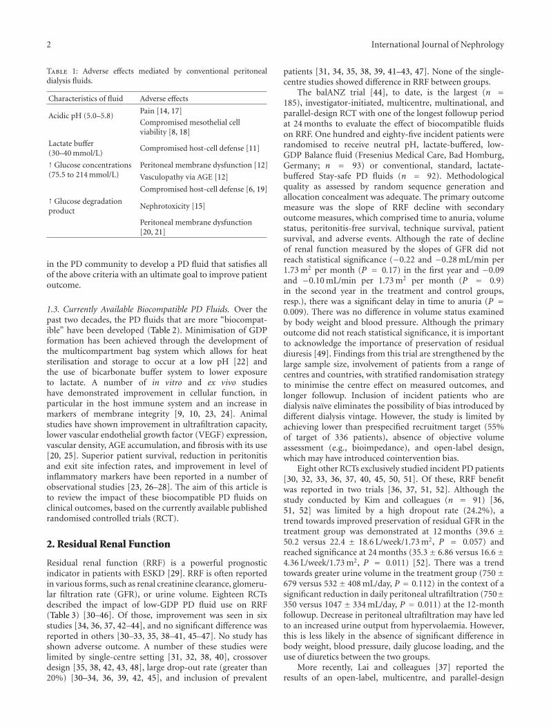
2 International Journal of Nephrology
Table 1: Adverse effects mediated by conventional peritonealdialysis fluids.
Characteristics of fluid Adverse effects
Acidic pH (5.0–5.8)Pain [14, 17]
Compromised mesothelial cellviability [8, 18]
Lactate buffer(30–40 mmol/L)
Compromised host-cell defense [11]
↑ Glucose concentrations(75.5 to 214 mmol/L)
Peritoneal membrane dysfunction [12]
Vasculopathy via AGE [12]
Compromised host-cell defense [6, 19]
↑ Glucose degradationproduct
Nephrotoxicity [15]
Peritoneal membrane dysfunction[20, 21]
in the PD community to develop a PD fluid that satisfies allof the above criteria with an ultimate goal to improve patientoutcome.
1.3. Currently Available Biocompatible PD Fluids. Over thepast two decades, the PD fluids that are more “biocompat-ible” have been developed (Table 2). Minimisation of GDPformation has been achieved through the development ofthe multicompartment bag system which allows for heatsterilisation and storage to occur at a low pH [22] andthe use of bicarbonate buffer system to lower exposureto lactate. A number of in vitro and ex vivo studieshave demonstrated improvement in cellular function, inparticular in the host immune system and an increase inmarkers of membrane integrity [9, 10, 23, 24]. Animalstudies have shown improvement in ultrafiltration capacity,lower vascular endothelial growth factor (VEGF) expression,vascular density, AGE accumulation, and fibrosis with its use[20, 25]. Superior patient survival, reduction in peritonitisand exit site infection rates, and improvement in level ofinflammatory markers have been reported in a number ofobservational studies [23, 26–28]. The aim of this article isto review the impact of these biocompatible PD fluids onclinical outcomes, based on the currently available publishedrandomised controlled trials (RCT).
2. Residual Renal Function
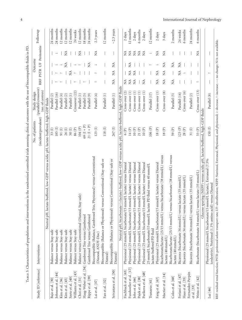
Residual renal function (RRF) is a powerful prognosticindicator in patients with ESKD [29]. RRF is often reportedin various forms, such as renal creatinine clearance, glomeru-lar filtration rate (GFR), or urine volume. Eighteen RCTsdescribed the impact of low-GDP PD fluid use on RRF(Table 3) [30–46]. Of those, improvement was seen in sixstudies [34, 36, 37, 42–44], and no significant difference wasreported in others [30–33, 35, 38–41, 45–47]. No study hasshown adverse outcome. A number of these studies werelimited by single-centre setting [31, 32, 38, 40], crossoverdesign [35, 38, 42, 43, 48], large drop-out rate (greater than20%) [30–34, 36, 39, 42, 45], and inclusion of prevalent
patients [31, 34, 35, 38, 39, 41–43, 47]. None of the single-centre studies showed difference in RRF between groups.
The balANZ trial [44], to date, is the largest (n =185), investigator-initiated, multicentre, multinational, andparallel-design RCT with one of the longest followup periodat 24 months to evaluate the effect of biocompatible fluidson RRF. One hundred and eighty-five incident patients wererandomised to receive neutral pH, lactate-buffered, low-GDP Balance fluid (Fresenius Medical Care, Bad Homburg,Germany; n = 93) or conventional, standard, lactate-buffered Stay-safe PD fluids (n = 92). Methodologicalquality as assessed by random sequence generation andallocation concealment was adequate. The primary outcomemeasure was the slope of RRF decline with secondaryoutcome measures, which comprised time to anuria, volumestatus, peritonitis-free survival, technique survival, patientsurvival, and adverse events. Although the rate of declineof renal function measured by the slopes of GFR did notreach statistical significance (−0.22 and −0.28 mL/min per1.73 m2 per month (P = 0.17) in the first year and −0.09and −0.10 mL/min per 1.73 m2 per month (P = 0.9)in the second year in the treatment and control groups,resp.), there was a significant delay in time to anuria (P =0.009). There was no difference in volume status examinedby body weight and blood pressure. Although the primaryoutcome did not reach statistical significance, it is importantto acknowledge the importance of preservation of residualdiuresis [49]. Findings from this trial are strengthened by thelarge sample size, involvement of patients from a range ofcentres and countries, with stratified randomisation strategyto minimise the centre effect on measured outcomes, andlonger followup. Inclusion of incident patients who aredialysis naıve eliminates the possibility of bias introduced bydifferent dialysis vintage. However, the study is limited byachieving lower than prespecified recruitment target (55%of target of 336 patients), absence of objective volumeassessment (e.g., bioimpedance), and open-label design,which may have introduced cointervention bias.
Eight other RCTs exclusively studied incident PD patients[30, 32, 33, 36, 37, 40, 45, 50, 51]. Of these, RRF benefitwas reported in two trials [36, 37, 51, 52]. Although thestudy conducted by Kim and colleagues (n = 91) [36,51, 52] was limited by a high dropout rate (24.2%), atrend towards improved preservation of residual GFR in thetreatment group was demonstrated at 12 months (39.6 ±50.2 versus 22.4 ± 18.6 L/week/1.73 m2, P = 0.057) andreached significance at 24 months (35.3± 6.86 versus 16.6±4.36 L/week/1.73 m2, P = 0.011) [52]. There was a trendtowards greater urine volume in the treatment group (750±679 versus 532 ± 408 mL/day, P = 0.112) in the context of asignificant reduction in daily peritoneal ultrafiltration (750±350 versus 1047 ± 334 mL/day, P = 0.011) at the 12-monthfollowup. Decrease in peritoneal ultrafiltration may have ledto an increased urine output from hypervolaemia. However,this is less likely in the absence of significant difference inbody weight, blood pressure, daily glucose loading, and theuse of diuretics between the two groups.
More recently, Lai and colleagues [37] reported theresults of an open-label, multicentre, and parallel-design

International Journal of Nephrology 3
Table 2: Selected peritoneal dialysis fluids currently available in Australia.
Solution(manufacturer)
pH Chambers Buffer Glucose degradation products(3-desoxyglycosone) [20, 53, 54]
Conventional PD fluids
Dianeal (Baxter) 5.2 Single Lactate (35–40 mmol/L) ↑↑↑ (525 µmol/L)
Stay-safe (Fresenius) 5.5 Single Lactate (40 mmol/L) ↑↑ (172–324 µmol/L)
Biocompatible PD fluids
Physioneal (Baxter) 7.4 Double Lactate (10–15 mmol/L)/bicarbonate (25 mmol/L) ↓ (253 µmol/L)
Balance (Fresenius) 7.0 Double Lactate (35 mmol/L) ↓↓ (42 µmol/L)
BicaVera (Fresenius) 7.4 Double Bicarbonate (34/39 mmol/L) ↓↓ (42 µmol/L)
Gambrosol Trio (Fresenius) 6.5 Triple Lactate (39–41 mmol/L) ↓↓ (65 µmol/L)
RCT involving 125 incident PD patients. Patients wereassigned to either treatment (Gambrosol Trio, GambroLundia AB, Lund, Sweden (n = 41); Physioneal 40,Baxter Healthcare Corporation, Deerfield, IL, USA (n =12); Balance (n = 5)) or control group (Dianeal PD-2, Baxter Healthcare Corporation (n = 43); ANDY-Disc,Fresenius Medical Care (n = 24)) for an average periodof 3.6 years. Randomisation was instituted by the patient’straining nursing officer at the individual renal centre, whichraises concern for selection and allocation bias. Moreover,informed consent was obtained after the commencement ofstudy at a median period of 30 months. In spite of using PDfluids with variable content of GDP (Table 2), the treatmentgroup had higher urine output (745.7±107.57 versus 475.1±77.69 mL/day, P = 0.04) and slower median decline of bothurine output (0.01 versus 0.33 mL/day, P = 0.004) and resid-ual GFR (0.2 versus 0.56 L/min/1.73 m2/year, P = 0.05) atapproximately 15 months. This study is limited by significantmethodological flaws, and obtainment of informed consentafter commencement of the trial is concerning.
In contrast, lack of benefit in RRF with the use ofbiocompatible PD fluid was reported by Kim and colleagues[45] in their open-label, multi-centre, parallel-design RCTinvolving 26 incident PD patients over 12 months (2.3 ± 0.3versus 1.8 ± 0.7 mL/min, P = NS). There was paucity indescription of methodological process, including absenceof clear reporting of randomisation technique, allocationconcealment, and patient flow to assess for dropout rates.The study analysed the data from 26 patients, but 64 wereinitially recruited, and it was not possible to determine ifthese patients were randomised or even the reasons that ledto their dropout.
A recent open-label, multicentre, parallel-design RCTfrom Hong Kong [46] assessed the effect of NEPP regimen(two exchanges of Physioneal, one Nutrineal, and oneexchange of Extraneal (Baxter); n = 77) against conventionalPD fluids (Dianeal (Baxter); n = 73) in 150 incident CAPDpatients. Although the study observed better preservationof daily urine volume in the treatment group (959 ± 515versus 798 ± 615 mL/day, P = 0.02), they did not identifyany significant difference in RRF (3.24 ± 1.98 versus 2.88 ±2.43 mL/min/1.73 m2, P = NS) or the rate of decline in RRF(−0.76± 1.77 versus −0.91± 1.92 mL/min/1.73 m2/year, P =NS) at 12 months. Adequate randomisation technique andallocation concealment were adopted in this RCT.
Inclusion of prevalent PD patients can cloud the inter-pretation of the outcome when the variable of interest is timedependent, such as RRF. A couple of RCTs included bothincident and prevalent PD patients [34, 39], whereas onlyprevalent PD patients were involved in others [35, 38, 40–43, 47]. Of the three studies that showed benefit on RRF [34,42, 43], the DIUREST study [34] was a parallel-design RCTconducted across three European countries with followupduration of 18 months (n = 80). Patients were centrallyrandomised to a treatment group to receive Gambrosol Trio(Gambro AB, Lund, Sweden) or conventional PD fluidsfrom different manufacturers in single-compartment bags(Gambrosol for 50% of patients (Gambro AB), Stay-safefor 31% (Fresenius Medical Care, Bad Homburg, Germany)or Dianeal for 19% (Baxter GmbH, Unterschleißheim, Ger-many)). A significant benefit in preservation of monthly RRFchange (−1.5%, 95% CI = −3.07%, +0.03% versus −4.3%,95% CI = −6.8%, −2.06%, P = 0.0437) and urine volume(12 versus 38 mL/month, P = 0.0241) in the treatment groupwas reported; however, this should be interpreted cautiouslyin the context of inclusion of both incident and prevalentpatients, unclear allocation concealment, high patient drop-out rate (51%), and use of per-protocol analysis.
3. Peritoneal Solute Transport Rate
Higher peritoneal solute transport rate (PSTR), assessedby the dialysate : plasma creatinine ratio (D : P Cr) from aperitoneal equilibration test (PET) [55], has been recognizedas a significant risk factor for both mortality and techniquefailure in a number of large observational studies [56–60].Although the exact mechanisms that lead to poor survivalremain uncertain, rapid absorption of glucose with removalof osmotic gradient could contribute to impaired soluteand fluid removal. Higher PSTR has been associated withgreater appearance rate of interleukin-6 (IL-6) in PD effluent[61, 62], accumulation of advanced glycation end product(AGE), presence of GDP [63], and use of hypertonic glucosePD fluids [64]. This is biologically plausible, as a rise invascularity followed by an increase in blood flow shouldresult in greater PSTR. Intuitively, the use of biocompatiblePD fluid has been postulated to slow the increase in PSTR.
Nineteen RCTs have reported the effect of biocompatiblePD fluid on PSTR [30–38, 41–45, 48, 51, 52, 65, 69]. Theoutcomes are conflicting, with a number of studies showing
4 International Journal of Nephrology
Ta
ble
3:C
har
acte
rist
ics
ofpo
pula
tion
san
din
terv
enti
ons
inth
era
ndo
mis
edco
ntr
olle
dtr
ials
asse
ssin
gcl
inic
alou
tcom
esw
ith
the
use
ofbi
ocom
pati
ble
flu
ids
inP
D.
Stu
dyID
[ref
eren
ce]
Inte
rven
tion
sN
o.of
pati
ents
Stu
dyde
sign
Ou
tcom
eFo
llow
up
(in
cide
nt/
prev
alen
t)(p
aral
lel/
cros
sove
r)(n
o.of
cen
tres
)R
RF
PST
RU
FPe
rito
nit
is
Neu
tral
pH,l
acta
te-b
uff
ered
,low
-GD
Pve
rsu
sac
idic
pH,l
acta
te-b
uff
ered
,hig
h-G
DP
flu
ids
Baj
oet
al.[
30]
Bal
ance
vers
us
Stay
-saf
e33
(I)
Para
llel(
2)—
——
—24
mon
ths
Joh
nso
net
al.[
44]
Bal
ance
vers
us
Stay
-saf
e18
5(I
)Pa
ralle
l(16
)↑
↑↓
↓24
mon
ths
Kim
etal
.[36
]B
alan
ceve
rsu
sSt
ay-s
afe
91(I
)Pa
ralle
l(4)
↑↑
↓—
12m
onth
sK
imet
al.[
45]
Bal
ance
vers
us
Stay
-saf
e26
(I)
Para
llel(
2)—
—N
AN
A12
mon
ths
Szet
oet
al.[
40]
Bal
ance
vers
us
Stay
-saf
e50
(I)
Para
llel(
1)—
NA
——
12m
onth
sW
illia
ms
etal
.[43
]B
alan
ceve
rsu
sSt
ay-s
afe
86(P
)C
ross
over
(22)
↑↑
↓—
24w
eeks
Ch
oiet
al.[
31]
Bal
ance
vers
us
Con
ven
tion
al(D
ian
eal,
Stay
-saf
e)10
4(P
)Pa
ralle
l(1)
——
↑N
A12
mon
ths
Haa
g-W
eber
etal
.[34
]G
ambr
osol
Trio
vers
us
Gam
bros
ol69
(I+
P)
Para
llel(
5)↑
——
—18
mon
ths
Rip
pe
etal
.[39
]G
ambr
osol
Trio
vers
us
Gam
bros
ol21
(I+
P)
Para
llel(
9)—
—N
A—
24m
onth
s
Laie
tal
.[37
]B
ioco
mpa
tibl
e(B
alan
ce,G
ambr
osol
Trio
,Phy
sion
eal)
vers
us
Con
ven
tion
al(D
ian
eal,
AN
DY-
Dis
c)12
5(I
)Pa
ralle
l(4)
↑↑
↓N
A2.
3ye
ars
Fan
etal
.[32
]B
ioco
mpa
tibl
e(B
alan
ceor
Phy
sion
eal)
vers
us
Con
ven
tion
al(S
tay-
safe
orD
ian
eal)
118
(I)
Para
llel(
1)—
——
—12
mon
ths
Sriv
asta
vaet
al.[
50]
Bio
com
pati
ble
(Bal
ance
orP
hysi
onea
l)ve
rsu
sC
onve
nti
onal
(Sta
y-sa
feor
Dia
nea
l)26
7(I
)Pa
ralle
l(1)
NA
NA
NA
—∼2
.3ye
ars
Neu
tral
pH,b
icar
bon
ate
(±la
ctat
e)-b
uff
ered
,low
-GD
Pve
rsu
sac
idic
pH,l
acta
te-b
uff
ered
,hig
h-G
DP
flu
ids
Fisc
hba
cket
al.[
65]
Phy
sion
eal(
25m
mol
/Lbi
carb
onat
e/15
mm
ol/L
lact
ate)
vers
us
Dia
nea
l6
(P)
Cro
ss-o
ver
(2)
NA
——
NA
2da
ysFu
ssh
oelle
ret
al.[
17]
Phy
sion
eal(
25m
mol
/Lbi
carb
onat
e/15
mm
ol/L
lact
ate)
vers
us
Dia
nea
l14
(P)
Cro
ss-o
ver
(1)
NA
NA
NA
NA
12m
onth
sJo
hn
etal
.[66
]P
hysi
onea
l(25
mm
ol/L
bica
rbon
ate/
15m
mol
/Lla
ctat
e)ve
rsu
sD
ian
eal
10(P
)C
ross
-ove
r(1
)N
AN
A—
NA
2da
ysPa
jek
etal
.[38
]P
hysi
onea
l(25
mm
ol/L
bica
rbon
ate/
15m
mol
/Lla
ctat
e)ve
rsu
sD
ian
eal
21(P
)C
ross
-ove
r(1
)—
——
NA
6m
onth
sPa
riko
vaet
al.[
48]
Phy
sion
eal(
25m
mol
/Lbi
carb
onat
e/15
mm
ol/L
lact
ate)
vers
us
Dia
nea
l10
(P)
Cro
ss-o
ver
(1)
NA
——
NA
2da
ys
Tran
aeu
s[4
1]25
mm
ol/L
bica
rbon
ate/
15m
mol
/Lla
ctat
eP
Dfl
uid
vers
us
40m
mol
/Lla
ctat
e-bu
ffer
edP
Dfl
uid
106
(P)
Para
llel(
17)
——
↑↓
12m
onth
s
Fan
get
al.[
67]
Phy
sion
eal(
25m
mol
/Lbi
carb
onat
e/15
mm
ol/L
lact
ate)
vers
us
Dia
nea
l18
(P)
Cro
ss-o
ver
(1)
NA
NA
↓N
A2
days
Mac
tier
etal
.[14
]B
icar
bon
ate/
lact
ate
(25/
15m
mol
/L)
vers
us
bica
rbon
ate
(38
mm
ol/L
)ve
rsu
sla
ctat
e(4
0m
mol
/L)
18(P
)C
ross
-ove
r(8
)N
AN
AN
AN
A3
days
Col
eset
al.[
68]
Bic
arbo
nat
e/la
ctat
e(2
5/15
mm
ol/L
)ve
rsu
sbi
carb
onat
e(3
8m
mol
/L)
vers
us
lact
ate
(40
mm
ol/L
)59
(P)
Para
llel(
5)N
A—
NA
—2
mon
ths
Feri
anie
tal
.[47
]B
icav
era
(bic
arbo
nat
e34
mm
ol/L
)ve
rsu
sla
ctat
e(3
5m
mol
/L)
123
(P)
Para
llel(
14)
—N
AN
A—
24w
eeks
Haa
set
al.[
35]
Bic
aver
a(b
icar
bon
ate
34m
mol
/L)
vers
us
lact
ate
(35
mm
ol/L
)28
(P)
Cro
ss-o
ver
(6)
—↓
——
6m
onth
sFe
rnan
dez-
Perp
enet
al.[
33]
Bic
aver
a(b
icar
bon
ate
34m
mol
/L)
vers
us
lact
ate
(35
mm
ol/L
)31
(I)
Para
llel(
2)—
——
—24
mon
ths
Wei
sset
al.[
42]
Bic
arbo
nat
e(b
icar
bon
ate
34m
mol
/L)
vers
us
lact
ate
(35
mm
ol/L
)53
(P)
Cro
ss-o
ver
(13)
↑—
—N
A6
mon
ths
Glu
cose
spar
ing
(NE
PP
)ve
rsu
sac
idic
pH,l
acta
te-b
uff
ered
,hig
h-G
DP
flu
ids
Lui[
46]
Phy
sion
eal(
25m
mol
/Lbi
carb
onat
e/15
mm
ol/L
lact
ate)
,Ext
ran
eal(
7.5%
icod
extr
in),
Nu
trin
eal(
1.1%
amin
oac
id)
vers
us
Dia
nea
l15
0(I
)Pa
ralle
l(8)
—↑
——
RR
F:re
sidu
alre
nal
fun
ctio
n;P
STR
:per
iton
eals
olu
tetr
ansp
ort
rate
;UF:
ult
rafi
ltra
tion
;NE
PP
:Nu
trin
eal,
Ext
ran
eal,
Phy
sion
eal,
and
phys
ion
eal;↓:
decr
ease
,↑:i
ncr
ease
;—:n
och
ange
;N/A
:not
avai
labl
e.
International Journal of Nephrology 5
a decrease [35], an increase [37, 43, 44, 46, 51, 52], and nochange [30, 32–34, 38, 41, 42, 45, 48, 65, 69]. In general,PSTR increases with time on PD, therefore, similar to RRF,inclusion of prevalent PD patients [31, 34, 35, 38, 41–43, 48,65, 69] and crossover design [35, 38, 42, 43, 48, 65] creates adilemma in the understanding of the outcome. Furthermore,interpretation of studies that showed greater PSTR in thetreatment group should be done carefully as the differencewas already present at the baseline (or month 1) in threestudies [36, 37, 44, 51, 52, 70]. Kim and colleagues [36,51, 52] reported a significant difference between treatmentand control groups at baseline (0.72 ± 0.1 versus 0.67 ± 0.1,P = 0.001) and at 12 months (0.72 ± 0.11 versus 0.64 ±0.08, P = 0.001). However, within-group analysis failed toshow significant difference over the 12-month period. A largevariation in PSTR between PD patients is well recognised[71]. Therefore, a difference at baseline may not be due tothe biocompatible PD fluid, and the trend in PSTR over timemay be of greater importance.
The trend in PSTR was reported in the Euro-Balance Trial[43]. In this multicentre, open-label, crossover design RCT,86 prevalent PD patients from 22 centres in 11 Europeancountries were randomly allocated to conventional, acidic,lactate-buffered fluid (Stay-safe; Fresenius Medical Care, BadHomburg, Germany) or neutral pH, lactate-buffered, low-GDP fluid (Balance; Fresenius Medical Care, Bad Homburg,Germany) for 12 weeks. There was no washout periodbetween the two study periods. Per-protocol analysis wasperformed in 71 patients who completed the trial. Patientsin the group I started receiving conventional fluids for thefirst 12 weeks followed by biocompatible fluid, and the orderwas reversed for patients in group II. In group I (n = 36),PSTR was higher whilst receiving biocompatible PD fluid(0.63 [0.34–0.89] versus 0.59 [0.35–0.80], P = 0.008) andsimilar outcome was reported in group II (0.60 [0.38–0.80]versus 0.56 [0.42–0.80], P = 0.0003). The decrease in PSTRwith the use of biocompatible PD fluid has been reportedby only one trial [35]. This study was a multicentre, open-label, crossover design RCT involving 28 prevalent patients.Following a 4-week run-in period, patients underwent twoconsecutive 12-week study periods, in randomised order,in which PD was performed with a neutral-pH PD fluidcontaining 34 mmol/L bicarbonate (BicaVera 170/180/190;Fresenius Medical Care, Bad Homburg, Germany) or aconventional PD fluid with 35 mmol/L lactate buffer (pH5.5, CAPD 17/18/19; Fresenius Medical Care). The twotreatment phases were separated by a 4-week washout periodwith a lactate-buffered PD fluid. Per-protocol analysis wasperformed in the twenty patients who completed bothphases. A significant decrease in 4-hour D : P Cr during thetreatment phase (0.67±0.14 versus 0.70±0.12, P < 0.05) wasreported. Although these two trials were multinational andmulticentre, they suffered from methodological problemsincluding relatively small sample size, per protocol analysis,crossover design, and short followup duration. The lattertwo issues are particularly relevant given the time-dependentnature of PSTR and the risk of carryover effect of the PD
fluids used. Therefore, the effect or lack of effect posed withthe use of biocompatible PD fluid remains to be unknown.
4. Peritoneal Ultrafiltration
The decrease in peritoneal ultrafiltration (UF) is an impor-tant cause of technique failure [4]. Although it is largelydriven by loss of osmotic gradient from higher PSTR withtime on PD, disproportionate decrease in UF capacity canoccur [72]. This is thought to result from an increase inmembrane fibrosis, thereby compromising osmotic conduc-tance independent of PSTR [73]. Severe fibrosis in the peri-toneum from morphologic examination has been attributedas a consequence of continuous exposure to “unphysiologic”PD fluids [13]. However, accurate interpretation of theimplication of UF volume as a clinical outcome is complex,as there are many variables that can affect its level, such asbody’s fluid status, urine volume, PSTR, glucose load, andthe use of 7.5% icodextrin.
Of the eighteen RCTs [30–38, 40–44, 48, 51, 52, 65–67]reporting UF, six studies showed a decrease in UF with theuse of biocompatible PD fluids [36, 37, 43, 44, 51, 52, 67].Interestingly, five RCTs within this category reported anincrease in the urine volume with the use of biocompatiblePD fluids [36, 37, 43, 44, 51, 52]. This highlights theimportance of interpreting data in the context of otherparameters present.
An increase in UF with the use of biocompatible fluidwas reported in only two RCTs [31, 41]. Both studies wereperformed in prevalent PD patients, and neither of thestudies showed any difference in RRF between groups. Choiand colleagues [31] performed a single-centre, open-label,parallel-design RCT over 12 months. Of the 104 patientswho were randomised, 66 patients were anuric at the timeof enrolment with median PD duration of 67 months inthe treatment group (n = 51) and 70.4 months in thecontrol group (n = 53). Daily UF was significantly greaterin the treatment group (1301.3 ± 597.6 versus 981.7 ±538.8 mL/day, P < 0.05) in spite of similar glucose load(151.4 ± 54.5 versus 167.3 ± 38.8 g/day). Randomisa-tion technique or allocation concealment were not clearlydescribed, and the study suffered from a moderately highdropout rate (35%).
Similarly, Tranaeus [41] conducted an open-label, paral-lel-design RCT across 17 European nephrology centres in106 prevalent PD patients with mean baseline RRF of2.8 mL/min/1.73 m2 over 12 months. Statistically significantdifference (P < 0.05) in favour of biocompatible PDfluid was demonstrated (numerical data is not reported inthe study). Stratified randomisation block technique wasadopted; however, allocation concealment method was notclearly described. Less than half of the patients (n =44) completed the study, which raises the possibility ofattrition bias. Based on these two studies, perhaps the useof biocompatible PD fluid may be favoured to improveperitoneal UF in prevalent PD patients. However, thesefindings were not reproduced in other trials which includedprevalent patients [35, 38, 42, 43, 48, 65–67]. Interestingly,all of those eight RCTs were crossover in design.
6 International Journal of Nephrology
5. Peritonitis
The use of biocompatible PD fluids has been associated withreduction in peritonitis in an observational study [74]. Thisis supported by a number of in vitro and ex vivo studiesthat have demonstrated improvement in cellular function,in particular in the host immune system and an increase inmarkers of membrane integrity with their use [9, 10, 23, 24].Peritonitis is an important cause of higher technique failurein Australia and New Zealand [4] and has been associatedwith greater mortality [5].
Disappointingly, however, of the 14 RCTs that reportedperitonitis [30, 32–36, 38–44, 46, 47, 50–52, 68], only twoshowed significant benefit with the use of biocompatible PDfluids. The balANZ trial reported a significant delay in timeto the first peritonitis episode (P = 0.01) and lower overallrates of peritonitis in the treatment group (0.30 versus 0.49episodes per year, P = 0.01). Likewise, a significant reductionin peritonitis rate was demonstrated in the treatment group(1 : 51 patient-months versus 1 : 19 patient-months, P <0.05) by Tranaeus [41]. No study has reported significantincrease in peritonitis risk with the use of biocompatible PDfluids.
Of the trials that showed no benefit, only the RCTconducted by Srivastava and colleagues [50] was poweredadequately to examine the peritonitis. This was an extensionstudy of an open-label, parallel-design, single-centre RCT(n = 118, dropout 21.7%) with initial followup of 12 months[32]. Enrolment into the study continued to achieve suffi-cient power to report any statistically significant difference inperitonitis episodes, which resulted in the inclusion of a largenumber of incident patients (n = 267). The treatment groupreceived biocompatible PD solutions (either Physioneal orBalance) and control group received conventional PD solu-tions (either Dianeal or Stay-safe). The patients who usedBaxter system (85% overall) were additionally allowed to useExtraneal or Nutrineal during the study duration. Patientswere allowed to use different connectology (1-2 connections)that was felt to be best suited to each individual. Therewere 227 peritonitis episodes suffered by the patients, withan at-risk period of 7408 patient-months. Peritonitis ratefor the treatment group was 1 : 34.7 versus 1 : 31.5 monthsin the control group (P = 0.61). Although this studywas strengthened by large patient numbers, allowance ofsystems requiring different number of connections, therebyintroducing variable risk of contamination and a varietyof PD fluid types with varying contents (e.g., GDP, buffersystem), could have introduced bias.
6. Pain
Inflow pain is generally attributed to the acidity (pH 5.2 to5.5) of conventional lactate-buffered PD fluids. Although itis often temporary, it can be a troublesome complication insome PD patients to result in discontinuation of PD. FiveRCTs assessed the effect of biocompatible PD fluids on inflowpain [14, 17, 41, 42, 47], with the majority of the studiesreporting favourable result with the use of bicarbonate-buffered PD fluids.
Mactier and colleagues [14] performed a double-blind,multicentre, multicountry, crossover design RCT in patientswho had previously experienced inflow pain using con-ventional lactate-buffered PD fluids. Eighteen patients wererecruited, and 17 completed the study protocol whichcomprised of two dialysis exchanges with each test solutiondetermined by random allocation. Three visits were requiredto complete six exchanges in total (i.e., two exchangesper test solution). All tested fluids were of same glucosestrength (3.86%), and pain was assessed by two methods(five-point verbal scale and the McGill Pain Questionnaire).Bicarbonate-buffered PD fluids were associated with sig-nificant reductions in inflow pain using both assessmentmethods. Bicarbonate/lactate-buffered PD fluid performedthe best in terms of improving alleviating pain when all painvariables were assessed. However, there was a large variationwithin the eight participating centres in the frequency ofinflow pain, which raises the concern for centre-relatedeffects.
Three other RCTs also reported significant benefit withthe use of bicarbonate- or bicarbonate/lactate-buffered PDfluids [17, 41, 47]. Level of pain was measured using differenttools devised during each trial in a form of questionnaire.For instance, Fusshoeller and colleagues conducted a single-centre, open-label, crossover design RCT in 14 prevalent PDpatients [17]. Patients were randomised to have automatedPD with either conventional fluid (Dianeal; Baxter Health-care SA, County Mayo, Ireland) or a bicarbonate/lactate-based neutral fluid (Physioneal; Baxter Healthcare SA,County mayo, Ireland). After 6 months, both groups changedfluids. There was no washout period. Dialysate inflowpain was assessed with the use of a patient questionnaireconducted at baseline visit (1 = no pain; 5 = very intense) andat the end of the 5 months of treatment with each of the PDfluids. Similar findings were reported by Tranaeus [41]; therewas a significant reduction in dialysate inflow pain in thetreatment group (0.46 ± 0.93 versus 1.67 ± 0.70; P = 0.05).
Feriani and colleagues [47] conducted a multicentre,open-label, parallel-design RCT over a 24-week period inprevalent PD patients (n = 123). Patients were randomlyallocated to receive either a bicarbonate- or lactate-bufferedPD fluid. Adverse symptoms were recorded using a stan-dardized questionnaire (higher score indicating increase inseverity) assessing local (pain during infusion, constipation,and diarrhoea), uraemic (itching, headache, restless legs,tiredness, and loss of appetite), and volume (thirst, ankleswelling, abdominal fullness, difficulty in maintaining cor-rect weight, circulatory troubles, and shortness of breath)effects. Significant improvement in “local effects” was shownin the treatment group (0.25 ± 0.60 versus 0.45 ± 0.87,P < 0.01). The results from these three RCTs shouldbe interpreted with caution as they were open-label RCTsleading to possible performance bias.
A multicentre, open-label, cross-over design RCT con-ducted across three European countries including 53 preva-lent PD patients was conducted by Weiss and colleagues [42].Following a 2-week run-in phase, patients were randomisedto receive either standard lactate-buffered PD fluids or purelybicarbonate-buffered PD fluids (Fresenius Medical Care,
International Journal of Nephrology 7
Bad Homburg, Germany) for 12 weeks, following which thetreatment fluids were switched and continued for further12 weeks. After completing this phase, pain assessment wasperformed under blinded administration condition of fourexchanges in a randomised order. Twenty-seven patientswho completed both treatment phases were included foranalyses, and twenty-three proceeded to pain assessment.Pain intensity was assessed using McGill Pain Questionnaire,with similar outcomes between the two groups. In specific,4 of 23 patients reported pain with both solutions duringinflow.
7. Conclusion
There has been an increase in a number of publishedRCTs that compare the clinical outcome from the use ofbiocompatible PD fluids over the past decade. The resultsare generally in favour of or at least neutral with regards toRRF, PD peritonitis, and inflow pain in those who receivedbiocompatible PD fluids. Its impact on peritoneal membranefunction (i.e., PSTR and UF) remains uncertain. Some ofthe variability in the reported outcomes stem from flawsin study design, inclusion of patients from different dialysisvintage, inadequate statistical power to assess hard endpoints(e.g., mortality, technique failure), high dropout rates, andadoption of inappropriate analytical methods. Predominantuse of open-label designs introduce cointervention andobserver biases. Meta-analysis of all RCTs to clarify whetherthe use of biocompatible fluids translates into importantclinical benefits is currently in progress [75]. The outcomeof the analyses may provide further evidence for or againstthe use of these products. In the future, a large RCTwith adequate statistical power to assess hard endpointssuch as patient and technique survivals with the use ofbiocompatible PD fluids is needed.
Conflict of Interests
D. W. Johnson is a principal investigator of the balANZstudy (funded by Fresenius Medical Care). He has receivedconsulting fees from Baxter and Gambro; research grants,payment for lectures, and travel grants from Baxter andFresenius. C. M. Hawley has received consulting fees fromFresenius and research grant from Baxter. The remainingauthors have reported that they have no potential conflict ofinterests to declare.
References
[1] S. J. Davies, W. Van Biesen, J. Nicholas, and N. Lameire,“Integrated care,” Peritoneal Dialysis International, vol. 21,Supplement 3, pp. S269–S274, 2001.
[2] S. P. McDonald, M. R. Marshall, D. W. Johnson, and K. R.Polkinghorne, “Relationship between dialysis modality andmortality,” Journal of the American Society of Nephrology, vol.20, no. 1, pp. 155–163, 2009.
[3] P. G. Blake, “Integrated end-stage renal disease care: the role ofperitoneal dialysis,” Nephrology Dialysis Transplantation, vol.16, Supplement 5, pp. 61–66, 2001.
[4] F. G. Brown, A. H. Dent, K. Hurst, and S. McDonald,Peritoneal Dialysis, Chapter 6, Australia & New ZealandDialysis & Transplantation (ANZDATA) Registry, 2011.
[5] N. Boudville, A. Kemp, P. Clayton et al., “Recent peritonitisassociates with mortality among patients treated with peri-toneal dialysis,” Journal of the American Society of Nephrology,vol. 23, no. 8, pp. 1398–1405, 2012.
[6] M. P. Catalan, B. Santamarıa, A. Reyero, A. Ortiz, J. Egido, andA. Ortiz, “3,4-Di-deoxyglucosone-3-ene promotes leukocyteapoptosis,” Kidney International, vol. 68, no. 3, pp. 1303–1311,2005.
[7] N. Topley, R. Mackenzie, M. M. Petersen et al., “In vitrotesting of a potentially biocompatible continuous ambulatoryperitoneal dialysis fluid,” Nephrology Dialysis Transplantation,vol. 6, no. 8, pp. 574–581, 1991.
[8] J. Witowski, N. Topley, A. Jorres, T. Liberek, G. A. Coles, andJ. D. Williams, “Effect of lactate-buffered peritoneal dialysisfluids on human peritoneal mesothelial cell interleukin-6 andprostaglandin synthesis,” Kidney International, vol. 47, no. 1,pp. 282–293, 1995.
[9] S. Mortier, A. S. De Vriese, R. M. McLoughlin et al., “Effects ofconventional and new peritoneal dialysis fluids on leukocyterecruitment in the rat peritoneal membrane,” Journal of theAmerican Society of Nephrology, vol. 14, no. 5, pp. 1296–1306,2003.
[10] E. Boulanger, M. P. Wautier, J. L. Wautier et al., “AGEsbind to mesothelial cells via RAGE and stimulate VCAM-1expression,” Kidney International, vol. 61, no. 1, pp. 148–156,2002.
[11] H. T. Schambye, “Effect of different buffers on the biocom-patibility of CAPD solutions,” Peritoneal Dialysis International,vol. 16, Supplement 1, pp. S130–S136, 1996.
[12] S. Mortier, A. S. De Vriese, and N. Lameire, “Recent conceptsin the molecular biology of the peritoneal membrane—implications for more biocompatible dialysis solutions,” BloodPurification, vol. 21, no. 1, pp. 14–23, 2003.
[13] J. D. Williams, K. J. Craig, N. Topley et al., “Morphologicchanges in the peritoneal membrane of patients with renaldisease,” Journal of the American Society of Nephrology, vol. 13,no. 2, pp. 470–479, 2002.
[14] R. A. Mactier, T. S. Sprosen, R. Gokal et al., “Bicarbonateand bicarbonate/lactate peritoneal dialysis solutions for thetreatment of infusion pain,” Kidney International, vol. 53, no.4, pp. 1061–1067, 1998.
[15] P. Justo, A. B. Sanz, J. Egido, and A. Ortiz, “3,4-Dideoxyglu-cosone-3-ene induces apoptosis in renal tubular epithelialcells,” Diabetes, vol. 54, no. 8, pp. 2424–2429, 2005.
[16] T. Tanikawa, Y. Okada, R. Tanikawa, and Y. Tanaka, “Advancedglycation end products induce calcification of vascular smoothmuscle cells through rage/p38 MAPK,” Journal of VascularResearch, vol. 46, no. 6, pp. 572–580, 2009.
[17] A. Fusshoeller, M. Plail, B. Grabensee, and J. Plum, “Biocom-patibility pattern of a bicarbonate/lactate-buffered peritonealdialysis fluid in APD: a prospective, randomized study,”Nephrology Dialysis Transplantation, vol. 19, no. 8, pp. 2101–2106, 2004.
[18] N. Topley, “In vitro biocompatibility of bicarbonate-basedperitoneal dialysis solutions,” Peritoneal Dialysis International,vol. 17, no. 1, pp. 42–47, 1997.
[19] S. Mortier, N. H. Lameire, and A. S. De Vriese, “The effectsof peritoneal dialysis solutions on peritoneal host defense,”Peritoneal Dialysis International, vol. 24, no. 2, pp. 123–138,2004.
8 International Journal of Nephrology
[20] S. Mortier, D. Faict, C. G. Schalkwijk, N. H. Lameire, andA. S. De Vriese, “Long-term exposure to new peritonealdialysis solutions: effects on the peritoneal membrane,” KidneyInternational, vol. 66, no. 3, pp. 1257–1265, 2004.
[21] J. Witowski, J. Wisniewska, K. Korybalska et al., “Prolongedexposure to glucose degradation products impairs viabilityand function of human peritoneal mesothelial cells,” Journal ofthe American Society of Nephrology, vol. 12, no. 11, pp. 2434–2441, 2001.
[22] J. Passlick-Deetjen and C. Lage, “Lactate-buffered andbicarbonate-buffered solutions with less glucose degradationproducts in a two-chamber system,” Peritoneal Dialysis Inter-national, vol. 20, Supplement 2, pp. S42–S47, 2000.
[23] O. Devuyst, N. Topley, and J. D. Williams, “Morphologicaland functional changes in the dialysed peritoneal cavity:impact of more biocompatible solutions,” Nephrology DialysisTransplantation, vol. 17, Supplement 3, pp. 12–15, 2002.
[24] R. Mackenzie, C. J. Holmes, S. Jones, J. D. Williams, and N.Topley, “Clinical indices of in vivo biocompatibility: the role ofex vivo cell function studies and effluent markers in peritonealdialysis patients,” Kidney International, vol. 64, no. 88, pp.S84–S93, 2003.
[25] S. Mortier, D. Faict, N. H. Lameire, and A. S. De Vriese,“Benefits of switching from a conventional to a low-GDPbicarbonate/lactate- buffered dialysis solution in a rat model,”Kidney International, vol. 67, no. 4, pp. 1559–1565, 2005.
[26] J. D. Furkert, M. Zeier, and V. Schwenger, “Effects of Peritonealdialysis solutions low in GDPs on peritonitis and exit-siteinfection rates,” Peritoneal Dialysis International, vol. 28, no.6, pp. 637–640, 2008.
[27] H. Y. Lee, H. C. Park, B. J. Seo et al., “Superior patient survivalfor continuous ambulatory peritoneal dialysis patients treatedwith a peritoneal dialysis fluid with neutral pH and low glu-cose degradation product concentration (balance),” PeritonealDialysis International, vol. 25, no. 3, pp. 248–255, 2005.
[28] V. Stankovic-Popovic et al., “Effects of conventional versusbiocompatible peritoneal dialysis solutions on peritoneal andsystemic inflammation, malnutrition and atherosclerosis inCAPD patients,” Clinical Nephrology, vol. 76, no. 4, pp. 314–322, 2011.
[29] J. M. Bargman, K. E. Thorpe, and D. N. Churchill, “Relativecontribution of residual renal function and peritoneal clear-ance to adequacy of dialysis: a reanalysis of the CANUSAstudy,” Journal of the American Society of Nephrology, vol. 12,no. 10, pp. 2158–2162, 2001.
[30] M. A. Bajo, M. L. Prıez-Lozano, P. Albar-Vizcaino et al., “Low-GDP peritoneal dialysis fluid (‘balance’) has less impact invitro and ex vivo on epithelial-to-mesenchymal transition(EMT) of mesothelial cells than a standard fluid,” NephrologyDialysis Transplantation, vol. 26, no. 1, pp. 282–291, 2011.
[31] H. Y. Choi, D. K. Kim, T. H. Lee et al., “The clinical usefulnessof peritoneal dialysis fluids with neural pH and low glucosedegradation product concentration: an open randomizedprospective trial,” Peritoneal Dialysis International, vol. 28, no.2, pp. 174–182, 2008.
[32] S. L. S. Fan, T. Pile, S. Punzalan, M. J. Raftery, and M.M. Yaqoob, “Randomized controlled study of biocompatibleperitoneal dialysis solutions: effect on residual renal function,”Kidney International, vol. 73, no. 2, pp. 200–206, 2008.
[33] A. Fernandez-Perpen, M. L. Perez-Lozano, M. A. Bajo et al.,“Influence of Bicarbonate/Low-Gdp peritoneal dialysis fluid(Bicavera) on in vitro and ex vivo epithelial-to-mesenchymaltransition of mesothelial cells,” Peritoneal Dialysis Interna-tional, vol. 32, no. 3, pp. 292–304, 2012.
[34] M. Haag-Weber, R. Kramer, R. Haake et al., “Low-GDP fluid(Gambrosol trio) attenuates decline of residual renal functionin PD patients: a prospective randomized study,” NephrologyDialysis Transplantation, vol. 25, no. 7, pp. 2288–2296, 2010.
[35] S. Haas, C. P. Schmitt, K. Arbeiter et al., “Improved acidosiscorrection and recovery of mesothelial cell mass with neutral-pH bicarbonate dialysis solution among children undergoingautomated peritoneal dialysis,” Journal of the American Societyof Nephrology, vol. 14, no. 10, pp. 2632–2638, 2003.
[36] S. Kim, J. Oh, S. Kim et al., “Benefits of biocompatible PD fluidfor preservation of residual renal function in incident CAPDpatients: a 1-year study,” Nephrology Dialysis Transplantation,vol. 24, no. 9, pp. 2899–2908, 2009.
[37] K. N. Lai et al., “A study of the clinical and biochemicalprofile of peritoneal dialysis fluid low in glucose degradationproducts,” Peritoneal Dialysis International, vol. 32, no. 3, pp.280–291, 2012.
[38] J. Pajek, R. Kveder, A. Bren et al., “Short-term effects ofbicarbonatelactate-buffered and conventional lactate-buffereddialysis solutions on peritoneal ultrafiltration: a comparativecrossover study,” Nephrology Dialysis Transplantation, vol. 24,no. 5, pp. 1617–1625, 2009.
[39] B. Rippe, O. Simonsen, O. Heimburger et al., “Long-termclinical effects of a peritoneal dialysis fluid with less glucosedegradation products,” Kidney International, vol. 59, no. 1, pp.348–357, 2001.
[40] C. Szeto, C. Lam, C. Leung, B. Kwan, K. Chung et al., “Clinicalbiocompatibility of a neutral peritoneal dialysis solution withminimal glucose-degradation products—a 1-year randomizedcontrol trial,” Nephrology Dialysis Transplantation, vol. 22, no.2, pp. 552–559, 2007.
[41] A. Tranaeus, “A long-term study of a bicarbonate/lactate-based peritoneal dialysis solution—clinical benefits,” Peri-toneal Dialysis International, vol. 20, no. 5, pp. 516–523, 2000.
[42] L. Weiss, B. Stegmayr, G. Malmsten et al., “Biocompatibilityand tolerability of a purely bicarbonate-buffered peritonealdialysis solution,” Peritoneal Dialysis International, vol. 29, no.6, pp. 647–655, 2009.
[43] J. D. Williams, N. Topley, K. J. Craig et al., “The Euro-BalanceTrial: the effect of a new biocompatible peritoneal dialysis fluid(balance) on the peritoneal membrane,” Kidney International,vol. 66, no. 1, pp. 408–418, 2004.
[44] D. W. Johnson et al., “Effects of Biocompatible versus standardfluid on peritoneal dialysis outcomes,” Journal of the AmericanSociety of Nephrology, vol. 23, no. 6, pp. 1097–1107, 2012.
[45] Y. L. Kim, J. Do, S. H. Park et al., “Low glucose degradationproducts dialysis solution modulates the levels of surrogatemarkers of peritoneal inflammation, integrity, and angiogene-sis: preliminary report,” Nephrology, vol. 8, Supplement s2, pp.S28–S32, 2003.
[46] S. L. Lui, S. Yung, A. Yim et al., “A combination ofbiocompatible peritoneal dialysis solutions and residual renalfunction, peritoneal transport, and inflammation markers:a randomized clinical trial,” American Journal of KidneyDiseases, vol. 60, no. 6, pp. 966–975, 2012.
[47] M. Feriani, J. Kirchgessner, G. La Greca et al., “Randomizedlong-term evaluation of bicarbonate-buffered CAPD solu-tion,” Kidney International, vol. 54, no. 5, pp. 1731–1738, 1998.
[48] A. Parikova, D. G. Struijk, M. M. Zweers et al., “Does thebiocompatibility of the peritoneal dialysis solution matterin assessment of peritoneal function?” Peritoneal DialysisInternational, vol. 27, no. 6, pp. 691–696, 2007.
International Journal of Nephrology 9
[49] J. M. Bargman, K. E. Thorpe, and D. N. Churchill, “Relativecontribution of residual renal function and peritoneal clear-ance to adequacy of dialysis: a reanalysis of the CANUSAstudy,” Journal of the American Society of Nephrology, vol. 12,no. 10, pp. 2158–2162, 2001.
[50] S. Srivastava, S. Hildebrand, and S. L. S. Fan, “Long-term follow-up of patients randomized to biocompatible orconventional peritoneal dialysis solutions show no differencein peritonitis or technique survival,” Kidney International, vol.80, no. 9, pp. 986–991, 2011.
[51] S. G. Kim, S. Kim, Y. H. Hwang et al., “Could solutionslow in glucose degradation products preserve residual renalfunction in incident peritoneal dialysis patients? A 1-yearmulticenter prospective randomized controlled trial (Balnetstudy),” Peritoneal Dialysis International, vol. 28, Supplement3, pp. S117–S122, 2008.
[52] S. J. Kim, W. K. Chung, K. H. Oh, and S. G. Kim, “Effectof biocompatible PD fluid on preservation of residual renalfunction incident CAPD patients: two-year extended follow-up study,” Nephrology Dialysis Transplantation, vol. 3, pp.iii175–iii176, 2010.
[53] M. Feriani and R. T. Krediet, “New peritoneal dialysissolutions and solutions on the horizon,” in Nolph and Gokal’STextbook of Peritoneal DialySiS, R. Khanna and R. T. Krediet,Eds., Chapter 11, Springer, New York, NY, USA, 2009.
[54] C. Lage, M. Pischetsrieder, C. Aufricht, A. Jorres, H. Schilling,and J. Passlick-Deetjen, “First in vitro and in vivo experienceswith stay·safe balance, a pH-neutral solution in a dual-chambered bag,” Peritoneal Dialysis International, vol. 20,Supplement 5, pp. S28–S32, 2001.
[55] Z. J. Twardowski, “PET—a simpler approach for determiningprescriptions for adequate dialysis therapy,” Advances inPeritoneal Dialysis, vol. 6, pp. 186–191, 1990.
[56] L. Fried, “Higher membrane permeability predicts poorerpatient survival,” Peritoneal Dialysis International, vol. 17, no.4, pp. 387–389, 1997.
[57] M. Rumpsfeld, S. P. McDonald, and D. W. Johnson, “Higherperitoneal transport status is associated with higher mortalityand technique failure in the Australian and New Zealand peri-toneal dialysis patient populations,” Journal of the AmericanSociety of Nephrology, vol. 17, no. 1, pp. 271–278, 2006.
[58] D. N. Churchill, K. E. Thorpe, K. D. Nolph, P. R. Keshaviah, D.G. Oreopoulos, and D. Page, “Increased peritoneal membranetransport is associated with decreased patient and techniquesurvival for continuous peritoneal dialysis patients,” Journalof the American Society of Nephrology, vol. 9, no. 7, pp. 1285–1292, 1998.
[59] T. Wang, O. Heimburger, J. Waniewski, J. Bergstrom, andB. Lindholm, “Increased peritoneal permeability is associatedwith decreased fluid and small-solute removal and highermortality in CAPD patients,” Nephrology Dialysis Transplan-tation, vol. 13, no. 5, pp. 1242–1249, 1998.
[60] S. J. Davies, L. Phillips, and G. I. Russell, “Peritoneal solutetransport predicts survival on CAPD independently of resid-ual renal function,” Nephrology Dialysis Transplantation, vol.13, no. 4, pp. 962–968, 1998.
[61] R. Pecoits-Filho, P. Stenvinkel, B. Lindholm, and O. Heim-burger, “Systemic and intraperitoneal interleukin-6 systemduring the first year of peritoneal dialysis,” Peritoneal DialysisInternational, vol. 26, no. 1, pp. 53–63, 2006.
[62] K. H. Oh, J. Y. Jung, M. O. Yoon et al., “Intra-peritonealinterleukin-6 system is a potent determinant of the baseline
peritoneal solute transport in incident peritoneal dialysispatients,” Nephrology, vol. 25, no. 5, pp. 1639–1646, 2010.
[63] M. Numata, M. Kawakami, B. Lindholm, and M. Nakayama,“Peritoneal microvessels and high peritoneal transport rate(PSTR),” in Microvascular Research, D. Schepro, Ed., Chapter67, Elsevier Science, 2006.
[64] S. J. Davies, L. Phillips, P. F. Naish, and G. I. Russell,“Peritoneal glucose exposure and changes in membrane solutetransport with time on peritoneal dialysis,” Journal of theAmerican Society of Nephrology, vol. 12, no. 5, pp. 1046–1051,2001.
[65] M. Fischback, J. Terzic, S. Chauve, V. Laugel, A. Muller, and B.Haraldsson, “Effect of peritoneal dialysis fluid composition onperitoneal area available for exchange in children,” NephrologyDialysis Transplantation, vol. 19, no. 4, pp. 925–932, 2004.
[66] S. G. John, N. M. Selby, and C. W. McIntyre, “Effectsof peritoneal dialysis fluid biocompatibility on baroreflexsensitivity,” Kidney international, no. 108, pp. S119–S124,2008.
[67] W. Fang, R. Mullan, H. Shah, S. Mujais, J. M. Bargman, andD. G. Oreopoulos, “Comparison between bicarbonate/lactateand standard lactate dialysis solution in peritoneal trans-port and ultrafiltration: a prospective, crossover single-dwellstudy,” Peritoneal Dialysis International, vol. 28, no. 1, pp. 35–43, 2008.
[68] G. A. Coles, D. J. O’Donoghue, N. Pritchard et al., “Acontrolled trial of two bicarbonate-containing dialysis fluidsfor CAPD—final report,” Nephrology Dialysis Transplantation,vol. 13, no. 12, pp. 3165–3171, 1998.
[69] B. Rippe, A. Wieslander, and B. Musi, “Long-term results withlow glucose degradation product content in peritoneal dialysisfluids,” Contributions to Nephrology, vol. 140, pp. 47–55, 2003.
[70] J. H. Cho, I. K. Hur, C. D. Kim et al., “Impact of systemic andlocal peritoneal inflammation on peritoneal solute transportrate in new peritoneal dialysis patients: a 1-year prospectivestudy,” Nephrology Dialysis Transplantation, vol. 25, no. 6, pp.1964–1973, 2010.
[71] M. Rumpsfeld, S. P. McDonald, D. M. Purdie, J. Collins, andD. W. Johnson, “Predictors of Baseline Peritoneal TransportStatus in Australian and New Zealand Peritoneal DialysisPatients,” American Journal of Kidney Diseases, vol. 43, no. 3,pp. 492–501, 2004.
[72] S. J. Davies, “Longitudinal relationship between solutetransport and ultrafiltration capacity in peritoneal dialysispatients,” Kidney International, vol. 66, no. 6, pp. 2437–2445,2004.
[73] S. J. Davies, L. Mushahar, Z. Yu, and M. Lambie, “Determi-nants of peritoneal membrane function over time,” Seminarsin Nephrology, vol. 31, no. 2, pp. 172–182, 2011.
[74] J. Montenegro, R. Saracho, I. Gallardo, I. Martnnez, R.Munoz, and N. Quintanilla, “Use of pure bicarbonate-buffered peritoneal dialysis fluid reduces the incidence ofCAPD peritonitis,” Nephrology Dialysis Transplantation, vol.22, no. 6, pp. 1703–1708, 2007.
[75] K. J. Wiggins, J. C. Craig, D. W. Johnson, and G. F. M. Strip-poli, “Biocompatible dialysis fluids for peritoneal dialysis,”Cochrane Database of Systematic Reviews, no. 1, Article IDCD007554, 2009.

Submit your manuscripts athttp://www.hindawi.com
Stem CellsInternational
Hindawi Publishing Corporationhttp://www.hindawi.com Volume 2014
Hindawi Publishing Corporationhttp://www.hindawi.com Volume 2014
MEDIATORSINFLAMMATION
of
Hindawi Publishing Corporationhttp://www.hindawi.com Volume 2014
Behavioural Neurology
EndocrinologyInternational Journal of
Hindawi Publishing Corporationhttp://www.hindawi.com Volume 2014
Hindawi Publishing Corporationhttp://www.hindawi.com Volume 2014
Disease Markers
Hindawi Publishing Corporationhttp://www.hindawi.com Volume 2014
BioMed Research International
OncologyJournal of
Hindawi Publishing Corporationhttp://www.hindawi.com Volume 2014
Hindawi Publishing Corporationhttp://www.hindawi.com Volume 2014
Oxidative Medicine and Cellular Longevity
Hindawi Publishing Corporationhttp://www.hindawi.com Volume 2014
PPAR Research
The Scientific World JournalHindawi Publishing Corporation http://www.hindawi.com Volume 2014
Immunology ResearchHindawi Publishing Corporationhttp://www.hindawi.com Volume 2014
Journal of
ObesityJournal of
Hindawi Publishing Corporationhttp://www.hindawi.com Volume 2014
Hindawi Publishing Corporationhttp://www.hindawi.com Volume 2014
Computational and Mathematical Methods in Medicine
OphthalmologyJournal of
Hindawi Publishing Corporationhttp://www.hindawi.com Volume 2014
Diabetes ResearchJournal of
Hindawi Publishing Corporationhttp://www.hindawi.com Volume 2014
Hindawi Publishing Corporationhttp://www.hindawi.com Volume 2014
Research and TreatmentAIDS
Hindawi Publishing Corporationhttp://www.hindawi.com Volume 2014
Gastroenterology Research and Practice
Hindawi Publishing Corporationhttp://www.hindawi.com Volume 2014
Parkinson’s Disease
Evidence-Based Complementary and Alternative Medicine
Volume 2014Hindawi Publishing Corporationhttp://www.hindawi.com