retinaldehyde, a potent inhibitor of gap junctional intercellular communication
TRANSCRIPT

Cell Communication and Adhesion, 11: 25–33, 2004Copyright C© Taylor & Francis Inc.ISSN: 1541-9061 print / 1543-5180 onlineDOI: 10.1080/15419060490471784
Retinaldehyde, a Potent Inhibitor of Gap JunctionalIntercellular Communication
SADHONA PULUKURI and ARI SITARAMAYYAEye Research Institute, Oakland University, Rochester, MI, USA
Retinaldehyde and retinoic acid are derivatives of vitamin A, and retinaldehyde is the pre-cursor for the synthesis of retinoic acid, a well-known inhibitor of gap junctional intercellularcommunication. In this investigation, we asked the question if retinaldehyde has similar effectson gap junctions. Gap junctional intercellular communication was measured by scrape-loadingand preloading dye-transfer methods, and studies were carried out mainly on cultured liverepithelial cells. Retinaldehyde was found to be a more potent inhibitor (dye transfer reduced by50% at 2.8 µM) than retinoic acid (dye transfer reduced by 50% at 30 µM) and glycyrrhetinicacid (dye transfer reduced by 50% at 65 µM). Both the 11-cis and all-trans forms of retinalde-hyde were equally effective. Retinaldehyde inhibited dye transfer of both anionic Lucifer yellowand cationic Neurobiotin. Inhibition by retinaldehyde developed in less than two minutes at50 µM, but unlike the reported case with retinoic acid, recovery was slower, though full. Inaddition to liver epithelial cells, retinaldehyde inhibited gap junctional communication in lensepithelial cells, retinal pigment epithelial cells and retinal ganglion cells.
Keywords. gap junctions, glycyrrhetinic acid, retina, retinaldehyde, retinoic acid
INTRODUCTION
Gap junctions are clusters of intercellular channelsthat permit passage of small molecules of up to onethousand Daltons between neighboring cells (5, 13,17). A channel is formed by pairing two half chan-nels in the apposed membranes of adjacent cells,each made up of six units of gap junction proteinscalled connexins. Gap junctions permit rapid pas-sage of ions, metabolites, and signaling moleculesbetween the connected cells and permit the cells toact synchronously.
Gap junctional intercellular communication(GJIC) can be increased or decreased by phosphory-
Received 17 July 2003; accepted 21 April 2004.Address correspondence to Ari Sitaramayya, 423 DHE, Eye Research Institute, Oakland University, Rochester, MI 48309-4480, USA.
E-mail: [email protected]
lation of connexins by specific protein kinases (18).In addition, a number of lipophilic agents are known,retinoic acid (all-trans form) among them, which in-hibit GJIC (13, 28, 32). Retinoic acid is a vitamin Aderivative that inhibits GJIC in a nonspecific man-ner, that is, the inhibition occurs on a wide variety ofcells, and apparently does not depend upon the typeof connexin protein that makes up the channels (28,30, 36).
Retinaldehyde is another vitamin A derivative,and a precursor of retinoic acid (22, 25). Its rolein vision, as the chromophore of the light recep-tor protein, rhodopsin, is well known (33). In addi-tion, in the last few years retinaldehyde has been
25
Cel
l Com
mun
Adh
es D
ownl
oade
d fr
om in
form
ahea
lthca
re.c
om b
y M
ichi
gan
Uni
vers
ity o
n 10
/30/
14Fo
r pe
rson
al u
se o
nly.

26 S. PULUKURI AND A. SITARAMAYYA
shown to inhibit epithelial cell proliferation (29),to inhibit mammalian replicative DNA polymerase(24), to have antibacterial activity (27), and to in-hibit cyclic nucleotide-gated cation channels (7). Inview of this broadening role of retinaldehyde as aphysiological and pharmacological agent, and giventhe known role of retinoic acid as an inhibitor ofgap junctions, we tested the effect of retinaldehydeon gap junctions in a variety of cultured cells, andshow here that retinaldehyde is a potent inhibitorof GJIC, about 10-fold more effective than retinoicacid.
MATERIALS AND METHODS
Cell Culture
WB-F344 rat liver epithelial cells (20), humanlens epithelial cells (15), untransformed human reti-nal pigment epithelial cells (1, 9), and RGC-5 ratretinal ganglion cells (16) were cultured at 37◦C inminimum essential medium (GIBCO), pH 7.4, sup-plemented with 10% fetal bovine serum, in a humid-ified incubator with 5% CO2/air mixture. Cells werecultured in 35-mm plastic petri dishes (Corning) andused as confluent monolayers.
Solutions
Stock solutions of all-trans-retinoic acid, all-trans-retinaldehyde, 18α-glycyrrhetinic acid andglycyrrhizic acid (all from Sigma) were madein dimethyl sulfoxide. Stock solution of 11-cis-retinaldehyde (gift from Roche) was made inethanol. Actual concentrations of retinoic acid andretinaldehyde stocks were determined by measuringoptical density of solutions diluted into hexane. Thefollowing molar extinction coefficients were usedin calculating the concentrations: all-trans-retinoicacid, 45,000 at 351 nm (14); 11-cis-retinaldehyde,26,360 at 365 nm (3); and all-trans-retinaldehyde,48,000 at 368 nm (31).
Dye-Transfer Measurements
Scrape-loading of the dye was done according toa standard procedure (8). Briefly, cells were washedtwice in PBS (phosphate-buffered saline contain-ing calcium chloride and magnesium chloride, bothat 0.1 g/liter) and incubated for 30 minutes (un-less indicated otherwise) at 37◦C with the desiredconcentration of the test substance. For each treat-ment, three 35-mm culture dishes were used. Thecells were washed and overlaid with Lucifer yel-low (0.5 mg/ml) (Sigma) or Neurobiotin (25 mg/ml)(Vector) and three to four scrapes were made in eachdish and dye transfer was allowed to occur for fourminutes. In some experiments, Texas Red-dextranwas also included in the dye solution to serve as animpermeant fluorophore. The dye solution was thenaspirated out and the dishes were rinsed twice withPBS. The cells were fixed in 4% paraformaldehydefor 15–30 minutes. After fixing, the cells treated withLucifer yellow were washed with PBS, cover slippedand photographed with a digital camera in phaseand UV modes using the 20x or 40x objective ofa Nikon epifluorescence microscope. Cells scrape-loaded with Neurobiotin were visualized accordingto a reported method (2); briefly, cells were perme-abilized with 2% BSA/0.25% Triton x-100 in PBSfor 15 minutes at 37◦C, washed twice with PBS andincubated with 100-fold diluted FITC-conjugatedstreptavidin (Pierce) for 30 minutes at 37◦C, finallywashed with PBS, cover slipped and photographed.The cells remained confluent after the treatment andthe processing.
Preloading was done according to a standard pro-cedure (10). Briefly, a confluent monolayer of liverepithelial cells in a 35-mm dish was labeled for30 minutes at 37◦C in a solution of 5 µM cal-cein AM (calcein acetoxymethyl ester; MolecularProbes) and 10 µM DiI (1,1′-dioctadecyl-3,3,3′-3′-tetramethylindocarbocyanine perchlorate; Molecu-lar Probes) in 0.3 M glucose. The cells were thenwashed twice with 0.3 M glucose, trypsinized for3 min, and suspended in growth medium (DMEM+10% fetal bovine serum). Labeled cells were
Cel
l Com
mun
Adh
es D
ownl
oade
d fr
om in
form
ahea
lthca
re.c
om b
y M
ichi
gan
Uni
vers
ity o
n 10
/30/
14Fo
r pe
rson
al u
se o
nly.
RETINALDEHYDE, INHIBITOR OF INTERCELLULAR COMMUNICATION 27
diluted 1:1000 with unlabeled cells in growthmedium without or with a desired concentration ofall-trans-retinaldehyde and about two million cellswere plated in each of three 35-mm dishes so thatthey formed a confluent monolayer when settled. Af-ter 90 min, the cells were fixed for 10 min in 4%paraformaldehyde, cover slipped and photographed.
Data Analysis
Data analysis was done on digital images of la-beled cells using UTHSCSA Image Tool (Univer-sity of Texas Health Sciences Center, San Antonio,Texas, USA). For dishes in which scrape-loading wasdone with both Texas Red-dextran and Lucifer yel-low, cells labeled with either dye were counted in16–24 randomly picked 7-cm × 2-cm fields of thedigital images and the extent of dye coupling wasdetermined by the ratio of labeling with permeantdye (Lucifer yellow) to impermeant dye (Texas Red-dextran). Coupling was considered inhibited whenthe ratio was reduced, and 100% inhibited when itwas reduced to one. In some experiments, an imper-meant dye was not used and the data shown repre-sent mean and standard deviation of the number oflabeled cells. For preloading experiments, dye cou-pling was defined as the average number of recipientcells (labeled with calcein only) per donor cell (la-beled with DiI and calcein).
RESULTS
Retinaldehyde Inhibits Dye Transfer BetweenLiver Epithelial Cells
Figure 1A shows dye transfer in cells scrape-loaded with Lucifer yellow and Texas Red-dextran.The cells were either untreated or treated with 10 µMall-trans-retinaldehyde. As shown, retinaldehyde in-hibited dye transfer almost completely, reducing dyecoupling from 7.1 ± 1.0 in the control to 1.1 ± 0.1in the treated (n = 3). Dimethyl sulfoxide, in whichretinaldehyde was dissolved, did not have any effecton dye transfer at 0.5%. The concentration of the sol-
vent carried into the assays from stock solutions wasbelow this concentration in all experiments. Neitherthe solvent nor the inhibitor changed the pH of theassay medium.
Retinaldehyde Inhibits Transfer of Neurobiotin
In order to test if retinaldehyde’s effect is lim-ited to the transfer of anionic molecules like Luciferyellow, we measured its influence on the transfer ofcationic Neurobiotin. As shown in Figure 1B, 10 µMretinaldehyde strongly inhibited the transfer of Neu-robiotin, as it did with Lucifer yellow (Figure 1A).
Verification of Inhibition by PreloadingMethod
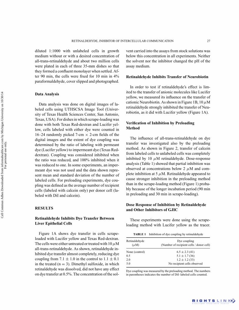
The influence of all-trans-retinaldehyde on dyetransfer was investigated also by the preloadingmethod. As shown in Figure 2, transfer of calceinfrom labeled cells to unlabeled cells was completelyinhibited by 10 µM retinaldehyde. Dose-responseanalysis (Table 1) showed that partial inhibition wasobserved at concentrations below 2 µM and com-plete inhibition at 5 µM. Retinaldehyde appeared tocause stronger inhibition in the preloading methodthan in the scrape-loading method (Figure 1) proba-bly because of the longer incubation period (90 minin preloading and 30 min in scrape-loading).
Dose Response of Inhibition by Retinaldehydeand Other Inhibitors of GJIC
These experiments were done using the scrape-loading method with Lucifer yellow as the tracer.
TABLE 1 Inhibition of dye coupling by retinaldehyde
Retinaldehyde Dye coupling(µM) (Number of recipient cells / donor cell)
None (control) 6.5 ± 2.3 (41)0.5 5.1 ± 1.7 (36)2.0 1.2 ± 1.2 (33)5.0 No recipient cells observed
Dye coupling was measured by the preloading method. The numbersin parentheses indicates the number of DiI–labeled cells counted.
Cel
l Com
mun
Adh
es D
ownl
oade
d fr
om in
form
ahea
lthca
re.c
om b
y M
ichi
gan
Uni
vers
ity o
n 10
/30/
14Fo
r pe
rson
al u
se o
nly.

28 S. PULUKURI AND A. SITARAMAYYA
Figure 1. Inhibition of dye transfer by retinaldehyde as measured by the scrape-loading method. (A) Dye transfer was measured betweenliver epithelial cells treated for 30 min with vehicle (control) or with 10 µM all-trans-retinaldehyde and scrape-loaded with Lucifer yellow andTexas Red-dextran. (B) Dye transfer of scrape-loaded Neurobiotin was measured between liver epithelial cells treated for 30 min with vehicle(control) or 10 µM retinaldehyde. (See Color Plate V).
Figure 3 shows reduction of dye transfer in re-sponse to treatment with different concentrationsof retinaldehyde as well as retinoic acid and gly-cyrrhetinic acid, two well-known inhibitors of GJIC.Retinaldehyde reduced the dye transfer by 50% at2.8 µM, retinoic acid, at 30 µM, and glycyrrhetinic
acid, at 65 µM. Retinaldehyde inhibited dye transferfully at concentrations below 20 µM, similar to theobservation made in Figure 1A; however, consistentwith earlier reports, dye transfer was not comparablyreduced by glycyrrhetinic acid or retinoic acid evenat higher concentrations (4, 28, 32). Glycyrrhizic
Cel
l Com
mun
Adh
es D
ownl
oade
d fr
om in
form
ahea
lthca
re.c
om b
y M
ichi
gan
Uni
vers
ity o
n 10
/30/
14Fo
r pe
rson
al u
se o
nly.

RETINALDEHYDE, INHIBITOR OF INTERCELLULAR COMMUNICATION 29
Figure 2. Inhibition of dye transfer by retinaldehyde as measured by the preloading method. Liver epithelial cells preloaded with DiI andcalcein AM were mixed with unlabeled cells and plated in the absence or presence of 10 µM retinaldehyde. Dye transfer was measured90 min later in the monolayers formed. (See Color Plate VI).
Figure 3. Dose dependence of inhibition of GJIC. Liver epithelial cells were treated for 30 min with different concentrations of retinaldehyde(�), retinoic acid (•) or glycyrrhetinic acid (�), followed by measurement of GJIC by the scrape-loading dye-transfer method. Lucifer yellowwas used as the dye.
Cel
l Com
mun
Adh
es D
ownl
oade
d fr
om in
form
ahea
lthca
re.c
om b
y M
ichi
gan
Uni
vers
ity o
n 10
/30/
14Fo
r pe
rson
al u
se o
nly.

30 S. PULUKURI AND A. SITARAMAYYA
acid, an analog of glycyrrhetinic acid, was used as acontrol for glycyrrhetinic acid (32); it had no effecton dye transfer at concentrations up to 100 µM.
Both All-Trans and 11-Cis Isomers ofRetinaldehyde were Effective Inhibitors
Retinaldehyde is present in the body mostly in twoisoforms, the 11-cis and the all-trans. All of the abovestudies were done with all-trans-retinaldehyde. Inorder to test if the inhibition of GJIC is isoformspecific, we also tested the influence of 11-cis-retinaldehyde and observed that it was as effec-tive as all-trans-retinaldehyde. At 10 µM, all-trans-retinaldehyde reduced dye coupling from 7.1 ± 1.0cells to 1.1 ± 0.1 cells and 11-cis-retinaldehyde re-duced it to 1.2 ± 0.2 cells (n = 3).
Retinaldehyde Did Not Influence Cell Viability
The effect of retinaldehyde on GJIC could havebeen due to cytotoxicity. In order to test this, a mono-layer of cultured cells was treated with 10 µM all-trans-retinaldehyde or vehicle for 30 min, washed
Figure 4. Kinetics of inhibition and recovery. Dye transfer was measured between liver epithelial cells following treatment with 50 µMretinaldehyde for different periods of time up to 30 min. Cells treated for 30 min with retinaldehyde were washed (washout), incubated inretinaldehyde-free growth medium for various periods of time and dye transfer was measured. Lucifer yellow was used as the dye.
with PBS and cell viability was determined by theTrypan blue exclusion method. No difference wasfound between treated and untreated cells: with morethan two million cells counted per sample, 94.7 ±2.4% of cells were found viable in the control and95.3 ± 2.3% in the treated (n = 3). Also, thereappeared to be no long-term toxic effect of thetreatment as both control and treated cells appearedhealthy 24 hr after the treatment and their dye trans-fer ability was similar.
The effect of retinaldehyde could possibly be sim-ply due to the aldehyde group. In order to test thispossibility, dye transfer was measured followingtreatment for 30 min with 100 µM formaldehyde.It was observed that formaldehyde had no influenceon dye transfer: dye coupling was 7.1 ± 1.0 in thecontrol and 7.0 ± 0.2 in the treated (n = 3).
Time Course of Inhibition and Recovery
In order to gain insight into the mechanism ofaction of retinaldehyde, the kinetics of inhibitionand recovery from inhibition were investigated. Asshown in Figure 4, inhibition was very rapid; dye
Cel
l Com
mun
Adh
es D
ownl
oade
d fr
om in
form
ahea
lthca
re.c
om b
y M
ichi
gan
Uni
vers
ity o
n 10
/30/
14Fo
r pe
rson
al u
se o
nly.

RETINALDEHYDE, INHIBITOR OF INTERCELLULAR COMMUNICATION 31
transfer was reduced by about 50% in about 2 minand 85% in 6 min. However, upon removal ofretinaldehyde and incubation in retinaldehyde-freegrowth medium, the ability to transfer Lucifer yel-low recovered more slowly; half-maximal activityrecovered in about 2 hours, and near complete re-covery in about 6 hours.
Effect of Retinaldehyde on GJIC in OtherCultured Cells
In order to determine if the effect of retinalde-hyde is limited to liver epithelial cells, its effect ondye transfer was investigated also on cultured hu-man lens and pigment epithelial cells and on rat reti-nal ganglion cells. These experiments were done byscrape-loading Lucifer yellow in the case of lensand pigment epithelial cells. Neurobiotin was usedfor ganglion cells which did not transfer Lucifer yel-low. Table 2 shows that retinaldehyde reduced dyetransfer between all three cell types.
DISCUSSION
Vitamin A and its derivatives, including retinalde-hyde, are usually present in the body bound tocarrier proteins or receptors (11, 21, 25, 33, 35). Reti-naldehyde, in its 11-cis form, is present in the outersegments of retinal photoreceptor cells at 2–3 mMconcentration (19), most of it bound to rhodopsinand converted to the all-trans form upon light ac-tivation. When dark adapted retina is activated by
TABLE 2 Effect of retinaldehyde on dye transfer in different celltypes
Retinaldehyde (µM) HLE HRPE RGC
None, control 6.2 ± 1.8 4.8 ± 2.0 14.5 ± 1.610 2.9 ± 1.4 1.9 ± 1.1 5.9 ± 1.225 1.7 ± 1.0 1.9 ± 1.0 3.0 ± 0.950 1.5 ± 1.4 0.9 ± 0.9 1.4 ± 0.5
HLE, Human lens epithelial cells; HRPE, human retinal pigmentepithelial cells; RGC, rat retinal ganglion cells. Dye transfer wasmeasured by the scrape-loading method using Lucifer yellow forHLE and HRPE and Neurobiotin for RGC. Data shown are meanand standard deviation of the number of labeled cells from 24 fieldsfor HLE and HRPE and 12 fields for RGC.
bright light, there is a potential for rapid release ofa large amount of this all-trans-retinaldehyde (26,34). Our goal in the present experiments was to testif free retinaldehyde could influence GJIC.
The most significant observation from these ex-periments is that retinaldehyde is a potent inhibitorof GJIC. With dye transfer reduced by half at about3 µM concentration, it is 10 and 20 times more ef-fective than retinoic acid (30 µM) and glycyrrhetinicacid (65 µM), respectively. Our results on retinoicacid and glycyrrhetinic acid are in agreement withearlier reports of their effects on GJIC, though theywere shown to be more or less effective in somereports (4, 6, 28, 32, 36–38). In general, the mag-nitude of inhibition depends upon concentration ofthe inhibitor as well as the length of incubation. Atand below 10 µM concentration and after longer pe-riods of incubation, retinoic acid is also known tostimulate GJIC (4, 23). In the present study, all threeinhibitors were tested under identical conditions andretinaldehyde appeared to be a much more effectiveinhibitor than retinoic acid and glycyrrhetinic acid.Unlike retinoic acid, retinaldehyde did not stimulateGJIC at low concentrations (between 2 and 10 µM).
The molecular mechanism of inhibition of GJICby retinaldehyde is not clear. It is, however, not dueto cytotoxicity, and also not simply due to the alde-hyde group. Retinaldehyde may function like otherlipid-soluble substances that inhibit gap junctions,by partitioning into the membrane and affectingits physical properties (13, 32). The rapid kineticsof inhibition and relatively slower recovery resem-bles the effects of oleamide, another inhibitor ofGJIC, thought to function by perturbing the lipidenvironment of connexins (12). It is, however, alsopossible that retinaldehyde binds with high affinity tosome connexins, explaining the slow recovery afterwashout.
Retinaldehyde inhibits dye transfer in differentcell types tested. However, whether gap junctionsmade of different connexins exhibit distinct sensi-tivity to retinaldehyde remains to be investigated.
Both the 11-cis and all-trans forms of retinalde-hyde inhibited GJIC just as they both were shown toinhibit cyclic GMP-gated channels in photoreceptor
Cel
l Com
mun
Adh
es D
ownl
oade
d fr
om in
form
ahea
lthca
re.c
om b
y M
ichi
gan
Uni
vers
ity o
n 10
/30/
14Fo
r pe
rson
al u
se o
nly.

32 S. PULUKURI AND A. SITARAMAYYA
cells (7). This lack of stereospecificity permits bothto be used as pharmacological tools in the study ofgap junctions, but the physiological significance maybe restricted to the effects of the all-trans form. The11-cis form is present at 2–3 mM concentration inthe photoreceptor cells of retina, bound to rhodopsin(19). When released after exposure of retina to brightlight, it is in the all-trans form. Given the potentialfor release in large amounts, and its accumulationfor a period of time before conversion back to the11-cis form (26, 34), all-trans-retinaldehyde mightinfluence GJIC between neighboring photoreceptorand pigment epithelial cells. In the present study, wehave already shown that all-trans-retinaldehyde in-hibits GJIC between pigment epithelial cells; futurestudies will determine its potential effects on gapjunctions between photoreceptor cells.
ACKNOWLEDGEMENTS
This study was supported by grants from the Na-tional Eye Institute (EY 07158 and EY 014803).We thank Drs. James E. Trosko and Brad L. Up-ham of Michigan State University, East Lansing,MI, USA, for demonstrating to us the technique ofscrape-loading dye-transfer and its use as a quanti-tative tool. We also thank Dr. Diane F. Matesic ofMercer University, Atlanta, GA, USA, for manyhelpful discussions.
REFERENCES
1. Bradford DJ, Hartzer MK, Cheng M, Blumenkranz MS(1990). Light damage thresoldsof retinal pigment epitheliumare decreased by thioridazine. Invest Ophthalmol Vis Sci 31:294.
2. Brissette JL, Kumar NM, Gilula NB, Hall JE, Dotto GP (1994).Switch in gap junction protein expression is associated withselective changes in junctional permeability during keratinocytedifferentiation. Proc Natl Acad Sci USA 91: 6453–6457.
3. Brown PK, Wald G (1956). The neo-b isomer of vitamin A andretinene. J Biol Chem 222: 865–877.
4. Brummer F, Zempel G, Buhle P, Stein JC, Hulser DF(1991). Retinoic acid modulates gap junctional permeability: Acomparative study of dye spreading and ionic coupling in cul-tured cells. Exp Cell Res 196: 158–163.
5. Bruzzone R, White TW, Paul DL (1996). Connections with con-nexins: The molecular basis of direct intercellular signaling. EurJ Biochem 238: 1–27.
6. Davidson JS, Baumgarten IM, Harley EH (1986). Reversibleinhibition of intercellular junctional communication by gly-cyrrhetinic acid. Biochem Biophys Res Commun 134: 29–36.
7. Dean DM, Nguitragool W, Miri A, McCabe SL, ZimmermanAL (2002). All-trans-retinal shuts down rod cyclic nucleotide-gated ion channels: A novel role for photoreceptor retinoids inthe response to bright light? Proc Natl Acad Sci USA 99: 8372–8377.
8. el Fouly MH, Trosko JE, Chang CC (1987). Scrape-loadingand dye transfer. A rapid and simple technique to study gapjunctional intercellular communication. Exp Cell Res 168: 422–430.
9. Flood MT, Gouras P, Kjeldbye H (1980). Growth characteristicsand ultrastructure of human retinal pigment epithelium in vitro.Invest Ophthalmol Vis Sci 19: 1309–1320.
10. Goldberg GS, Bechberger JF, Naus CC (1995). A pre-loadingmethod of evaluating gap junctional communication by fluores-cent dye transfer. Biotechniques 18: 490–497.
11. Gonzalez-Fernandez F (2002). Evolution of the visual cycle: Therole of retinoid-binding proteins. J Endocrinol 175: 75–88.
12. Guan X, Cravatt BF, Ehring GR, Hall JE, Boger DL, Lerner RA,Gilula NB (1997). The sleep-inducing lipid oleamide deconvo-lutes gap junction communication and calcium wave transmis-sion in glial cells. J Cell Biol 139: 1785–1792.
13. Harris AL (2001). Emerging issues of connexin channels: Bio-physics fills the gap. Q Rev Biophys 34: 325–472.
14. Hubbard R, Brown PK, Bownds D (1971). Methodology of vi-tamin A and visual pigments. Meth Enzymol 18: 615–653.
15. Ibaraki N, Chen SC, Lin LR, Okamoto H, Pipas JM, ReddyVN (1998). Human lens epithelial cell line. Exp Eye Res 67:577–585.
16. Krishnamoorthy RR, Agarwal P, Prasanna G, Vopat K, LambertW, Sheedlo HJ, Pang IH, Shade D, Wordinger RJ, Yorio T, ClarkAF, Agarwal N (2001). Characterization of a transformed ratretinal ganglion cell line. Brain Res Mol Brain Res 86: 1–12.
17. Kumar NM, Gilula NB (1992). Molecular biology and geneticsof gap junction channels. Semin Cell Biol 3: 3–16.
18. Lampe PD, Lau AF (2000). Regulation of gap junctions byphosphorylation of connexins. Arch Biochem Biophys 384: 205–215.
19. Liebman PA (1962). In situ microspectrophotometric studies onthe pigments of single retinal rods. Biophys J 2: 161–178.
20. Matesic DF, Rupp HL, Bonney WJ, Ruch RJ, Trosko JE (1994).Changes in gap-junction permeability, phosphorylation, andnumber mediated by phorbol ester and non-phorbol-ester tu-mor promoters in rat liver epithelial cells. Mol Carcinog 10:226–236.
21. McBee JK, Palczewski K, Baehr W, Pepperberg DR (2001).Confronting complexity: The interlink of phototransduction andretinoid metabolism in the vertebrate retina. Prog Retin Eye Res20: 469–529.
22. McCaffery P, Mey J, Drager UC (1996). Light-mediated retinoicacid production. Proc Natl Acad Sci USA 93: 12570–12574.
23. Mehta PP, Bertram JS, Loewenstein WR (1989). The actions ofretinoids on cellular growth correlate with their actions on gapjunctional communication. J Cell Biol 108: 1053–1065.
24. Murakami C, Takemura M, Sugiyama Y, Kamisuki S, AsaharaH, Kawasaki M, Ishidoh T, Linn S, Yoshida S, SugawaraF, Yoshida H, Sakaguchi K, Mizushina Y (2002). VitaminA-related compounds, all-trans retinal and retinoic acids,
Cel
l Com
mun
Adh
es D
ownl
oade
d fr
om in
form
ahea
lthca
re.c
om b
y M
ichi
gan
Uni
vers
ity o
n 10
/30/
14Fo
r pe
rson
al u
se o
nly.

RETINALDEHYDE, INHIBITOR OF INTERCELLULAR COMMUNICATION 33
selectively inhibit activities of mammalian replicative DNApolymerases. Biochim Biophys Acta 1574: 85–92.
25. Napoli JL (1999). Interactions of retinoid binding proteins andenzymes in retinoid metabolism. Biochim Biophys Acta 1440:139–162.
26. Okajima TL, Pepperberg DR (1997). Retinol kinetics in the iso-lated retina determined by retinoid extraction and HPLC. ExpEye Res 65: 331–340.
27. Pechere M, Germanier L, Siegenthaler G, Pechere JC, Saurat JH(2002). The antibacterial activity of topical retinoids: The caseof retinaldehyde. Dermatology 205: 153–158.
28. Pitts JD, Hamilton AE, Kam E, Burk RR, Murphy JP (1986).Retinoic acid inhibits junctional communication between animalcells. Carcinogenesis 7: 1003–1010.
29. Purup S, Jensen SK, Sejrsen K (2001). Differential effects ofretinoids on proliferation of bovine mammary epithelial cells incollagen gel culture. J Dairy Res 68: 157–164.
30. Rivedal E, Yamasaki H, Sanner T (1994). Inhibition of gap junc-tional intercellular communication in Syrian hamster embryocells by TPA, retinoic acid and DDT. Carcinogenesis 15: 689–694.
31. Robeson CD, Cawley JD, Weisler L, Stern MH, EddingerCC, Chechak AJ (2003). Chemistry of vitamin A. XXIV. The
synthesis of geometric isomers of Vitamin A via methyl beta-methylglutaconate. J Am Chem Soc 77: 4111–4119.
32. Rozental R, Srinivas M, Spray DC (2001). How to close a gapjunction channel. Efficacies and potencies of uncoupling agents.Methods Mol Biol 154: 447–476.
33. Saari JC (2000). Biochemistry of visual pigment regeneration:The Friedenwald lecture. Invest Ophthalmol Vis Sci 41: 337–348.
34. Saari JC, Garwin GG, Van Hooser JP, Palczewski K (1998). Re-duction of all-trans-retinal limits regeneration of visual pigmentin mice. Vision Res 38: 1325–1333.
35. Vieira AV, Schneider WJ, Vieira PM (1995). Retinoids: Trans-port, metabolism, and mechanisms of action. J Endocrinol 146:201–207.
36. Weiler R, He S, Vaney DI (1999). Retinoic acid modulates gapjunctional permeability between horizontal cells of the mam-malian retina. Eur J Neurosci 11: 3346–3350.
37. Yamamoto Y, Fukuta H, Nakahira Y, Suzuki H (1998). Blockadeby 18beta-glycyrrhetinic acid of intercellular electrical couplingin guinea-pig arterioles. J Physiol 511(Pt 2): 501–508.
38. Zhang DQ, McMahon DG (2001). Gating of retinal horizontalcell hemi gap junction channels by voltage, Ca2+, and retinoicacid. Mol Vis 7: 247–252.
Cel
l Com
mun
Adh
es D
ownl
oade
d fr
om in
form
ahea
lthca
re.c
om b
y M
ichi
gan
Uni
vers
ity o
n 10
/30/
14Fo
r pe
rson
al u
se o
nly.