retina surgery goba perspetives use of 27 …oshimaganka.com/pdf/0915rt_global_27gdiabetic...
TRANSCRIPT
RETINA SURGERY GLOBAL PERSPECTIVES Section Editors: Stanislao Rizzo, MD; Albert Augustin, MD;
J. Fernando Arevalo, MD; and Masahito Ohji, MD
SEPTEMBER 2015 RETINA TODAY 33
dept headline headline headline headlineDeck
DEPT BYLINE
A pilot survey demonstrated anatomic success and visual improvement.
BY YUSUKE OSHIMA, MD
Use of 27-Gauge Vitrectomy for Diabetic TRD
In 2010, we reported preliminary results using a first-generation 27-gauge vitrectomy system.1 Although at that time it was used only in highly selected cases, mainly of macular disease and simple
vitreous hemorrhage, both the anatomic and visual results were promising. The most remarkable findings were that there was no need to transition to a larger gauge, no suturing was required, and no hypotony was observed in any of the study cases.
To explore the potential for further widespread use of this ultrasmall-gauge system, we have moved to the next stage. We recently confirmed the system’s feasibility for treating more challenging conditions, such as advanced proliferative diabetic retinopathy (PDR). Current indications for pars plana vitrectomy in patients with PDR include vitreous hemorrhage, tractional retinal detachment (TRD), diabetic macular edema associated with posterior hyaloidal traction, and anterior segment neovascularization with media opacities. Of these, TRDs with prominent fibrovascu-lar membranes are the most challenging situations.
In combination with recent advances in vitrectomy technologies and pharmacologic agents, the use of small-gauge vitrectomy systems has made it possible to achieve anatomic stability and improvement of visual acuity in eyes with diabetic TRD.
In this article, we describe preliminary results using 27-gauge vitrectomy for treating these challenging situations.
BENEFITS OF SMALL GAUGESWith adoption of chandelier endoillumination and
wideangle viewing systems, vitreous shaving with a recently improved high-speed 27-gauge vitreous cut-ter can be smoothly achieved without undue concern about fragility or cutting efficiency. Based on our previous experiences with 23- and 25-gauge systems, we knew that a smaller-gauge cutter would be more useful for membrane dissection in diabetic eyes.2 The 27-gauge vitreous cutter is currently the best device for this indication because it can be easily inserted into the tiny spaces between membranes without the need for complex instruments or special techniques (Figure 1).
Substantial benefits have been reported with the use of small-gauge systems in previous literature. For diabetic
At a Glance• The 27-gauge vitreous cutter is the best device for
membrane dissection in diabetic eyes because it can be easily inserted between membranes.
• An improved, high-speed 27-gauge vitrectomy system offers anatomic success in patients with diabetic TRD similar to that of 23- and 25-gauge systems.
• Surgical procedures are simplified and operating times are shortened with the 27-gauge system.
”With adoption of chandelier
endoillumination and wideangle
viewing systems, vitreous shaving
with a recently improved high-speed
27-gauge vitreous cutter can be
smoothly achieved... .”

RETINA SURGERY GLOBAL PERSPECTIVES
34 RETINA TODAY SEPTEMBER 2015
patients, in addition to these reported benefits, it is impor-tant to ensure rigid wound construction to maintain intra-ocular pressure stability in order to prevent postoperative hypotony-related bleeding complications. Preservation of the conjunctiva is another important goal. For all of these reasons, we believe the use of a 27-gauge system to treat challenging diabetic TRD cases makes a lot of sense.
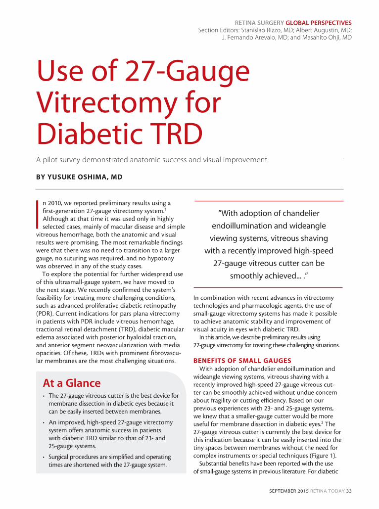
Currently, a variety of 27-gauge instruments are commercially available for diabetic vitrectomy, includ-ing a high-speed cutter, a curved laser probe, a dia-thermy probe, membrane forceps, curved scissors, and a soft-tipped cannula. Of these, the most recently developed 27-gauge double-port cutter, with a second port in the internal guillotine blade of the cutter, has aspiration flow almost equal to or even a little bit bet-ter than that of a standard 25-gauge cutter (Figure 2, Video; eyetube.net/v.asp?f=eevim).3
RETROSPECTIVE REVIEWIn a pilot survey, we performed a retrospective chart
review of a consecutive series of 40 patients who under-went primary 27-gauge vitrectomy for diabetic TRD or macula-threatening fibrovascular proliferation. The surgical results were compared with our previously reported case series treated with bevacizumab (Avastin, Genentech)-assisted 25- or 23-gauge vitrectomy.4
The study results are shown in Tables 1 and 2. Of the 80 study eyes, there was no significant difference in baseline characteristics between groups. However,
in the 27-gauge group, no eyes needed transition to larger size instruments, even in the most challenging cases, and no eyes needed suturing even with exten-sive peripheral vitreous shaving.
Overall anatomic success was comparable between groups. Operating time was a bit shorter for the 27-gauge group, possibly because use of additional instruments was less necessary for 27-gauge membrane dissection. We also had less need to perform bimanual manipulation with the 27-gauge device, suggesting that use of the 27-gauge cutter with one hand is sufficient to play several roles. This point has also been made by Beroccal.4
Surprisingly, despite a much lower need for beva-cizumab as a preoperative adjunct, there were few bleeding-related complications in the 27-gauge group. Intraocular pressure was stable from postoperative
Figure 1. Microincision vitrectomy: 27-gauge three-port setting with 27-gauge twin-light chandelier endoillumination fibers
(Twinlight chandelier illumination system; A); vitreous hemorrhage removal with 27-gauge twin-duty cycle (TDC) cutter (B);
bimanual manipulation using TDC cutter with membrane forceps for membrane dissection (C); internal limiting membrane
peeling with 27-gauge forceps (D); panretinal endophotocoagulation with a 27-gauge curved endolaser probe (E); external
view of eye immediately after conclusion of 27-gauge diabetic vitrectomy (F).
A B C
D E F
”[I]t is important to ensure
rigid wound construction to
maintain intraocular pressure
stability in order to prevent
postoperative hypotony-related
bleeding complications.”

RETINA SURGERY GLOBAL PERSPECTIVES
SEPTEMBER 2015 RETINA TODAY 35
TABLE 1. SURGICAL OUTCOMES OF SMALL-GAUGE DIABETIC VITRECTOMY: 27-GAUGE VS. 23- OR 25-GAUGE
27-gauge Group 23- or 25-gauge GroupP Value
n = 42 n = 38
Preoperative bevacizumab injection [n, (%)] 6 (14) 38 (100)
Operating time (min) 77 ± 37 85 ± 47 .35
Bimanual manipulation 15 (38) 23 (61) .027
Instrumentation for FVM removal [n, (%)]
Vitreoretinal forceps 18 (43) 23 (61) .18
Membrane scissors and/or MPC 3 (7) 8 (21) .041
Tamponade [n, (%)]
Gas (air or long-acting gas) 18 (43) 18 (47) .76
Silicone oil 0 (0) 1 (3) .48
Self-sealing [n, (%)] 42 (100) 28 (74) <.001
Initial reattachment [n, (%)] 40 (93) 36 (95) 1.00
Final reattachment [n, (%)] 42 (100) 38 (100) 1.00
Postoperative BCVA
Mean (range) LogMAR ± SD
0.34 (CF-1.0)0.46 ± 0.51
0.30 (HM-1.2)0.51 ± 0.71
.08
Mean BCVA changes (logMAR ± SD) -0.63 ± 0.66 -0.81 ± 1.16 .14
Abbreviations: BCVA, best corrected visual acuity; CF, counting fingers; FVM, fibrovascular membrane; HM, hand motion; MPC, membrane peeler-cutter; SD, standard deviation
TABLE 2. INTRAOPERATIVE AND POSTOPERATIVE COMPLICATIONS: 27-GAUGE VS. 23- OR 25-GAUGE
27-gauge Group 23- or 25-gauge GroupP Value
n = 42 n = 38
Intraoperative complications
Iatrogenic retinal breaks [n, (%)] 7 (17) 8 (21) .83
Mean scores of intraoperative bleeding 2.2 ± 1.1 2.7 ± 0.9 .75
Postoperative complications [n, (%)]
Hypotony 0 (0) 1 (3) .48
Progressing or persistent NVG Progressive fibrovascular proliferation Persistent or recurrent VH
1 (2.4)1 (2.4)2 (5)
3 (8)2 (5)5 (13)
.34
.601.00
Recurrent retinal detachment 2 (5) 2 (5) 1.00
Vitreoretinal reoperation [n, (%)] 4 (10) 4 (11) 1.00Abbreviations: NVG, neovascular glaucoma; VH, vitreous hemorrhage
No eyes needed transition to larger-size instruments even in the most challenging cases, and no eyes needed suturing even
with extensive peripheral vitreous shaving. The overall anatomic success was comparable between the groups, but the
operating time was a bit shorter for the 27-gauge group. Surprisingly, despite the lower chance of needing bevacizumab as
a preoperative adjunct, there were few bleeding-related complications in the 27-gauge group.
RETINA SURGERY GLOBAL PERSPECTIVES
36 RETINA TODAY SEPTEMBER 2015
day 1 throughout the 6-month follow-up period. As expected, visual acuity was significantly improved at 1 month after surgery. Changes in visual acuity at final examination were also comparable between the groups.
SNAPSHOT OF 27-GAUGE DIABETIC VITRECTOMY Following is a description of a representative case of
27-gauge diabetic vitrectomy. After perpendicular inser-tion of a 27-gauge valved trocar cannula and a 29-gauge dual chandelier fiber, there is sufficient endoillumina-tion to perform fibrovascular membrane dissection as usual, even with 27-gauge instruments. The 27-gauge cutter can be much more easily inserted into the tiny
space between the membrane and the retina. With the use of wide-angle fundus viewing, it is also easy to carry out bimanual membrane dissection with the 27-gauge system. After extensive peripheral vitreous shaving with scleral indentation and panretinal laser photocoagula-tion with a curved endolaser probe, all cannulas and optical fibers are removed, and surgery is soon complete.
CONCLUSIONAlthough the study design of the pilot survey pre-
viously described had several limitations, the results help to confirm that the 27-gauge vitrectomy sys-tem offers at least comparable anatomic success in patients with diabetic TRD, with fewer intraoperative complications and favorable visual recovery compared with 23- and 25-gauge systems. The 27-gauge system not only simplified surgical procedures and shortened operating time, but it also significantly eliminated the wound-sealing concerns sometimes seen with current 23- and 25-gauge vitrectomy systems. The latest developments in 27-gauge instruments, in con-junction with new vitrectomy machines, has made it possible to treat the full spectrum of vitreoretinal diseases, including challenging diabetic TRDs, with this ultrasmall-gauge technology. n
Yusuke Oshima, MD, is founder and director of the Oshima Eye Clinic in Takatsuki, a consultant and visiting surgeon at the Nishikasai Inoue Eye Hospital, a vis-iting lecturer of ophthalmology at Kyoto Prefectural University of Medicine, Kyoto, all in Japan; and an honorary director of the vitreoretinal division at the Tianjin Eye Hospital and visiting professor of oph-thalmology at the Nankai University in Tianjin, China. He is a member of the Retina Today Editorial Board. Dr. Oshima states that he is a consultant for Synergetics. He may be reached at [email protected].
1. Oshima Y, Wakabayashi T, Sato T, et al. A 27-gauge instrument system for transconjunctival sutureless microinci-sion vitrectomy surgery. Ophthalmology. 2010;117(1):93-102. 2. Wakabayashi T, Oshima Y. Microincision vitrectomy surgery for diabetic retinopathy. Retinal Physician. October 2011.3. Osawa S, Oshima Y. Innovations in 27-gauge vitrectomy for sutureless microincision vitrectomy surgery. Retina Today. 2014;9(5):42-45.4. Oshima Y, Shima C, Wakabayashi T, et al. Microincision vitrectomy surgery and intravitreal bevacizumab as a surgical adjunct to treat diabetic traction retinal detachment. Ophthalmology. 2009;116(5): 927-938.5. Berrocal M. A minimalist approach to surgery for diabetic retinal detachment. Retina Today. 2014;9(3):65-68.
Figure 2. The cutting efficiency of the 27-gauge TDC cutter
(yellow dashed line) in porcine vitreous at a variety of
cutting rates is almost equal to or a little better than that of
a conventional 25-gauge high-speed vitreous cutter.
Video: Diabetic Vitrectomy With a 27-Gauge TDC Cutter
”The latest developments in 27-gauge
instruments, in conjunction with new
vitrectomy machines, has made it
possible to treat the full spectrum
of vitreoretinal diseases ... with this
ultrasmall-gauge technology.”