restricted feeding uncouples circadian oscillators in...
TRANSCRIPT
Restricted feeding uncouples circadianoscillators in peripheral tissuesfrom the central pacemakerin the suprachiasmatic nucleus
Francesca Damiola,1 Nguyet Le Minh,1 Nicolas Preitner, Benoıt Kornmann, Fabienne Fleury-Olela,and Ueli Schibler2
Departement de Biologie Moleculaire, Sciences II, Universite de Geneve, CH-1211 Geneva, Switzerland
In mammals, circadian oscillators exist not only in the suprachiasmatic nucleus, which harbors the centralpacemaker, but also in most peripheral tissues. It is believed that the SCN clock entrains the phase ofperipheral clocks via chemical cues, such as rhythmically secreted hormones. Here we show that temporalfeeding restriction under light–dark or dark–dark conditions can change the phase of circadian gene expressionin peripheral cell types by up to 12 h while leaving the phase of cyclic gene expression in the SCN unaffected.Hence, changes in metabolism can lead to an uncoupling of peripheral oscillators from the central pacemaker.Sudden large changes in feeding time, similar to abrupt changes in the photoperiod, reset the phase ofrhythmic gene expression gradually and are thus likely to act through a clock-dependent mechanism.Food-induced phase resetting proceeds faster in liver than in kidney, heart, or pancreas, but after 1 wk ofdaytime feeding, the phases of circadian gene expression are similar in all examined peripheral tissues.
[Key Words: Circadian clock; restricted feeding; SCN; liver; clock genes; mouse]
Received June 26, 2000; revised version accepted October 9, 2000.
In mammals, most physiology and behavior are subjectto well-controlled daily oscillations. Thus, sleep–wakecycles, heartbeat frequency, blood pressure, body tem-perature, renal activity, liver metabolism, and the secre-tion of many hormones are controlled by an endogenoustime measuring system called the circadian clock (Por-taluppi et al. 1996; Rabinowitz 1996; Hastings 1997;Schibler and Lavery 1999). On the basis of surgical abla-tion and transplantation experiments, it is believed thatthe suprachiasmatic nucleus (SCN) in the hypothalamuscoordinates most if not all daily rhythms in behavior andphysiology (Rusak and Zucker 1979; Ralph et al. 1990).The SCN consists of two small, bilateral groups of neu-rons and, as indicated by its name, is located directlyabove the optical chiasma. SCN neurons receive infor-mation about the light intensity in the environment viadirect synaptic connections with the retina, whichadapts the phase of SCN oscillator to the photoperiod(Takahashi 1995; Hastings 1997). The SCN clock thensynchronizes overt rhythms in physiology and behavior,probably through both synaptic connections and hu-
moral signals (Shibata and Tominaga 1991; Silver et al.1996).
The molecular makeup of circadian clocks is subjectto intense genetic and biochemical investigation in vari-ous organisms, including cyanobacteria, Neurospora,higher plants, Drosophila, and mammals (for review, seeReppert 1998; Dunlap 1999). In most examined systems,autoregulatory feedback loops of gene expression are be-lieved to provide the rhythm-generating mechanisms. Asseveral clock genes are homologous in flies and mam-mals, it is likely that these mechanisms have beenlargely conserved during animal evolution (Young 2000).In mammals, Clock and Bmal1 are part of the positivelimb of the feedback circuitry, whereas two crypto-chrome isoforms, Cry1 and Cry2, and perhaps the periodisoforms, Per1, Per2, and Per3, are part of the negativelimb (Brown and Schibler 1999; Jin et al. 1999; Shearmanet al. 2000). At present, the negative feedback loop in theexpression of timekeeper genes is the most widely usedmolecular model in explaining how circadian clock-works operate. Although it cannot be formally excludedthat circadian gene expression is a manifestation, ratherthan the motor of, rhythm generation (Lakin-Thomas2000), the cyclic mRNA and protein accumulation pro-files can be used as convenient reporters for the phase ofcircadian oscillators.
1These authors contributed equally to this work.2Corresponding author.E-MAIL [email protected]; FAX 41-22-702-6868.Article and publication are at www.genesdev.org/cgi/doi/10.1101/gad.183500.
2950 GENES & DEVELOPMENT 14:2950–2961 © 2000 by Cold Spring Harbor Laboratory Press ISSN 0890-9369/00 $5.00; www.genesdev.org
Cold Spring Harbor Laboratory Press on March 13, 2020 - Published by genesdev.cshlp.orgDownloaded from
Molecular oscillators do not exist solely in pacemakercells such as lateral head neurons in Drosophila, pine-alocytes in cold-blooded vertebrates and birds, and SCNneurons in mammals (for review, see Schibler and Lavery1999). Rather, oscillators capable of generating severalconsecutive cycles of circadian gene expression are alsofound in peripheral, nonneuronal tissues of several ani-mals, including Drosophila (Emery et al. 1997; Krishnanet al. 1999; Giebultowicz et al. 2000), zebrafish (Cahill1996; Whitmore et al. 2000), and mammals (Balsalobre etal. 1998; Yamazaki et al. 2000). In Drosophila and ze-brafish embryos, which are semitransparent, these pe-ripheral clocks can be entrained directly by light via no-nocular mechanisms (Plautz et al. 1997; Whitmore et al.2000). However, in mammals, which are opaque, suchmechanisms are unlikely to be operative. Rather, lightresets the time of the central pacemaker in the SCN viaocular mechanisms (see above), and the SCN clock thensynchronizes peripheral oscillators via neuronal connec-tions and/or chemical signals (Shibata and Tominaga1991; Sakamoto et al. 1998; Yamazaki et al. 2000). Ex-periments with tissue-culture cells are in support of arole of blood-borne substances as time-resetting cues.Thus, a brief treatment of immortalized fibroblasts withhigh concentrations of serum induces circadian gene ex-pression persisting for several days (Balsalobre et al.1998). Similar results are obtained with cells incubatedfor a short time period with chemicals activating a vari-ety of known signal transduction pathways. Thus, TPA,a tumor promoter activating protein kinase C and MAPkinases; FGF, a chemokine activating MAP kinases; for-skolin and butyryl cAMP, substances that activate pro-tein kinase A; and dexamethasone, a glucocorticoid hor-mone analog, all provoke circadian gene expression intissue-culture cells (Akashi and Nishida 2000; Balsalobreet al. 2000b; Yagita and Okamura 2000). Given the re-sponsiveness of these oscillators to the multiple signals,it appears likely that the central pacemaker may exploitseveral chemical entrainment pathways to synchronizeperipheral clocks.
A major question in mammalian chronobiology con-cerns the physiological purpose of circadian gene expres-sion in peripheral cells. In liver, most known genes withrhythmic expression encode enzymes or regulatory pro-teins involved in food processing and energy homeosta-sis. These include cholesterol 7� hydroxylase (Mitropou-los et al. 1972; Noshiro et al. 1990; Lavery and Schibler1993), the rate-limiting enzyme in the synthesis of bileacids, a number of cytochrome P450 enzymes involvedin detoxification and elimination of food components(e.g., coumarin hydroxylase, Cyp2a5; Lavery et al. 1999),enzymes involved in carbohydrate metabolism (e.g.,PEPCK, glycogen synthase, glycogen phosphorylase;Ishikawa and Shimazu 1976; Roesler and Khandelwal1985; Frederiks et al. 1987), and transcription factorsgoverning fatty acid metabolism (e.g., PPAR and spot 14;Kinlaw et al. 1987; Lemberger et al. 1996). At least inliver, the coordination of physiological needs during theabsorptive and postabsorptive phase may be the majorfunction of circadian oscillators. Previous reports on re-
stricted feeding are in keeping with this conjecture. Forexample, in the liver of (nocturnal) rats, the phase of thedaily Dbp mRNA accumulation profile is severely al-tered when food is offered exclusively during the day(Ogawa et al. 1997).
As nocturnal animals, mice consume most of theirfood during the night. Here we show that feeding of miceexclusively during the day completely inverses the phaseof circadian oscillators in peripheral cells, but has little ifany effect on the central oscillator in the suprachias-matic nucleus. Interestingly, feeding during the subjec-tive day under constant dark (DD) conditions uncoupledperipheral from central oscillators to a similar extent asunder light–dark (LD) conditions. Hence, the feeding-time-induced uncoupling of circadian phases in periph-eral cells and SCN neurons cannot be explained by adominant Zeitgeber effect of light in the SCN. Rather,feeding elicits entrainment cues that act specifically onoscillators in peripheral tissues and that are ineffectivein resetting time in the SCN.
Results
Restricted feeding during the day uncouples circadianliver gene expression from circadian gene expressionin the suprachiasmatic nucleus
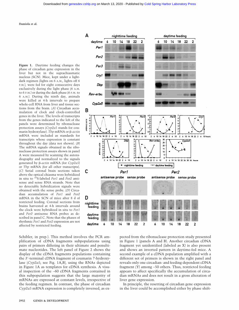
As mentioned above, a major task of circadian oscillatorsin liver cells (and perhaps other peripheral cell types)may be to anticipate and adapt the physiological condi-tions required for food processing. As nocturnal animals,mice consume ∼80% of their food during the active darkphase if kept under a 12-h light/12-h dark cycle (LD) andif food is offered ad libitum. To examine whether feedingtime can affect the phase of circadian liver gene expres-sion, mice were fed for 9 d exclusively during the nightor during the day. At the conclusion of the entrainmentperiod, the phase of mRNA expression for the four clockcomponents, PER1, PER2, PER3, and CRY1, the two cir-cadian transcription factors DBP and Rev-erb�, and thecytochrome P450 enzyme coumarin 7-hydroxylase(Cyp2a5) was determined. As shown in Figure 1A and B,the phases of all examined mRNA accumulation profilesdiffer by 8–12 h between mice fed during the day or dur-ing the night. As expected, livers of mice fed exclusivelyduring the night (Fig. 1A,B) or ad libitum displayed asimilar phase angle of cyclic liver gene expression (formice fed ad libitum, see Lopez-Molina et al. 1997; Laveryet al. 1999; Balsalobre et al. 2000a). However, feedingduring the day almost entirely inversed the phase of theliver oscillator. Hence, feeding time appears to be a po-tent Zeitgeber for circadian liver gene expression.
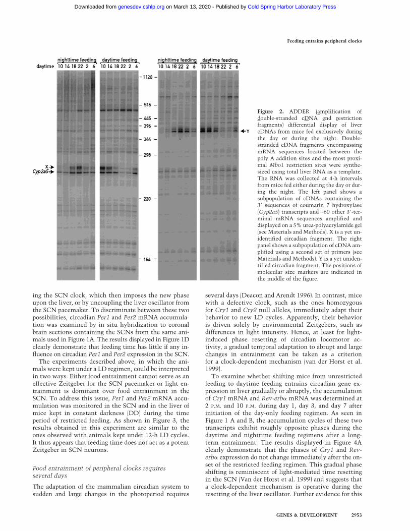
From the data presented thus far, the effects of feedingregimen on circadian phase could be either caused by aspecific change in circadian gene expression or by a moregeneral perturbation of liver transcription. To differenti-ate between these possibilities, we examined the expres-sion profiles of ∼110 random genes using a novel cDNAdisplay method, dubbed ADDER (amplification ofdouble-stranded cDNA end restriction fragments; B.Kornmann, N. Preitner, D. Rifat, F. Fleury-Olela, and U.
Feeding entrains peripheral clocks
GENES & DEVELOPMENT 2951
Cold Spring Harbor Laboratory Press on March 13, 2020 - Published by genesdev.cshlp.orgDownloaded from
Schibler, in prep.). This method involves the PCR am-plification of cDNA fragments subpopulations usingpairs of primers differing in their ultimate and penulti-mate nucleotides. The left panel of Figure 2 shows thedisplay of the cDNA fragments populations containingthe 3�-terminal cDNA fragment of coumarin 7-hydroxy-lase (Cyp2a5; see Fig. 1A,B), using the RNAs depictedin Figure 1A as templates for cDNA synthesis. A visu-al inspection of the ∼60 cDNA fragments contained inthis subpopulation suggests that the large majority ofmRNAs are expressed at constant levels, irrespective ofthe feeding regimen. In contrast, the phase of circadianCyp2a5 mRNA expression is completely inversed, as ex-
pected from the ribonuclease protection study presentedin Figure 1 (panels A and B). Another circadian cDNAfragment yet unidentified (labeled as X) is also presentand shows an inverted pattern in daytime-fed mice. Asecond example of a cDNA population amplified with adifferent set of primers is shown in the right panel andreveals only one circadian- and feeding-dependent cDNAfragment (Y) among ∼50 others. Thus, restricted feedingappears to affect specifically the accumulation of circa-dian mRNAs and does not result in a gross alteration ofliver gene expression.
In principle, the resetting of circadian gene expressionin the liver could be accomplished either by phase shift-
Figure 1. Daytime feeding changes thephase of circadian gene expression in theliver but not in the suprachiasmaticnucleus (SCN). Mice, kept under a light-dark regimen (lights on 6 A.M., lights off 6P.M.), were fed for eight consecutive daysexclusively during the light phase (6 A.M.to 6 P.M.) or during the dark phase (6 P.M. to6 A.M.). During the ninth day, animalswere killed at 4-h intervals to preparewhole-cell RNA from liver and tissue sec-tions from the brain. (A) Circadian accu-mulation of clock and clock-controlledgenes in the liver. The levels of transcriptsfrom the genes indicated to the left of thepanels were determined by ribonucleaseprotection assays (Cyp2a5 stands for cou-marin hydroxylase). Tbp mRNA or �-actinmRNA were included as standards fortranscripts whose expression is constantthroughout the day (data not shown). (B)The mRNA signals obtained in the ribo-nuclease protection assays shown in panelA were measured by scanning the autora-diography and normalized to the signalsgenerated by �-actin mRNA (for Cyp2a5)or Tbp mRNA (for all other transcripts).(C) Serial coronal brain sections takenabove the optical chiasma were hybridizedin situ to 35S-labeled Per1 and Per2 anti-sense and sense RNA strands. Note thatno detectable hybridization signals wereobtained with the sense probe. (D) Circa-dian accumulation of Per1 and Per2mRNA in the SCN of mice after 8 d ofrestricted feeding. Coronal sections frombrains harvested at 4-h intervals aroundthe clock were hybridized in situ to Per1and Per2 antisense RNA probes as de-scribed in panel C. Note that the phases ofrhythmic Per1 and Per2 expression are notaffected by restricted feeding.
Damiola et al.
2952 GENES & DEVELOPMENT
Cold Spring Harbor Laboratory Press on March 13, 2020 - Published by genesdev.cshlp.orgDownloaded from
ing the SCN clock, which then imposes the new phaseupon the liver, or by uncoupling the liver oscillator fromthe SCN pacemaker. To discriminate between these twopossibilities, circadian Per1 and Per2 mRNA accumula-tion was examined by in situ hybridization to coronalbrain sections containing the SCNs from the same ani-mals used in Figure 1A. The results displayed in Figure 1Dclearly demonstrate that feeding time has little if any in-fluence on circadian Per1 and Per2 expression in the SCN.
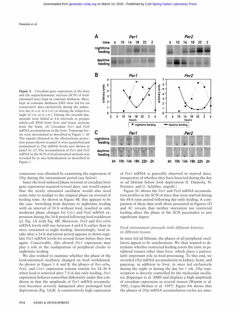
The experiments described above, in which the ani-mals were kept under a LD regimen, could be interpretedin two ways. Either food entrainment cannot serve as aneffective Zeitgeber for the SCN pacemaker or light en-trainment is dominant over food entrainment in theSCN. To address this issue, Per1 and Per2 mRNA accu-mulation was monitored in the SCN and in the liver ofmice kept in constant darkness (DD) during the timeperiod of restricted feeding. As shown in Figure 3, theresults obtained in this experiment are similar to theones observed with animals kept under 12-h LD cycles.It thus appears that feeding time does not act as a potentZeitgeber in SCN neurons.
Food entrainment of peripheral clocks requiresseveral days
The adaptation of the mammalian circadian system tosudden and large changes in the photoperiod requires
several days (Deacon and Arendt 1996). In contrast, micewith a defective clock, such as the ones homozygousfor Cry1 and Cry2 null alleles, immediately adapt theirbehavior to new LD cycles. Apparently, their behavioris driven solely by environmental Zeitgebers, such asdifferences in light intensity. Hence, at least for light-induced phase resetting of circadian locomotor ac-tivity, a gradual temporal adaptation to abrupt and largechanges in entrainment can be taken as a criterionfor a clock-dependent mechanism (van der Horst et al.1999).
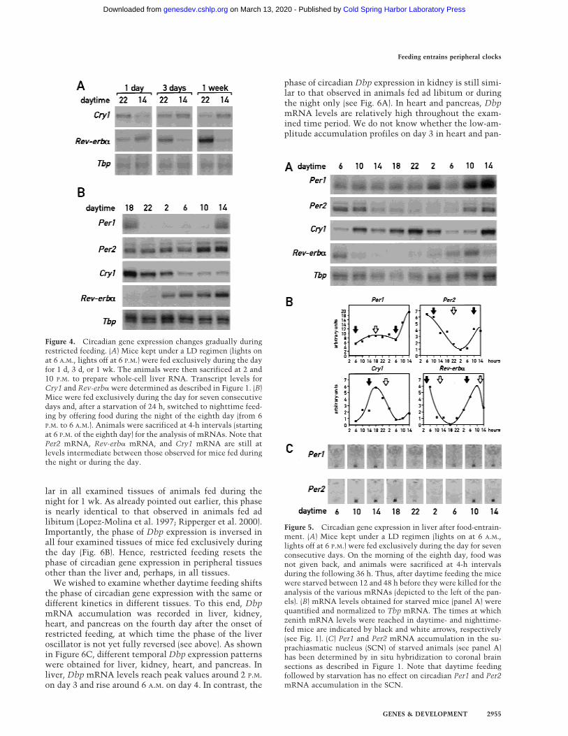
To examine whether shifting mice from unrestrictedfeeding to daytime feeding entrains circadian gene ex-pression in liver gradually or abruptly, the accumulationof Cry1 mRNA and Rev-erb� mRNA was determined at2 P.M. and 10 P.M. during day 1, day 3, and day 7 afterinitiation of the day-only feeding regimen. As seen inFigure 1 A and B, the accumulation cycles of these twotranscripts exhibit roughly opposite phases during thedaytime and nighttime feeding regimens after a long-term entrainment. The results displayed in Figure 4Aclearly demonstrate that the phases of Cry1 and Rev-erb� expression do not change immediately after the on-set of the restricted feeding regimen. This gradual phaseshifting is reminiscent of light-mediated time resettingin the SCN (Van der Horst et al. 1999) and suggests thata clock-dependent mechanism is operative during theresetting of the liver oscillator. Further evidence for this
Figure 2. ADDER (amplification ofdouble-stranded cDNA end restrictionfragments) differential display of livercDNAs from mice fed exclusively duringthe day or during the night. Double-stranded cDNA fragments encompassingmRNA sequences located between thepoly A addition sites and the most proxi-mal Mbo1 restriction sites were synthe-sized using total liver RNA as a template.The RNA was collected at 4-h intervalsfrom mice fed either during the day or dur-ing the night. The left panel shows asubpopulation of cDNAs containing the3� sequences of coumarin 7 hydroxylase(Cyp2a5) transcripts and ∼60 other 3�-ter-minal mRNA sequences amplified anddisplayed on a 5% urea-polyacrylamide gel(see Materials and Methods). X is a yet un-identified circadian fragment. The rightpanel shows a subpopulation of cDNA am-plified using a second set of primers (seeMaterials and Methods). Y is a yet uniden-tified circadian fragment. The positions ofmolecular size markers are indicated inthe middle of the figure.
Feeding entrains peripheral clocks
GENES & DEVELOPMENT 2953
Cold Spring Harbor Laboratory Press on March 13, 2020 - Published by genesdev.cshlp.orgDownloaded from
conjecture was obtained by examining the expression ofDbp during the entrainment period (see below).
Since the food-induced phase reversal of circadian livergene expression required several days, one would expectthat the newly entrained oscillator would also needsome time to readapt to the original phase on reversal offeeding time. As shown in Figure 4B, this appears to bethe case. Switching from daytime to nighttime feedingwith an interval of 24 h without food, resulted in onlymoderate phase changes for Cry1 and Per2 mRNA ex-pression during the 24-h period following food readdition(cf. Fig. 1A with Fig. 4B). Moreover, Per2 and Rev-erb�mRNA levels still rise between 4 and 8 h earlier than inmice entrained to night feeding. Interestingly, food in-take after a 24-h starvation period appears to down-regu-late Per1 mRNA levels for several hours before they riseagain. Conceivably, this altered Per1 expression mayplay a role in the readaptation of peripheral clocks tonighttime feeding.
We also wished to examine whether the phase of thefood-entrained oscillator changed on food withdrawal.As shown in Figure 5 A and B, the phases of Rev-erb�,Per2, and Cry1 expression remain similar for 24–36 hwhen food is removed after 7 d of day-only feeding. Per1expression behaves somewhat differently under this con-dition in that the amplitude of Per1 mRNA accumula-tion becomes severely dampened after prolonged fooddeprivation (Fig. 5A,B). A constitutively high expression
of Per1 mRNA is generally observed in starved mice,irrespective of whether they have been fed during the dayor ad libitum before food deprivation (F. Damiola, N.Preitner, and U. Schibler, unpubl.).
Figure 5C shows the Per1 and Per2 mRNA accumula-tion profiles in the SCN of mice that were starved duringthe 48-h time period following day-only feeding. A com-parison of these data with those presented in Figures 1Dand 3C reveals that neither starvation nor restrictedfeeding affect the phase of the SCN pacemaker to anysignificant degree.
Food entrainment proceeds with different kineticsin different tissues
In mice fed ad libitum, the phases of all peripheral oscil-lators appear to be synchronous. We thus wanted to de-termine whether restricted feeding resets the time in pe-ripheral tissues other than liver, which plays a particu-larly important role in food processing. To this end, werecorded Dbp mRNA accumulation in kidney, heart, andpancreas, in addition to liver, in mice fed exclusivelyduring the night or during the day for 1 wk. Dbp tran-scription is directly controlled by the molecular oscilla-tor (Ripperger et al. 2000) and displays a high amplitudeof circadian expression in several tissues (Wuarin et al.1992; Lopez-Molina et al. 1997). Figure 6A shows thatthe phases of Dbp mRNA accumulation cycles are simi-
Figure 3. Circadian gene expression in the liverand the suprachiasmatic nucleus (SCN) of food-entrained mice kept in constant darkness. Mice,kept in constant darkness (DD) were fed for sixconsecutive days exclusively during the subjec-tive day (6 A.M. to 6 P.M.) or during the subjectivenight (6 P.M. to 6 A.M.). During the seventh day,animals were killed at 4-h intervals to preparewhole-cell RNA from liver and tissue sectionsfrom the brain. (A) Circadian Per1 and Per2mRNA accumulation in the liver. Transcript lev-els were determined as described in Figure 1. (B)The signals obtained in the ribonuclease protec-tion assays shown in panel A were quantified andnormalized to Tbp mRNA levels (not shown inpanel A). (C) The accumulation of Per1 and Per2mRNA in the SCN of food entrained animals wasrevealed by in situ hybridization as described inFigure 1.
Damiola et al.
2954 GENES & DEVELOPMENT
Cold Spring Harbor Laboratory Press on March 13, 2020 - Published by genesdev.cshlp.orgDownloaded from
lar in all examined tissues of animals fed during thenight for 1 wk. As already pointed out earlier, this phaseis nearly identical to that observed in animals fed adlibitum (Lopez-Molina et al. 1997; Ripperger et al. 2000).Importantly, the phase of Dbp expression is inversed inall four examined tissues of mice fed exclusively duringthe day (Fig. 6B). Hence, restricted feeding resets thephase of circadian gene expression in peripheral tissuesother than the liver and, perhaps, in all tissues.
We wished to examine whether daytime feeding shiftsthe phase of circadian gene expression with the same ordifferent kinetics in different tissues. To this end, DbpmRNA accumulation was recorded in liver, kidney,heart, and pancreas on the fourth day after the onset ofrestricted feeding, at which time the phase of the liveroscillator is not yet fully reversed (see above). As shownin Figure 6C, different temporal Dbp expression patternswere obtained for liver, kidney, heart, and pancreas. Inliver, Dbp mRNA levels reach peak values around 2 P.M.on day 3 and rise around 6 A.M. on day 4. In contrast, the
phase of circadian Dbp expression in kidney is still simi-lar to that observed in animals fed ad libitum or duringthe night only (see Fig. 6A). In heart and pancreas, DbpmRNA levels are relatively high throughout the exam-ined time period. We do not know whether the low-am-plitude accumulation profiles on day 3 in heart and pan-
Figure 5. Circadian gene expression in liver after food-entrain-ment. (A) Mice kept under a LD regimen (lights on at 6 A.M.,lights off at 6 P.M.) were fed exclusively during the day for sevenconsecutive days. On the morning of the eighth day, food wasnot given back, and animals were sacrificed at 4-h intervalsduring the following 36 h. Thus, after daytime feeding the micewere starved between 12 and 48 h before they were killed for theanalysis of the various mRNAs (depicted to the left of the pan-els). (B) mRNA levels obtained for starved mice (panel A) werequantified and normalized to Tbp mRNA. The times at whichzenith mRNA levels were reached in daytime- and nighttime-fed mice are indicated by black and white arrows, respectively(see Fig. 1). (C) Per1 and Per2 mRNA accumulation in the su-prachiasmatic nucleus (SCN) of starved animals (see panel A)has been determined by in situ hybridization to coronal brainsections as described in Figure 1. Note that daytime feedingfollowed by starvation has no effect on circadian Per1 and Per2mRNA accumulation in the SCN.
Figure 4. Circadian gene expression changes gradually duringrestricted feeding. (A) Mice kept under a LD regimen (lights onat 6 A.M., lights off at 6 P.M.) were fed exclusively during the dayfor 1 d, 3 d, or 1 wk. The animals were then sacrificed at 2 and10 P.M. to prepare whole-cell liver RNA. Transcript levels forCry1 and Rev-erb� were determined as described in Figure 1. (B)Mice were fed exclusively during the day for seven consecutivedays and, after a starvation of 24 h, switched to nighttime feed-ing by offering food during the night of the eighth day (from 6P.M. to 6 A.M.). Animals were sacrificed at 4-h intervals (startingat 6 P.M. of the eighth day) for the analysis of mRNAs. Note thatPer2 mRNA, Rev-erb� mRNA, and Cry1 mRNA are still atlevels intermediate between those observed for mice fed duringthe night or during the day.
Feeding entrains peripheral clocks
GENES & DEVELOPMENT 2955
Cold Spring Harbor Laboratory Press on March 13, 2020 - Published by genesdev.cshlp.orgDownloaded from
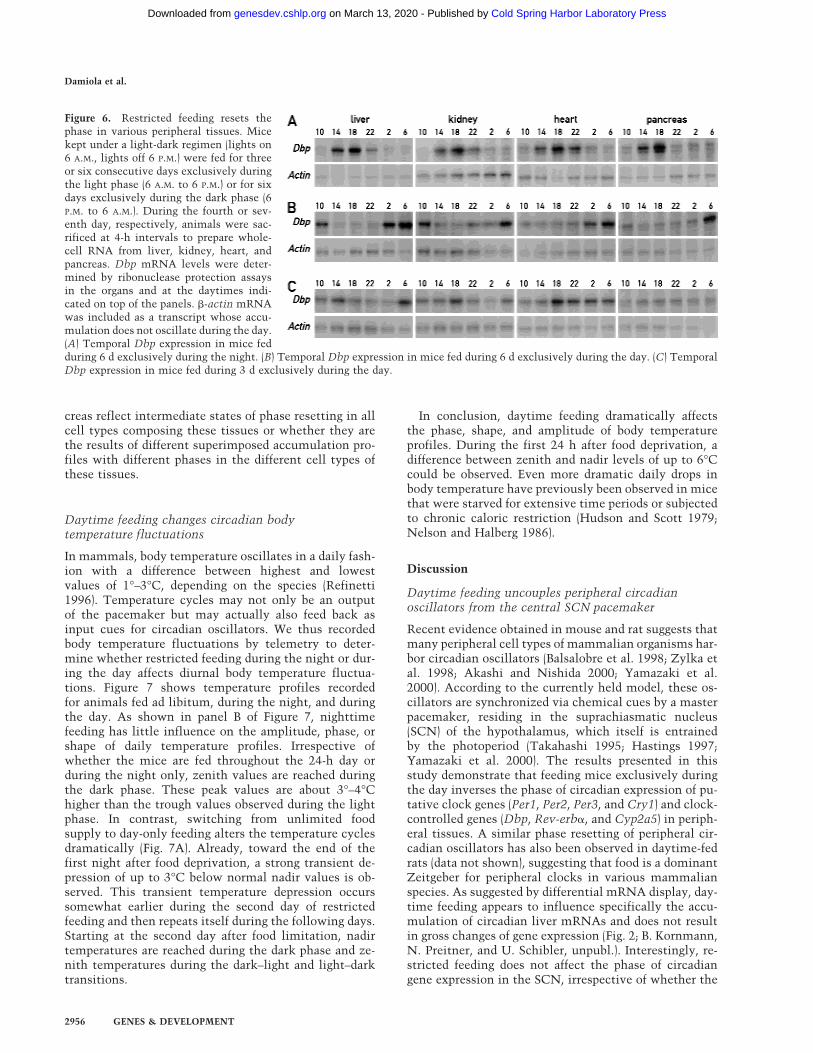
creas reflect intermediate states of phase resetting in allcell types composing these tissues or whether they arethe results of different superimposed accumulation pro-files with different phases in the different cell types ofthese tissues.
Daytime feeding changes circadian bodytemperature fluctuations
In mammals, body temperature oscillates in a daily fash-ion with a difference between highest and lowestvalues of 1°–3°C, depending on the species (Refinetti1996). Temperature cycles may not only be an outputof the pacemaker but may actually also feed back asinput cues for circadian oscillators. We thus recordedbody temperature fluctuations by telemetry to deter-mine whether restricted feeding during the night or dur-ing the day affects diurnal body temperature fluctua-tions. Figure 7 shows temperature profiles recordedfor animals fed ad libitum, during the night, and duringthe day. As shown in panel B of Figure 7, nighttimefeeding has little influence on the amplitude, phase, orshape of daily temperature profiles. Irrespective ofwhether the mice are fed throughout the 24-h day orduring the night only, zenith values are reached duringthe dark phase. These peak values are about 3°–4°Chigher than the trough values observed during the lightphase. In contrast, switching from unlimited foodsupply to day-only feeding alters the temperature cyclesdramatically (Fig. 7A). Already, toward the end of thefirst night after food deprivation, a strong transient de-pression of up to 3°C below normal nadir values is ob-served. This transient temperature depression occurssomewhat earlier during the second day of restrictedfeeding and then repeats itself during the following days.Starting at the second day after food limitation, nadirtemperatures are reached during the dark phase and ze-nith temperatures during the dark–light and light–darktransitions.
In conclusion, daytime feeding dramatically affectsthe phase, shape, and amplitude of body temperatureprofiles. During the first 24 h after food deprivation, adifference between zenith and nadir levels of up to 6°Ccould be observed. Even more dramatic daily drops inbody temperature have previously been observed in micethat were starved for extensive time periods or subjectedto chronic caloric restriction (Hudson and Scott 1979;Nelson and Halberg 1986).
Discussion
Daytime feeding uncouples peripheral circadianoscillators from the central SCN pacemaker
Recent evidence obtained in mouse and rat suggests thatmany peripheral cell types of mammalian organisms har-bor circadian oscillators (Balsalobre et al. 1998; Zylka etal. 1998; Akashi and Nishida 2000; Yamazaki et al.2000). According to the currently held model, these os-cillators are synchronized via chemical cues by a masterpacemaker, residing in the suprachiasmatic nucleus(SCN) of the hypothalamus, which itself is entrainedby the photoperiod (Takahashi 1995; Hastings 1997;Yamazaki et al. 2000). The results presented in thisstudy demonstrate that feeding mice exclusively duringthe day inverses the phase of circadian expression of pu-tative clock genes (Per1, Per2, Per3, and Cry1) and clock-controlled genes (Dbp, Rev-erb�, and Cyp2a5) in periph-eral tissues. A similar phase resetting of peripheral cir-cadian oscillators has also been observed in daytime-fedrats (data not shown), suggesting that food is a dominantZeitgeber for peripheral clocks in various mammalianspecies. As suggested by differential mRNA display, day-time feeding appears to influence specifically the accu-mulation of circadian liver mRNAs and does not resultin gross changes of gene expression (Fig. 2; B. Kornmann,N. Preitner, and U. Schibler, unpubl.). Interestingly, re-stricted feeding does not affect the phase of circadiangene expression in the SCN, irrespective of whether the
Figure 6. Restricted feeding resets thephase in various peripheral tissues. Micekept under a light-dark regimen (lights on6 A.M., lights off 6 P.M.) were fed for threeor six consecutive days exclusively duringthe light phase (6 A.M. to 6 P.M.) or for sixdays exclusively during the dark phase (6P.M. to 6 A.M.). During the fourth or sev-enth day, respectively, animals were sac-rificed at 4-h intervals to prepare whole-cell RNA from liver, kidney, heart, andpancreas. Dbp mRNA levels were deter-mined by ribonuclease protection assaysin the organs and at the daytimes indi-cated on top of the panels. �-actin mRNAwas included as a transcript whose accu-mulation does not oscillate during the day.(A) Temporal Dbp expression in mice fedduring 6 d exclusively during the night. (B) Temporal Dbp expression in mice fed during 6 d exclusively during the day. (C) TemporalDbp expression in mice fed during 3 d exclusively during the day.
Damiola et al.
2956 GENES & DEVELOPMENT
Cold Spring Harbor Laboratory Press on March 13, 2020 - Published by genesdev.cshlp.orgDownloaded from

animals are kept under LD or DD conditions. Hence, itappears that the unresponsiveness of the SCN clock tofeeding time reflects an inherent property of the centralpacemaker, rather than a dominance of photic entrain-ment over food entrainment.
In contrast to restricted feeding during the day, night-time feeding has little effect on the phase of circadiangene expression. This may not be surprising, given thatmice (as nocturnal animals) consume ∼80% of the foodduring the night (N. Preitner and F. Damiola, unpubl.)when they are fed ad libitum. Nighttime feeding alsodoes not significantly alter circadian body temperaturerhythms. Thus, in mice fed exclusively during the night,there is no sign of a core temperature depressionbelow the nadir values recorded for animals fed ad libitum.
We also examined whether restricted feeding influ-
ences voluntary locomotor activity, as measured by cir-cadian wheel-running activity (data not shown). Micekept in a 12-h light/dark regimen displayed similar ac-tograms irrespective of whether food was offeredthroughout the day, during the dark phase, or during thelight phase. However, when mice were kept under DDconditions, we did observe some notable differences be-tween the actograms of animals fed ad libitum or exclu-sively during the subjective night or during the subjec-tive day. In constant darkness, restricted feeding duringthe subjective night kept the animals on a precise 24-hschedule and prevented them from free running with theperiod dictated by their endogenous pacemaker. Hence,in accordance with previously reported data (White andTimberlake 1995; Holmes and Mistlberger 2000), feedingtime can serve as a Zeitgeber in the daily resetting of the
Figure 7. Daytime feeding causes body temperature depressions during the night. Temperature probes were implanted in the ab-dominal cavities of mice and body temperature rhythms were recorded by telemetry in animals fed either ad libitum or exclusivelyduring the night or during the day. The feeding periods are indicated by solid bars on top of the panels. The dark phases are depictedin blue. (A) Body temperature recordings from two individuals fed during 5 d exclusively during the day. Note the dramatic tempera-ture depressions during the dark phases of daytime feeding. (B) Recordings from two individuals fed during 7 d exclusively during thenight. In some animals (e.g., the one shown in the bottom panel), the minimal temperature values observed during the light phasebecame slightly lower after several days of nighttime feeding.
Feeding entrains peripheral clocks
GENES & DEVELOPMENT 2957
Cold Spring Harbor Laboratory Press on March 13, 2020 - Published by genesdev.cshlp.orgDownloaded from
central circadian clock by a few minutes. The wheel-running data obtained for mice kept under DD condi-tions and fed exclusively during the day were more dif-ficult to interpret. While most of the wheel running wasstill restricted to the subjective night, most examinedanimals were more active during the second half of thenight after ∼3 d. As this altered behavior is not accom-panied by phase changes in circadian Per1 and Per2 geneexpression in the SCN, the shifting of wheel-runningactivity toward the second half of the night may bebrought about by noncircadian mechanisms of behavior(e.g., homeostatic sleep components; see Borbely andAchermann 1999).
How does restricted feeding entrain peripheraloscillators?
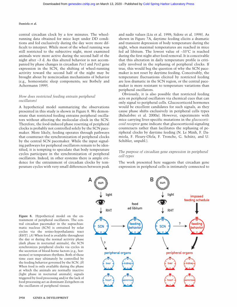
A hypothetical model summarizing the observationspresented in this study is shown in Figure 8. We demon-strate that restricted feeding entrains peripheral oscilla-tors without affecting the molecular clock in the SCN.Therefore, the food-induced phase resetting of peripheralclocks is probably not controlled solely by the SCN pace-maker. More likely, feeding operates through pathwaysthat counteract the synchronization of peripheral clocksby the central SCN pacemaker. While the input signal-ing pathways for peripheral oscillators remain to be iden-tified, it is tempting to speculate that body temperaturecycles participate in the synchronization of peripheraloscillators. Indeed, in other systems there is ample evi-dence for the entrainment of circadian clocks by tem-perature cycles with very small differences between peak
and nadir values (Liu et al. 1998; Sidote et al. 1998). Asshown in Figure 7A, daytime feeding elicits a dramaticand transient depression of body temperature during thenight, when maximal temperatures are reached in micefed ad libitum. The lowest value of ∼33°C is reachedduring the first night after food removal. It is conceivablethat this alteration in daily temperature profile is criti-cally involved in the rephasing of peripheral clocks. Iftrue, this would beg the question of why the SCN pace-maker is not reset by daytime feeding. Conceivably, thetemperature fluctuations elicited by restricted feedingare less dramatic in the SCN region, or the central pace-maker is more resistant to temperature variations thanperipheral oscillators.
Obviously, it is also possible that restricted feedingacts on peripheral oscillators via chemical cues that canonly signal to peripheral cells. Glucocorticoid hormoneswould be excellent candidates for such signals, as theycause phase shifts exclusively in peripheral cells types(Balsalobre et al. 2000a). However, experiments withmice carrying liver-specific mutations in the glucocorti-coid receptor gene indicate that glucocorticoid-signalingcounteracts rather than facilitates the rephasing of pe-ripheral clocks by daytime feeding (N. Le Minh, F. Da-miola, F. Fleury-Olela, F. Tronche, G. Schutz, and U.Schibler, unpubl.).
The purpose of circadian gene expression in peripheralcell types
The work presented here suggests that circadian geneexpression in peripheral cells is intimately connected to
Figure 8. Hypothetical model on the en-trainment of peripheral oscillators. The cen-tral circadian pacemaker in the suprachias-matic nucleus (SCN) is entrained by solarcycles via the retino-hypothalamic tract(RHT). (A) When food is available throughoutthe day or during the normal activity phase(dark phase in nocturnal animals), the SCNsynchronizes peripheral clocks via cycles inthe secretion of blood-borne factors (e.g., hor-mones) or temperature rhythms. Both of thesetime cues may ultimately be controlled bythe feeding behavior governed by the SCN. (B)When food is only available during the phaseat which the animals are normally inactive(light phase in nocturnal animals), signalstriggered by food processing and/or the lack offood processing act as dominant Zeitgebers onthe oscillators of peripheral tissues.
Damiola et al.
2958 GENES & DEVELOPMENT
Cold Spring Harbor Laboratory Press on March 13, 2020 - Published by genesdev.cshlp.orgDownloaded from
feeding. In keeping with this conjecture, liver is the or-gan reacting most rapidly to the temporal feeding regi-men. Indeed, the liver plays a dominant role in the me-tabolism and processing of food components, such as car-bohydrates, proteins, and lipids. For example, during theabsorptive phase, liver cells polymerize glucose into gly-cogen stores, while during the postabsorptive phase theydegrade glycogen into glucose. This helps in establishinga constant supply of glucose to cells that are incapable ofgluconeogenesis. The temporal regulation of glucose ho-meostasis demands that the glycogen synthase activityis high during the absorptive phase and low during thepostabsorptive phase and that the converse applies to theactivity of glycogen phosphorylase. At least in part, thistemporal regulation is under the control of the circadiantime-keeping system (N. Preitner and U. Schibler, un-publ.; Ishikawa and Shimazu 1976). Another example isthe conversion of cholesterol to bile acids, detergentsrequired for the emulsification of food lipids. Thus, theexpression of the gene encoding cholesterol 7� hydroxy-lase, the rate-limiting enzyme in bile acid synthesis, isunder stringent circadian control (Lavery and Schibler1993, and references therein). Rhythmic expression hasalso been observed for enzymes involved in protein andamino acid metabolism, such as serine dehydratase(Ogawa et al. 1994), and transcription factors involved infat metabolism, such as PPAR� and spot 14 (Kinlaw et al.1987; Lemberger et al. 1996; Kersten et al. 2000). As foodalso contains numerous toxins, the modification andelimination of hazardous substances demand a high ac-tivity of detoxifying enzymes during the absorptivephase. Indeed, several genes specifying cytochromeP-450 enzymes involved in such processes or enzymesinvolved in the synthesis and regeneration of cyto-chrome P-450 enzymes are subject to circadian regula-tion. Examples include coumarin hydroxylase (Lavery etal. 1999; this article), NADPH-cytochrom-p450-reduc-tase (Belanger 1996), and �-aminolevulinate synthase(Rodriguez et al. 1996).
A connection between circadian gene regulation andmetabolism/energy homeostasis has been established fornumerous organisms across many phyla, including cya-nobacteria, marine algae, fungi, higher plants, and fruitflies (Portaluppi et al. 1996; Rabinowitz 1996; Hastings1997; Roenneberg and Merrow 1999; Schibler and Lavery1999). In fact, during the evolution of lower organisms,the adaptation of the metabolism to solar cycles mayhave been the major selective force in establishing cir-cadian physiology. We propose that in mammals, foodprocessing and its anticipation are the major purposes ofcircadian gene expression in liver and perhaps in otherperipheral organs. This rhythmic gene expression is anoutput of peripheral oscillators that, under normal con-ditions, are probably synchronized via signals governedby the central pacemaker in the SCN. However, whenfood availability becomes restricted and remains in along-lasting temporal conflict with the activity phasedictated by the SCN, peripheral oscillators become un-coupled from the central pacemaker. We speculate thatthis uncoupling may be facilitated by changes in body
temperature rhythms that accompany the altered feed-ing behavior. As soon as the food availability returns tonormal, the SCN clock, whose phase angle remained un-affected by temporally restricted feeding, resynchronizesthe peripheral oscillators. It will be an enticing challengeto identify the signal transduction pathways participat-ing in the entrainment of peripheral clocks by the SCNand by feeding.
Materials and methods
Animal care and handling
All experiments were done with mice between 10 and 16 wk ofage. The animals were kept in a 12-h light/dark regimen (lighton at 6 A.M.). Mice fed during the day received food from 6 A.M.to 6 P.M., whereas mice fed during the night received food from6 P.M. to 6 A.M.
Telemetric body temperature recordings
To introduce the temperature probes, mice were anesthesizedfor 30 min by intraperitoneal injection of ketarom (7 µL/g ofbody weight). Ketarom is prepared by mixing 2.4 mL of 50 mg/mL ketasol (E. Graub AG), 0.8 mL of 2% rompun (Provet AG),and 6.8 mL of 0.9 % NaCl. The temperature probe was intro-duced into the abdominal cavity behind the gut via a 1.5-cmincision made in the skin and linea abla. The muscle wall andthe skin were then closed by five stitches and two staples, re-spectively. All the components of the telemetric system, in-cluding the probes (PDT-4000 E-mitters), the receivers detect-ing the probe signals (ER-4000 Energizer-Receivers), and the Vi-talview Software and Hardware Interface Card for dataacquisition and analysis, were purchased from Minimitter. Themice were housed in cages in ventilated light-tight cabinets.Inside the cabinets, the timing of light/dark regimens was con-trolled by the computer software Chronobiology Kit (StandfordSoftware System).
Ribonuclease protection experiments
Mouse tissues, except pancreas, were removed within 4 minafter decapitation, frozen in N2 liquid, and stored at −70°C untiluse. The pancreas (which contains exceedingly high levels ofribonuclease) was immediately homogenized in the guanidiumthiocyanate lysis buffer used for RNA extraction. The extrac-tion of whole-cell RNA and its analysis by ribonuclease protec-tion assays were performed as described (Schmidt and Schibler1995). The antisense Rev-erb� RNA probe was transcribed froma pBluescript-KS+ vector containing an RT–PCR product ofmouse Rev-erb� mRNA (+376 to +614; N. Preitner and U.Schibler, unpubl.). The Dbp and Tef antisense RNA probes arecomplementary to rat Dbp mRNA (+1126 to +1221) and rat TefmRNA (+598 to +693; Fonjallaz et al. 1996). The Tbp probe iscomplementary to mouse Tbp mRNA (+36 to +135; Schmidtand Schibler 1995). The Per3 and Cry1 probes (kindly providedby A. Balsalobre) are complementary to mouse Per3 mRNA(+367 to +572) and mouse Cry1 mRNA (+83 to +312), respec-tively (Balsalobre et al. 2000a). The Per2 probe is complemen-tary to mouse Per2 mRNA (+165 to +287; Balsalobre et al. 1998).The Per1 probe is complementary to mouse Per1 mRNA (+2397to +2523; gift from J. Ripperger). The �-actin and coumarin hy-droxylase (Cyp2a5) are described by Lavery et al. (1999).
In all cases, the plasmids were linearized with a suitable re-striction enzyme and the antisense RNA probes were prepared
Feeding entrains peripheral clocks
GENES & DEVELOPMENT 2959
Cold Spring Harbor Laboratory Press on March 13, 2020 - Published by genesdev.cshlp.orgDownloaded from
by in vitro transcription of the linearized templates with T7 orT3 RNA polymerase using (�32P)-UTP. Autoradiography wasperformed with an intensifying screen (FUJI) at −70°C for 1–5 d.
The signals obtained in ribonuclease protection experimentswere quantified by scanning the autoradiographies, using a HPScanJet 6100C/T scanner, and normalized to the signals ob-tained for Tbp mRNA. Tbp mRNA accumulation is not subjectto circadian regulation (Balsalobre et al. 1998).
Differential display of mRNA sequences by ADDER
The experimental details of the ADDER will be described else-where (B. Kornmann, N. Preitner, D. Rifat, F. Fleury-Olela, andU. Schibler, in prep.) and will be provided on request. Briefly,double-stranded cDNA fragments were synthesized using totalliver RNAs as templates. These cDNA fragments encompassedmRNA sequences located between the poly A addition sites andthe most proximal Mbo1 restriction sites and contained differ-ent PCR linker sequences at both ends. PCR amplification ofthese cDNA fragments with upstream and downstream primersthat contain different ultimate and penultimate nucleotidesyielded cDNA subpopulations complementary to ∼50–100 indi-vidual cDNA species each. These cDNA fragments were thenvisualized by electrophoretic size fractionation on 5% urea-polyacrylamide gels. The upstream primer 5�-AACGATCCC-3�
(Mbo1 site underlined) and the downstream primer 5�-AGCTTTTTTTTTTTTGC-3� were used for the display shown in the leftpanel of Figure 2. The upstream primer 5�-AACGATCAG-3�
and the downstream primer 5�-AGCTTTTTTTTTTTTCC-3�
were used for the display shown in the right panel of the samefigure.
In situ hybridization on coronal brain sections
Immediately after removal, brains were frozen in isopentane (4min at −20°C) and stored at −70°C until use. Serial coronal braincryosections of 12 microns above the optical chiasma were pre-pared using standard procedures. In situ hybridizations withsections though the central SCN were performed as describedpreviously (Nef et al. 1996). The Per1 and Per2 riboprobes usedin these experiments were prepared from the templates pKS-mPer1-Fl and pKS-mPer2-nqFl (gift from S. Brown), using RNApolymerases T3 and T7 for the antisense and sense strands,respectively. The pKS-mPer1-Fl and pKS-mPer2-nqFl were ob-tained by cloning the inserts of the plasmids pcDNA3.1-P1 andpcDNA3.1-P2, respectively, into the plasmid vector pBS-KS. Tothis end, the inserts of pcDNA3.1-P1 and pcDNA3.1-P2 wereexcised with Xba1 and Cla1/Not1, respectively. The plasmidspcDNA3.1-P1 and pcDNA3.1-P2 (Jin et al. 1999) were providedby Steven Reppert.
Acknowledgments
We thank Steven Reppert for his generous gift of the Per1 andPer2 cDNA plasmids pcDNA3.1-P1 and pcDNA3.1-P2, StevenBrown and Juergen Ripperger for their valuable discussions, andNicolas Roggli for expert preparation of the illustrations. Thiswork was supported by the Swiss National Science Foundation(grant no. 47314.96), the State of Geneva, the Bonizzi-ThelerStiftung, and the Louis-Jeantet Foundation for Medicine.
The publication costs of this article were defrayed in part bypayment of page charges. This article must therefore be herebymarked “advertisement” in accordance with 18 USC section1734 solely to indicate this fact.
References
Akashi, M. and Nishida, E. 2000. Involvement of the MAP ki-nase cascade in resetting of the mammalian circadian clock.Genes & Dev. 14: 645–649.
Balsalobre, A., Damiola, F., and Schibler, U. 1998. A serumshock induces circadian gene expression in mammalian tis-sue culture cells. Cell 93: 929–937.
Balsalobre, A., Brown, S.A., Marcacci, L., Tronche, F., Kellen-donk, C., Reichardt, H.M., Schutz, G., and Schibler, U.2000a. Resetting of circadian time in peripheral tissues byglucocorticoid signaling. Science 289: 2344–2347.
Balsalobre, A., Marcacci, L., and Schibler, U. 2000b. Multiplesignaling pathways elicit circadian gene expression in cul-tured rat-1 fibroblasts. Curr. Biol. 10: 1291–1294.
Belanger, P.M. 1996. Circadian rhythms in hepatic biotransfor-mation of drugs. Pathol. Biol. (Paris) 44: 564–570.
Borbely, A.A. and Achermann, P. 1999. Sleep homeostasis andmodels of sleep regulation. J. Biol. Rhythms 14: 557–568.
Brown, S.A. and Schibler, U. 1999. The ins and outs of circadiantimekeeping. Curr. Opin. Genet. Dev. 9: 588–594.
Cahill, G.M. 1996. Circadian regulation of melatonin produc-tion in cultured zebrafish pineal and retina. Brain Res.708: 177–181.
Deacon, S. and Arendt, J. 1996. Adapting to phase shifts. I. Anexperimental model for jet lag and shift work. Physiol. Be-hav. 59: 665–673.
Dunlap, J.C. 1999. Molecular bases for circadian clocks. Cell96: 271–290.
Emery, I.F., Noveral, J.M., Jamison, C.F., and Siwicki, K.K.1997. Rhythms of Drosophila period gene expression in cul-ture. Proc. Natl. Acad. Sci. 94: 4092–4096.
Fonjallaz, P., Ossipow, V., Wanner, G., and Schibler, U. 1996.The two PAR leucine zipper proteins, TEF and DBP, dis-play similar circadian and tissue-specific expression, but havedifferent target promoter preferences. EMBO J. 15: 351–362.
Frederiks, W.M., Marx, F., and Bosch, K.S. 1987. Diurnal varia-tion in glycogen phosphorylase activity in rat liver: A quan-titative histochemical study. Eur. J. Cell Biol. 43: 339–341.
Giebultowicz, J.M., Stanewsky, R., Hall, J.C., and Hege, D.M.2000. Transplanted Drosophila excretory tubules maintaincircadian clock cycling out of phase with the host. Curr.Biol. 10: 107–110.
Hastings, M.H. 1997. Circadian clocks. Curr. Biol. 7: R670–R672.
Holmes, M.M. and Mistlberger, R.E. 2000. Food anticipatoryactivity and photic entrainment in food-restricted BALB/cmice. Physiol. Behav. 68: 655–666.
Hudson, J.W. and Scott, I.R. 1979. Daily torpor in the laboratorymouse Mus musculus. Physiol. Zool. 79: 205–218.
Ishikawa, K., and Shimazu, T. 1976. Daily rhythms of glycogensynthetase and phosphorylase activities in rat liver: Influ-ence of food and light. Life Sci. 19: 1873–1878.
Jin, X., Shearman, L.P., Weaver, D.R., Zylka, M.J., de Vries, G.J.,and Reppert, S.M. 1999. A molecular mechanism regulatingrhythmic output from the suprachiasmatic circadian clock.Cell 96: 57–68.
Kersten, S., Desvergne, B., and Wahli, W. 2000. Roles of PPARsin health and disease. Nature 405: 421–424.
Kinlaw, W.B., Fish, L.H., Schwartz, H.L., and Oppenheimer, J.H.1987. Diurnal variation in hepatic expression of the rat S14gene is synchronized by the photoperiod. Endocrinology120: 1563–1567.
Krishnan, B., Dryer, S.E., and Hardin, P.E. 1999. Circadianrhythms in olfactory responses of Drosophila melanogaster.Nature 400: 375–378.
Damiola et al.
2960 GENES & DEVELOPMENT
Cold Spring Harbor Laboratory Press on March 13, 2020 - Published by genesdev.cshlp.orgDownloaded from
Lakin-Thomas, P.L. 2000. Circadian rhythms: New functionsfor old clock genes. Trends Genet. 16: 135–142.
Lavery, D.J. and Schibler, U. 1993. Circadian transcription ofthe cholesterol 7 � hydroxylase gene may involve the liver-enriched bZIP protein DBP. Genes & Dev. 7: 1871–1884.
Lavery, D.J., Lopez-Molina, L., Margueron, R., Fleury-Olela, F.,Conquet, F., Schibler, U., and Bonfils, C. 1999. Circadianexpression of the steroid 15 �-hydroxylase (Cyp2a4) and cou-marin 7-hydroxylase (Cyp2a5) genes in mouse liver is regu-lated by the PAR leucine zipper transcription factor DBP.Mol. Cell Biol. 19: 6488–6499.
Lemberger, T., Saladin, R., Vazquez, M., Assimacopoulos, F.,Staels, B., Desvergne, B., Wahli, W., and Auwerx, J. 1996.Expression of the peroxisome proliferator-activated receptor� gene is stimulated by stress and follows a diurnal rhythm.J. Biol. Chem. 271: 1764–1769.
Liu, Y., Merrow, M., Loros, J.J., and Dunlap, J.C. 1998. Howtemperature changes reset a circadian oscillator. Science281: 825–829.
Lopez-Molina, L., Conquet, F., Dubois-Dauphin, M., andSchibler, U. 1997. The DBP gene is expressed according to acircadian rhythm in the suprachiasmatic nucleus and influ-ences circadian behavior. EMBO J. 16: 6762–6771.
Mitropoulos, K.A., Balasubramaniam, S., Gibbons, G.F., andReeves, B.E. 1972. Diurnal variation in the activity of cho-lesterol 7-hydroxylase in the livers of fed and fasted rats.FEBS Lett. 27: 203–206.
Nef, S., Allaman, I., Fiumelli, H., De Castro, E., and Nef, P.1996. Olfaction in birds: Differential embryonic expressionof nine putative odorant receptor genes in the avian olfactorysystem. Mech. Dev. 55: 65–77.
Nelson, W. and Halberg, F. 1986. Meal-timing, circadianrhythms and life span of mice. J. Nutr. 116: 2244–2253.
Noshiro, M., Nishimoto, M., and Okuda, K. 1990. Rat livercholesterol 7 �-hydroxylase: Pretranslational regulation forcircadian rhythm. J. Biol. Chem. 265: 10036–10041.
Ogawa, A., Yano, M., Tsujinaka, T., Morimoto, T., Morita, S.,Taniguchi, M., Shiozaki, H., Okamoto, K., Sato, S., and Mon-den, M. 1997. Modulation of circadian expression of D-sitebinding protein by the schedule of parenteral nutrition in ratliver. Hepatology 26: 1580–1586.
Ogawa, H., Pitot, H.C., and Fujioka, M. 1994. Diurnal variationof the serine dehydratase mRNA level in rat liver. Arch.Biochem. Biophys. 308: 285–291.
Plautz, J.D., Kaneko, M., Hall, J.C., and Kay, S.A. 1997. Inde-pendent photoreceptive circadian clocks throughout Dro-sophila. Science 278: 1632–1635.
Portaluppi, F., Vergnani, L., Manfredini, R., and Fersini, C. 1996.Endocrine mechanisms of blood pressure rhythms. Ann. NYAcad. Sci. 783: 113–131.
Rabinowitz, L. 1996. Aldosterone and potassium homeostasis.Kidney Int. 49: 1738–1742.
Ralph, M.R., Foster, R.G., Davis, F.C., and Menaker, M. 1990.Transplanted suprachiasmatic nucleus determines circadianperiod. Science 247: 975–978.
Refinetti, R. 1996. Comparison of the body temperature rhythmsof diurnal and nocturnal rodents. J. Exp. Zool. 275: 67–70.
Reppert, S.M. 1998. A clockwork explosion! Neuron 21: 1–4.Ripperger, J.A., Shearman, L.P., Reppert, S.M., and Schibler, U.
2000. CLOCK, an essential pacemaker component, controlsexpression of the circadian transcription factor DBP. Genes& Dev. 14: 679–689.
Rodriguez, C., Kotler, M., Antolin, I., Sainz, R.M., and Menen-dez-Pelaez, A. 1996. Regulation of the aminolevulinate syn-thase gene in the Syrian hamster Harderian gland: Changesduring development and circadian rhythm and role of some
hormones. Microsc. Res. Tech. 34: 65–70.Roenneberg, T. and Merrow, M. 1999. Circadian systems and
metabolism. J. Biol. Rhythms 14: 449–459.Roesler, W.J. and Khandelwal, R.L. 1985. Diurnal variations in
the activities of the glycogen metabolizing enzymes inmouse liver. Int. J. Biochem. 17: 81–85.
Rusak, B. and Zucker, I. 1979. Neural regulation of circadianrhythms. Physiol. Rev. 59: 449–526.
Sakamoto, K., Nagase, T., Fukui, H., Horikawa, K., Okada, T.,Tanaka, H., Sato, K., Miyake, Y., Ohara, O., Kako, K., et al.1998. Multitissue circadian expression of rat period homolog(rPer2) mRNA is governed by the mammalian circadianclock, the suprachiasmatic nucleus in the brain. J. Biol.Chem. 273: 27039–27042.
Schibler, U. and Lavery, D.J. 1999. Circadian timing in animals.In Development-genetics, epigenetcs, and environmentalregulation (ed. E. Russo et al.). pp. 487–505. Springer, Heidel-berg 1999.
Schmidt, E.E. and Schibler, U. 1995. High accumulation of com-ponents of the RNA polymerase II transcription machineryin rodent spermatids. Development 121: 2373–2383.
Shearman, L.P., Sriram, S., Weaver, D.R., Maywood, E.S.,Chaves, I., Zheng, B., Kume, K., Lee, C.C., van der Horst,G.T., Hastings, M.H., et al. 2000. Interacting molecular loopsin the mammalian circadian clock. Science 288: 1013–1019.
Shibata, S. and Tominaga, K. 1991. Brain neuronal mechanismsof circadian rhythms in mammalians. Yakugaku Zasshi111: 270–283.
Sidote, D., Majercak, J., Parikh, V., and Edery, I. 1998. Differen-tial effects of light and heat on the Drosophila circadianclock proteins PER and TIM. Mol. Cell Biol. 18: 2004–2013.
Silver, R., LeSauter, J., Tresco, P.A., and Lehman, M.N. 1996. Adiffusible coupling signal from the transplanted suprachias-matic nucleus controlling circadian locomotor rhythms. Na-ture 382: 810–813.
Takahashi, J.S. 1995. Molecular neurobiology and genetics ofcircadian rhythms in mammals. Annu. Rev. Neurosci. 18:531–553.
van der Horst, G.T., Muijtjens, M., Kobayashi, K., Takano, R.,Kanno, S., Takao, M., de Wit, J., Verkerk, A., Eker, A.P., vanLeenen, D., et al. 1999. Mammalian Cry1 and Cry2 are es-sential for maintenance of circadian rhythms. Nature398: 627–630.
White, W. and Timberlake, W. 1995. Two meals promote en-trainment of rat food-anticipatory and rest-activity rhythms.Physiol. Behav. 57: 1067–1074.
Whitmore, D., Foulkes, N.S., and Sassone-Corsi, P. 2000. Lightacts directly on organs and cells in culture to set the verte-brate circadian clock. Nature 404: 87–91.
Wuarin, J., E. Falvey, D. Lavery, D. Talbot, E. Schmidt, V. Os-sipow, P. Fonjallaz, and U. Schibler. 1992. The role of thetranscriptional activator protein DBP in circadian liver geneexpression. J. Cell Sci. (Suppl.) 16: 123–127.
Yagita, K. and Okamura, H. 2000. Forskolin induces circadiangene expression of rPer1, rPer2 and dbp in mammalian rat-1fibroblasts. FEBS Lett. 465: 79–82.
Yamazaki, S., Numano, R., Abe, M., Hida, A., Takahashi, R.,Ueda, M., Block, G.D., Sakaki, Y., Menaker, M., and Tei, H.2000. Resetting central and peripheral circadian oscillatorsin transgenic rats. Science 288: 682–685.
Young, M.W. 2000. Circadian rhythms: Marking time for akingdom. Science 288: 451–453.
Zylka, M.J., Shearman, L.P., Weaver, D.R., and Reppert, S.M.1998. Three period homologs in mammals: Differentiallight responses in the suprachiasmatic circadian clock and os-cillating transcripts outside of brain. Neuron 20: 1103–1110.
Feeding entrains peripheral clocks
GENES & DEVELOPMENT 2961
Cold Spring Harbor Laboratory Press on March 13, 2020 - Published by genesdev.cshlp.orgDownloaded from
10.1101/gad.183500Access the most recent version at doi: 14:2000, Genes Dev.
Francesca Damiola, Nguyet Le Minh, Nicolas Preitner, et al. tissues from the central pacemaker in the suprachiasmatic nucleusRestricted feeding uncouples circadian oscillators in peripheral
References
http://genesdev.cshlp.org/content/14/23/2950.full.html#ref-list-1
This article cites 59 articles, 18 of which can be accessed free at:
License
ServiceEmail Alerting
click here.right corner of the article or
Receive free email alerts when new articles cite this article - sign up in the box at the top
Cold Spring Harbor Laboratory Press
Cold Spring Harbor Laboratory Press on March 13, 2020 - Published by genesdev.cshlp.orgDownloaded from