respiratory effort measurements during psg effort... · learning objectives •learn about...
TRANSCRIPT

Respiratory Effort Measurements During Polysomnography
September 2015
Michael Coleman, BA, RST, RPSGT

Conflicts of Interest
• Inspire Medical Systems
• No other conflicts
Learning Objectives
• Learn about available AASM approved tools for measurement of respiratory effort during polysomnography
• Discuss the history of Sleep Research and Belt Technology
• Discuss RIP belt technology
• Discuss PVDF belt technology
• Discuss Piezo-electric technology
• Discuss diaphragmatic EMG (dEMG) and intercostal EMG

Technological History – Research Driven
1990’s: Inductance plethysmography is adapted for human polysomnography (initially used only for pediatric and Asthma research)
1913: French scientist Henri Pieron (1881–1964) publishes his work entitled “Le probleme ` physiologique du sommeil”.
1939: Nathaniel Kleitman (1895–1999) authors the seminal book: Sleep and Wakefulness. He is recognized as the father of American sleep research
1952: William Dement (born 1928), made all-night, continuous recordings (EEG, EOG and movement channels) during sleep. He would later go on to found the American Sleep Disorders Association (now the AASM)
1967: A committee of investigators with experience in scoring sleep, led by Allan Rechtschaffen and Anthony Kales, developed a terminology and scoring system to be universally used by sleep specialists (the R&K Manual)
2007: The AASM Manual for the Scoring of Sleep and Associated Events was published by the American Academy of Sleep Medicine (AASM)
How do you determine which respiratory effort tool to use?
• AASM Guidelines
• Quantitative versus qualitative measurements
• Patient comfort
• Cost
• Replacement frequency
• Susceptibility to artifact
• Hardware compatibility
• Industry driven

AASM Guidelines
• Chapter VIII. Respiratory Rules• Part 1: Rules for Adults

Esophageal Manometry
• Correct placement of the sensor is difficult and can cause patient discomfort and sleep disturbance
• Rarely used in sleep laboratories for routine diagnostic PSGs
Garrick W. Don, Karen A. Waters; Journal of Applied Physiology Published 1 June 2003 Vol. 94 no. 6, 2456-2464
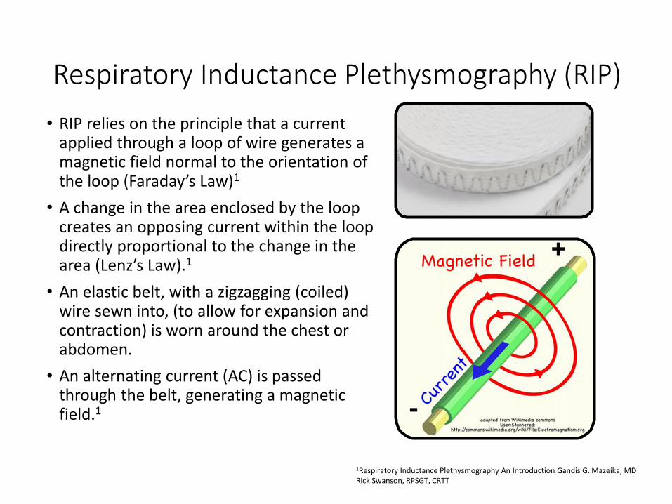
Respiratory Inductance Plethysmography (RIP)
• RIP relies on the principle that a current applied through a loop of wire generates a magnetic field normal to the orientation of the loop (Faraday’s Law)1
• A change in the area enclosed by the loop creates an opposing current within the loop directly proportional to the change in the area (Lenz’s Law).1
• An elastic belt, with a zigzagging (coiled) wire sewn into, (to allow for expansion and contraction) is worn around the chest or abdomen.
• An alternating current (AC) is passed through the belt, generating a magnetic field.1
1Respiratory Inductance Plethysmography An Introduction Gandis G. Mazeika, MD Rick Swanson, RPSGT, CRTT

RIP Measurements
• The act of breathing changes the cross-sectional area of the patient’s body, and thus changes the shape of the magnetic field generated by the belt, “inducing” an opposing current that can be measured, most easily as a change in the frequency of the applied current.1
• With RIP, no electrical current passes through the body (a weak magnetic field is present that does not affect the patient or any surrounding equipment).1
• The signal is linear and is a fairly accurate representation of the change in cross-sectional area.1
1Respiratory Inductance Plethysmography An Introduction Gandis G. Mazeika, MD Rick Swanson, RPSGT, CRTT
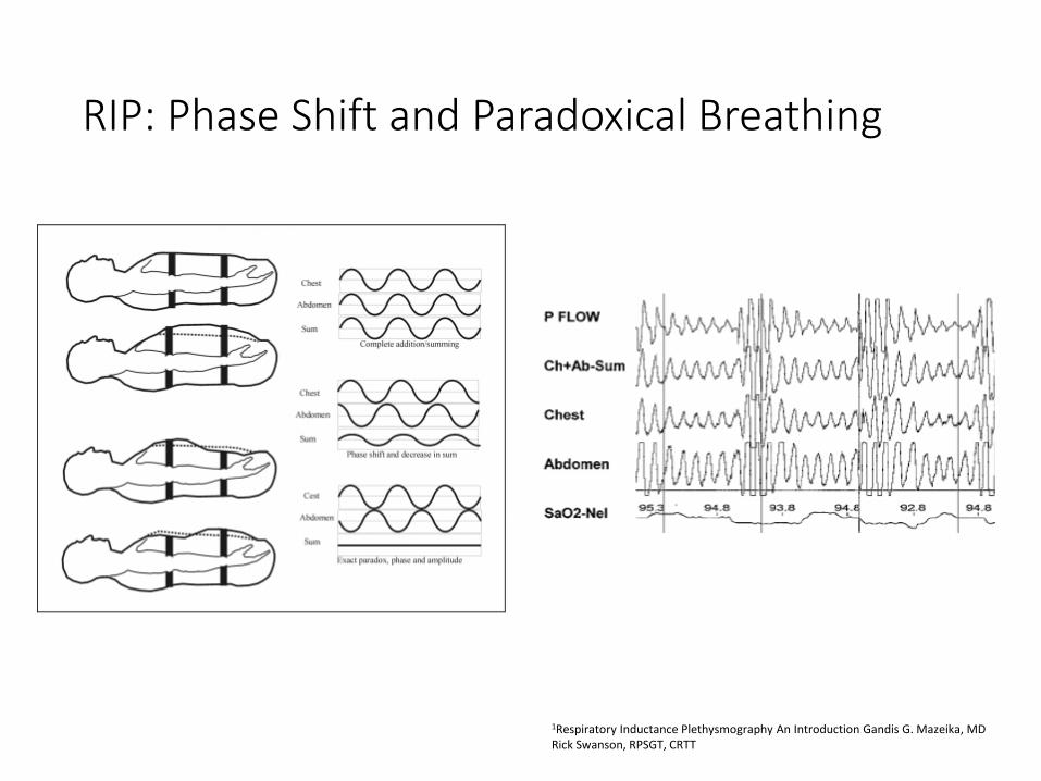
RIP: Phase Shift and Paradoxical Breathing
1Respiratory Inductance Plethysmography An Introduction Gandis G. Mazeika, MD Rick Swanson, RPSGT, CRTT

Polyvinylidene Fluoride (PVDF)
• Polyvinylidene difluoride (PVDF) is a highly non-reactive and pure thermoplastic fluoropolymer produced by the polymerization of vinylidene difluoride.
• PVDF has flexibility, low weight, low thermal conductivity, high chemical corrosion resistance, and heat resistance.

PVDF
• There are many commercial applications of PVDF• Insulation on electrical wires
• Tactile sensor arrays
• Strain gauges
• Audio transducers
• Measuring dust density in the solar system
• Binder material in lithium ion batteries
• Specialty fishing lines (fluorocarbon)
• Sterilizing filter in biomedical science

PVDF Belts
• Provides linear, accurate effort signal and maintains polarity
• Infant, Pediatric, and Adult Sizes are available
• Sum Channel Option
• Compatible with most current PSG Systems
• Easy to clean with machine washable straps
• No Batteries to Change
On December 19, 2011 the AASM made changes to the Scoring Manual, effective immediately, to include Polyvinylidene Fluoride (PVDF) effort sensors using thoracoabdominal belts.

Piezoelectric Technology
• The sensing element, a piezo crystal, on a piezo effort belt is located only on a very small section of the belt’s length.
• The effort signal can be dampened, not detected (like when the patient is lying on the sensor), producing erroneous readings or unexplained changes in polarity (false paradoxing).
• Susceptible to trapping artifact.
• No longer recommended as an AASM standard.

Diaphragmatic EMG (dEMG) & Intercostal EMG
1 Differential Sensitivity of Abdominal Muscles and the Diaphragm to Mivacurium: An Electromyographic StudyKrassen Kirov, M.D.; Cyrus Motamed, M.D.; Gilles Dhonneur, M.D., Ph.D., Anesthesiology 12 2001, Vol.95, 1323-1328.
2 Discharge frequencies of single motor units in human diaphragm and parasternal muscles in lying and standing. J. E. Butler, D. K. McKenzie, S. C. Gandevia. Journal of Applied Physiology Published 1 January 2001 Vol. 90 no. 1, 147-154
1 2

dEMG / Intercostals
• Diaphragm EMG can be recorded from the chest wall surface electrodes in most subjects and can be used to demonstrate the presence of respiratory effort during apnoea/hypopnea events.
Respiratory Action of the Intercostal Muscles. André De Troyer, Peter A. Kirkwood, Theodore A. Wilson. Physiological Reviews Published 1 April 2005 Vol. 85 no. 2, 717-756

dEMG / Intercostals
Notice the Intercostal Channel shows a lack of effort. Ballistocardiogram artifact can be seen in the RIP (abdomen) belt. Verification of central apnea.
dEMG / Intercostals
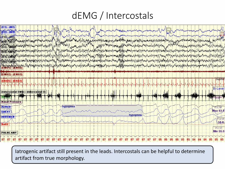
Iatrogenic artifact still present in the leads. Intercostals can be helpful to determine artifact from true morphology.
dEMG / Intercostals
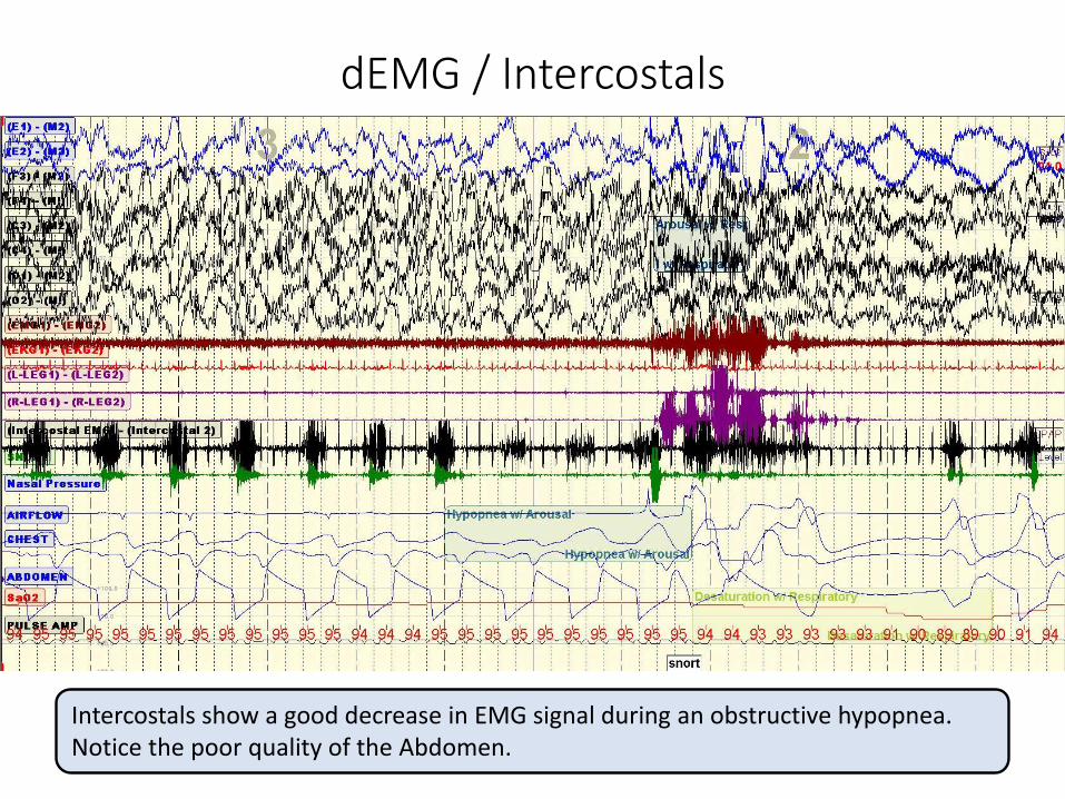
Intercostals show a good decrease in EMG signal during an obstructive hypopnea. Notice the poor quality of the Abdomen.
dEMG / Intercostals
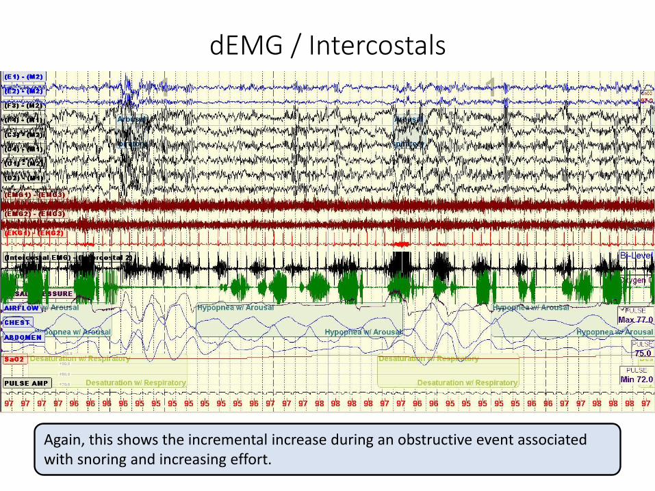
Again, this shows the incremental increase during an obstructive event associated with snoring and increasing effort.

dEMG / Intercostals
Strong intercostal signals help with identification of obstructive events.

dEMG / Intercostals
Ambiguity from the chest and abdomen belt make this type of even difficult to differentiate between central and obstructive. The intercostals provide help here.

Summary
• The AASM gold standard for monitoring of respiratory effort is esophageal Manometry, but this technique is rarely used in commercial sleep laboratories today due to patient discomfort, technique difficulty and cost.
• RIP belts are the primary tool used to measure respiratory effort.
• PVDF are an AASM approved acceptable alternative to RIP belts.
• Piezoelectric belts are not recommended for measurement of respiratory effort.
• dEMG and intercostal EMG leads can be a helpful tool for monitoring respiratory effort, but more research is needed to determine accuracy and standardized placement procedures.