respiratory burst oxidasefromhuman neutrophils ... · the respiratory burst oxidase was first...
TRANSCRIPT

Proc. Nati. Acad. Sci. USAVol. 82, pp. 3144-3148, May 1985Biochemistry
Respiratory burst oxidase from human neutrophils: Purification andsome properties
(phagocytes/superoxide/oxygen radicals/host defense mechanisms)
MICHELE MARKERT*, G. ALLISON GLASS, AND BERNARD M. BABIORBlood Research Laboratory and Department of Medicine, Tufts-New England Medical Center, Boston, MA 02111
Communicated by Irwin Fridovich, January 16, 1985
ABSTRACT The respiratory burst oxidase of human neu-trophils was purified by "dye-affinity" chromatography overa red agarose column. Electrophoresis of the purified enzymeon NaDodSO4 gel showed a single major band at 64,000-66,000 daltons, together with some minor contaminants. On anondenaturing gel, the enzyme ran as two closely spacedbands, the faster of which contained flavin. When these twobands were rerun separately on a NaDodSO4 gel, they gaveidentical patterns, each showing a major band at ca. 65,00Qdaltons. The specific activity (mean ± SEM) of the purifiedenzyme was 8.8 ± 3.5 ,umol of O2 per min/mg of protein.
When exposed to appropriate stimuli, neutrophils are in-duced to express the "respiratory burst," a profound alter-ation in oxygen metabolism whose purpose is the generationof microbicidal oxidants by the partial reduction of oxygen.This alteration results from the activation of a membrane-bound oxidase, dormant in resting cells, that catalyzes thereduction of oxygen to °- at the expense of NADPH (1-11):
202 + NADPH -, 2 °- + NADP'.
The °2 is then converted in a series of secondary reactionsto OCl- and reactive oxidizing radicals (including OH-), theproximate microbicidal oxidants. The respiratory burst andother aspects of the oxygen-dependent microbicidal mecha-nisms of phagocytes have recently been reviewed (12-16).The respiratory burst oxidase was first solubilized from
the plasma membranes of activated human neutrophils in1978 (17). We now report the purification of this oxidase anddescribe some of the properties of the purified enzyme.
MATERIALS AND METHODS
Human neutrophils were prepared by dextran sedimentationand differential centrifugation as described (18). Zymosan(Sigma) was boiled for 10 min in 1 M NaOH, then washedthree times with Hanks' balanced salt solution (GIBCO), andfinally opsonized as described elsewhere (3). Superoxidedismutase, horse heart cytochrome c (type VI), phosphati-dylethanolamine (ovine brain), Lubrol PX, sodium deoxy-cholate, phenylmethylsulfonyl fluoride, leupeptin, pepsta-tin, FAD, and NADPH were purchased from Sigma. The so-dium deoxycholate was purified by recrystallization fromabsolute ethanol. Red Sepharose CL-6B (agarose coupled toreactive red 120) was obtained from Pharmacia. The concen-tration of dye in the lots purchased from Pharmacia rangedfrom 2.0 to 3.0 Afmol/ml of packed gel. Other reagents werethe best grade commercially available apd were used withoutfurther purification.
Gel Electrophoresis. Electrophoresis on 7.5% nondenatur-ing slab gels was performed by a modification of the methodof Davis (19) in which the solution for preparing the lower gelcontained 4% glycerol plus twice the usual concentration ofTris, that for preparing the upper gel contained half the usualconcentration of bisacrylamide, and polymerization was ac-complished with 0.035% (wt/vol) ammonium persulfate,which was removed by preelectrophoresis for 1 hr before thesamples were applied to the slab. Nondenaturing gel electro-phoresis was carried out at 40C. NaDodSO4/polyacrylamidegel electrophoresis (NaDodSO4/PAGE) was performed bythe method of Laemmli (20) using 9o slab gels. Where nec-essary, samples were concentrated before electrophoresis inCF 25A ultrafiltration cones (Amicon) and then centrifugedfor 30 min at 100,000 x g to remove aggregated proteins.Proteins were visualized with Coomassie blue R-250 or witha silver stain kit purchased from Bio-Rad.
Localization of Flavin in the Nondenaturing Gel. The loca-tion of flavin in the nondenaturing gel was determined fluori-metrically. The track of interest was excised from the gel,and the region between the top of the running gel and thetracking dye band was cut into 1-mm slices. Each slice washomogenized by hand (6-8 strokes in a Potter-Elvehjem ho-mogenizer) in 3 ml of elution buffer without detergent (seebelow). The fluorescence spectrum of each homogenate wasobtained with a Perkin-Elmer model MPF-3 spectrofluorim-eter, exciting at 470 ± 5 nm and measuring the emission be-tween 480 and 540 nm. An excitation spectrum was obtainedon the sample showing maximum fluorescence; for this, theexcitation wavelength was varied between 380 and 480 nm,and emission was measured at 525 nm. The measurement offlavin fluorescence was not affected by the presence of gelfragments in the sample.
Assays. Superoxide production was measured as described(21), except that FAD was added to a final concentration of20 ,uM, and incubations were carried out for only 5 min. Ini-tial rates were calculated by dividing the 5-min value for O2production by 3, a figure that was determined experimental-ly by comparing initial rates of O2 production as measuredin a continuous assay (22) with the 5-min values obtainedwith the same enzyme preparations. Protein concentrationsof the membrane suspensions were measured by using theBradford reagent (Bio-Rad). Protein concentrations of deter-gent-containing preparations were determined by the meth-od of Schaffner and Weissmann (23). Bovine serum albuminwas used as standard for both methods.
RESULTS
Purification of the Oxidase. The oxidase was purified bysolubilizing the O-forming activity from the plasma mem-branes of zymosan-activated neutrophils and then chromato-
*Present address: Laboratoire Central de Chimie Clinique, CHUV,Lausanne, Switzerland.
3144
The publication costs of this article were defrayed in part by page chargepayment. This article must therefore be hereby marked "advertisement"in accordance with 18 U.S.C. §1734 solely to indicate this fact.
Proc. NatL. Acad Sci. USA 82 (1985) 3145
graphing the solubilized material over a column of red agar-ose.Preparation of neutrophil membranes. Neutrophils were
isolated from 300 ml of blood. The cells [resting, or activatedwith opsonized zymosan (8) with or without a 10-min prein-cubation with 0.1 gM fMet-Leu-Phe (24)] were suspended ata concentration of 5 x 10⁷ cells per ml in 0.34 M sucrosecontaining 0.5 mM phenylmethylsulfonyl fluoride, 2 p.Mpepstatin, and 2 kkM leupeptin. Six milliliters of this suspen-sion was placed in a 100-ml glass beaker and sonicated at fullpower for two 30-sec intervals 1 min apart at 00C in a HeatSystems model W220F sonifier fitted with a cup horn. Thesonicate was centrifuged at 160 x g for 5 min at 40C to re-move whole cells and nuclei, and the entire supernatant waslayered over 10 ml of 30% (wt/vol) sucrose, which, in turn,rested on 20 ml of 50% (wt/vol) sucrose in a 2.5 x 8.9 cmpolyallomer tube. This discontinuous gradient was centri-fuged at 140,000 x g for 45 min at 40C using a Spinco SW 28head in a Beckman model L3-50 preparative ultracentrifuge.The membranes sedimented to the interface between the twosucrose layers. This interface was carefully aspirated with aPasteur pipet, diluted with 3 vol of distilled water, and cen-trifuged in a Sorvall RC-5 high-speed centrifuge at 27,000 xg for 30 minutes at 4°C. The resulting membrane pellet wassuspended in 1.0 ml of 0.34 M sucrose and assayed for pro-tein and O-forming activity.
Solubilization of the oxidase. The membrane suspensionwas mixed with an equal volume of extraction buffer [2%(wt/vol) sodium deoxycholate in 20 mM sodium glycinatebuffer (pH 8.0) containing 1 mM NaN3, 1.7 ,uM CaCl2, and50% (vol/vol) glycerol]. This mixture was incubated on icefor 30 min with occasional gentle agitation on a Vortex mix-er. The extract was centrifuged at 100,000 x g for 30 min at4°C (Spinco fixed-angle Ti 60 head). The supernatant wasthen assayed for protein and for O-forming activity. Theprotein concentration in the extract averaged 2.3 ± 0.4mg/ml (mean ± SEM; n = 5). Thirty to 40% of the originalO-forming activity could routinely be recovered in the ex-tract.Chromatography over red agarose. Chromatography was
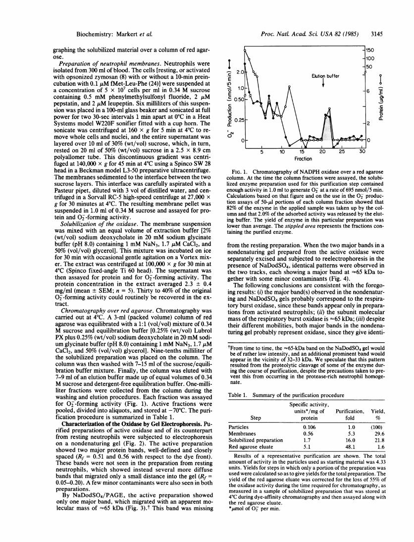
carried out at 4°C. A 3-ml (packed volume) column of redagarose was equilibrated with a 1:1 (vol/vol) mixture of 0.34M sucrose and equilibration buffer [0.25% (wt/vol) LubrolPX plus 0.25% (wt/vol) sodium deoxycholate in 20 mM sodi-um glycinate buffer (pH 8.0) containing 1 mM NaN3, 1.7 ,uMCaCl2, and 50% (vol/vol) glycerol]. Nine-tenths milliliter ofthe solubilized preparation was placed on the column. Thecolumn was then washed with 7-15 ml of the sucrose/equili-bration buffer mixture. Finally, the column was eluted with7-9 ml of an elution buffer made up of equal volumes of 0.34M sucrose and detergent-free equilibration buffer. One-milli-liter fractions were collected from the column during thewashing and elution procedures. Each fraction was assayedfor 0--forming activity (Fig. 1). Active fractions werepooled, divided into aliquots, and stored at -70°C. The puri-fication procedure is summarized in Table 1.
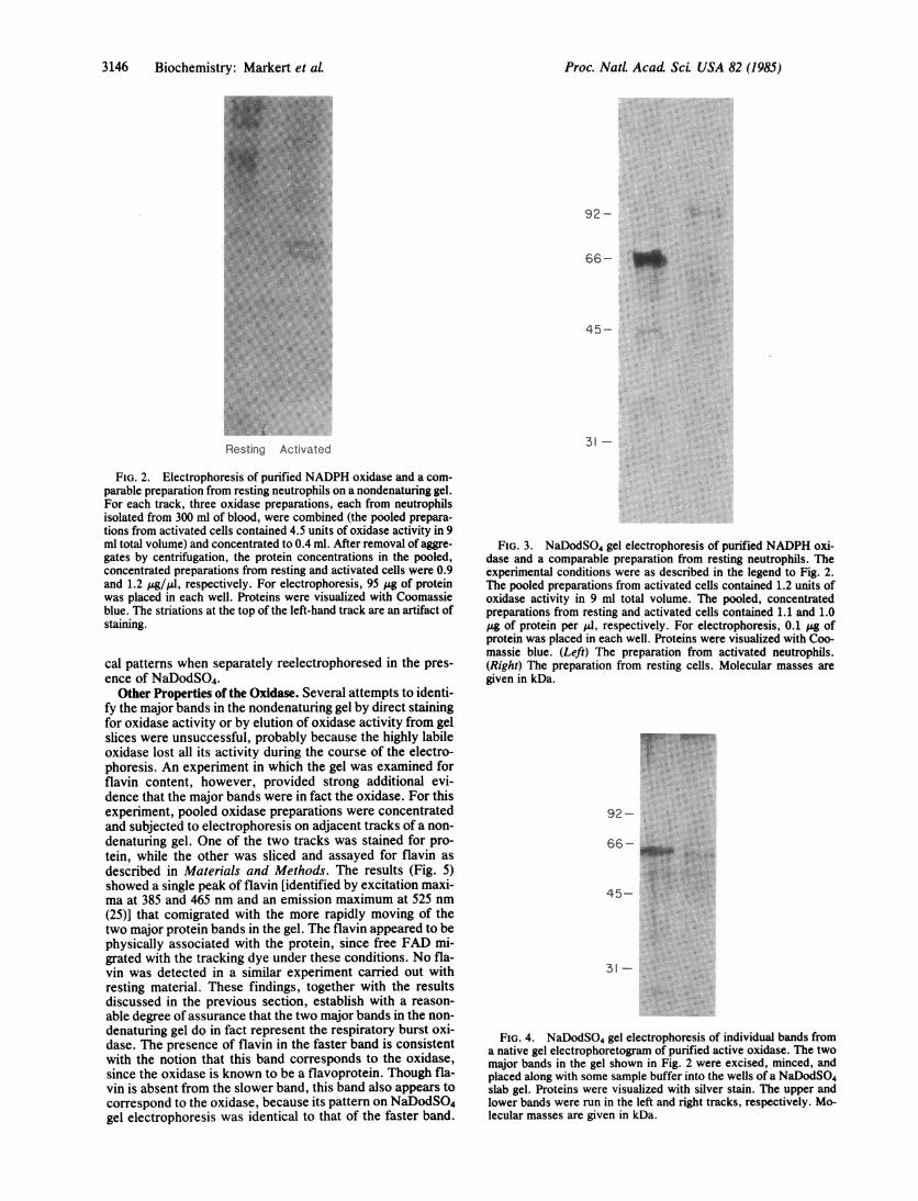
Characterization of the Oxidase by Gel Electrophoresis. Pu-rified preparations of active oxidase and of its counterpartfrom resting neutrophils were subjected to electrophoresison a nondenaturing gel (Fig. 2). The active preparationshowed two major protein bands, well-defined and closelyspaced (Rf = 0.51 and 0.56 with respect to the dye front).These bands were not seen in the preparation from restingneutrophils, which showed instead several more diffusebands that migrated only a small distance into the gel (Rf =
0.05-0.20). A few minor contaminants were also seen in bothpreparations.By NaDodSO4/PAGE, the active preparation showed
only one major band, which migrated with an apparent mo-lecular mass of -65 kDa (Fig. 3).t This band was missing
-12CE
if)
E
C:0
a.
Ny
15
Fraction
"I
C0'
a-
E.
FIG. 1. Chromatography of NADPH oxidase over a red agarosecolumn. At the time the column fractions were assayed, the solubi-lized enzyme preparation used for this purification step containedenough activity in 1.0 ml to generate O2 at a rate of 695 nmol/5 min.Calculations based on that figure and on the use in the O2 produc-tion assays of 50-II portions of each column fraction showed that82% of the enzyme in the applied sample was taken up by the col-umn and that 2.0% of the adsorbed activity was released by the elut-ing buffer. The yield of enzyme in this particular preparation waslower than average. The stippled area represents the fractions con-taining the purified enzyme.
from the resting preparation. When the two major bands in anondenaturing gel prepared from the active oxidase wereseparately excised and subjected to reelectrophoresis in thepresence of NaDodSO4, identical patterns were observed inthe two tracks, each showing a major band at =65 kDa to-gether with some minor contaminants (Fig. 4).The following conclusions are consistent with the forego-
ing results: (i) the major band(s) observed in the nondenatur-ing and NaDodSO4 gels probably correspond to the respira-tory burst oxidase, since these bands appear only in prepara-tions from activated neutrophils; (ii) the subunit molecularmass of the respiratory burst oxidase is =65 kDa; (iii) despitetheir different mobilities, both major bands in the nondena-turing gel probably represent oxidase, since they give identi-
tFrom time to time, the -65-kDa band on the NaDodSO4 gel wouldbe of rather low intensity, and an additional prominent band wouldappear in the vicinity of 32-33 kDa. We speculate that this patternresulted from the proteolytic cleavage of some of the enzyme dur-ing the course of purification, despite the precautions taken to pre-vent this from occurring in the protease-rich neutrophil homoge-nate.
Table 1. Summary of the purification procedure
Specific activity,units*/mg of Purification, Yield,
Step protein fold S
Particles 0.106 1.0 (100)Membranes 0.56 5.3 29.6Solubilized preparation 1.7 16.0 21.8Red agarose eluate 5.1 48.1 1.6
Results of a representative purification are shown. The totalamount of activity in the particles used as starting material was 4.33units. Yields for steps in which only a portion of the preparation wasused were calculated so as to give yields for the total preparation. Theyield of the red agarose eluate was corrected for the loss of 55% ofthe oxidase activity during the time required for chromatography, asmeasured in a sample of solubilized preparation that was stored at4°C during dye-affinity chromatography and then assayed along withthe red agarose eluate.*,umol of O2 per min.
Biochemistry: Markert et aL
3146 Biochemistry: Markert et aL
92-
66-
45-
31 -
FIG. 2. Electrophoresis of purified NADPH oxidase and a com-parable preparation from resting neutrophils on a nondenaturing gel.For each track, three oxidase preparations, each from neutrophilsisolated from 300 ml of blood, were combined (the pooled prepara-tions from activated cells contained 4.5 units of oxidase activity in 9ml total volume) and concentrated to 0.4 ml. After removal of aggre-gates by centrifugation, the protein concentrations in the pooled,concentrated preparations from resting and activated cells were 0.9and 1.2 pyg/IA, respectively. For electrophoresis, 95 P8 of proteinwas placed in each well. Proteins were visualized with Coomassieblue. The striations at the top of the left-hand track are an artifact ofstaining.
cal patterns when separately reelectrophoresed in the pres-ence of NaDodSO4.
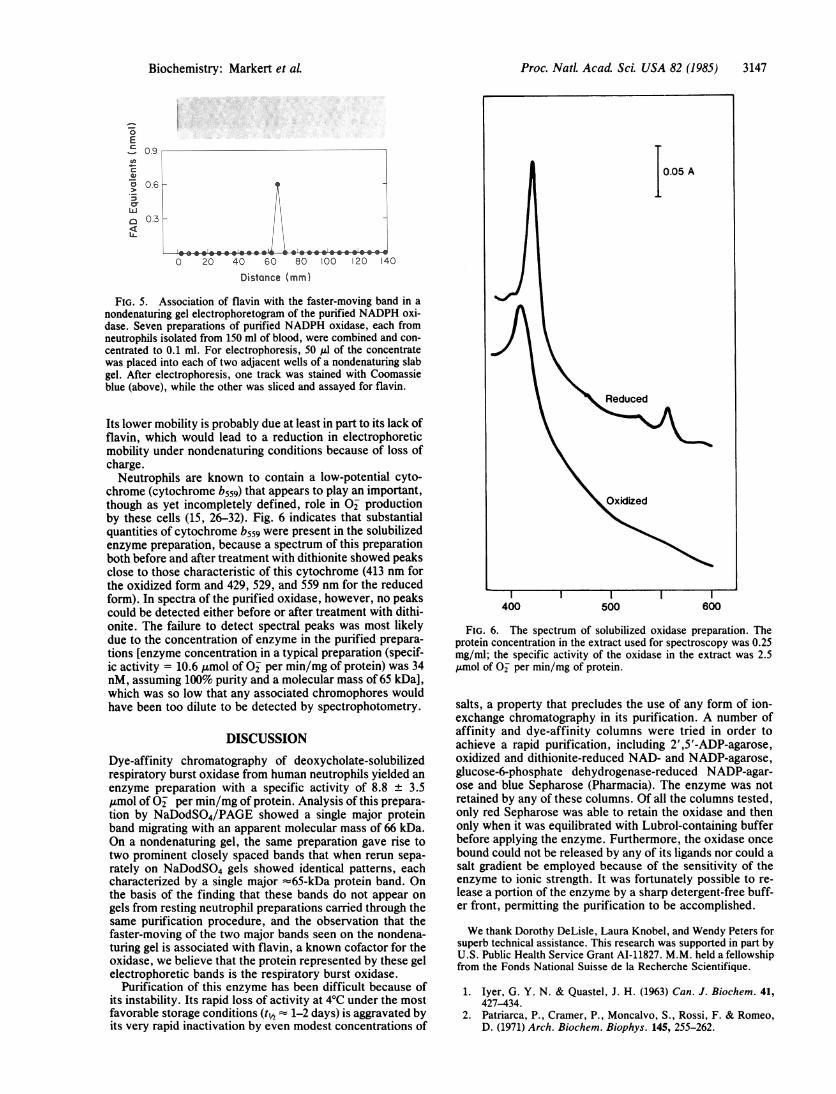
Other Properties of the Oxidase. Several attempts to identi-fy the major bands in the nondenaturing gel by direct stainingfor oxidase activity or by elution of oxidase activity from gelslices were unsuccessful, probably because the highly labileoxidase lost all its activity during the course of the electro-phoresis. An experiment in which the gel was examined forflavin content, however, provided strong additional evi-dence that the major bands were in fact the oxidase. For thisexperiment, pooled oxidase preparations were concentratedand subjected to electrophoresis on adjacent tracks of a non-denaturing gel. One of the two tracks was stained for pro-tein, while the other was sliced and assayed for flavin asdescribed in Materials and Methods. The results (Fig. 5)showed a single peak of flavin [identified by excitation maxi-ma at 385 and 465 nm and an emission maximum at 525 nm(25)] that comigrated with the more rapidly moving of thetwo major protein bands in the gel. The flavin appeared to bephysically associated with the protein, since free FAD mi-grated with the tracking dye under these conditions. No fla-vin was detected in a similar experiment carried out withresting material. These findings, together with the resultsdiscussed in the previous section, establish with a reason-able degree of assurance that the two major bands in the non-denaturing gel do in fact represent the respiratory burst oxi-dase. The presence of flavin in the faster band is consistentwith the notion that this band corresponds to the oxidase,since the oxidase is known to be a flavoprotein. Though fla-vin is absent from the slower band, this band also appears tocorrespond to the oxidase, because its pattern on NaDodSO4gel electrophoresis was identical to that of the faster band.
FIG. 3. NaDodSO4 gel electrophoresis of purified NADPH oxi-dase and a comparable preparation from resting neutrophils. Theexperimental conditions were as described in the legend to Fig. 2.The pooled preparations from activated cells contained 1.2 units ofoxidase activity in 9 ml total volume. The pooled, concentratedpreparations from resting and activated cells contained 1.1 and 1.0Ag of protein per Al, respectively. For electrophoresis, 0.1 Ag ofprotein was placed in each well. Proteins were visualized with Coo-massie blue. (Left) The preparation from activated neutrophils.(Right) The preparation from resting cells. Molecular masses aregiven in kDa.
92-
66-
A_-t J >r.... ..s
.:.:.:...
*..:
3 1-.. :: .......... . .......
:: ... ::
:::.:: :
FIG. 4. NaDodSO4 gel electrophoresis of individual bands froma native gel electrophoretogram of purified active oxidase. The twomajor bands in the gel shown in Fig. 2 were excised, minced, andplaced along with some sample buffer into the wells of a NaDodSO4slab gel. Proteins were visualized with silver stain. The upper andlower bands were run in the left and right tracks, respectively. Mo-lecular masses are given in kDa.
Proc. NatL Acad Sci. USA 82 (1985)
Proc. Natl. Acad Sci. USA 82 (1985) 3147
0EC0.9
vI.
hU
a 03r<LL
!H_1
I-~---.--~ 1--- I
0 20 40 60 80 l00 20 140Distance (mm)
FIG. 5. Association of flavin with the faster-moving band in anondenaturing gel electrophoretogram of the purified NADPH oxi-dase. Seven preparations of purified NADPH oxidase, each fromneutrophils isolated from 150 ml of blood, were combined and con-centrated to 0.1 ml. For electrophoresis, 50 1.d of the concentratewas placed into each of two adjacent wells of a nondenaturing slabgel. After electrophoresis, one track was stained with Coomassieblue (above), while the other was sliced and assayed for flavin.
Its lower mobility is probably due at least in part to its lack offlavin, which would lead to a reduction in electrophoreticmobility under nondenaturing conditions because of loss ofcharge.Neutrophils are known to contain a low-potential cyto-
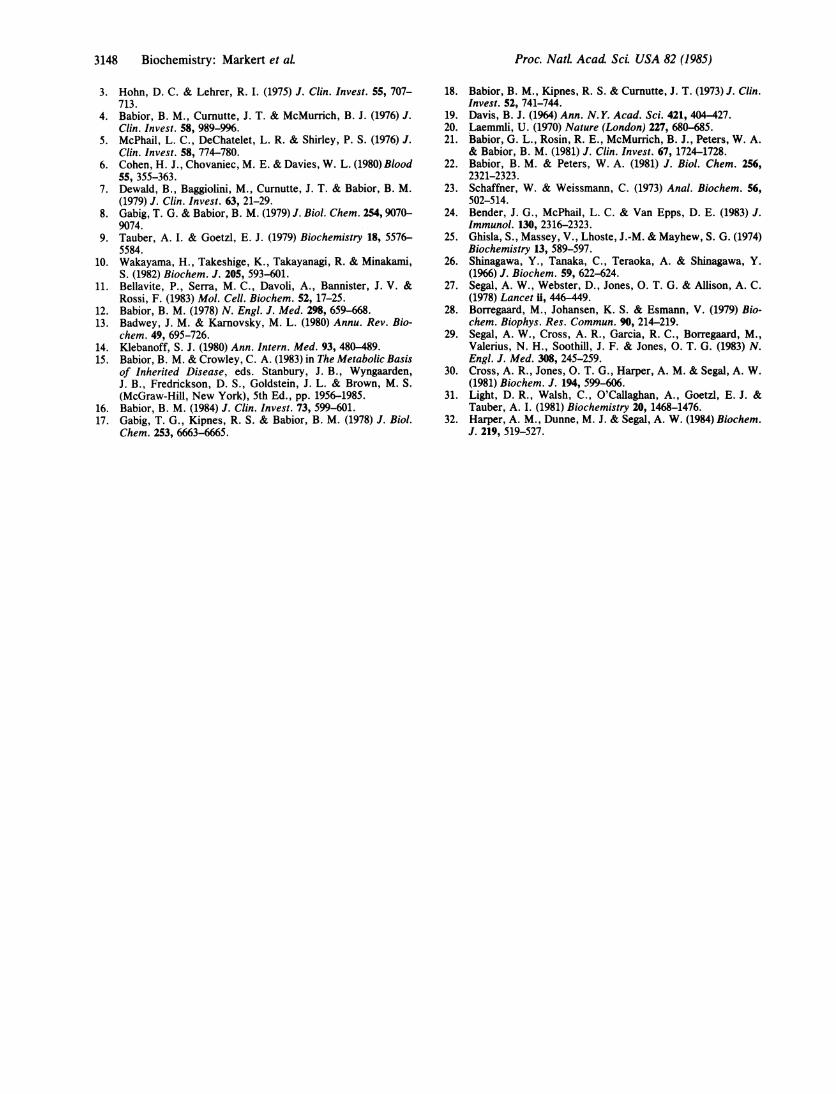
chrome (cytochrome b559) that appears to play an important,though as yet incompletely defined, role in O2 productionby these cells (15, 26-32). Fig. 6 indicates that substantialquantities of cytochrome b559 were present in the solubilizedenzyme preparation, because a spectrum of this preparationboth before and after treatment with dithionite showed peaksclose to those characteristic of this cytochrome (413 nm forthe oxidized form and 429, 529, and 559 nm for the reducedform). In spectra of the purified oxidase, however, no peakscould be detected either before or after treatment with dithi-onite. The failure to detect spectral peaks was most likelydue to the concentration of enzyme in the purified prepara-tions [enzyme concentration in a typical preparation (specif-ic activity = 10.6 ,umol of O2 per min/mg of protein) was 34nM, assuming 100% purity and a molecular mass of 65 kDa],which was so low that any associated chromophores wouldhave been too dilute to be detected by spectrophotometry.
DISCUSSION
Dye-affinity chromatography of deoxycholate-solubilizedrespiratory burst oxidase from human neutrophils yielded anenzyme preparation with a specific activity of 8.8 ± 3.5,umol of O2 per min/mg of protein. Analysis of this prepara-tion by NaDodSO4/PAGE showed a single major proteinband migrating with an apparent molecular mass of 66 kDa.On a nondenaturing gel, the same preparation gave rise totwo prominent closely spaced bands that when rerun sepa-rately on NaDodSO4 gels showed identical patterns, eachcharacterized by a single major =65-kDa protein band. Onthe basis of the finding that these bands do not appear ongels from resting neutrophil preparations carried through thesame purification procedure, and the observation that thefaster-moving of the two major bands seen on the nondena-turing gel is associated with flavin, a known cofactor for theoxidase, we believe that the protein represented by these gelelectrophoretic bands is the respiratory burst oxidase.
Purification of this enzyme has been difficult because ofits instability. Its rapid loss of activity at 4°C under the mostfavorable storage conditions (t½ = 1-2 days) is aggravated byits very rapid inactivation by even modest concentrations of
I I I
400 500 600
FIG. 6. The spectrum of solubilized oxidase preparation. Theprotein concentration in the extract used for spectroscopy was 0.25mg/ml; the specific activity of the oxidase in the extract was 2.5,umol of O2 per min/mg of protein.
salts, a property that precludes the use of any form of ion-exchange chromatography in its purification. A number ofaffinity and dye-affinity columns were tried in order toachieve a rapid purification, including 2',5'-ADP-agarose,oxidized and dithionite-reduced NAD- and NADP-agarose,glucose-6-phosphate dehydrogenase-reduced NADP-agar-ose and blue Sepharose (Pharmacia). The enzyme was notretained by any of these columns. Of all the columns tested,only red Sepharose was able to retain the oxidase and thenonly when it was equilibrated with Lubrol-containing bufferbefore applying the enzyme. Furthermore, the oxidase once
bound could not be released by any of its ligands nor could asalt gradient be employed because of the sensitivity of theenzyme to ionic strength. It was fortunately possible to re-lease a portion of the enzyme by a sharp detergent-free buff-er front, permitting the purification to be accomplished.
We thank Dorothy DeLisle, Laura Knobel, and Wendy Peters forsuperb technical assistance. This research was supported in part byU.S. Public Health Service Grant AI-11827. M.M. held a fellowshipfrom the Fonds National Suisse de la Recherche Scientifique.
1. Iyer, G. Y. N. & Quastel, J. H. (1963) Can. J. Biochem. 41,427-434.
2. Patriarca, P., Cramer, P., Moncalvo, S., Rossi, F. & Romeo,D. (1971) Arch. Biochem. Biophys. 145, 255-262.
0.o5 A
Reduced
L
Biochemistry: Markert et aL
3148 Biochemistry: Markert et al.
3. Hohn, D. C. & Lehrer, R. I. (1975) J. Clin. Invest. 55, 707-713.
4. Babior, B. M., Curnutte, J. T. & McMurrich, B. J. (1976) J.Clin. Invest. 58, 989-996.
5. McPhail, L. C., DeChatelet, L. R. & Shirley, P. S. (1976) J.Clin. Invest. 58, 774-780.
6. Cohen, H. J., Chovaniec, M. E. & Davies, W. L. (1980) Blood55, 355-363.
7. Dewald, B., Baggiolini, M., Curnutte, J. T. & Babior, B. M.(1979) J. Clin. Invest. 63, 21-29.
8. Gabig, T. G. & Babior, B. M. (1979) J. Biol. Chem. 254, 9070-9074.
9. Tauber, A. I. & Goetzl, E. J. (1979) Biochemistry 18, 5576-5584.
10. Wakayama, H., Takeshige, K., Takayanagi, R. & Minakami,S. (1982) Biochem. J. 205, 593-601.
11. Bellavite, P., Serra, M. C., Davoli, A., Bannister, J. V. &Rossi, F. (1983) Mol. Cell. Biochem. 52, 17-25.
12. Babior, B. M. (1978) N. Engl. J. Med. 298, 659-668.13. Badwey, J. M. & Karnovsky, M. L. (1980) Annu. Rev. Bio-
chem. 49, 695-726.14. Klebanoff, S. J. (1980) Ann. Intern. Med. 93, 480-489.15. Babior, B. M. & Crowley, C. A. (1983) in The Metabolic Basis
of Inherited Disease, eds. Stanbury, J. B., Wyngaarden,J. B., Fredrickson, D. S., Goldstein, J. L. & Brown, M. S.(McGraw-Hill, New York), 5th Ed., pp. 1956-1985.
16. Babior, B. M. (1984) J. Clin. Invest. 73, 599-601.17. Gabig, T. G., Kipnes, R. S. & Babior, B. M. (1978) J. Biol.
Chem. 253, 6663-6665.
18. Babior, B. M., Kipnes, R. S. & Curnutte, J. T. (1973) J. Clin.Invest. 52, 741-744.
19. Davis, B. J. (1964) Ann. N. Y. Acad. Sci. 421, 404 427.20. Laemmli, U. (1970) Nature (London) 227, 680-85.21. Babior, G. L., Rosin, R. E., McMurrich, B. J., Peters, W. A.
& Babior, B. M. (1981) J. Clin. Invest. 67, 1724-1728.22. Babior, B. M. & Peters, W. A. (1981) J. Biol. Chem. 256,
2321-2323.23. Schaffner, W. & Weissmann, C. (1973) Anal. Biochem. 56,
502-514.24. Bender, J. G., McPhail, L. C. & Van Epps, D. E. (1983) J.
Immunol. 130, 2316-2323.25. Ghisla, S., Massey, V., Lhoste, J.-M. & Mayhew, S. G. (1974)
Biochemistry 13, 589-597.26. Shinagawa, Y., Tanaka, C., Teraoka, A. & Shinagawa, Y.
(1966) J. Biochem. 59, 622-624.27. Segal, A. W., Webster, D., Jones, 0. T. G. & Allison, A. C.
(1978) Lancet U, 446-449.28. Borregaard, M., Johansen, K. S. & Esmann, V. (1979) Bio-
chem. Biophys. Res. Commun. 90, 214-219.29. Segal, A. W., Cross, A. R., Garcia, R. C., Borregaard, M.,
Valerius, N. H., Soothill, J. F. & Jones, 0. T. G. (1983) N.Engl. J. Med. 308, 245-259.
30. Cross, A. R., Jones, 0. T. G., Harper, A. M. & Segal, A. W.(1981) Biochem. J. 194, 599-606.
31. Light, D. R., Walsh, C., O'Callaghan, A., Goetzl, E. J. &Tauber, A. I. (1981) Biochemistry 20, 1468-1476.
32. Harper, A. M., Dunne, M. J. & Segal, A. W. (1984) Biochem.J. 219, 519-527.
Proc. NatL Acad ScL USA 82 (1985)