researcharticle developmentalblockandprogrammedcell ... · researcharticle...
TRANSCRIPT

RESEARCH ARTICLE
Developmental Block and Programmed CellDeath in Bos indicus Embryos: Effects ofProtein Supplementation Source andDevelopmental KineticsSheila Merlo Garcia1, Luciana Simões Rafagnin Marinho2, Paula Alvares Lunardelli2,Marcelo Marcondes Seneda2*, Flávio Vieira Meirelles1
1 São Paulo University (USP), Faculdade de Zootecnia e Engenharia de Alimentos, Pirassununga, SP,Brazil, 2 State University of Londrina (UEL), Laboratório de Reprodução Animal, DCV, CCA, Londrina, PR,Brazil
AbstractThe aims of this study were to determine if the protein source of the medium influences
zebu embryo development and if developmental kinetics, developmental block and pro-
grammed cell death are related. The culture medium was supplemented with either fetal
calf serum or bovine serum albumin. The embryos were classified as Fast (n = 1,235) or
Slow (n = 485) based on the time required to reach the fourth cell cycle (48 h and 90 h post
insemination - hpi -, respectively). The Slow group was further separated into two groups:
those presenting exactly 4 cells at 48 hpi (Slow/4 cells) and those that reached the fourth
cell cycle at 90 hpi (Slow). Blastocyst quality, DNA fragmentation, mitochondrial membrane
potential and signs of apoptosis or necrosis were evaluated. The Slow group had higher in-
cidence of developmental block than the Fast group. The embryos supplemented with fetal
calf serum had lower quality. DNA fragmentation and mitochondrial membrane potential
were absent in embryos at 48 hpi but present at 90 hpi. Early signs of apoptosis were more
frequent in the Slow and Slow/4 cell groups than in the Fast group. We concluded that fetal
calf serum reduces blastocyst development and quality, but the mechanism appears to be
independent of DNA fragmentation. The apoptotic cells detected at 48 hpi reveal a possible
mechanism of programmed cell death activation prior to genome activation. The apoptotic
cells observed in the slow-developing embryos suggested a relationship between pro-
grammed cell death and embryonic developmental kinetics in zebu in vitro-
produced embryos.
IntroductionIn vitro embryo production (IVEP) is used in bovine herds around the world. IVEP was ini-tially used as a final option for donor cows that could not establish pregnancies through other
PLOSONE | DOI:10.1371/journal.pone.0119463 March 11, 2015 1 / 16
OPEN ACCESS
Citation: Garcia SM, Marinho LSR, Lunardelli PA,Seneda MM, Meirelles FV (2015) DevelopmentalBlock and Programmed Cell Death in Bos indicusEmbryos: Effects of Protein Supplementation Sourceand Developmental Kinetics. PLoS ONE 10(3):e0119463. doi:10.1371/journal.pone.0119463
Academic Editor: Peter James Hansen, Universityof Florida, UNITED STATES
Received: August 28, 2014
Accepted: January 21, 2015
Published: March 11, 2015
Copyright: © 2015 Garcia et al. This is an openaccess article distributed under the terms of theCreative Commons Attribution License, which permitsunrestricted use, distribution, and reproduction in anymedium, provided the original author and source arecredited.
Data Availability Statement: All relevant data arewithin the paper and its Supporting Information files.
Funding: This study was supported by Fundação deAmparo à Pesquisa do Estado de São Paulo(FAPESP): U$ 7000,00 (http://www.fapesp.br/),Conselho Nacional de Desenvolvimento Científico eTecnológico (CNPq): U$ 3000,00 (http://www.cnpq.br/).Authors who recieved the funding: SMG and FVM.The funders had no role in study design, datacollection and analysis, decision to publish, orpreparation of the manuscript.
means. However, improvement in this technology has enabled it to become a very practicaland competitive reproductive tool, and large-scale IVF programs have been successfully imple-mented [1,2].
Zebu cattle greatly contribute to large-scale IVEP in both milk and beef herds [1,2]. Bos in-dicus cows usually have four-fold more ovarian antral follicles than Bos taurus cows, with aver-ages ranging from 18 to 25 recovered oocytes per OPU session [3]. Furthermore, Bos indicusbreeds are well adapted to regions with warm weather and high humidity and can maintainproductivity under stressful conditions.
Although current IVF results for bovine species are considered satisfactory, approximately60% of fertilized oocytes do not complete the pre-implantation phase. This period is character-ized by the cleavage of a one-cell embryo until just beyond the blastocyst stage and representsan extremely dynamic period of embryogenesis. At this point, the embryo must undergo sever-al cell divisions, epigenetic reprogramming, activation of the embryonic genome, compaction,differentiation into two cell types and development of the blastocoel cavity [4,5].
Among these events, the activation of the embryonic genome is particularly demanding.After activation of the embryonic genome, which primarily occurs at the eight- to 16-cell stagein bovines, the embryo becomes dependent on new transcripts produced by the nucleus to con-tinue development [6]. Embryos that fail to accomplish this task do not survive beyond theeight-cell stage; this phenomenon is known as developmental block. At this stage, a low level ofDNA fragmentation [7] and rapid development [8] are indicative of embryonic quality.
The addition of fetal calf serum (FCS) to the culture medium has been studied extensivelyand appears to affect embryo viability. Despite its undefined and variable composition, FCS iscommonly used as a component of culture media for IVEP because it may provide higher ratesof blastocysts [9]. However, FCS is known to cause adverse effects such as lipid accumulation[10], changes in mitochondrial structure [11], induction of apoptosis [10] and modifications ingene expression [12]. These findings suggest that FCS may be involved in blastomere apoptosisand fragmentation and, possibly, developmental block.
Embryo fragmentation is a morphological feature that has been recognized as a determinantof viability and appears to be highly related to cellular apoptosis [13]. Increased DNA fragmen-tation may be related to infertility and implantation failure [14].
In the present study, we investigated whether the ability of Bos indicus embryos to overcomedevelopmental block and reach the blastocyst stage is influenced by the protein source in theculture medium (FCS or BSA). In addition, we investigated the association between the speedof development, embryonic block and the activation of programmed cell death (PCD) in thefirst cell cycles. Our hypotheses were as follows: (1) embryos cultured with FCS have higherrates of developmental block, lower quality and reduced probability of reaching the blastocyststage; (2) slow-developing embryos have higher rates of developmental block, lower qualityand a reduced probability of reaching the blastocyst stage; (3) the developmental block inducedby FCS leads to DNA fragmentation of eight-cell embryos; and (4) the speed of embryo cleav-age is negatively correlated with developmental block and PCD.
FCS decreased blastocyst rates, embryo quality and the number of cell cycles completed.However, there is no evidence that the effects of FCS on embryo development involve DNAbreakage. Fast-developing embryos had higher blastocyst rates, lower rates of developmentalblock and PCD and presented better quality than slow-developing embryos.
Material and MethodsAll chemicals used in this study were purchased from Sigma-Aldrich (St Louis, MO, USA) un-less stated otherwise.
Embryonic Developmental Block and Apoptosis
PLOS ONE | DOI:10.1371/journal.pone.0119463 March 11, 2015 2 / 16
Competing Interests: The authors have declaredthat no competing interests exist.
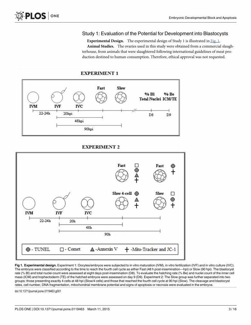
Study 1: Evaluation of the Potential for Development into BlastocystsExperimental Design. The experimental design of Study 1 is illustrated in Fig. 1.Animal Studies. The ovaries used in this study were obtained from a commercial slaugh-
terhouse, from animals that were slaughtered following international guidelines of meat pro-duction destined to human consumption. Therefore, ethical approval was not requested.
Fig 1. Experimental design. Experiment 1: Oocytes/embryos were subjected to in vitro maturation (IVM), in vitro fertilization (IVF) and in vitro culture (IVC).The embryos were classified according to the time to reach the fourth cell cycle as either Fast (48 h post-insemination—hpi) or Slow (90 hpi). The blastocystrate (% Bl) and total nuclei count were assessed at eight days post-insemination (D8). To evaluate the hatching rate (% Be) and nuclei count of the inner cellmass (ICM) and trophectoderm (TE) of the hatched embryos were assessed on day 9 (D9). Experiment 2: The Slow group was further separated into twogroups: those presenting exactly 4 cells at 48 hpi (Slow/4 cells) and those that reached the fourth cell cycle at 90 hpi (Slow). The cleavage and blastocystrates, cell number, DNA fragmentation, mitochondrial membrane potential and signs of apoptosis or necrosis were evaluated in the embryos.
doi:10.1371/journal.pone.0119463.g001
Embryonic Developmental Block and Apoptosis
PLOS ONE | DOI:10.1371/journal.pone.0119463 March 11, 2015 3 / 16

Ovary Collection and In VitroMaturation (IVM). Ovaries from zebu cows were collect-ed at Raja slaughterhouse in Piracicaba, São Paulo, stored in saline solution at 25°C to 30°Cand transported to the laboratory. Oocytes with at least three layers of compact cumulus cellsand homogeneous cytoplasm were matured in TCM 199 containing Earle’s salts and L-gluta-mine (Gibco Labs) supplemented with 10% fetal calf serum (FCS; Gibco Life Technologies), so-dium pyruvate (22 μg/ml), gentamycin (50 μg/ml), FSH (0.5 μg/ml), LH (0.5 μg/ml) andestradiol (1 μg/ml). IVM was performed in drops of 100 μL covered in mineral oil at 38.5°Cand 5% atmospheric CO2 for 22 to 24 h.
In Vitro Fertilization (IVF). IVF was performed in drops of 100 μL of TALP mediumsupplemented with 10 μg/mL heparin, 22 μg/ml sodium pyruvate, 50 μg/ml gentamycin, 6 mg/ml fatty acid-free bovine serum albumin (FAF BSA) and PHE (2 μMpenicillamine, 1 μM hypo-taurine and 0.25 μM epinephrine). Previously tested, frozen-thawed sperm of a Nelore sirekindly offered by CRV Lagoa, Sertãozinho, São Paulo, was centrifuged at 200 X g for 30 min ina 90–45% Percoll gradient. After a visual assessment of motility, the sperm was diluted to afinal concentration of 2 x 10⁶ motile sperm/ml. Fertilization was performed in drops in whichsperm and oocytes (30 oocytes/drop) were co-cultured for 20 h under the same conditionsused for IVM.
In Vitro Culture (IVC). After gamete co-incubation, the cumulus cells were removed viasuccessive pipetting. The presumptive zygotes were washed and moved to 100-μL drops (20embryos per drop) of synthetic oviduct fluid (SOF [15]) supplemented with 10% FCS or 0.8%FAF BSA. In vitro culture was performed in a modular incubator in a low-O2 atmosphere (5%CO2, 5% O2 and 90% N2).
At 48 h post-insemination (hpi), the cleavage rate was assessed, and the embryos that werenot cleaved were removed. At this time, the culture medium was renewed (feeding). The em-bryos were separated based on the speed of development: Fast—embryos that reached thefourth cell cycle at 48 hpi and therefore had between five and eight cells; and Slow—embryosthat reached the fourth cell cycle at 90 hpi. Embryo development was assessed at days 8 and 9post-insemination and 383 blastocysts were observed for evaluation of hatching rates. Expand-ed and hatched blastocysts were used for cell number analysis.
Qualitative Embryo Evaluation. For the estimation of cell numbers, 75 expanded D8-blastocysts were fixed in 3.7% paraformaldehyde with 10% Triton X-100 for 1 h and thentransferred to a PBS solution supplemented with 0.3% BSA for 1 h. The embryos were stainedvia immersion in glycerol containing the vital dye Hoechst 33342, and nuclear counting wasperformed by epifluorescence microscopy.
The hatched D9-embryos (n = 66) were qualitatively analyzed based on the number of cellsof the inner cell mass (ICM) and the ratio of ICM and trophectoderm (TE) cells, which wasbased on differential staining by fluorochrome [16]. The zona pellucida (ZP) was removedfrom the embryos, and the embryos were washed in TCM-199 Hepes with 10% FCS and inTCM-199 Hepes without FCS. The embryos were then incubated on ice for 10 min in a picricacid solution (10 mM) and polyvinyl pyrrolidone (PVP; 3 mg/ml) in PBS and washed in TCM-199 Hepes. The embryos were subsequently incubated at 39°C for 15 min in an inactivatedanti-bovine rabbit serum and diluted 1:10 with TCM-199 medium containing bicarbonate.After washing in TCM-199 medium with 10% FCS, the embryos were incubated at 39°C for 15min in guinea pig complement diluted 1:10 in TCM-199 Hepes containing 2 μg/ml Hoechst33342 and 1 μg/ml propidium iodide (PI). Finally, the embryos were washed in PBS containing0.3% BSA and fixed in blades with glycerol.
The number of live and pyknotic nuclei in the ICM and TE of the embryos was evaluated byepifluorescence microscopy. All nuclei were stained with Hoechst 33342 (vital dye; blue
Embryonic Developmental Block and Apoptosis
PLOS ONE | DOI:10.1371/journal.pone.0119463 March 11, 2015 4 / 16
fluorescence). Nuclei with pink fluorescence due to staining with PI (non-vital dye) were con-sidered TE cells and the number of ICM cells was obtained by subtraction.
Study 2: Evaluation of Developmental block and ApoptosisExperimental Design. To analyze embryonic development block and apoptosis, the em-
bryos were separated based on their speed of development. Those that reached the fourth cellcycle (between 5 and 8 cells) at 48 hpi were classified as Fast; those presenting exactly 4 cells at48 hpi were classified as Slow/4 cell, and those that reached the fourth cell cycle at 90 hpi wereclassified as Slow (Fig. 1).
For qualitative analysis, the embryos from the Fast group were evaluated using Comet andTUNEL assays and Annexin V and Mito Tracker/JC-1 staining at 48 hpi. A portion of the Fastgroup was maintained in culture for up to 90 hpi for evaluation by the TUNEL technique andstained with Mito Tracker and JC-1. The embryos from the Slow/4 cell group were analyzed byTUNEL assay and Annexin V and Mito Tracker/JC-1 staining at 48 hpi. The Slow group wasevaluated by the Comet assay at 90 hpi.
Comet Assay. Embryos from the Slow (at 90 hpi) and Fast (at 48 hpi) groups were sub-jected to the Comet assay to detect DNA damage in isolated blastomeres. From the embryostreated with BSA, 58 of the Fast group and 47 of the Slow group were evaluated; from the em-bryos treated with FCS, 33 of the Fast group and 53 of the Slow group. Embryos were first pre-pared by removing the ZP with Tyrode’s solution, and the blastomeres were isolated in asolution free of calcium and magnesium. Then, the blastomeres were transferred to a slide witha thin layer of 1% agarose, covered with a low melting point agarose gel [17] and incubated at50°C for 2 h in lysis solution (10 mM Tris, pH 10, with 2.5 mMNaCl, 100 mMNa2-EDTA, 1%Triton X-100 and 10 μg/ml K proteinase). After 20 min of equilibration in the electrophoresissolution (1 mMNa2-EDTA and 300 mMNaOH), the slides were subjected to electrophoresisfor 20 min at 25 V to separate the degraded DNA.
The slides were stained with ethidium bromide, and the DNA damage was assessed by fluo-rescent microscopy by evaluating the tail length (measured from the cell membrane to the end ofthe tail) and the proportion of damaged DNAmeasured using KS 400 software (Carl Zeiss, Inc.).
Tunel Assay. In order to evaluate a possible influence of time of culture on DNA fragmen-tation, the embryos from the Fast and Slow/4 cell groups were analyzed at 48 hpi and at 90 hpi.Of the embryos treated with BSA, 32 from the Fast group and 30 from the Slow group wereevaluated; of the embryos treated with FCS, 28 from the Fast group and 18 from the Slowgroup. The embryos were washed in PBS containing 1 mg/ml PVP and fixed in 3.7% parafor-maldehyde for 1 h. The embryos were then permeabilized for 1 h in 0.5% Triton X-100 and0.1% sodium citrate diluted in PBS and washed in PBS with PVP. The embryos were then incu-bated in a humidity chamber for 1 h at 37°C in a buffer solution of TDT 10X, CoCl2, 2 mMdATP, 0.5 units/μl terminal deoxynucleotidyl transferase enzyme and 0.5 mM Cy3-dUTP andwere washed in PBS with PVP.
For DNA visualization by epifluorescence microscopy, slides were prepared by stainingwith Hoechst 33342 diluted in glycerol (1 μg/ml). The blue fluorescent nuclei (stained usingHoechst 33342) indicated total cell number, and red fluorescent staining (stained usingCy3) indicated cells with fragmented DNA. For each replicate, a few embryos were incubatedfor 1 h in buffer containing 50 U/ml DNase, thus establishing a positive control group. Theresults were evaluated based on the number of embryos with more than 50% TUNEL-positive nuclei.
Annexin V Staining. The Annexin V (Molecular Probes, Inc.) staining technique enablesthe differentiation of cell death by apoptosis from cell death by necrosis. In viable cells,
Embryonic Developmental Block and Apoptosis
PLOS ONE | DOI:10.1371/journal.pone.0119463 March 11, 2015 5 / 16

phosphatidylserine (PS) is located on the inner surface of the cytoplasmic membrane; in cellsin the death process, the PS is displaced from the inner to the outer membrane. Annexin V,which is bound to a fluorescent label, binds to PS in the presence of Ca+, enabling its visualiza-tion by epifluorescence microscopy. Propidium iodide (PI), a dye that is permeable to damagedmembranes and therefore identifies dead cells, assists in distinguishing cells undergoing apo-ptosis (in which no membrane has been compromised) from necrotic cells. Thus, the resultsare analyzed as follows: living cells are not stained with PI (neither the cytoplasm nor nucleus);cells undergoing apoptosis are externally stained with Annexin V (blue); and the cytoplasm ofnecrotic cells is stained with Annexin V, and their nuclei are stained with PI (red). A failure ofthis method concerns anucleate cells, in which no marking occurs, making it impossible to de-termine whether they are in the process of apoptosis or necrosis.
The embryos from the Fast and Slow/4 cell groups (both at 48 hpi) were washed in PBS andtransferred to the Annexin 1X buffer (200 μL of Annexin 5X buffer in 800 μL of deionizedwater). Subsequently, they were incubated in a solution of Biotin-X Annexin V (2.5 μL Biotin-X Annexin V in 50 μL 1X Annexin buffer) at 37°C for 45 min. The embryos were then trans-ferred to an Alexa Fluor 350 solution (0.5 μL of 1 mg/ml streptavidin Alexa Fluor 350 in 50 μLof 1X Annexin buffer) for 30 min at 37°C. After this period, the embryos were washed in 1XAnnexin buffer and stained with PI (0.2μL of 1 mg/ml PI in 200 μL of Annexin 1X buffer) for10 min. The embryos were then evaluated by fluorescence microscopy to determine the num-ber of embryos in which more than 50% of cells were stained blue.
Mito Tracker Green and JC-1. Changes in membrane potential were qualitatively evaluat-ed using the marker Mitotracker Green as well as JC-1 (Molecular Probes, Inc.) in Fast andSlow/4 cell embryos at 48 and 90 hpi to investigate possible alterations in mitochondrial oxida-tive phosphorylation. The embryos were incubated with 7.5 μM JC-1 for 40 min and 0.05 μMMitotracker Green for 30 min and then observed by fluorescence microscopy.
Statistical Analysis. For statistical analysis of the cleavage, blastocyst and hatching rates,as well as data obtained using TUNEL and Annexin V techniques, the variable responses werepresented as percentage and subjected to a logistic regression test using the Car statistical pack-age of “R” software [18,18]. The average number of cells and differences in the intensity of nu-clei damage in the blastomeres (measured using the Comet assay) were presented as meanand standard error and were evaluated by analysis of variance followed by Tukey’s t test(JMP software version 2.0.4; SAS Institute). In Study 1, the number of nuclei at D8 and thehatching rates were conducted separately. In Study 2, the Comet assay, TUNEL, AnnexinV and JC-1 were conducted separately. Differences were considered significant when P�0.05. Changes in the distribution of mitochondria and membrane potential wereevaluated qualitatively.
Results
Study 1Embryos supplemented with BSA had higher cleavage (P< 0.01) and blastocyst rates (P< 0.01)than those cultured with FCS (93.5% vs. 84.0% and 31.9/% vs. 26.2%, respectively). Blastocystrates were higher among Fast embryos than both slow-developing groups in groups culturedwith either BSA (54.2% vs. 32.1%; P< 0.01) or FCS (49.4% vs. 31.8%; P< 0.01). There was nosignificant difference in the blastocyst rates of slow-developing embryos in the groups culturedwith different protein sources (32.1% for BSA and 31.8% for FCS; P = 0.910; Table 1).
The hatching rate of the BSA group was higher than that of the FCS group for both fast-(78.4% vs. 38.9%; P< 0.01) and slow-developing (69.7% vs. 37.9%; P = 0.01) embryos.
Embryonic Developmental Block and Apoptosis
PLOS ONE | DOI:10.1371/journal.pone.0119463 March 11, 2015 6 / 16
However, there were no differences in the hatching rates of Fast and Slow embryos culturedwith BSA (P = 0.274) or FCS (P = 0.920).
Of the slow-developing embryos, 68% in both groups (BSA, 32.1% ± 3.0 and FCS, 31.8% ±3.0) suffered developmental block after reaching the fourth cell cycle. Of the fast-developingembryos, 46% in the BSA group (54.2% ± 1.9) and 51% of those in the FCS group (49.4% ± 2.1)underwent developmental block at this stage.
Approximately 50.7 (1,235/2,434) and 19.9 (485/2,434) % of the cleaved embryos reachedthe fourth cell cycle at 48 hpi (fast developing group) and 90 hpi (slow-developing group), re-spectively; the remaining embryos (n = 714) did not reach the fourth cycle prior to 90 hpi.
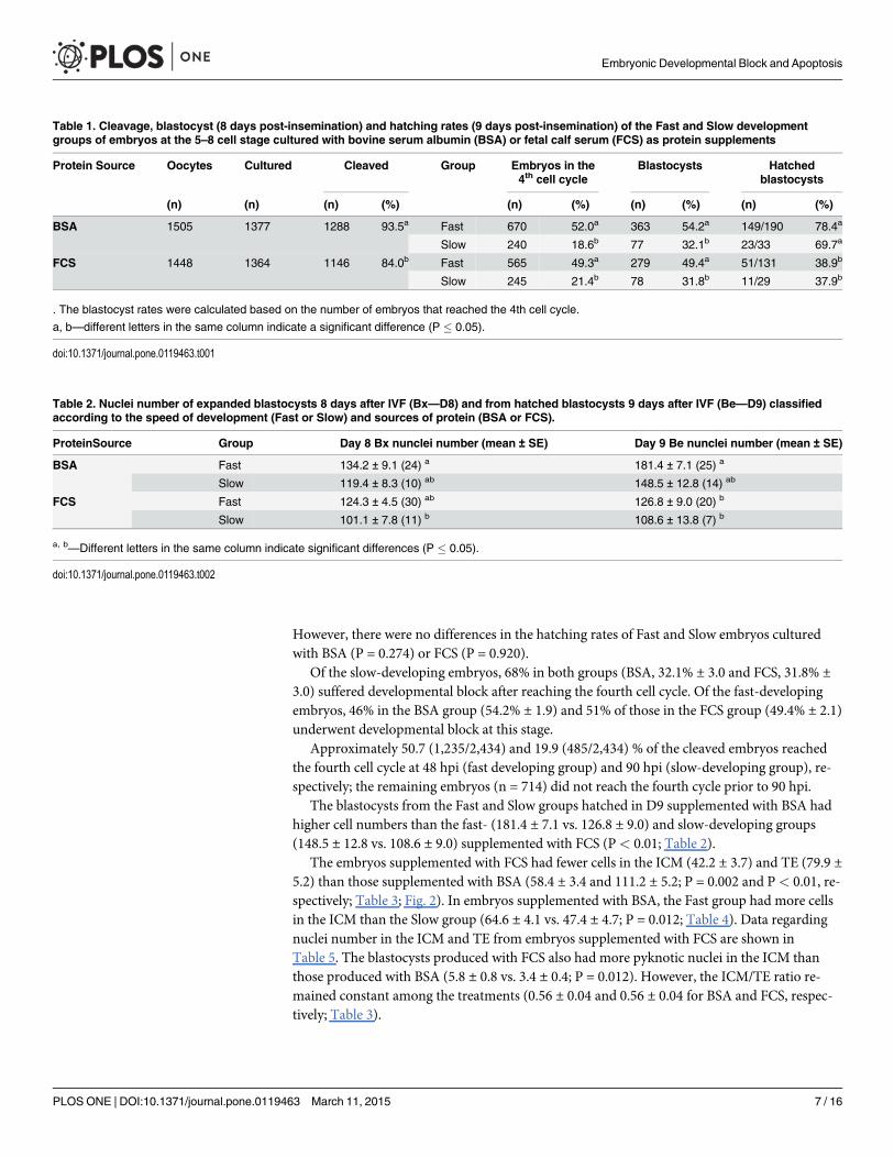
The blastocysts from the Fast and Slow groups hatched in D9 supplemented with BSA hadhigher cell numbers than the fast- (181.4 ± 7.1 vs. 126.8 ± 9.0) and slow-developing groups(148.5 ± 12.8 vs. 108.6 ± 9.0) supplemented with FCS (P< 0.01; Table 2).
The embryos supplemented with FCS had fewer cells in the ICM (42.2 ± 3.7) and TE (79.9 ±5.2) than those supplemented with BSA (58.4 ± 3.4 and 111.2 ± 5.2; P = 0.002 and P< 0.01, re-spectively; Table 3; Fig. 2). In embryos supplemented with BSA, the Fast group had more cellsin the ICM than the Slow group (64.6 ± 4.1 vs. 47.4 ± 4.7; P = 0.012; Table 4). Data regardingnuclei number in the ICM and TE from embryos supplemented with FCS are shown inTable 5. The blastocysts produced with FCS also had more pyknotic nuclei in the ICM thanthose produced with BSA (5.8 ± 0.8 vs. 3.4 ± 0.4; P = 0.012). However, the ICM/TE ratio re-mained constant among the treatments (0.56 ± 0.04 and 0.56 ± 0.04 for BSA and FCS, respec-tively; Table 3).
Table 1. Cleavage, blastocyst (8 days post-insemination) and hatching rates (9 days post-insemination) of the Fast and Slow developmentgroups of embryos at the 5–8 cell stage cultured with bovine serum albumin (BSA) or fetal calf serum (FCS) as protein supplements
Protein Source Oocytes Cultured Cleaved Group Embryos in the4th cell cycle
Blastocysts Hatchedblastocysts
(n) (n) (n) (%) (n) (%) (n) (%) (n) (%)
BSA 1505 1377 1288 93.5a Fast 670 52.0a 363 54.2a 149/190 78.4a
Slow 240 18.6b 77 32.1b 23/33 69.7a
FCS 1448 1364 1146 84.0b Fast 565 49.3a 279 49.4a 51/131 38.9b
Slow 245 21.4b 78 31.8b 11/29 37.9b
. The blastocyst rates were calculated based on the number of embryos that reached the 4th cell cycle.
a, b—different letters in the same column indicate a significant difference (P � 0.05).
doi:10.1371/journal.pone.0119463.t001
Table 2. Nuclei number of expanded blastocysts 8 days after IVF (Bx—D8) and from hatched blastocysts 9 days after IVF (Be—D9) classifiedaccording to the speed of development (Fast or Slow) and sources of protein (BSA or FCS).
ProteinSource Group Day 8 Bx nunclei number (mean ± SE) Day 9 Be nunclei number (mean ± SE)
BSA Fast 134.2 ± 9.1 (24) a 181.4 ± 7.1 (25) a
Slow 119.4 ± 8.3 (10) ab 148.5 ± 12.8 (14) ab
FCS Fast 124.3 ± 4.5 (30) ab 126.8 ± 9.0 (20) b
Slow 101.1 ± 7.8 (11) b 108.6 ± 13.8 (7) b
a, b—Different letters in the same column indicate significant differences (P � 0.05).
doi:10.1371/journal.pone.0119463.t002
Embryonic Developmental Block and Apoptosis
PLOS ONE | DOI:10.1371/journal.pone.0119463 March 11, 2015 7 / 16
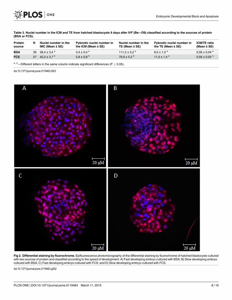
Table 3. Nuclei number in the ICM and TE from hatched blastocysts 9 days after IVF (Be—D9) classified according to the sources of protein(BSA or FCS).
Proteinsource
N Nuclei number in theIMC (Mean ± SE)
Pyknotic nuclei number inthe ICM (Mean ± SE)
Nuclei number in theTE (Mean ± SE)
Pyknotic nuclei number inthe TE (Mean ± SE)
ICM/TE ratio(Mean ± SE)
BSA 39 58,4 ± 3,4 a 3,4 ± 0,4 a 111,2 ± 5,2 a 8,0 ± 1,0 a 0,56 ± 0,04 a
FCS 27 42,2 ± 3,7 b 5,8 ± 0,8 b 79,9 ± 5,2 b 11,0 ± 1,4 a 0,56 ± 0,05 a
a, b—Different letters in the same column indicate significant differences (P � 0.05).
doi:10.1371/journal.pone.0119463.t003
Fig 2. Differential staining by fluorochrome. Epifluorescence photomicrography of the differential staining by fluorochrome of hatched blastocysts culturedwith two sources of protein and classified according to the speed of development: A) Fast developing embryo cultured with BSA; B) Slow developing embryocultured with BSA; C) Fast developing embryo cultured with FCS; and D) Slow developing embryo cultured with FCS.
doi:10.1371/journal.pone.0119463.g002
Embryonic Developmental Block and Apoptosis
PLOS ONE | DOI:10.1371/journal.pone.0119463 March 11, 2015 8 / 16

Study 2The Comet assay revealed that the Slow group supplemented with either FCS or BSA exhibitedsignificantly higher tail length and degraded DNA density than the Fast group (P< 0.01 in allcomparisons; Fig. 3; Table 6).
Fragmented nuclei were not observed by TUNEL assay in any group (Fast and Slow/4 cellsupplemented with BSA or FCS) at 48 hpi. However, at 90 hpi, TUNEL-positive nuclei wereobserved (Fig. 4). In the Fast and Slow/4 cell groups, 25.0% and 41.7% of the embryos,
Table 4. Nuclei number in the ICM and TE from hatched blastocysts 9 days after IVF (Be—D9), supplemented with BSA, classified according tothe speed of development (Fast or Slow).
Speed ofdevelopment
N Nuclei number in theIMC (Mean ± SE)
Pyknotic nuclei number inthe ICM (Mean ± SE) *
Nuclei number in theTE (Mean ± SE) *
Pyknotic nuclei numberin the TE (Mean ± SE) *
ICM/TE ratio(Mean ± SE) *
Fast 25 64,6 ± 4,1a 3,6 ± 0,5 116,8 ± 4,9 6,9 ± 1,0 0,57 ± 0,03
Slow 14 47,4 ±4,7b 3,1 ± 0,8 101,8 ± 11,3 9,8 ± 2,3 0,55 ± 0,08
a,b—Different letters in the same column indicate significant differences (P � 0.05).
*—There were no significant differences between treatments (P � 0.05).
doi:10.1371/journal.pone.0119463.t004
Table 5. Nuclei number in the ICM and TE from hatched blastocysts 9 days after IVF (Be—D9), supplemented with FCS, classified according tothe speed of development (Fast or Slow).
Speed ofdevelopment
N Nuclei number in theIMC (Mean ± SE) *
Pyknotic nuclei number inthe ICM (Mean ± SE) *
Nuclei number in theTE (Mean ± SE) *
Pyknotic nuclei numberin the TE (Mean ± SE) *
ICM/TE ratio(Mean ± SE) *
Fast 20 45,7 ± 4,5 6,5 ± 1,1 81,1 ± 6,0 11,3 ± 1,7 0,60 ± 0,07
Slow 7 32,1 ± 3,7 4,0 ± 0,6 76,4 ± 10,5 10,0 ± 2,1 0,43 ± 0,03
*—There were no significant differences between treatments (P � 0.05).
doi:10.1371/journal.pone.0119463.t005
Fig 3. Comet assay. Epifluorescence photomicrography of the results for blastomeres isolated from embryos during the fourth cell cycle, which wereclassified according to the speed of development as either Fast developing (A) or Slow developing (B). The arrow indicates migrated DNA.
doi:10.1371/journal.pone.0119463.g003
Embryonic Developmental Block and Apoptosis
PLOS ONE | DOI:10.1371/journal.pone.0119463 March 11, 2015 9 / 16

Table 6. Assessment of DNA fragmentation using the Comet assay (tail length and density) and nuclear diameter of the blastomeres ofembryos in the fourth cell cycle classified according to the speed of development as Fast (at 48 hpi) or Slow (at 90 hpi) and cultured in mediawith different sources of protein.
ProteinSupplementation
Group Embryos in the 4th cellcycle (n)
Tail density (%)± Standard error
Tail length (μm)± Standard error
Nucleus Diameter (μm)± Standard error
BSA Fast 58 12.67 ± 1.26 b 27.89 ± 2.55 b 67.65 ± 3.29
Slow 47 43.38 ± 3.08 a 65.66 ± 5.05 a 63.59 ± 4.09
FCS Fast 33 14.45 ± 2.57 b 23.08 ± 2.72 b 71.83 ± 3.89
Slow 53 44.72 ± 3.10 a 74.59 ± 5.84 a 70.30 ± 3.31
a, b—Different letters in the same column indicate significant differences (P � 0.05).
doi:10.1371/journal.pone.0119463.t006
Fig 4. TUNEL assay. Epifluorescence photomicrography of embryos at 48 and 90 hpi classified according to the speed of development as either Fast(reached the fourth cell cycle at 48 hpi) or Slow/4 cells (exactly 4 cells and prior to the fourth cell cycle at 48 hpi). A) Fast embryo at 48 h of culture; B) Slow/4cell embryo at 48 h of culture; C) embryo at 48 h of culture submitted to DNA fragmentation via exposure to DNase (technique positive control); D) Fastembryo at 90 h of culture; E) Slow/4 cell embryo at 90 h of culture; and F) embryo at 90 h of culture submitted to DNA fragmentation via exposure to DNase.The blue fluorescent nuclei indicate total cell number and red fluorescent staining indicates cells with fragmented DNA.
doi:10.1371/journal.pone.0119463.g004
Embryonic Developmental Block and Apoptosis
PLOS ONE | DOI:10.1371/journal.pone.0119463 March 11, 2015 10 / 16

respectively, exhibited greater than 50% TUNEL-positive nuclei (P = 0.068); there were also nosignificant differences between the embryos supplemented with BSA and those supplementedwith FCS (32.3 vs. 32.6%; P = 0.969).
The results of Annexin V staining in the Fast and Slow/4 cell groups to assess apoptosis andnecrosis are illustrated in Fig. 5. The number of Annexin-positive blastomeres was higher inthe Slow/4 cell embryos than the Fast embryos (14.3% vs 1.2%; P< 0.01). There were no differ-ences between the rates of positive cells in the embryos supplemented with BSA (7.9%) andFCS (7.1%). No necrotic nuclei (stained with PI) were observed; however, there were manyfragments of anucleated cytoplasm, complicating the results analysis. Among the embryos sup-plemented with BSA, 15% of the Fast group and 21% of the Slow/4 cell group had at least oneanucleated blastomere. Among the embryos supplemented with FCS, 9% and 7% of the Fastand Slow/4 cell groups, respectively, had at least one anucleated blastomere.
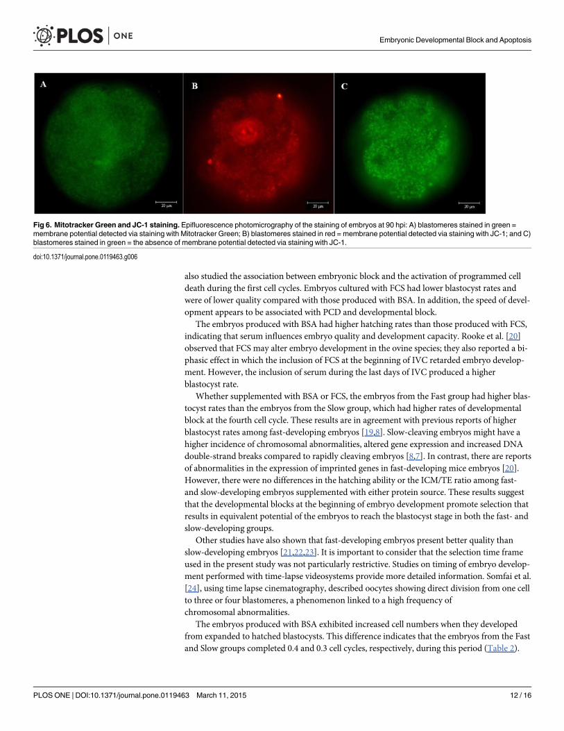
No membrane potential was detected via JC-1 at 48 hpi regardless of the speed of develop-ment. However, the organelles exhibited a low mitochondrial membrane potential, as demon-strated by staining with Mitotracker Green. Both markers indicated the existence of membranepotential at 90 hpi, at which time nuclear fragmentation was also observed (Fig. 6).
DiscussionIn this study, we investigated the influence of the protein source in the culture medium on theability of bovine embryos to overcome developmental block and reach the blastocyst stage. We
Fig 5. Annexin V staining. Epifluorescence photomicrography of embryos during the fourth cell cycle at 48 hpi: A) embryo with blastomeres undergoingapoptosis; B) blastomeres with blue staining indicating a positive reaction to the Annexin V antibody; and C) blastomeres without membrane permeability topropidium iodide.
doi:10.1371/journal.pone.0119463.g005
Embryonic Developmental Block and Apoptosis
PLOS ONE | DOI:10.1371/journal.pone.0119463 March 11, 2015 11 / 16
also studied the association between embryonic block and the activation of programmed celldeath during the first cell cycles. Embryos cultured with FCS had lower blastocyst rates andwere of lower quality compared with those produced with BSA. In addition, the speed of devel-opment appears to be associated with PCD and developmental block.
The embryos produced with BSA had higher hatching rates than those produced with FCS,indicating that serum influences embryo quality and development capacity. Rooke et al. [20]observed that FCS may alter embryo development in the ovine species; they also reported a bi-phasic effect in which the inclusion of FCS at the beginning of IVC retarded embryo develop-ment. However, the inclusion of serum during the last days of IVC produced a higherblastocyst rate.
Whether supplemented with BSA or FCS, the embryos from the Fast group had higher blas-tocyst rates than the embryos from the Slow group, which had higher rates of developmentalblock at the fourth cell cycle. These results are in agreement with previous reports of higherblastocyst rates among fast-developing embryos [19,8]. Slow-cleaving embryos might have ahigher incidence of chromosomal abnormalities, altered gene expression and increased DNAdouble-strand breaks compared to rapidly cleaving embryos [8,7]. In contrast, there are reportsof abnormalities in the expression of imprinted genes in fast-developing mice embryos [20].However, there were no differences in the hatching ability or the ICM/TE ratio among fast-and slow-developing embryos supplemented with either protein source. These results suggestthat the developmental blocks at the beginning of embryo development promote selection thatresults in equivalent potential of the embryos to reach the blastocyst stage in both the fast- andslow-developing groups.
Other studies have also shown that fast-developing embryos present better quality thanslow-developing embryos [21,22,23]. It is important to consider that the selection time frameused in the present study was not particularly restrictive. Studies on timing of embryo develop-ment performed with time-lapse videosystems provide more detailed information. Somfai et al.[24], using time lapse cinematography, described oocytes showing direct division from one cellto three or four blastomeres, a phenomenon linked to a high frequency ofchromosomal abnormalities.
The embryos produced with BSA exhibited increased cell numbers when they developedfrom expanded to hatched blastocysts. This difference indicates that the embryos from the Fastand Slow groups completed 0.4 and 0.3 cell cycles, respectively, during this period (Table 2).
Fig 6. Mitotracker Green and JC-1 staining. Epifluorescence photomicrography of the staining of embryos at 90 hpi: A) blastomeres stained in green =membrane potential detected via staining with Mitotracker Green; B) blastomeres stained in red = membrane potential detected via staining with JC-1; and C)blastomeres stained in green = the absence of membrane potential detected via staining with JC-1.
doi:10.1371/journal.pone.0119463.g006
Embryonic Developmental Block and Apoptosis
PLOS ONE | DOI:10.1371/journal.pone.0119463 March 11, 2015 12 / 16

This phenomenon was not observed among embryos supplemented with FCS, which did notexhibit an increase in the number of nuclei from one stage to the next. This lack of increase inthe number of nuclei may explain the low hatchability of these embryos, indicating again thatFCS leads to lower development capacity and embryonic quality. Another possibility is thatembryos supplemented with FCS can accelerate the mechanism of programmed cell death bystimulating factors such as the apoptosis-inducing factor (AIF), which is required for the for-mation of the blastocoel [25]. Thus, the reduced cell number could be due to the presence offewer cells at the induction of apoptosis for the formation of the blastocoel. Consequently, thenumber of remaining cells (that are able to replicate) that are prepared for the opening of theblastocoel cavity would be lower in the embryos treated with FCS. Despite the fact that embry-os produced with BSA had higher quality than the embryos produced with serum, it is impor-tant to consider that BSA is essentially a protein derived from serum. Therefore, othercomponents of FCS must be harmful for early development of bovine embryos.
The differential staining technique highlights characteristics that are of great importance forembryo survival, such as the ICM/TE ratio and the distribution of morphologically altered nu-clei. The embryos supplemented with FCS exhibited lower numbers of total nuclei in the ICMand TE as well as higher number of pyknotic nuclei in the ICM. However, the ICM/TE ratio re-mained constant among the treatments, indicating that the difference in cell number and em-bryo quality did not occur due to any effect induced by FCS on differentiation but instead mostlikely occurred at an earlier stage of development. Alternatively, the total cell number of blasto-cysts cultured with FCS could be influenced by growth factors. Growth factors can acceleratethe mechanism of PCD by stimulating factors such as apoptosis-inducing factor (AIF). Thus,the smaller number of cells reported here could be due to a reduced number of cells available atthe induction of apoptosis for the opening of the blastocoel. Consequently, the number of re-maining cells (that are able to replicate) would be lower in the embryos that were treated withFCS, which exhibited early induction of cavity formation.
The pyknotic nuclei observed may be related to embryonic cell death; however, this may bea survival and not necessarily a destruction mechanism [26]. The apoptosis of abnormal cellscan be a mechanism for the removal of damaged cells and may not be lethal in embryos thatalso have normal nuclei [27].
Nuclear fragmentation was estimated using the Comet assay, and the Slow group exhibitedhigher DNA degradation than the Fast group. This DNA damage may reduce the speed of de-velopment and increase the developmental block occurring at this phase. The embryos supple-mented with BSA or FCS exhibited no difference in the amount of degraded DNA. These datasuggest that the effects of FCS are not directly involved in DNA breakage. Serum may cause in-creased sensitivity to apoptosis induction systems involving cytokines or other constituents,therefore exerting an indirect effect in the blocking of embryo development.
However, the duration of the culture could have exerted an effect on the DNA damage ob-served in the Comet assay; the embryos of the Fast group reach the fourth cell cycle 48 h afterIVF, and the embryos of the Slow group required up to 90 h to reach the same stage. Thus,DNA damage was also analyzed in the Slow/4 cell group using the TUNEL technique beforethese embryos reached the fourth cell cycle. In this case, the embryos of the Fast and Slow/4cell groups were not in the same cell cycle and had blastomeres of varying sizes, preventing acomparison of their Comet assays results.
Fragmented nuclei were not observed in any group assessed within 48 hpi; however, a signif-icant increase in the number of TUNEL-positive nuclei was observed at 90 hpi. These data arein accordance with other reports indicating that TUNEL-positive cells are first observed in em-bryos cultured in vitro between the six- and eight-cell stages [30]. Together, these data indicatethat there may be some resistance to nuclear fragmentation during the first three days of
Embryonic Developmental Block and Apoptosis
PLOS ONE | DOI:10.1371/journal.pone.0119463 March 11, 2015 13 / 16
culture. Brad et al. [26] reported that the block to apoptosis occurs in two-cell embryos at twopoints in the apoptotic cascade: at the activation of caspase-9 activity and during caspase-mediated DNA damage.
Annexin V staining was performed to detect early signs of apoptosis (PS exposure) and ne-crosis (membrane permeability). The exposure of PS observed at 48 hpi may indicate the acti-vation of a cell death mechanism prior to nuclear fragmentation because no DNAfragmentation was detected via the TUNEL assay during the same period. There is limited in-formation in the literature to correlate early embryonic development with PS exposure; howev-er, when combined with other techniques, this could be an interesting tool to understand themechanism of apoptosis in embryos.
Staining with Mitotracker Green and JC-1 permitted us to assess the changes in membranepotential and also helped us determine whether a change in mitochondrial oxidative phosphor-ylation of the embryos is associated with DNA fragmentation. The absence of membrane po-tential at 48 hpi may indicate that low ATP production limits the action of certain enzymesinvolved in the apoptosis process. After 90 hpi, a mitochondrial membrane potential was veri-fied, which coincided with the appearance of apoptotic nuclei. ATP is necessary for the releaseof cytochrome C, an enzyme involved in caspase activation that can cause cell death by apopto-sis [28]. There is evidence that mitochondria play a central role in regulating programmed celldeath and that the release of proapoptotic factors such as cytochrome C and AIF (from the mi-tochondria) is a primary event in caspase activation [29]. Thus, the absence of membrane po-tential at 48 hpi could prevent or delay death by apoptosis and could be a mechanismunderlying the absence of apoptosis observed via the TUNEL technique in embryos at 48 hpi.
Some of the nuclear fragmentation may have been triggered by oxidative stress caused at themoment of handling the embryos at 48 hpi, when they are subjected to a sudden change in theO2 rate. There is evidence that oxidative stress can cause mitochondrial dysfunction [30] andincrease DNA damage in embryos [31], thus potentially influencing apoptotic cell death in-duced by oxidative stress [32].
The specific characteristics of Bos indicus breeds must also be considered. In addition to dif-fering numbers of antral follicles, zebu and taurine cows diverge in several aspects, such as es-trus manifestation [33], progesterone concentration, follicle size [34] and IGF-I and insulinconcentrations [35]. Furthermore, zebu embryos differ from taurine embryos with respect tolipid amount and tolerance to cryopreservation [36]. Therefore, the findings obtained in thisstudy may not extend to Bos taurus embryos.
ConclusionsThe results obtained in this study lead us to conclude that, supplementation with 10% FCS dur-ing the culture period decreases blastocyst rates and the quality of the embryos produced, thusreducing the number of cell cycles completed as well as hatching ability. Activation of pro-grammed cell death can be detected in early-stage embryos (before the embryos have the abilityto perform DNA fragmentation) via Annexin V staining. Lastly, fast-cleaving embryos exhibithigher blastocyst rates, and the speed of development appears to be negatively associated withprogrammed cell death and the blocking of embryo development.
Author ContributionsConceived and designed the experiments: FVM. Performed the experiments: SMG. Analyzedthe data: LSRM PALMMS. Contributed reagents/materials/analysis tools: SMG FVM LSRMPALMMS. Wrote the paper: LSRM PALMMS.
Embryonic Developmental Block and Apoptosis
PLOS ONE | DOI:10.1371/journal.pone.0119463 March 11, 2015 14 / 16
References1. Pontes JHF, Silva KCF, Basso AC, Ferreira CR, Rigo AG, Santos GMG, et al. Large-scale in vitro em-
bryo production and pregnancy rates from Bos taurus, Bos indicus, and indicus-taurus dairy cowsusing sexed sperm. Theriogenology. 2010; 74:1349–1355. doi: 10.1016/j.theriogenology.2010.06.004PMID: 20708245
2. Pontes JHF, Sterza FAM, Basso AC, Ferreira CR, Sanches BV, Rubin KCP, et al. Ovum pick up, invitro embryo production, and pregnancy rates from a large-scale commercial program using Nelore cat-tle (Bos indicus) donors. Theriogenology. 2011; 75:1640–1646. doi: 10.1016/j.theriogenology.2010.12.026 PMID: 21334055
3. Pontes JHF, Nonato-Junior I, Sanches BV, Ereno-Junior JC, Uvo S, Barreiros TRR, et al. Comparisonof embryo yield and pregnancy rate between in vivo and in vitro methods in the same Nelore (Bos indi-cus) donor cows. Theriogenology. 2009; 71:690–697. doi: 10.1016/j.theriogenology.2008.09.031PMID: 18995895
4. ZhengW, Liu K. Maternal control of mouse preimplantation development. Results Probl Cell Differ.2012; 55:115–139. doi: 10.1007/978-3-642-30406-4_7 PMID: 22918804
5. Cantone I, Fisher AG. Epigenetic programming and reprogramming during development. Nat StructMol Biol. 2013; 20:282–289. doi: 10.1038/nsmb.2489 PMID: 23463313
6. Wasler CB, Lipshitz HD. Transcript clearance during the maternal-to-zygotic transition. Curr OpinGenet Dev. 2011; 21:431–443. doi: 10.1016/j.gde.2011.03.003 PMID: 21497081
7. Bohrer RC, Che L, Gonçalves PBD, Duggavathi R, Bordignon V. Phosphorylated histone H2A.x in por-cine embryos produced by IVF and somatic cell nuclear transfer. Reproduction. 2013; 146:325–333.doi: 10.1530/REP-13-0271 PMID: 23858475
8. Sugimura S, Akai T, Hashiyada Y, Somfai T, Inaba Y, Hirayama M, et al. Promising system for selectinghealthy in vitro-fertilized embryos in cattle. Plos One. 2012; 7:e36627. doi: 10.1371/journal.pone.0036627 PMID: 22590579
9. Leivas FG, Brum DS, Fialho SS, SalibaWP, Alvim MTT, Bernardi Ml, et al. Fetal calf serum enhancesin vitro production of Bos taurus indicus embryos. Theriogenology. 2011; 75:429–433. doi: 10.1016/j.theriogenology.2010.08.017 PMID: 20961608
10. Sudano MJ, Paschoal DM, Rascado TS, Magalhães LC, Crocomo LF, Neto JFL, et al. Lipid contentand apoptosis of in vitro-produced bovine embryos as determinants of susceptibility to vitrification.Theriogenology. 2011; 75:1211–1220. doi: 10.1016/j.theriogenology.2010.11.033 PMID: 21247620
11. Crosier AE, Farin PW, Dykstra MJ, Alexander JE, Farin CE. Ultrastructural morphometry of bovinecompact morulae produced in vivo or in vitro. Biol Reprod. 2000; 62:1459–1365. PMID: 10775201
12. Kuzmany A, Havlicek V, Wrenzycki C, Wilkening S, Brem G, Besenfelder U. Expression of mRNA, be-fore and after freezing, in bovine blastocysts cultured under different conditions. Theriogenology. 2011;75:482–494. doi: 10.1016/j.theriogenology.2010.09.016 PMID: 21144573
13. Stigliani S, Anserini P, Venturini PL, Scaruffi P. Mitochondrial DNA content in embryo culture medium issignificantly associated with human embryo fragmentation. Hum Reprod. 2013; 0:1–9.
14. Pons I, Cercas R, Villas C, Braña C, Fernández-Shaw S. One abstinence day decreases sperm DNAfragmentation in 90% of selected patients. J Assist Reprod Genet. 2013; 30:1211–1218. doi: 10.1007/s10815-013-0089-8 PMID: 23996278
15. Gardner DK, Lane M, Spitzer A, Batt PA. Enhanced rates of cleavage and development for sheep zy-gotes cultured to the blastocyst stage in vitro in the absence of serum and somatic cells: amino acids,vitamins, and culturing embryos in groups stimulate development. Biol Reprod. 1994; 50:390. PMID:8142556
16. Thouas GA, Korfiatis NA, French AJ, Jones GM, Trounson AO. Simplified technique for differentialstaining of inner cell mass and trophectoderm cells of mouse and bovine blastocysts. Reprod BiomedOnline. 2001; 3: 25–29. PMID: 12513888
17. Singh NP, McCoy MT, Tice RR, Scheider EL. A simple technique for quantitation of low levels of DNAdamage in individual cells. Exp Cell Res. 1988; 175:184–191. PMID: 3345800
18. R Development Core Team. R: A language and environment for statistical computing. R Foundation forStatistical Computing, Vienna, Austria. ISBN 3–900051–07–0. 2008. Available: http://www.R-project.org.
19. Fox J, Weisberg S. An {R} Companion to Applied Regression, Second Edition. Thousand Oaks CA:Sage.2011. Available: http://socserv.socsci.mcmaster.ca/jfox/Books/Companion.
20. Rooke JA, McEvoy TG, Ashworth CJ, Robinson JJ, Wilmut I, Young LE, et al. Ovine fetal developmentis more sensitive to perturbation by the presence of serum in embryo culture before than after compac-tion. Theriogenology. 2007; 67:639–647. PMID: 17070902
Embryonic Developmental Block and Apoptosis
PLOS ONE | DOI:10.1371/journal.pone.0119463 March 11, 2015 15 / 16
21. Bastos GM, Gonçalves PBD, Bordignon V. Immunolocalization of the high-mobility group N2 proteinand acetylated histone H3K14 in early developing parthenogenetic bovine embryos derived from oo-cytes of high and low developmental competence. Mol Reprod Dev. 2008; 75:282–290. PMID:17712799
22. Market Velker BA, DenommeMM, Mann MR. Loss of genomic imprinting in mouse embryos with fastrates of preimplantation development in culture. Biol Reprod. 2012; 86:1–16.
23. Van Soom A, Ysebaert MT, de Kruif A. Relationship between timing of development, morula morpholo-gy, and cell allocation to inner cell mass and trophectoderm in in vitro-produced bovine embryos. MolReprod Dev. 1997; 47:47–56. PMID: 9110314
24. Gutiérrez-Adán A, Rizos D, Fair T, Moreira PN, Pintado B, Fuente J, et al. Effect of speed of develop-ment on mRNA expression pattern in early bovine embryos cultured in vivo or in vitro. Mol Reprod Dev.2004; 68:441–448. PMID: 15236328
25. Pers-Kamczyc E, Pawlak P, Rubes J, Lechniak D. Early cleaved bovine embryos show reduced inci-dence of chromosomal aberrations and higher developmental potential on day 4.5 post-insemination.Reprod Domest Anim. 2012; 47:899–906. doi: 10.1111/j.1439-0531.2012.01987.x PMID: 22304363
26. Somfai T, Inaba Y, Aikawa Y, Ohtake M, Kobayashi S, Konishi K, et al. Relationship between the lengthof cell cycles, cleavage pattern and developmental competence in bovine embryos generated by invitro fertilization or parthenogenesis. J Reprod Dev. 2010; 56:200–207. PMID: 20035110
27. Joza N, Susin SA, Daugas E, StanfordWL, Cho SK, Li CY, et al. Essential role of the mitochondrial apo-ptosis-inducing factor in programmed cell death. Nature. 2011; 410:549–554. doi: 10.1016/j.bbrc.2011.06.022 PMID: 21683060
28. Brad AM, Hendricks KEM, Hansen PJ. The block to apoptosis in bovine two-cell embryos involves inhi-bition of caspase-9 activation and caspase-mediated DNA damage. Reproduction. 2007; 134:789–797. PMID: 18042636
29. Jousan FD, Hansen PJ. Insulin-like growth factor-I promotes resistance of bovine preimplantation em-bryos to heat shock through actions independent of its anti-apoptotic actions requiring PI3K signaling.Mol Reprod Dev. 2007; 74:189–196. PMID: 16955404
30. Gjørret JO, Fabian D, Avery B, Maddox-Hyttel P. Active caspase-3 and ultrastructural evidence of apo-ptosis in spontaneous and induced cell death in bovine in vitro produced pre-implantation embryos. MolProd Dev. 2007; 74:961–971. PMID: 17393434
31. Patriarca A, Eliseo T, Sinibaldi F, Piro MC, Melis R, Paci M, et al. ATP acts as a regulatory effector inmodulating structural transitions of cytochrome c: implications for apoptotic activity. Biochemistry.2009; 48:3279–3287. doi: 10.1021/bi801837e PMID: 19231839
32. Norberg E, Orrenius S, Zhivotovsky B. Mitochondrial regulation of cell death: Processing of apoptosis-inducing factor (AIF). Biochem Bioph Res Co. 2010; 396:95–100.
33. Rose S, Frye RE, Slattery J, Wynne R, Tippett M, Pavliv O, et al. Oxidative stress induces mitochondrialdysfunction in a subset of autism lymphoblastoid cell lines in a well-matched case control cohort. PlosOne. 2014; 9: e85436. doi: 10.1371/journal.pone.0085436 PMID: 24416410
34. Hwang IS, Bae HK, Cheong HT. Mitochondrial and DNA damage in bovine somatic cell nuclear transferembryos. J Vet Sci. 2013; 14:235–240. PMID: 23820170
35. Chaudhari M, Jayaraj R, Bhaskar ASB, Lakshmana Rao PV. Oxidative stress induction by T-2 toxincauses DNA damage and triggers apoptosis via caspase pathway in human cervical cancer cells. Toxi-cology. 2009; 262:153–161. doi: 10.1016/j.tox.2009.06.002 PMID: 19524637
36. Bó GA, Baruselli PS, Martinez MF. Pattern and manipulation of follicular development in Bos indicuscattle. Anim Reprod Sci. 2003; 78:307–326. PMID: 12818651
37. Carvalho JPB, Carvalho NA, Reis EL, Nichi M, Souza AH, Baruselli PS. Effect of early luteolysis in pro-gesterone-based timed AI protocols in Bos indicus, Bos indicus x Bos taurus, and Bos taurus heifers.Theriogenology. 2008; 69:167–175. PMID: 17980904
38. Alvarez P, Spicer LJ, Chase CC Jr, Payton ME, Hamilton TD, Stewart RE, et al. Ovarian and endocrinecharacteristics during and estrous cycle in Angus, Brahman, and Senepol cows in a subtropical envi-ronment. J Anim Sci. 2000; 78:1291–1302. PMID: 10834585
39. Sudano MJ, Santos VG, Tata A, Ferreira CR, Paschoal DM, Machado R, et al. Phosphatidylcholineand Sphingomyelin Profiles Vary in Bos taurus indicus and Bos taurus taurus In Vitro- and In Vivo-Produced Blastocysts. Biol Reprod. 2012; 87:1–11.
Embryonic Developmental Block and Apoptosis
PLOS ONE | DOI:10.1371/journal.pone.0119463 March 11, 2015 16 / 16