research paper interleukin-22 ameliorates cerulein …ameliorates cerulein-induced pancreatitis by...
TRANSCRIPT
Int. J. Biol. Sci. 2012, 8
http://www.biolsci.org
249
IInntteerrnnaattiioonnaall JJoouurrnnaall ooff BBiioollooggiiccaall SScciieenncceess 2012; 8(2):249-257. doi: 10.7150/ijbs.3967
Research Paper
Interleukin-22 Ameliorates Cerulein-Induced Pancreatitis in Mice by In-
hibiting the Autophagic Pathway
Dechun Feng1#, Ogyi Park1#, Svetlana Radaeva2, Hua Wang1, Shi Yin1, Xiaoni Kong1, Mingquan Zheng3, Sam Zakhari2, Jay K. Kolls3, Bin Gao1
1. Laboratory of Liver Diseases, National Institute on Alcohol Abuse and Alcoholism, NIH, Bethesda, MD 20892, USA; 2. Division of Metabolism and Health Effects, National Institute on Alcohol Abuse and Alcoholism, NIH, Bethesda, MD 20892, USA; 3. Department of Genetics, Louisiana State University Health Sciences Center, New Orleans, LA 70112, USA.
# DF and OP contributed equally to this work.
Corresponding author: Bin Gao, M.D., Ph.D., NIAAA/NIH, 5625 Fishers Lane, Bethesda, MD 20892. E-mail: [email protected]. This work was supported by the intramural program of NIAAA, NIH.
© Ivyspring International Publisher. This is an open-access article distributed under the terms of the Creative Commons License (http://creativecommons.org/ licenses/by-nc-nd/3.0/). Reproduction is permitted for personal, noncommercial use, provided that the article is in whole, unmodified, and properly cited.
Received: 2011.12.16; Accepted: 2012.01.01; Published: 2012.01.06
Abstract
Pancreatitis occurs when digestive enzymes are activated in the pancreas. Severe pancreatitis has a 10-30% mortality rate. No specific treatments for pancreatitis exist now. Here, we discovered that interleukin-22 (IL-22) may have therapeutic potential in treating acute and chronic pancreatitis. Wild-type and IL-22 knockout mice were equally susceptible to ceru-lein-induced acute and chronic pancreatitis, whereas liver-specific IL-22 transgenic mice were completely resistant to cerulein-induced elevation of serum digestive enzymes, pancreatic necrosis and apoptosis, and inflammatory cell infiltration. Treatment of wild-type mice with recombinant IL-22 or adenovirus IL-22 markedly attenuated the severity of cerulein-induced acute and chronic pancreatitis. Mechanistically, we show that the protective effect of IL-22 on pancreatitis was mediated via the induction of Bcl-2 and Bcl-XL, which bind to Beclin-1 and subsequently inhibit autophagosome formation to ameliorate pancreatitis. In conclusion, IL-22 ameliorates cerulein-induced pancreatitis by inhibiting the autophagic pathway. IL-22 could be a promising therapeutic drug to treat pancreatitis.
Key words: IL-22, pancreatitis, autophagy, Bcl-2, Beclin-1.
Introduction
Pancreatitis is inflammation of the pancreas that can occur in acute or chronic forms [1, 2]. Acute pan-creatitis is marked by the sudden onset of right upper quadrant or epigastric pain, whereas chronic pancre-atitis is characterized by a recurring or persistent ab-dominal pain with or without steatorrhea or diabetes mellitus [1, 2]. Alcohol consumption is a leading cause for both acute and chronic pancreatitis, followed by gallstones and autoimmune diseases etc [1-3]. Many alcoholic patients develop chronic pancreatitis soon after surviving a second attack of acute pancreatitis [3, 4]. Mild pancreatitis is usually self-limited, with less than 5% mortality, whereas severe acute pancreatitis
is associated with 10-30% mortality [5, 6]. The current treatment for pancreatitis is supportive and consists of fluid and salt replacement and pain management. No specific treatments are available at this time [1, 2]. It is generally believed that pancreatitis is caused by the self-digestion of pancreatic acinar cells following the conversion of the inactive trypsinogen to the active trypsin [1, 2, 7]. However, the mechanisms underlying the pathogenesis of pancreatitis are not fully under-stood.
Interleukin-22 (IL-22), a recently identified type II cytokine that belongs to the IL-10 family [8], can be
produced by many types of cells, including Th17, T,
Ivyspring International Publisher
Int. J. Biol. Sci. 2012, 8
http://www.biolsci.org
250
NK, and NKT cells, but it only targets epithelial cells due to the restricted expression of IL-22R1 on these cells [9-11]. Through binding to IL-22R1 on various types of epithelial cells, including hepatocytes and intestinal epithelial cells, IL-22 has been shown to protect against concanavalin A-induced hepatitis [12-14], inflammatory bowel disease [15], and ulcera-tive colitis [16]. Similarly, acinar cells, the pancreatic epithelial cells, also express high levels of IL-22R1 [17]. However, the functions of IL-22 in acinar cells remain largely unknown. Because IL-22 is a well-documented survival factor for hepatocytes and liver cancer cells [12, 14, 18], we hypothesize that IL-22 may also protect against acinar cell death and pancreatitis. In this paper, we tested this hypothesis in a model of pancreatitis induced by the injection of cerulein. Our results reveal that IL-22 treatment ame-liorates cerulein-induced pancreatitis by inhibiting autophagy, suggesting that IL-22 may have therapeu-tic potential for the treatment of pancreatitis.
Materials and Methods
Materials
Anti-STAT3, anti-phospho-STAT3, anti-Bcl-2, anti-LC3, anti-USP9x, anti-Bcl-XL, and anti-Beclin-1 antibodies were obtained from Cell Signaling Tech-nology (Danvers, MA). Anti-Mcl-1 antibody (Abcam, Cambridge, MA), anti-VMP1 antibody (Enzo Life Sciences, Farmingdale, NY), and anti-MPO antibody (Biocare Medical, Concord, CA) were also purchased. IL-22 ELISA kit was obtained from R&D Systems (Minneapolis, MN).
Mice
Liver-specific IL-22 transgenic mice in a C57BL/6 background were generated as described previously [14]. The IL-22TG heterozygous male mice were bred with female C57BL/6 mice, producing ap-proximately 50% IL-22TG heterozygous mice and 50% WT littermate controls. All animal experiments were approved by the NIAAA animal care and use com-mittee. IL-22 knockout (KO) mice on a C57BL/6 background were kindly provided by Dr. Wenjun Ouyang (Genentech, San Francisco, California).
Histological analysis and immunohistochem-
istry
Formalin-fixed pancreas samples were pro-
cessed, and 4-m thick paraffin sections were stained with hematoxylin and eosin (H&E) for histological analysis. To detect apoptotic cells, the terminal de-oxynucleotidyl transferase dUTP nick end labeling (TUNEL) assay was performed using a kit from Milipore (Temecula, CA). To detect myeloperoxidase
(MPO) and Microtubule-associated protein 1A/1B- light chain 3 (LC3), pancreas sections were incubated with primary antibodies after antigen retrieval and visualized by DAB.
Administration of mice with IL-22 adenovirus
IL-22 adenovirus and control adenovirus-empty vectors were made as described previously [14]. Mice were injected intravenously (i.v.) with adenovi-rus-IL-22 (5×10⁸ pfu) or adenovirus-empty vector (5×10⁸ pfu)
Experimental pancreatitis
Acute pancreatitis was induced by hourly in-traperitoneal (i.p.) injections of 50 μg/kg cerulein (Sigma) for 7 times as described [19]. Mice were sacri-ficed 7 hours after the first cerulein injection. For ex-ogenous recombinant IL-22 treatment, recombinant
IL-22 (1 g/g) (Genescript, Piscataway, NJ) was ad-ministered (i.p) to mice 2 hours before the first ceru-lein injection. Chronic pancreatitis was induced by 7 hourly injections (i.p) of 50 μg/kg cerulein once a week for 4 weeks. Mice received adenovirus-IL-22 or adenovirus-empty vector during the third week.
Blood chemistry
Serum amylase and lipase levels were deter-mined using the IDEXX analyzer system (IDEXX La-boratories, Westbrook, ME).
Western blot and immunoprecipitation
Western blots were performed as described previously [20]. For immunoprecipitation, protein was extracted from the pancreas of WT or IL-22TG mice and incubated with Bcl-2 or Bcl-XL antibodies overnight. Protein G beads were then added and in-cubated for 4 hours. Immunoprecipitates were ana-lyzed by Western blot.
Electron microscope (EM)
One-mm3 pancreatic tissue samples were fixed with 2.5% glutaraldehyde in 0.15 M sodium caco-dylate (pH 7.4) overnight, postfixed with 1% OsO4, stained en bloc with 1.5% uranyl acetate, dehydrated in ethanol, and embedded in epoxy resin. Ultrathin sections were stained with uranyl acetate and Reyn-old's lead citrate and examined with a Hitachi-600 electron microscope.
Statistical analysis
Data are expressed as the mean SD. To com-pare values obtained from three or more groups, one-factor analysis of variance (ANOVA) was used, followed by Tukey’s post hoc test. To compare values
Int. J. Biol. Sci. 2012, 8
http://www.biolsci.org
251
obtained from two groups, the Student’s t-test was performed. Statistical significance was assumed at the P<0.05 level.
Results
IL-22TG mice are resistant to cerulein-induced
acute and chronic pancreatitis
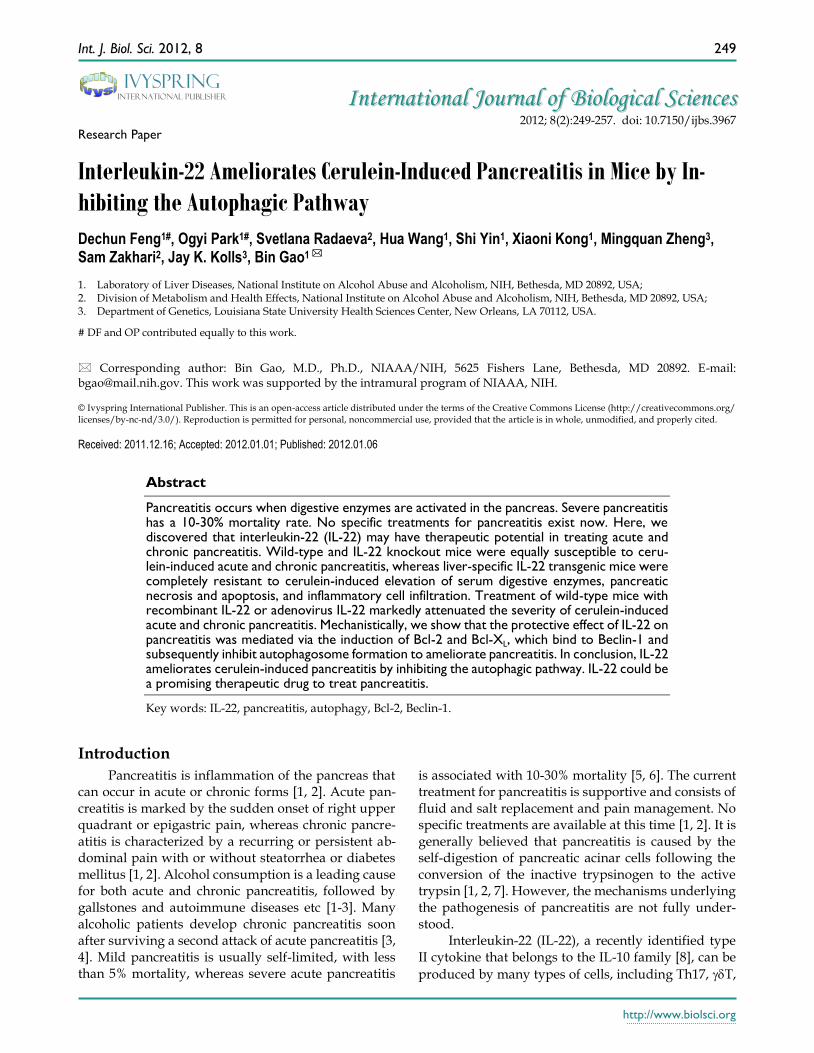
To determine the effects of IL-22 on pancreatitis, we compared cerulein-induced acute pancreatitis in WT, IL-22KO, and IL-22TG mice. As illustrated in Figure 1A, without cerulein treatment (phosphate buffered saline [PBS] -treated groups), pancreas ap-pearance and weight and serum amylase and lipase levels were similar between the WT, IL-22KO, and IL-22TG mice. Repeated injection of cerulein, which is a decapeptide analog of cholecystokinin, increased the pancreas and body weight ratio and serum amyl-
ase and lipase levels in the WT and IL-22KO mice, but not in the IL-22TG mice. In addition, as shown in Figure 1A, serum IL-22 levels were undetectable in the PBS-treated WT and IL-22KO mice but were de-tected at high levels in the IL-22TG mice. Repeated injection of cerulein did not elevate serum IL-22 levels in the WT or IL-22KO mice, nor did it affect serum IL-22 levels in the IL-22TG mice.
The histological analyses shown in Figure 1B in-dicate significant tissue necrosis in the ceru-lein-treated WT and IL-22KO mice, but not in the IL-22TG mice. The numbers of TUNEL+ apoptotic acinar cells and MPO+ neutrophils were elevated in the WT and IL-22KO mice after cerulein treatment, whereas this elevation was barely detectable in the cerulein-treated IL-22TG mice (Fig. 1B).
Figure 1. IL-22TG mice are resistant to cerulein-induced acute pancreatitis. WT, IL-22KO, and IL-22TG mice were treated
seven times with cerulein or PBS injections to induce acute pancreatitis. Mice were sacrificed one hour after the last injection. The ratio
of the pancreas/body weight, serum levels of IL-22, and amylase and lipase levels were measured (A). Representative H&E staining, TUNEL
staining, and MPO staining are shown in the left panel of (B). The pathology score and the number of TUNEL+ and MPO+ cells are
summarized in the right panel of (B). *** P < 0.001 compared to cerulein-treated WT mice. Data are representative of three different
experiments, 5~7 mice in each group. In panel A, UD: undetectable.
Int. J. Biol. Sci. 2012, 8
http://www.biolsci.org
252
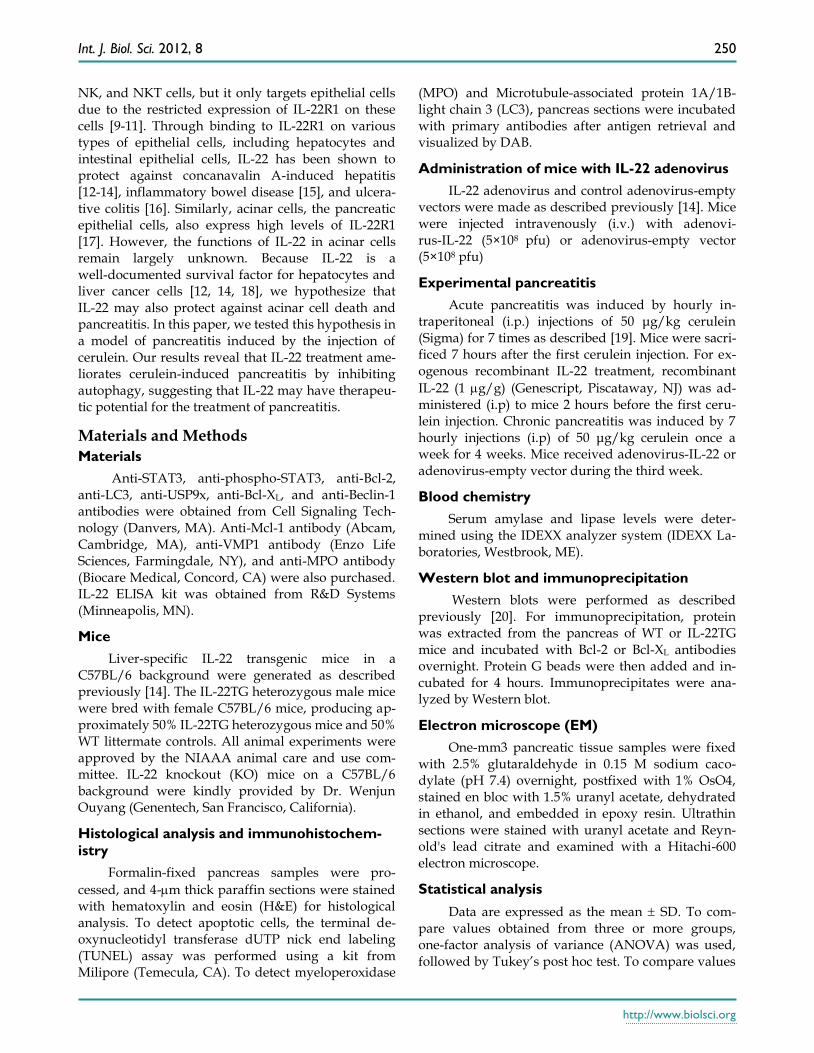
Figure 2. IL-22TG mice are resistant to cerulein-induced chronic pancreatitis. WT and IL-22TG mice received seven cerulein
or PBS injections weekly for four weeks to induce chronic pancreatitis. Mice were sacrificed one hour after the last injection. Repre-
sentative H&E staining, TUNEL staining, and MPO staining are shown (A). Serum levels of amylase and lipase were measured. The pa-
thology score and the number of TUNEL+ and MPO+ cells are summarized (B). ***P<0.001 compared to cerulein-treated WT mice. Data
are representative of three different experiments, 5~7 mice in each group.
We also tested the severity of cerulein-induced
chronic pancreatitis (4-week treatment) in the WT and IL-22TG mice. As illustrated in Figure 2A, 4-week repeated cerulein treatment markedly induced chronic pancreatitis, as evidenced by an elevation in serum amylase and lipase levels and of pancreatic necrosis, TUNEL+ cells, and MPO+ neutrophils in the pancreases of the WT mice. In contrast, such repeated cerulein treatment induced only mild pancreatitis in the IL-22TG mice (Fig. 2A and 2B).
Treatment with recombinant mouse IL-22
(rmIL-22) or adeno-IL-22 protects mice from
cerulein-induced acute and chronic pancreati-
tis
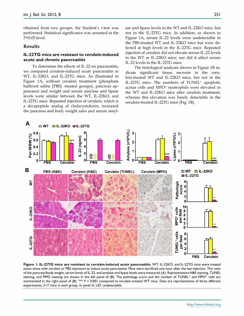
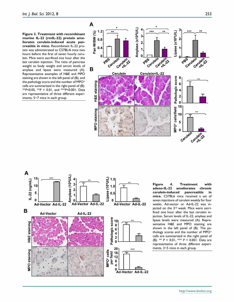
To evaluate the therapeutic potential of IL-22 in acute pancreatitis, we injected mice with rmIL-22 protein two hours before the first cerulein injection. As illustrated in Fig. 3A, pretreatment with rmIL-22 did not affect cerulein-mediated increase in the pan-creas/body weight ratio, but markedly diminished
the elevation of serum amylase and lipase levels. Consistently, cerulein-induced tissue damage and inflammatory cell infiltration were also greatly ame-liorated by rmIL-22 treatment, as indicated by H&E staining and MPO staining (Fig. 3B).
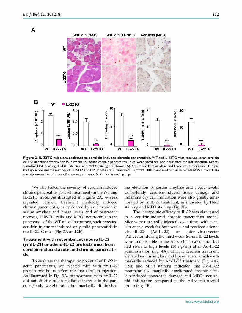
The therapeutic efficacy of IL-22 was also tested in a cerulein-induced chronic pancreatitis model. Mice were repeatedly injected seven times with ceru-lein once a week for four weeks and received adeno-virus-IL-22 (Ad-IL-22) or adenovirus-vector (Ad-vector) during the third week. Serum IL-22 levels were undetectable in the Ad-vector-treated mice but had risen to high levels (10 ng/ml) after Ad-IL-22 administration (Fig. 4A). Chronic cerulein treatment elevated serum amylase and lipase levels, which were markedly reduced by Ad-IL-22 treatment (Fig. 4A). H&E and MPO staining indicated that Ad-IL-22 treatment also markedly ameliorated chronic ceru-lein-induced pancreatic damage and MPO+ neutro-phil infiltration compared to the Ad-vector-treated group (Fig. 4B).
Int. J. Biol. Sci. 2012, 8
http://www.biolsci.org
253
Figure 3. Treatment with recombinant
murine IL-22 (rmIL-22) protein ame-
liorates cerulein-induced acute pan-
creatitis in mice. Recombinant IL-22 pro-
tein was administrated to C57BL/6 mice two
hours before the first of seven hourly ceru-
lein. Mice were sacrificed one hour after the
last cerulein injection. The ratio of pancreas
weight to body weight and serum levels of
amylase and lipase were measured (A).
Representative examples of H&E and MPO
staining are shown in the left panel of (B), and
the pathology score and the number of MPO+
cells are summarized in the right panel of (B).
**P<0.05, **P < 0.01, and ***P<0.001. Data
are representative of three different exper-
iments, 5~7 mice in each group.
Figure 4. Treatment with
adeno-IL-22 ameliorates chronic cerulein-induced pancreatitis in
mice. C57BL6 mice received a set of
seven injections of cerulein weekly for four
weeks. Ad-vector or Ad-IL-22 was in-
jected on the 3rd week. Mice were sacri-
ficed one hour after the last cerulein in-
jection. Serum levels of IL-22, amylase and
lipase levels were measured (A). Repre-
sentative H&E and MPO staining are
shown in the left panel of (B). The pa-
thology scores and the number of MPO+
cells are summarized in the right panel of
(B). ** P < 0.01, *** P < 0.001. Data are
representative of three different experi-
ments, 3~5 mice in each group.
Int. J. Biol. Sci. 2012, 8
http://www.biolsci.org
254
In summary, the above data clearly indicate that IL-22 treatment ameliorates cerulein-induced acute and chronic pancreatitis.
The protective role of IL-22 in pancreatitis is
mediated by the induction of Bcl-2 and Bcl-XL,
which bind to beclin-1 and subsequently inhibit
autophagy
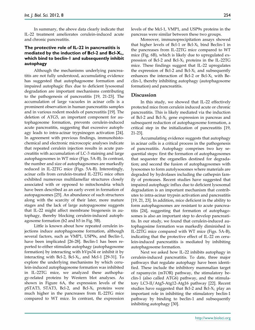
Although the mechanisms underlying pancrea-titis are not fully understood, accumulating evidence has suggested that autophagosome formation and impaired autophagic flux due to deficient lysosomal degradation are important mechanisms contributing to the pathogenesis of pancreatitis [19, 21-23]. The accumulation of large vacuoles in acinar cells is a prominent observation in human pancreatitis samples and in various rodent models of pancreatitis [19]. The deletion of ATG5, an important component for au-tophagosome formation, prevents cerulein-induced acute pancreatitis, suggesting that excessive autoph-agy leads to intra-acinar trypsinogen activation [24]. In agreement with previous findings, immunohisto-chemical and electronic microscopic analyses indicate that repeated cerulein injection results in acute pan-creatitis with accumulation of LC-3 staining and large autophagosomes in WT mice (Figs. 5A-B). In contrast, the number and size of autophagosomes are markedly reduced in IL-22TG mice (Figs. 5A-B). Interestingly, acinar cells from cerulein-treated IL-22TG mice often exhibited numerous multilamellar structures closely associated with or opposed to mitochondria which have been described as an early event in formation of autopagosomes [25]. An abundance of such structures along with the scarcity of their later, more mature stages and the lack of large autopagosome suggests that IL-22 might affect membrane biogenesis in au-tophagy, thereby blocking cerulein-induced autoph-agosome formation (b2 and b3 in Fig. 5B).
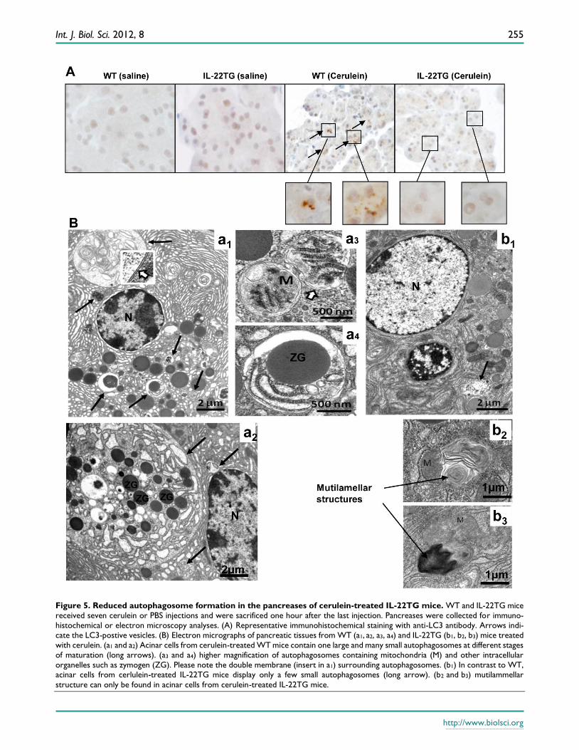
Little is known about how repeated cerulein in-jections induce autophagosome formation, although several factors, such as VMP1, USP9x, and Beclin-1, have been implicated [26-28]. Beclin-1 has been re-ported to either stimulate autophagy (autophagosome formation) by interacting with hVps34 or inhibit it by interacting with Bcl-2, Bcl-XL, and Mcl-1 [29-31]. To explore the underlying mechanisms by which ceru-lein-induced autophagosome formation was inhibited in IL-22TG mice, we analyzed these authopha-gy-related proteins by Western blot analyses. As shown in Figure 6A, the expression levels of the pSTAT3, STAT3, Bcl-2, and Bcl-XL proteins were much higher in the pancreases from IL-22TG mice compared to WT mice. In contrast, the expression
levels of the Mcl-1, VMP1, and USP9x proteins in the pancreas were similar between these two groups.
Moreover, immunoprecipitation assays showed that higher levels of Bcl-1 or Bcl-XL bind Beclin-1 in the pancreases from IL-22TG mice compared to WT mice (Fig. 6B), which is likely due to upregulated ex-pression of Bcl-2 and Bcl-XL proteins in the IL-22TG mice. These findings suggest that IL-22 upregulates the expression of Bcl-2 and Bcl-XL and subsequently enhances the interaction of Bcl-2 or Bcl-XL with Be-clin-1, thereby inhibiting autophagy (autophagosome formation) and pancreatitis.
Discussion
In this study, we showed that IL-22 effectively protected mice from cerulein induced acute or chronic pancreatitis. This is likely mediated via the induction of Bcl-2 and Bcl-XL gene expression in pancreas and subsequent reduction of autophagosome formation, a critical step in the initialization of pancreatitis [19, 21-23].
Accumulating evidence suggests that autophagy in acinar cells is a critical process in the pathogenesis of pancreatitis. Autophagy comprises two key se-quential steps: first the formation of autophagosomes that sequester the organelles destined for degrada-tion; and second the fusion of autophagosomes with lysosomes to form autolysosomes where materials are degraded by hydrolases including the cathepsin fam-ily of proteases. Recent studies have suggested that impaired autophagic influx due to deficient lysosomal degradation is an important mechanism that contrib-utes to intra-acinar trypsin activation and pancreatitis [19, 21, 23]. In addition, mice deficient in the ability to form autophagosomes are resistant to acute pancrea-titis [24], suggesting that formation of autophago-somes is also an important step to develop pancreati-tis. In our study, we found that cerulein-induced au-tophagosime formation was markedly diminished in IL-22TG mice compared with WT mice (Figs. 5A-B), indicating that the protective effect of IL-22 on ceru-lein-induced pancreatitis is mediated by inhibiting autophagosome formation.
Next we asked how IL-22 inhibits autophagy in cerulein-induced pancreatitis. To date, three major pathways that regulate autophagy have been identi-fied. These include the inhibitory mammalian target of rapamycin (mTOR) pathway, the stimulatory be-clin-1 (also called ATG6) pathway, and the stimula-tory LC3-II/Atg5-Atg12-Atg16 pathway [22]. Recent studies have suggested that Bcl-2 and Bcl-XL play an important role in inhibiting the stimulatory beclin-1 pathway by binding to beclin-1 and subsequently inhibiting autophagy [30].
Int. J. Biol. Sci. 2012, 8
http://www.biolsci.org
255
Figure 5. Reduced autophagosome formation in the pancreases of cerulein-treated IL-22TG mice. WT and IL-22TG mice
received seven cerulein or PBS injections and were sacrificed one hour after the last injection. Pancreases were collected for immuno-
histochemical or electron microscopy analyses. (A) Representative immunohistochemical staining with anti-LC3 antibody. Arrows indi-
cate the LC3-postive vesicles. (B) Electron micrographs of pancreatic tissues from WT (a1, a2, a3, a4) and IL-22TG (b1, b2, b3) mice treated
with cerulein. (a1 and a2) Acinar cells from cerulein-treated WT mice contain one large and many small autophagosomes at different stages
of maturation (long arrows). (a3 and a4) higher magnification of autophagosomes containing mitochondria (M) and other intracellular
organelles such as zymogen (ZG). Please note the double membrane (insert in a1) surrounding autophagosomes. (b1) In contrast to WT, acinar cells from cerlulein-treated IL-22TG mice display only a few small autophagosomes (long arrow). (b2 and b3) mutilammellar
structure can only be found in acinar cells from cerulein-treated IL-22TG mice.
Int. J. Biol. Sci. 2012, 8
http://www.biolsci.org
256
Figure 6. Upregulation of Bcl-2 and Bcl-XL, which bind Beclin-1, was associated reduced autophagosome formation in
the pancreases of cerulein-treated IL-22TG mice. (A) Western blot analyses of pancreatic protein extracts from WT and IL-22TG
mice. (B) Immunoprecipitation analyses of the interaction of Beclin-1 with Bcl-2 family proteins. (C) Scheme of the mechanisms underlying
IL-22 inhibition of autophagy and pancreatitis. Data are representative of three different experiments.
Several previous studies have demonstrated that
STAT3 activation induces the expression of Bcl-2 and Bcl-XL in a variety of cell types via transcriptional upregulation of these genes [32]. Thus, we speculated that upregulated Bcl-2 and Bcl-XL in the pancreases of IL-22TG mice are likely due to the enhanced IL-22 activation of STAT3. This hypothesis was supported by our experimental data that expression of pSTAT3, Bcl-2 and Bcl-XL levels was much higher in the pan-creas of IL-22TG mice than that in WT mice (Fig. 6A). Moreover, immunoprecipitation assays in Fig. 6B showed that binding of Bcl-2 or Bcl-XL with Beclin 1 was enhanced in the pancreas from IL-22TG mice compared with WT mice. Collectively, our findings suggest that IL-22 activates STAT3 and then upregu-lates the STAT3 downstream genes such as Bcl-2 and Bcl-XL. Increased Bcl-2 and Bcl-XL subsequently binds to Beclin1 and inhibits autophagosome formation, thereby ameliorating acinar cell damage in ceru-lein-induced pancreatitis (Fig. 6C).
Chronic alcohol drinking is a leading cause for both acute and chronic pancreatitis [1-3]. Thus, fur-ther studies of the protective effects of IL-22 on alco-holic pancreatitis are urgently needed. Alcoholic pancreatitis is attributed to its non-oxidative metabo-lism by phospholipase D (PLD) to fatty acid ethyl esters (FAEEs) generated in the pancreatic acinar cells [33, 34]. It will be interesting to determine whether IL-22 affects the interaction between PLD activation and formation of FAEEs (or phosphatidylethanol), thereby ameliorating alcoholic pancreatitis. In addi-
tion, rats fed ethanol diet for 14 weeks, then received a single lipopolysaccharide injection showed depletion of several lysosomal proteins including lysoso-mal-associated membrane protein-2 (Lamp-2), an es-sential lysosomal membrane protein that is required for the proper fusion of autophagosomes with lyso-somes [35]. Accordingly, Lamp-2 depletion is report-ed to inhibit the fusion of lysosomes and autophago-somes, thus causing the accumulation of autophago-somes [36, 37]. Importantly, patients with alcoholic pancreatitis also exhibit local Lamp-2 depletion, which highlights the important role of Lamp-2 in in-hibiting autophagy and pancreatic acinar cell death [35]. It will be important to examine whether IL-22 regulates Lamp-2 gene expression and subsequently ameliorates alcoholic pancreatitis.
In summary, IL-22 treatment appears to have beneficial effects on acute and chronic pancreatitis with the added benefit of potentially few side effects due to the restricted expression of IL-22R [38]. Clinical trials examining IL-22 treatment for pancreatitis are therefore a reasonable next step.
Abbreviations
adeno-IL-22: IL-22 adenovirus; adeno-vector: control vector adenovirus; IL-22TG: IL-22 transgenic mice; LC3: Microtubule-associated protein 1A/1B-light chain 3; EM: Electron microscope; FAEEs: fatty acid ethyl esters; MPO: myeloperoxidase; STAT3: signal transducer and activator of transcrip-tion 3; PLD: phospholipase D
Int. J. Biol. Sci. 2012, 8
http://www.biolsci.org
257
Acknowledgment
We thank the Electron Microscopy Facility at NINDS, NIH for technical support.
Conflict of Interests
The authors have declared that no conflict of in-terest exists.
References 1. Harper SJ, Cheslyn-Curtis S. Acute pancreatitis. Ann Clin Biochem
2011;48:23-37.
2. Braganza JM, Lee SH, McCloy RF, McMahon MJ. Chronic
pancreatitis. Lancet 2011;377:1184-1197.
3. Apte MV, Pirola RC, Wilson JS. Mechanisms of alcoholic
pancreatitis. J Gastroenterol Hepatol 2010;25:1816-1826.
4. Lankisch PG, Breuer N, Bruns A, Weber-Dany B, Lowenfels AB,
Maisonneuve P. Natural history of acute pancreatitis: a long-term
population-based study. Am J Gastroenterol 2009;104:2797-2805.
5. Dervenis C, Johnson CD, Bassi C, Bradley E, Imrie CW, McMahon
MJ, et al. Diagnosis, objective assessment of severity, and
management of acute pancreatitis. Santorini consensus conference.
Int J Pancreatol 1999;25:195-210.
6. Petrov MS, Shanbhag S, Chakraborty M, Phillips AR, Windsor JA.
Organ failure and infection of pancreatic necrosis as determinants
of mortality in patients with acute pancreatitis. Gastroenterology
2010;139:813-820.
7. Gaisano HY, Gorelick FS. New insights into the mechanisms of
pancreatitis. Gastroenterology 2009;136:2040-2044.
8. Dumoutier L, Louahed J, Renauld JC. Cloning and characterization
of IL-10-related T cell-derived inducible factor (IL-TIF), a novel
cytokine structurally related to IL-10 and inducible by IL-9. J
Immunol 2000;164:1814-1819.
9. Wolk K, Witte E, Witte K, Warszawska K, Sabat R. Biology of
interleukin-22. Semin Immunopathol 2010;32:17-31.
10. Zenewicz LA, Flavell RA. Recent advances in IL-22 biology. Int
Immunol 2011;23:159-163.
11. Ouyang W, Kolls JK, Zheng Y. The biological functions of T helper
17 cell effector cytokines in inflammation. Immunity
2008;28:454-467.
12. Radaeva S, Sun R, Pan HN, Hong F, Gao B. Interleukin 22 (IL-22)
plays a protective role in T cell-mediated murine hepatitis: IL-22 is a
survival factor for hepatocytes via STAT3 activation. Hepatology
2004;39:1332-1342.
13. Zenewicz LA, Yancopoulos GD, Valenzuela DM, Murphy AJ,
Karow M, Flavell RA. Interleukin-22 but not interleukin-17
provides protection to hepatocytes during acute liver inflammation.
Immunity 2007;27:647-659.
14. Park O, Wang H, Weng H, Feigenbaum L, Li H, Yin S, et al. In vivo
consequences of liver-specific interleukin-22 expression in mice:
Implications for human liver disease progression. Hepatology
2011;54:252-261.
15. Zenewicz LA, Yancopoulos GD, Valenzuela DM, Murphy AJ,
Stevens S, Flavell RA. Innate and adaptive interleukin-22 protects
mice from inflammatory bowel disease. Immunity 2008;29:947-957.
16. Sugimoto K, Ogawa A, Mizoguchi E, Shimomura Y, Andoh A, Bhan
AK, et al. IL-22 ameliorates intestinal inflammation in a mouse
model of ulcerative colitis. J Clin Invest 2008;118:534-544.
17. Aggarwal S, Xie MH, Maruoka M, Foster J, Gurney AL. Acinar cells
of the pancreas are a target of interleukin-22. J Interferon Cytokine
Res 2001;21:1047-1053.
18. Jiang R TZ, Deng L, Chen Y, Xia Y, Gao Y, Wang X, Sun B.
Interleukin-22 promotes human hepatocellular carcinoma by
activation of STAT3. Hepatology 2011;54:900-909.
19. Mareninova OA, Hermann K, French SW, O'Konski MS, Pandol SJ,
Webster P, et al. Impaired autophagic flux mediates acinar cell
vacuole formation and trypsinogen activation in rodent models of
acute pancreatitis. J Clin Invest 2009;119:3340-3355.
20. Wang H, Park O, Lafdil F, Shen K, Horiguchi N, Yin S, et al.
Interplay of hepatic and myeloid signal transducer and activator of
transcription 3 in facilitating liver regeneration via tempering innate
immunity. Hepatology 2010;51:1354-1362.
21. Fortunato F, Kroemer G. Impaired autophagosome-lysosome fusion
in the pathogenesis of pancreatitis. Autophagy 2009;5:850-853.
22. Czaja MJ. Functions of autophagy in hepatic and pancreatic
physiology and disease. Gastroenterology 2011;140:1895-1908.
23. Gukovsky I, Gukovskaya AS. Impaired autophagy underlies key
pathological responses of acute pancreatitis. Autophagy
2010;6:428-429.
24. Hashimoto D, Ohmuraya M, Hirota M, Yamamoto A, Suyama K,
Ida S, et al. Involvement of autophagy in trypsinogen activation
within the pancreatic acinar cells. J Cell Biol 2008;181:1065-1072.
25. Hailey DW, Rambold AS, Satpute-Krishnan P, Mitra K, Sougrat R,
Kim PK, et al. Mitochondria supply membranes for autophagosome
biogenesis during starvation. Cell 2010;141:656-667.
26. Grasso D, Ropolo A, Lo Re A, Boggio V, Molejon MI, Iovanna JL, et
al. Zymophagy, a novel selective autophagy pathway mediated by
VMP1-USP9x-p62, prevents pancreatic cell death. J Biol Chem
2011;286:8308-8324.
27. Ropolo A, Grasso D, Pardo R, Sacchetti ML, Archange C, Lo Re A, et
al. The pancreatitis-induced vacuole membrane protein 1 triggers
autophagy in mammalian cells. J Biol Chem 2007;282:37124-37133.
28. Liang XH, Jackson S, Seaman M, Brown K, Kempkes B, Hibshoosh
H, et al. Induction of autophagy and inhibition of tumorigenesis by
beclin 1. Nature 1999;402:672-676.
29. Pattingre S, Tassa A, Qu X, Garuti R, Liang XH, Mizushima N, et al.
Bcl-2 antiapoptotic proteins inhibit Beclin 1-dependent autophagy.
Cell 2005;122:927-939.
30. Levine B, Sinha S, Kroemer G. Bcl-2 family members: dual
regulators of apoptosis and autophagy. Autophagy 2008;4:600-606.
31. Maiuri MC, Le Toumelin G, Criollo A, Rain JC, Gautier F, Juin P, et
al. Functional and physical interaction between Bcl-X(L) and a
BH3-like domain in Beclin-1. EMBO J 2007;26:2527-2539.
32. Yu H, Jove R. The STATs of cancer--new molecular targets come of
age. Nat Rev Cancer 2004;4:97-105.
33. Werner J, Laposata M, Fernandez-del Castillo C, Saghir M, Iozzo
RV, Lewandrowski KB, et al. Pancreatic injury in rats induced by
fatty acid ethyl ester, a nonoxidative metabolite of alcohol.
Gastroenterology 1997;113:286-294.
34. Pandol SJ, Periskic S, Gukovsky I, Zaninovic V, Jung Y, Zong Y, et
al. Ethanol diet increases the sensitivity of rats to pancreatitis
induced by cholecystokinin octapeptide. Gastroenterology
1999;117:706-716.
35. Fortunato F, Burgers H, Bergmann F, Rieger P, Buchler MW,
Kroemer G, et al. Impaired autolysosome formation correlates with
Lamp-2 depletion: role of apoptosis, autophagy, and necrosis in
pancreatitis. Gastroenterology 2009;137:350-360.
36. Huynh KK, Eskelinen EL, Scott CC, Malevanets A, Saftig P,
Grinstein S. LAMP proteins are required for fusion of lysosomes
with phagosomes. Embo J 2007;26:313-324.
37. Saftig P, Tanaka Y, Lullmann-Rauch R, von Figura K. Disease
model: LAMP-2 enlightens Danon disease. Trends Mol Med
2001;7:37-39.
38. Wolk K, Sabat R. Interleukin-22: A novel T- and NK-cell derived
cytokine that regulates the biology of tissue cells. Cytokine Growth
Factor Rev 2006;17:367-380.