research open access intensity-modulated radiation therapy ... · research open access...
TRANSCRIPT

Murai et al. Radiation Oncology 2013, 8:68http://www.ro-journal.com/content/8/1/68
RESEARCH Open Access
Intensity-modulated radiation therapy using staticports of tomotherapy (TomoDirect): comparisonwith the TomoHelical modeTaro Murai1,2*, Yuta Shibamoto1, Yoshihiko Manabe1,2, Rumi Murata2, Chikao Sugie1, Akihiro Hayashi1,Hiroya Ito2 and Yoshihito Miyoshi2
Abstract
Purpose: With the new mode of Tomotherapy, irradiation can be delivered using static ports of the TomoDirectmode. The purpose of this study was to evaluate the characteristics of TomoDirect plans compared to conventionalTomoHelical plans.
Methods: TomoDirect and TomoHelical plans were compared in 46 patients with a prostate, thoracic wall or lungtumor. The mean target dose was used as the prescription dose. The minimum coverage dose of 95% of the target(D95%), conformity index (CI), uniformity index (UI), dose distribution in organs at risk and treatment time wereevaluated. For TomoDirect, 2 to 5 static ports were used depending on the tumor location.
Results: For the prostate target volume, TomoDirect plans could not reduce the rectal dose and required a longertreatment time than TomoHelical. For the thoracic wall target volume, the V5Gy of the lung or liver was lower inTomoDirect than in TomoHelical (p = 0.02). For the lung target volume, TomoDirect yielded higher CI (p = 0.009)but smaller V5Gy of the lung (p = 0.005) than TomoHelical. Treatment time did not differ significantly between thethoracic wall and lung plans.
Conclusion: Prostate cancers should be treated with the TomoHelical mode. Considering the risk of low-doseradiation to the lung, the TomoDirect mode could be an option for thoracic wall and lung tumors.
Keywords: Tomotherapy, TomoDirect, Intensity modulated radiotherapy, Lung cancer, Prostate cancer
IntroductionThe Tomotherapy Hi-Art system (Accuray Inc, Sunnyvale,CA, USA) is a radiation delivery system that combinesdynamic intensity-modulated radiation therapy (IMRT)and an on-board imaging systems [1,2]. Treatment is usu-ally delivered with 360-degree rotation of the 6-megavoltlinear accelerator gantry. This conventional treatmentmode is called TomoHelical. Recently, a new systemupgrade named TomoDirect has been introduced [3,4].TomoDirect allows the delivery of radiation at pre-established discrete angles with a fixed gantry. This newsystem is expected to reduce treatment time and reduce
* Correspondence: [email protected] of Radiology, Nagoya City University Graduate School ofMedical Sciences, 1 Kawasumi, Mizuho-cho, Mizuho-ku, Nagoya 467-8601,Japan2Department of Radiation Oncology, JA Suzuka General Hospital, Suzuka,Japan
© 2013 Murai et al.; licensee BioMed Central LCommons Attribution License (http://creativecreproduction in any medium, provided the or
critical organ dose. However, these expectations have notyet been proven in clinical situations.Clinically, localized prostate cancer is one of the most
common malignancies treated with IMRT [5]. UsingTomotherapy, irradiation usually requires 3–6 minutesin addition to 10–15 minutes of set-up to treat prostatetarget volume, so only a limited number of patients canbe treated with one Tomotherapy machine in one day. IfTomoDirect could reduce the treatment time, more pa-tients could be treated even in institutions possessingonly one machine. In addition, Tomotherapy has re-cently been demonstrated to be adequate for treatingmoving targets with a hypofractionated course of radio-therapy [6-8]. In chemoradiotherapy for thoracic tumorsusing Tomotherapy, some reports about lung cancer [9]or mesothelioma [10-12] recommended that low doseexposure of the lung should be reduced as much as
td. This is an Open Access article distributed under the terms of the Creativeommons.org/licenses/by/2.0), which permits unrestricted use, distribution, andiginal work is properly cited.

Murai et al. Radiation Oncology 2013, 8:68 Page 2 of 7http://www.ro-journal.com/content/8/1/68
possible in order to reduce pulmonary toxicity. Thus, ifTomoDirect could reduce the lung volume receiving lowdose radiation, this modality could be expected to re-place TomoHelical treatment in this situation.In the present study, dose distributions and treatment
times were compared between TomoDirect and Tomo-Helical plans in three clinical situations: a) prostate tar-get volume, b) thoracic wall target volume and c) lungtarget volume. The purposes of this planning study wereto examine 1) whether TomoDirect can reduce the treat-ment time and 2) whether TomoDirect can be an alter-native to TomoHelical for prostatic and thoracic targetvolume.
MethodsComparisons of treatment plans between TomoDirectand TomoHelical were carried out during the fixedperiod from September 2010 to July 2011 at SuzukaGeneral Hospital. All eligible patients seen during theperiod, i.e., 46 adults actually treated for prostate cancer,thoracic wall metastasis and lung tumor, were studied.
CT SimulationTo reduce breathing motion and set-up error, patientswere laid in the supine position on appropriate im-mobilization devices depending on the location of theradiotherapy target [13]. In planning computed tomog-raphy (CT), 2- to 3-mm slice thickness axial images wereacquired using a 64-row multi-detector CT (Aquilion CX,
Figure 1 Thoracic wall target volume. Upper panel; the target volume land TomoDirect mode (b). Lower panel; the target volume located near tmode (d).
Toshiba Medical, Otahara, Japan). Contrast-enhanced CTimages were acquired and fused to the planning CT imagesto delineate the target, but unenhanced CT images wereused for dose calculation to keep calculation accuracy [14].Contouring of target volumes and normal structures wasperformed on the Pinnacle (3) version 9 treatment planningsystem (Philips Medical System, Eindhoven, Netherlands).The contours created in the treatment planning systemwere exported to the Tomotherapy Hi-Art treatment plan-ning system v4.0, where TomoHelical and TomoDirectplans were generated.
PlanningThe prescription dose was defined as the mean dose ofthe planning target volume (PTV) (Dmean). As dose con-straints, 1) D95% > 90% of the prescribed dose and 2)V90% ≥ 95% were satisfied. D95% was defined as the mini-mum dose delivered to 95% of the PTV. V90% was de-fined as the percentage of the PTV receiving at least 90%of the prescribed dose. Appropriate dose constraints wereimplemented for inverse planning procedures. In theTomotherapy planning system, the parameters set beforeoptimization were field width, modulation factor andpitch. The same field width, pitch and modulation factorwere used in both TomoHelical and TomoDirect plans.To reduce dose to the critical organs, for example thelung, blocking structures were added appropriately in theTomoHelical plan. All optimization procedures were car-ried out until breaking the PTV dose constraints or
ocated on the lateral wall was treated with TomoHelical mode (a)he midline was treated with TomoHelical mode (c) and TomoDirect
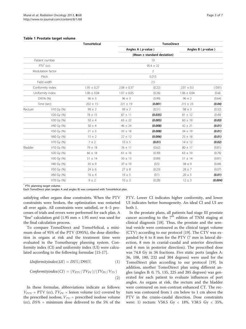
Table 1 Prostate target volume
TomoHelical TomoDirect
Angles A ( p-value ) Angles B ( p-value )
(Mean ± standard deviation)
Patient number 19
PTV* (cc) 95.4 ± 22
Modulation factor 2
Pitch 0.215
Field width 2.5
Conformity index 1.95 ± 0.27 2.08 ± 0.37 (0.22) 2.01 ± 0.5 ( 0.61)
Uniformity index 1.06 ± 0.04 1.07 ± 0.05 (0.26) 1.06 ± 0.04 (0.4)
D95% (%) 96 ± 3 96 ± 3 (0.99) 96 ± 2 (0.64)
Time (sec) 202 ± 15 221 ± 19 (0.001) 215 ± 23 (0.04)
Rectum V10 Gy (%) 99 ± 2 99 ± 2 (0.51) 98 ± 3 (0.32)
V20 Gy (%) 78 ± 15 87 ± 11 (0.035) 81 ± 12 (0.49)
V30 Gy (%) 50 ± 4 65 ± 22 (0.005) 60 ± 19 (0.03)
V40 Gy (%) 30 ± 4 46 ± 24 (0.008) 42 ± 21 (0.01)
V50 Gy (%) 21 ± 3 33 ± 18 (0.008) 34 ± 19 (0.01)
V60 Gy (%) 15 ± 2 22 ± 12 (0.006) 25 ± 16 (0.01)
V70 Gy (%) 7 ± 2 10 ± 5 (0.01) 14 ± 12 (0.02)
Bladder V10 Gy (%) 79 ± 18 76 ± 17 (0.62) 80 ± 17 (0.81)
V20 Gy (%) 66 ± 18 61 ± 16 (0.39) 63 ± 19 (0.76)
V30 Gy (%) 51 ± 14 50 ± 13 (0.89) 51 ± 14 (0.81)
V40 Gy (%) 35 ± 9 37 ± 10 (0.5) 38 ± 9 (0.44)
V50 Gy (%) 24 ± 6 27 ± 8 (0.23) 28 ± 7 (0.07)
V60 Gy (%) 16 ± 4 19 ± 5 (0.1) 20 ± 5 (0.01)
V70 Gy (%) 9 ± 2 10 ± 3 (0.28) 12 ± 3 (0.004)* PTV, planning target volume.Each TomoDirect plan (angles A and angles B) was compared with TomoHelical plan.
Murai et al. Radiation Oncology 2013, 8:68 Page 3 of 7http://www.ro-journal.com/content/8/1/68
satisfying other organs dose constraints. When the PTVconstraints were broken, the optimization was restartedall over again. All constraints were satisfied, or 4–5 pro-cesses of trials and errors were performed for each plan. A“fine” calculation grid (1.95 mm × 1.95 mm) was used forthe final calculation process.To compare TomoDirect and TomoHelical, a mini-
mum dose of 95% of the PTV (D95%), the dose distribu-tion in organs at risk and the treatment time wereevaluated in the Tomotherapy planning system. Con-formity index (CI) and uniformity index (UI) were calcu-lated according to the following formulae [15-17].
Uniformityindex UIð Þ ¼ D5%=D95% ð1Þ
Conformityindex CIð Þ ¼ VPTV=TVPVð Þ= TVPV=VTVð Þð2Þ
In these formulae, abbreviations indicate as follows:VPTV = PTV (cc), TVPV = lesion volume (cc) covered bythe prescribed isodose, VTV = prescribed isodose volume(cc), D5% = minimum dose delivered to the 5% of the
PTV. Lower CI indicates higher conformity, and lowerUI indicates better homogeneity. An ideal CI and UI areboth 1.In the prostate plans, all patients had stage III prostate
cancer according to the 7th edition of TNM staging atclinical diagnosis [18]. Thus, the prostate and the sem-inal vesicle were contoured as the clinical target volume(CTV) according to our protocol [19]. The CTV was ex-panded by 6 to 8 mm for the PTV (7 mm in lateral dir-ection, 8 mm in cranial-caudal and anterior directionsand 6 mm in posterior direction). The prescribed dosewas 74.8 Gy in 34 fractions. Five static ports (angles A:36, 108, 180, 232 and 304 degrees) were used for theTomoDirect plan according to our protocol [19]. Inaddition, another TomoDirect plan using different an-gles (angles B: 0, 75, 135, 225 and 285 degrees) was gen-erated for each patient to evaluate influences of portangles. As organs at risk, the rectum and the bladderwere contoured on non-contrast enhanced CT. The rec-tum was contoured from 1 cm below to 1 cm above thePTV in the cranio-caudal direction. Dose constraintswere: 1) rectum: V58.5 Gy < 18%, V38.5 Gy < 35%,

Figure 2 Prostate target volume. (a) TomoHelical plan. (b) TomoDirect plan – Angles A. (c) TomoDirect plan – Angles B.
Murai et al. Radiation Oncology 2013, 8:68 Page 4 of 7http://www.ro-journal.com/content/8/1/68
maximum dose (Dmax) < 75.1 Gy and 2) bladder: V60Gy < 20%, V40 Gy < 35%. The V Gy value represents thepercentage volume receiving the specified dose, e.g., V60Gy is the percentage volume receiving 60 Gy.In the thoracic wall plans, the visible enhanced lesion
was contoured as the CTV. The CTV was expanded by5 mm for the PTV and 39 Gy in 13 fractions wasprescribed. For TomoDirect, oblique or 3 directions wereused depending on the tumor location. Lesions locatednear the midline were treated with 3 ports of TomoDirect(Figure 1c, d). Organs at risk included the lung or liver,the skin and the spinal cord. The skin was contoured as a3–5 mm thick layer under the body surface, and evenwhen the PTV included the skin surface, it was spared toavoid skin toxicity. Spinal cord, lung and/or liver dosewere reduced as much as possible.In the lung plans, the visible enhanced lesion was
contoured as the CTV. The CTV was expanded by 5mm plus a patient-specific internal margin for the PTV.Each tumor motion was examined under 4-dimensional(4-D) CT during 2–3 respiratory cycles to make a max-imum projection of the phases and delineate contourson that. Contrast material was not used at 4-D CT. To
Figure 3 Dose volume histograms of TomoHelical and TomoDirect plplan – Angles A. (c) TomoDirect plan – Angles B.
the PTV, 59.4 Gy in 27 fractions was prescribed. Fourstatic ports were used for TomoDirect. Organs at riskincluded the lung, the skin and the spinal cord. Doseconstraints were: 1) lung: mean lung dose (MLD) < 17 Gy,V10 Gy < 40%, V20 Gy < 30% and 2) spinal cord + 5 mmmargin: Dmax < 50 Gy.Comparisons of dose-volume parameters and treat-
ment time between TomoDirect and TomoHelical planswere carried out using the two-tailed paired t-test. Weassumed that the study populations distributed normally.All statistical analyses were performed using Statview 5.0(SAS Institute Inc, Cary, NC). Statistical significance wasdefined as p ≤ 0.05. All planning and evaluation wasperformed by one radiation oncologist (T. M.). All dosesevaluated in this study were calculated physical doses onthe planning workstation.
ResultsA typical dose distribution and a typical dose volumehistogram of the prostate plans are shown in Table 1,Figure 2 and Figure 3. Table 1 summarizes the treat-ment parameters, dose-volume parameters and treat-ment times of the two plans in 19 patients. D95%, CI
ans for prostate target volume. (a) TomoHelical plan. (b) TomoDirect
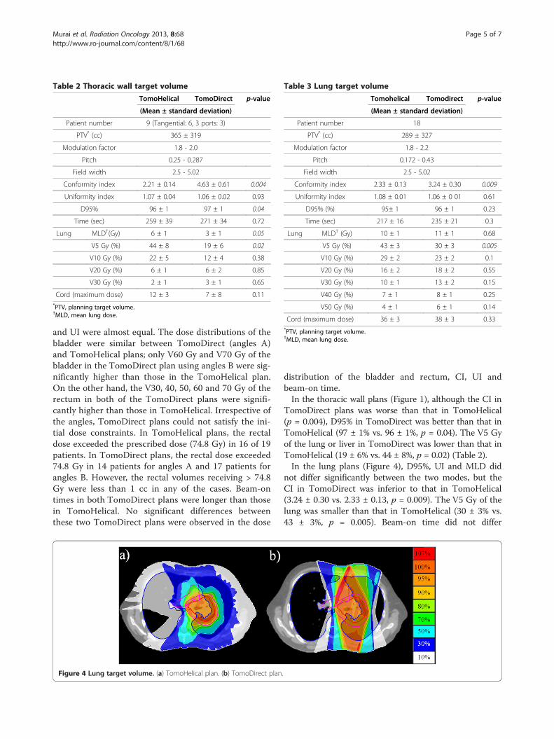
Table 3 Lung target volume
Tomohelical Tomodirect p-value
(Mean ± standard deviation)
Patient number 18
PTV* (cc) 289 ± 327
Modulation factor 1.8 - 2.2
Pitch 0.172 - 0.43
Field width 2.5 - 5.02
Conformity index 2.33 ± 0.13 3.24 ± 0.30 0.009
Uniformity index 1.08 ± 0.01 1.06 ± 0 01 0.61
D95% (%) 95± 1 96 ± 1 0.23
Time (sec) 217 ± 16 235 ± 21 0.3
Lung MLD† (Gy) 10 ± 1 11 ± 1 0.68
V5 Gy (%) 43 ± 3 30 ± 3 0.005
V10 Gy (%) 29 ± 2 23 ± 2 0.1
V20 Gy (%) 16 ± 2 18 ± 2 0.55
V30 Gy (%) 10 ± 1 13 ± 2 0.15
V40 Gy (%) 7 ± 1 8 ± 1 0.25
V50 Gy (%) 4 ± 1 6 ± 1 0.14
Cord (maximum dose) 36 ± 3 38 ± 3 0.33*PTV, planning target volume.†MLD, mean lung dose.
Table 2 Thoracic wall target volume
TomoHelical TomoDirect p-value
(Mean ± standard deviation)
Patient number 9 (Tangential: 6, 3 ports: 3)
PTV* (cc) 365 ± 319
Modulation factor 1.8 - 2.0
Pitch 0.25 - 0.287
Field width 2.5 - 5.02
Conformity index 2.21 ± 0.14 4.63 ± 0.61 0.004
Uniformity index 1.07 ± 0.04 1.06 ± 0.02 0.93
D95% 96 ± 1 97 ± 1 0.04
Time (sec) 259 ± 39 271 ± 34 0.72
Lung MLD†(Gy) 6 ± 1 3 ± 1 0.05
V5 Gy (%) 44 ± 8 19 ± 6 0.02
V10 Gy (%) 22 ± 5 12 ± 4 0.38
V20 Gy (%) 6 ± 1 6 ± 2 0.85
V30 Gy (%) 2 ± 1 3 ± 1 0.65
Cord (maximum dose) 12 ± 3 7 ± 8 0.11*PTV, planning target volume.†MLD, mean lung dose.
Murai et al. Radiation Oncology 2013, 8:68 Page 5 of 7http://www.ro-journal.com/content/8/1/68
and UI were almost equal. The dose distributions of thebladder were similar between TomoDirect (angles A)and TomoHelical plans; only V60 Gy and V70 Gy of thebladder in the TomoDirect plan using angles B were sig-nificantly higher than those in the TomoHelical plan.On the other hand, the V30, 40, 50, 60 and 70 Gy of therectum in both of the TomoDirect plans were signifi-cantly higher than those in TomoHelical. Irrespective ofthe angles, TomoDirect plans could not satisfy the ini-tial dose constraints. In TomoHelical plans, the rectaldose exceeded the prescribed dose (74.8 Gy) in 16 of 19patients. In TomoDirect plans, the rectal dose exceeded74.8 Gy in 14 patients for angles A and 17 patients forangles B. However, the rectal volumes receiving > 74.8Gy were less than 1 cc in any of the cases. Beam-ontimes in both TomoDirect plans were longer than thosein TomoHelical. No significant differences betweenthese two TomoDirect plans were observed in the dose
Figure 4 Lung target volume. (a) TomoHelical plan. (b) TomoDirect plan
distribution of the bladder and rectum, CI, UI andbeam-on time.In the thoracic wall plans (Figure 1), although the CI in
TomoDirect plans was worse than that in TomoHelical(p = 0.004), D95% in TomoDirect was better than that inTomoHelical (97 ± 1% vs. 96 ± 1%, p = 0.04). The V5 Gyof the lung or liver in TomoDirect was lower than that inTomoHelical (19 ± 6% vs. 44 ± 8%, p = 0.02) (Table 2).In the lung plans (Figure 4), D95%, UI and MLD did
not differ significantly between the two modes, but theCI in TomoDirect was inferior to that in TomoHelical(3.24 ± 0.30 vs. 2.33 ± 0.13, p = 0.009). The V5 Gy of thelung was smaller than that in TomoHelical (30 ± 3% vs.43 ± 3%, p = 0.005). Beam-on time did not differ
.

Murai et al. Radiation Oncology 2013, 8:68 Page 6 of 7http://www.ro-journal.com/content/8/1/68
significantly between TomoDirect and TomoHelical inthoracic wall and lung plans (Table 3).
DiscussionThe present study clarified three characteristics of theTomoDirect mode. First, in most cases, TomoDirectcannot reduce total treatment time. TomoDirect usesstatic gantry positions combined with simultaneous couchtranslation and multileaf collimator modulation. After apatient is treated from one gantry angle, the gantry is ro-tated to a different beam direction and the patient againpasses through the bore for delivery of the subsequentport [3]. As a result, it takes more time to deliver thebeams as the number of ports increases. Even 2- to 4-portTomoDirect plans needed almost equal beam-on time toTomoHelical plans. Thus, when 5 or more ports are usedin TomoDirect plans, the beam-on time may often exceedthat of TomoHelical.Secondly, prostate target volume should be treated with
TomoHelical. Davidson et al. [20] compared TomoHelicaland 7-static-port conventional IMRT plans for prostatetarget volume. In their report, conventional IMRT planswere similar to TomoHelical plans. In contrast, in thecurrent study, the dose to the rectum in TomoDirectplans was significantly higher than those in TomoHelicalplans (Table 2, Figure 3). These results can be explainedby a mechanical feature of Tomotherapy, which uses 64-leaf binary multileaf collimators. One leaf of the multileafcollimators is considered to have 51 beamlets associatedwith it during each gantry rotation [1,2]. Meanwhile, inTomoDirect plans, treatment delivery is limited to fewerdirections with a smaller set of beamlets [3,4]. Therefore,it was possible that 5-static-port TomoDirect plans hadfewer beamlets than 7-static-port conventional IMRTplans, resulting in TomoDirect mode being unable to re-duce dose to the rectum in most cases with stage III pros-tate cancer in this study. Port angles might not havestriking effects on the rectal dose in these situations. Fur-thermore, the treatment time in TomoDirect plans waslonger than that in TomoHelical plans (Table 1). These re-sults suggest that TomoHelical should be used for pros-tate target volume.Third, considering the risk of low-dose radiation to
the lung, TomoDirect mode is one option for thoracicwall and lung target volumes. In the current study, theTomoHelical plans delivered low-dose radiation to largerlung volumes than the TomoDirect plans. It was previ-ously reported that, in comparison with 3D conformalradiotherapy, the use of TomoHelical plans resulted indelivery of low dose to areas in the body that would nor-mally receive only scatter dose [4,21,22]. These resultsindicate the possibility that TomoDirect mode can re-duce the lung toxicity.
Combined chemoradiotherapy is becoming a standardof care for the non-operative management of a varietyof solid malignancies [23,24]. However, modeling tools toanalyze the possible interactions between these modalitiesare lacking. Vogelius et al. [25] indicated that chemother-apy might increase the lung toxicity of low dose radiationexposure when a certain level of chemotherapy-relatednormal tissue damage is exceeded. As an example of this,among the first 46 patients treated with TomoHelical in asingle-arm phase I/II dose-per-fraction escalation trial, theincidences of grade 2 and 3 radiation pneumonitis wereonly 13% and 0%, respectively, despite the large-volume ofdisease treated with very high dose [21]. In contrast, Songet al. [9] reported 7 cases with grade 3 or greater radiationpneumonitis among 37 patients (19%) also treated withTomoHelical, but 24 of the 37 patients had received con-current chemotherapy and 13 had received neoadjuvantchemotherapy, illustrating the impact of chemother-apy. Thus, in cases undergoing chemoradiotherapy fora thoracic tumor, low-dose exposure of the lung, such asV5 Gy, should be reduced to the level of 3D conformalradiotherapy. According to the QUANTEC report [26],doses of the lung should be limited to the levels ofV20 Gy < 30-35% and MLD < 20–23 Gy. In additionto these criteria, it is prudent to give attention to thelow dose exposure of the lung.In the current study, TomoDirect could also reduce
the low-dose-exposed volume of the lung in treatingthoracic wall and lung target volumes and achieve com-parable target dose coverage. Thus, the TomoDirectmode may be an alternative in these situations. Furtherinvestigation into the clinical outcomes of these patientstreated with TomoDirect mode is warranted.
ConclusionsIn conclusion, contrary to previous expectations, Tomo-Direct could not reduce treatment time in any of thesethree settings. Indeed, this modality should be used toreduce the critical organ dose. If chemotherapy is deliv-ered, thoracic wall and lung target volumes may be agood indication for use of TomoDirect.
Competing interestsThe authors declare that they have no competing interests.
Authors’ contributionsEach author had participated sufficiently in the work to take publicresponsibility for appropriate portions of the content. TM and YS designedthe study. TM, YM, RM and HI collected the data. CS and AH interpreted thedata and made some artworks. The manuscript was written by TM and YS;all other authors helped. All authors read and approved the final manuscript.
AcknowledgementsThe authors wish to thank Drs. Shiho Ayakawa, Hiromitsu Iwata and SatoshiIshikura and Mrs. Tomomi Inoue and Mr. Masayoshi Sugitani for theirvaluable help in this research. This work was supported in part by Grants-in-Aids for Scientific Research from the Japanese Ministry of Education, Culture,Sports, Science and Technology.

Murai et al. Radiation Oncology 2013, 8:68 Page 7 of 7http://www.ro-journal.com/content/8/1/68
Received: 27 August 2012 Accepted: 12 March 2013Published: 21 March 2013
References1. Mackie TR: History of tootherapy. Phys Med Biol 2006, 51:R427–53.2. Mackie TR, Holmes T, Swerdloff S, Reckwerdt P, Deasy JO, Yang J, et al:
Tomotherapy: a new concept for the delivery of dynamic conformalradiotherapy. Med Phys 1993, 20:1709–19.
3. Franco P, Catuzzo P, Cante D, La Porta MR, Sciacero P, Girelli G, et al:TomoDirect: an efficient means to deliver radiation at static angles withtomotherapy. Tumori 2011, 97:498–502.
4. Reynders T, Tournel K, De Coninck P, Heymann S, Vinh-Hung V, Van Parijs H,et al: Dosimetric assessment of static and helical TomoTherapy in theclinical implementation of breast cancer treatments. Radiother Oncol2009, 93:71–9.
5. Bhide SA, Nutting CM: Recent advances in radiotherapy. BMC Med 2010,8:25.
6. Kissick MW, Mo X, McCall KC, Schubert LK, Westerly DC, Mackie TR: Aphantom model demonstration of tomotherapy dose painting delivery,including managed respiratory motion without motion management.Phys Med Biol 2010, 55:2983–95.
7. Kissick MW, Flynn RT, Westerly DC, Hoban PW, Mo X, Soisson ET, et al: Onthe impact of longitudinal breathing motion randomness fortomotherapy delivery. Phys Med Biol 2008, 53:4855–73.
8. Kanagaki B, Read PW, Molloy JA, Larner JM, Sheng K: A motion phantomstudy on helical tomotherapy: the dosimetric impacts of deliverytechnique and motion. Phys Med Biol 2007, 52:243–55.
9. Song CH, Pyo H, Moon SH, Kim TH, Kim DW, Cho KH: Treatment-relatedpneumonitis and acute esophagitis in non-small-cell lung cancerpatients treated with chemotherapy and helical tomotherapy. Int J RadiatOncol Biol Phys 2010, 78:651–8.
10. Allen AM, Czerminska M, Janne PA, Sugarbaker DJ, Bueno R, Harris JR, et al:Fatal pneumonitis associated with intensity-modulated radiation therapyfor mesothelioma. Int J Radiat Oncol Biol Phys 2006, 65:640–5.
11. Rice DC, Smythe WR, Liao Z, Guerrero T, Chang JY, McAleer MF, et al: Dose-dependent pulmonary toxicity after postoperative intensity-modulatedradiotherapy for malignant pleural mesothelioma. Int J Radiat Oncol BiolPhys 2007, 69:350–7.
12. Kristensen CA, Nottrup TJ, Berthelsen AK, Kjaer-Kristoffersen F, Ravn J,Sorensen JB, et al: Pulmonary toxicity following IMRT after extrapleuralpneumonectomy for malignant pleural mesothelioma. Radiother Oncol2009, 92:96–9.
13. Baba F, Shibamoto Y, Tomita N, Ikeya-Hashizume C, Oda K, Ayakawa S, et al:Stereotactic body radiotherapy for stage I lung cancer and small lungmetastasis: evaluation of an immobilization system for suppression ofrespiratory tumor movement and preliminary results. Radiat Oncol 2009,4:15.
14. Shibamoto Y, Naruse A, Fukuma H, Ayakawa S, Sugie C, Tomita N: Influenceof contrast materials on dose calculation in radiotherapy planning usingcomputed tomography for tumors at various anatomical regions: aprospective study. Radiother Oncol 2007, 84:52–55.
15. Paddick I: A simple scoring ratio to index the conformity of radiosurgicaltreatment plans. Technical note. J Neurosurg 2000, 3(93):219–222.
16. Nakamura JL, Verhey LJ, Smith V, Petti PL, Lamborn KR, Larson DA, et al:Dose conformity of gamma knife radiosurgery and risk factors forcomplications. Int J Radiat Oncol Biol Phys 2001, 51:1313–9.
17. Joseph KJ, Syme A, Small C, Warkentin H, Quon H, Ghosh S, et al: Atreatment planning study comparing helical tomotherapy with intensity-modulated radiotherapy for the treatment of anal cancer. RadiotherOncol 2010, 94(1):60–6.
18. Edge SB, Byrd DR, Compton CC, et al: American Joint Committee on Cancer,Prostate. AJCC Cancer Staging Manual. 7th edition. New York: Springer;2010:457–468.
19. Takemoto S, Shibamoto Y, Ayakawa S, Nagai A, Hayashi A, Ogino H, et al:Treatment and prognosis of patients with late rectal bleeding afterintensity-modulated radiation therapy for prostate cancer. Radiat Oncol2012, 7:87.
20. Davidson MT, Blake SJ, Batchelar DL, Cheung P, Mah K: Assessing the roleof volumetric modulated arc therapy (VMAT) relative to IMRT and helicaltomotherapy in the management of localized, locally advanced, and
post-operative prostate cancer. Int J Radiat Oncol Biol Phys 2011,80:1550–8.
21. Adkison JB, Khuntia D, Bentzen SM, Cannon GM, Tome WA, Jaradat H, et al:Dose escalated, hypofractionated radiotherapy using helicaltomotherapy for inoperable non-small cell lung cancer: preliminaryresults of a risk-stratified phase I dose escalation study. Technol CancerRes Treat 2008, 7:441–7.
22. Chen YJ, Liu A, Han C, Tsai PT, Schultheiss TE, Pezner RD, et al: Helicaltomotherapy for radiotherapy in esophageal cancer: a preferred planwith better conformal target coverage and more homogeneous dosedistribution. Med Dosim 2007, 32:166–71.
23. Bentzen SM, Trotti A: Evaluation of early and late toxicities inchemoradiation trials. J Clin Oncol 2007, 25:4096–103.
24. Shibamoto Y, Jeremic B: Biologic premises of combined radiation therapyand chemotherapy in lung cancer. Hematol Oncol Clin N Am 2004, 18:29–40.
25. Vogelius IS, Westerly DC, Cannon GM, Mackie TR, Mehta MP, Sugie C, et al:Intensity-modulated radiotherapy might increase pneumonitis riskrelative to three-dimensional conformal radiotherapy in patientsreceiving combined chemotherapy and radiotherapy: a modeling studyof dose dumping. Int J Radiat Oncol Biol Phys 2011, 80:893–9.
26. Marks LB, Bentzen SM, Deasy JO, Kong FM, Bradley JD, Vogelius IS, et al:Radiation dose-volume effects in the lung. Int J Radiat Oncol Biol Phys2010, 76(3 Suppl):S70–6.
doi:10.1186/1748-717X-8-68Cite this article as: Murai et al.: Intensity-modulated radiation therapyusing static ports of tomotherapy (TomoDirect): comparison with theTomoHelical mode. Radiation Oncology 2013 8:68.
Submit your next manuscript to BioMed Centraland take full advantage of:
• Convenient online submission
• Thorough peer review
• No space constraints or color figure charges
• Immediate publication on acceptance
• Inclusion in PubMed, CAS, Scopus and Google Scholar
• Research which is freely available for redistribution
Submit your manuscript at www.biomedcentral.com/submit