research open access admission risk factors for … · ruptured brain arteriovenous malformations:...
TRANSCRIPT

RESEARCH Open Access
Admission risk factors for cerebral vasospasm inruptured brain arteriovenous malformations: Anobservational studyVibol Chhor1, Yannick Le Manach1,2, Fréderic Clarençon3, Aurélien Nouet4, Jean-Louis Daban1, Lamine Abdennour1
, Louis Puybasset1 and Thomas Lescot1*
Abstract
Introduction: Cerebral vasospasm is a well-documented complication of aneurismal subarachnoid hemorrhage buthas not been extensively studied in brain arteriovenous malformations (BAVMs). Here, our purpose was to identifyrisk factors for cerebral vasospasm after BAVM rupture in patients requiring intensive care unit (ICU) admission.
Methods: Patients admitted to our ICU from January 2003 to May 2010 for BAVM rupture were included in thisobservational study. Clinical, laboratory and radiological features from admission to ICU discharge were recorded.The primary endpoint was cerebral vasospasm by transcranial Doppler (TCD-VS) or cerebral infarction (CI)associated with vasospasm. Secondary endpoints included the Glasgow Outcome Scale (GOS) at ICU discharge.
Results: Of 2,734 patients admitted to our ICU during the study period, 72 (2.6%) with ruptured BAVM wereincluded. TCD-VS occurred in 12 (17%) and CI in 6 (8%) patients. All patients with CI had a previous diagnosis ofTCD-VS. A Glasgow Coma Scale score <8 was a risk factor for both TCD-VS (relative risk (RR), 4.7; 95% confidenceinterval (95% CI), 1.6 to 26) and CI (RR, 7.8; 95% CI, 0.1 to 63). Independent risk factors for TCD-VS by multivariateanalysis were lower Glasgow Coma Scale score (odds ratio (OR) per unit decrease, 1.38; 95% CI, 1.13 to 1.80),female gender (OR, 4.86; 95% CI, 1.09 to 25.85), and younger age (OR per decade decrease, 1.39; 95% CI, 1.05 to1.82). The risk of a poor outcome (GOS <4) at ICU discharge was non-significantly increased in the patients withTCD-VS (RR, 4.9; 95% CI, 0.7 to 35; P = 0.09). All six patients with CI had poor outcomes.
Conclusions: This is the first cohort study describing the incidence and risk factors for cerebral vasospasm afterBAVM rupture. Larger studies are needed to investigate the significance of TCD-vasospasm and CI in these patients.
IntroductionCerebral vasospasm has been extensively studied follow-ing aneurismal subarachnoid hemorrhage (SAH) andhas also been reported after traumatic brain injury [1]or neurosurgery [2]. After aneurismal SAH, several riskfactors present at admission have been identified, suchas younger age, cigarette smoking, poor clinical grade,arterial hypertension, intracerebral hemorrhage, andthick cisternal clot [3-5].Although rupture of a brain arteriovenous malforma-
tion (BAVM) is a cause of SAH, few data are available
on the incidence of cerebral vasospasm after BAVMrupture. In a series of 100 patients admitted between1957 and 1977, Parkinson et al. [6] found a single caseof symptomatic vasospasm. In recent years, however,transcranial Doppler (TCD) and CT/MRI cerebralangiography have contributed to improve the detectionof vasospasm. Severe vasospasm associated with delayedcerebral infarction (CI) was reported recently in youngadults [7-11] and children [10,12,13] with BAVM rup-ture. Medical treatments may be effective in minimizingthe adverse consequences of vasospasm and improvingoutcomes after aneurismal SAH [14]. These treatmentsmay also be effective in ruptured BAVM. Early vasos-pasm detection in patients with ruptured BAVM wouldallow evaluations of therapeutic interventions such as
* Correspondence: [email protected] of Anesthesiology and Critical Care, Groupe Hospitalier Pitié-Salpêtrière, Assistance Publique-Hôpitaux de Paris, Université Pierre et MarieCurie- Paris 6, 47-83 boulevard de l’hôpital, Paris, 75651, FranceFull list of author information is available at the end of the article
Chhor et al. Critical Care 2011, 15:R190http://ccforum.com/content/15/4/R190
© 2011 Chhor et al.; licensee BioMed Central Ltd. This is an open access article distributed under the terms of the Creative CommonsAttribution License (http://creativecommons.org/licenses/by/2.0), which permits unrestricted use, distribution, and reproduction inany medium, provided the original work is properly cited.

calcium-channel blockers and triple-H therapy. Theidentification of risk factors for vasospasm would beexpected to assist in early vasospasm detection.Here, our aim was to identify risk factors for cerebral
vasospasm present at admission to the intensive careunit (ICU) for intracerebral bleeding following BAVMrupture.
Materials and methodsThis observational study was conducted in compliancewith STROBE (Strengthening the Reporting of Observa-tional Studies in Epidemiology) guidelines [15], with theslight adjustments detailed below.
PatientsConsecutive patients admitted to our 25-bed neurosur-gical ICU with ruptured BAVM from January 2003 toMay 2010 were eligible. BAVM rupture was defined asSAH, intraventricular hemorrhage (IVH), or intracereb-ral hematoma visualized on the admission computedtomography (CT) scan with concomitant BAVM visuali-zation by digital subtraction angiography (DSA) or CT-angiography. Exclusion criteria were admission morethan four days after BAVM rupture suggesting subopti-mal initial care, death within four days after BAVM rup-ture (minimal time to vasospasm), BAVM rupture afterelective treatment, and age younger than 15 years. Forthis single-center retrospective observational study usinganonymized information, informed consent was waivedby our local ethics review board (Comité de Protectiondes Personnes - Ile de France VI Pitié-Salpêtrière) andaccording to the French law (Act n°78-17 of 6 January1978 on data processing, data files, and individualliberties).
Clinical managementThe timing and type of treatment (embolization, surgicalresection, or both) were decided by consensus betweenthe neurosurgeon and interventional neuroradiologistbased on the clinical presentation and on the location,size, and angioarchitecture of the BAVM. All patientswere admitted to the ICU. None received prophylacticnimodipine or statin therapy. A central venous line andan arterial catheter were inserted when required. Intra-venous isotonic saline was given routinely to maintainnormovolemia. After the BAVM was secured, systolicarterial blood pressure was maintained above 130 to 140mmHg, if needed by continuously infusing norepinephr-ine. Intracranial pressure (ICP) elevation was treated bycerebrospinal fluid drainage, mechanical ventilation,reinforced sedation, and, rarely, moderate hypothermia.CT was performed regularly during the ICU stay, routi-nely on the day of transfer from the ICU to the ward,and in the event of clinical deterioration, to look for
secondary complications such as hydrocephalus, re-bleeding, or ischemia. Patients diagnosed with TCDvasospasm (TCD-VS) were treated with continuousintravenous nimodipine (2 mg/h) and, if the BAVM wassecured, continuous norepinephrine infusion for arterialblood pressure elevation. DSA was performed in trans-portable patients. Selective intraarterial chemical vasodi-lation (nimodipine) and transarterial balloon dilationwere considered to be second-line treatments in patientswith secured BAVMs.
Study variablesAt admission, we recorded factors describing the popu-lation and factors potentially associated with outcomes,including age, gender, smoking history, arterial hyper-tension, diabetes, and Glasgow Coma Scale (GCS) score.The consequences of BAVM rupture identified on
the admission cerebral CT scan were recorded asintraventricular hemorrhage, intracerebral hematoma,and/or SAH. SAH was classified as diffuse (diffusedeposition or thin layer of blood <1 mm), focal (loca-lized clot >1 mm), or absent. A neuroradiologist (FC)examined the DSA images to determine the BAVMangioarchitectural features including location; size;venous drainage pattern; and presence of a Willis,intranidal, or feeding-vessel aneurism. The Spetzler-Martin grade [16] based on nidus size, venous drainagepattern, and neurological eloquence of adjacent brain(from 1 to 5, with a higher grade indicating a higherrisk of surgical complication) was also recorded.BAVM treatments such as surgical BAVM resection,embolization (microcatheter arterial occlusion usingocclusive materials), or both were recorded. Patientswith incomplete BAVM treatment were identified.Early BAVM treatment was defined as treatmentstarted within the first seven days after hospital admis-sion. Intracranial hypertension was defined as ICPgreater than 20 mmHg for more than 10 minutes.
EndpointsThe primary endpoint was vasospasm in the ICU, withvasospasm defined as either TCD-VS or CI. Transcranialcolor-coded Doppler sonography (Envisor, Philips Medi-cal Systems, Bothell, WA, USA) was performed daily bya neurointensivist in unconscious patients and in awakepatients with symptoms (deteriorating consciousness,focal deficit, headache, fever, confusion) as part of theroutine screening protocol used in our ICU. TCD-VSwas defined as blood flow velocity >120 cm/s in any cer-ebral vessel [4,17,18]. Velocities were measured at a dis-tance from the BAVM visualized by color-codedsonography, and the Aaslid index was determined toexclude a hyperemia-induced velocity increase [19]. Cer-ebral infarction was defined as CT or MRI evidence of
Chhor et al. Critical Care 2011, 15:R190http://ccforum.com/content/15/4/R190
Page 2 of 10

cerebral infarction associated with vasospasm with noother identifiable cause [20].The Glasgow Outcome Scale (GOS) at ICU discharge
was among the secondary endpoints. We consideredtwo categories: poor outcome (death (GOS = 1), vegeta-tive state (GOS = 2) or severe disability (GOS = 3)), andgood outcome (moderate disability (GOS = 4) or goodrecovery (GOS = 5)). Length of stay was the time fromadmission to discharge in survivors. Finally, to take earlydeaths into account, we recorded ICU-free days as thenumber of days spent outside the ICU within the first40 days; patients who died at any time were classified ashaving no ICU-free days.
Statistical analysesData are expressed as mean with standard deviation fornormal quantitative variables, median with the inter-quartile range (IQR) for non-normal quantitative vari-ables, and numbers (percentages) for qualitativevariables. Normality was assessed using the D’Agostino-Pearson omnibus test. The unpaired Student’s t test wasused to compare means, the Mann-Whitney U test tocompare medians, and Fisher’s exact method to com-pare proportions.Stepwise logistic regression was performed to identify
risk factors for TCD-VS. We used a semi-parsimoniousapproach, including only the available unbiased vari-ables (Table 1). Discrimination of the final models wasassessed using the c-statistic and calibration using theHosmer-Lemeshow statistic. Internal validation wasperformed using 10-fold cross-validation [21] and wasdescribed based on the difference (optimism) betweenthe c-statistic in the overall population and cross-vali-dation samples and on the optimism-corrected c-statis-tic. The number of patients with CI was too small fora separate multivariate analysis of risk factors for thisevent. P-values were two-tailed and P-values less than0.05 were considered significant. Statistical analysiswas performed using R software and specific packages[22].
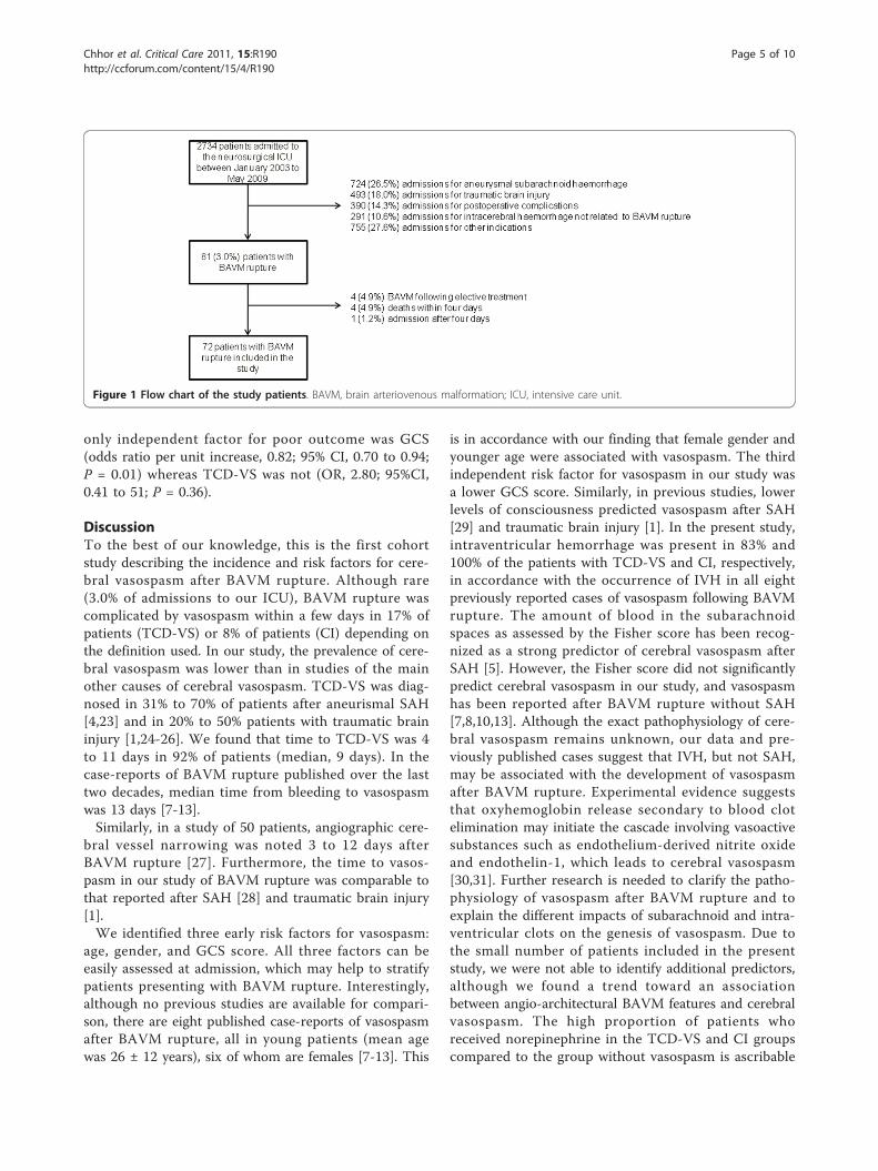
ResultsCohort descriptionFigure 1 shows the patient flowchart. During the seven-year study period, 2,734 patients were admitted to ourneurosurgical ICU including 81 (3.0%) with BAVM rup-ture. Of these 81 patients, 9 were excluded, for the fol-lowing reasons: BAVM rupture during electivetreatment (n = 4), death before Day 4 (n = 4), or admis-sion more than four days after BAVM rupture (n = 1).This left 72 patients for the study.Of the 72 study patients, 12 (17%) had TCD-VS and 6
(8%) had CI. DSA was performed in 4 of the 12 TCD-VS patients and showed diffuse vasospasm in all of
them. Figure 2 shows an example of BAVM and angio-graphic cerebral vasospasm associated with CI.Median time from BAVM rupture to TCD-VS diagno-
sis was nine days (IQR, 4 to 11). Figure 3 reports thecumulative incidence of TCD-VS according to timefrom BAVM rupture to TCD-VS diagnosis. All patientswith CI had a previous diagnosis of TCD-VS.
Admission risk factors for vasospasmTable 1 lists the admission features and ICU outcomesof patients diagnosed with TCD-VS and CI. The risk ofdeveloping TCD-VS was greater in young patients (P =0.05) and in patients with GCS scores <8 (P < 0.01).Neither TCD-VS nor CI was significantly associatedwith the amount of subarachnoid blood, intraventricularhemorrhage, or intracerebral hematoma. SAH was diag-nosed in 32 (44%) of the 72 study patients, including 7(58%) of the 12 patients with TCD-VS and 3 (50%) ofthe 6 patients with CI. IVH was present in 54% of thepatients. More specifically, isolated IVH was diagnosedin 7 of the 72 patients, including 6 of the 60 patientswithout vasospasm and 1 patient with TCD-VS and CI.Neither TCD-VS nor CI was associated with BAVMlocation, angioarchitectural features or treatment modal-ities. The risk of developing CI was greater in patientswith GCS scores <8 (P = 0.03). Details on the clinicaland radiological features and treatment of each patientwith cerebral vasospasm are given in Table 2.By multivariate analysis, three factors were associated
with TCD-VS, namely, a worse GCS score, female gen-der, and younger age (Table 3). The final model hadgood discrimination (c-statistic = 0.82) and calibration(Hosmer-Lemeshow statistic P-value = 0.16). The inter-nal validation procedure showed good robustness of thefinal model (optimism = 0.04).
Consequences of cerebral vasospasmPatients with TCD-VS had significantly fewer ICU-freedays. Of the 72 study patients, 50 had poor outcomes(GOS 1, 2 or 3) at ICU discharge. The poor outcomewas directly ascribable to the initial bleed in 42 patients,to CI in 6 patients, and to re-bleeding in 2 patients. Therisk of a poor outcome was non-significantly increasedin the patients who developed TCD-VS (relative risk,RR, 4.9; 95% confidence interval 95% CI, 0.7 to 35; P =0.09). All six patients with CI had poor outcomes. Inthe patients without vasospasm, ICU mortality was 17%and causes of death were as follows: initial bleed (n =5), refractory intracranial hypertension (n = 3), and re-bleeding (n = 2). Of the patients with TCD-VS, 42%died and death was considered directly related to CI intwo patients, intracranial hypertension in two patients,and the initial bleed in one patient (Table 2). Usinglogistic regression including age, GSC, TCD and CI, the
Chhor et al. Critical Care 2011, 15:R190http://ccforum.com/content/15/4/R190
Page 3 of 10

Table 1 Associations linking transcranial Doppler vasospasm and cerebral infarction to admission characteristics andoutcomes
All Patients N = 72 TCD-VS N = 12 CI N = 6
N (%) RR (95% CI) P N (%) RR (95% CI) P
Demographics and medical history
Age (years) 40 +/- 9 33 +/-10 - 0.05 38 +/- 13 - 0.98
Female sex 29 (40%) 7 (58%) 2.1 (0.7 to 5.9) 0.20 4 (67%) 2.9 (0.6 to 15.1) 0.22
Smokers 18 (25%) 1 (8%) 0.3 (0.03 to 2.0) 0.27 1 (17%) 0.6 (0.07 to 4.8) 0.99
Hypertension 8 (11%) 0 (0%) - 0.34 0 (0%) - 0.99
Diabetes 2 (3%) 0 (0%) - 0.99 0 (0%) - 0.99
Admission features
GCS score <8 28 (39%) 9 (75%) 4.7 (1.6 to 26) 0.001 5 (83%) 7.8 (0.1 to 63) 0.03
Seizure 9 13%) 2 (17%) 1.4 (0.4 to 5.4) 0.63 2 (33%) 3.5 (0.7 to 16.4) 0.16
Troponin elevation 18 (25%) 4 (33%) 1.5 (0.5 to 4.4) 0.48 1 (17%) 0.6 (0.07 to 4.8) 0.99
EKG abnormality 2 (3%) 1 (8%) 3.2 (0.7 to 14) 0.31 0 (0%) - 0.99
ICH 64 (89%) 10 (83%) 0.6 (0.2 to 2.4) 0.61 4 (67%) 0.2 (0.05 to 1.2) 0.14
IVH 54 (75%) 10 (83%) 1.7 (0.4 to 6.9) 0.72 6 (100%) - 0.33
SAH 32 (44%) 7 (58%) 1.7 (0.6 to 5.0) 0.34 3 (50%) 1.2 (0.3 to 5.8) 0.99
Diffuse 14 (19%) 4 (33%) 1 (17%)
Focal 18 (25%) 3 (25%) - 0.40 2 (33%) - 0.85
Absent 40 (56%) 5 (42%) 3 (50%)
BAVM characteristics and treatment
BAVM location
Frontal 25 (35%) 5 (43%) 4 (67%)
Temporal 10 (14%) 0 (0%) 0 (0%)
Parietal 15 (21%) 4 (33%) - 0.31 1 (17%) - 0.77
Occipital 8 (11%) 0 (0%) 0 (0%)
Posterior fossa 14 (19%) 3 (25% 1 (17%)
Size of BAVM*
<3 cm 43 (63%) 7 (58%) 5 (83%)
3 to 6 cm 19 (28%) 3 (25%) - 0.52 0 (0%) - 0.29
>6 cm 6 (8%) 2 (17%) 1 (17%)
Adjacent to eloquent brain * 50 (69%) 7 (58%) 0.5 (0.2 to 1.4) 0.27 3 (50%) 0.4 (0.07; 1.6) 0.32
Deep venous drainage* 31 (43%) 7 (58%) 1.7 (0.5 to 4.7) 0.36 4 (67%) 2.4 (0.5 to 12) 0.40
Co-existing aneurysm*
None 50 (73%) 11 (92%) 5 (83%)
Intranidal 8 (12%) 1 (8%) - 0.99 1 (17%) - 0.99
Feeding vessel 8 (12%) 0 (0%) 0 (0%)
Willis 2 (3%) 0 (0%) 0 (0%)
Surgical resection 35 (49%) 6 (50%) 1.0 (0.3 to 2.8) 0.99 2 (33%) 0.5 (0.1 to 2.5) 0.67
Embolization 15 (20%) 2 (17%) 0.7 (0.2 to 2.9) 0.99 1 (17%) 0.7 (0.1 to 5.6) 0.99
Mixed 6 (8%) 0(0%) to 0.58 0 (0%) - 0.99
Complete treatment 34 (47%) 5 (42%) 1.6 (0.5 to 4.4) 0.53 3 (50%) 1.2 (0.2 to 4.2) 0.99
Early treatment 43 (60%) 8 (67%) 1.4 (0.4 to 4.1) 0.75 3 (50%) 0.7 (0.1 to 3.1) 0.67
ICU outcomes
Norepinephrine 46 (64%) 11 (92%) 6.2 (0.8 to 45) 0.04 6 (100%) - 0.08
Intracranial hypertension 41 (57%) 10 (83%) 3.7 (0.9 to 16) 0.06 6 (100%) 3.8 (0.5 to 31) 0.22
Hypothermia 15 (21%) 4 (33%) 1.9 (0.7 to 5.5) 0.26 3 (50%) 3.8 ( 0.8 to 17) 0.10
CSF drainage 42 (59%) 10 (83%) 3.5 (0.8 to 15) 0.10 6 (100%) - 0.04
ICU LOS 28 (12 to 38) 34 (17 to 41) - 0.09 27 (14 to 36) - 0.74
ICU-free days 6 (0 to 28) 1 (0 to 3) - 0.01 0 (0 to 2) - 0.06
Unfavorable outcome (GOS 1-3) 50 (69%) 11 (92%) 4.9 (0.7 to 35) 0.09 6 (100%) - 0.17
ICU mortality 15 (21%) 5 (42%) 2.7 (1.0 to 7.3) 0.11 3 (50%) 3.8 (0.8 to 17) 0.10
BAVM, brain arteriovenous malformation; CI, cerebral infarction, CSF, cerebrospinal fluid; GCS, Glasgow Coma Scale; ICH, intracerebral hemorrhage; ICU, intensivecare unit; IVH, intraventricular hemorrhage; LOS, length of stay; RR (95%CI), relative risk with the 95% confidence interval; SAH, subarachnoid hemorrhage; TCD-VS, transcranial Doppler vasospasm. * n = 68 patients (data missing in four patients without vasospasm)
Values are median (IQR) for quantitative variables and N (%) for qualitative variables.
Chhor et al. Critical Care 2011, 15:R190http://ccforum.com/content/15/4/R190
Page 4 of 10
only independent factor for poor outcome was GCS(odds ratio per unit increase, 0.82; 95% CI, 0.70 to 0.94;P = 0.01) whereas TCD-VS was not (OR, 2.80; 95%CI,0.41 to 51; P = 0.36).
DiscussionTo the best of our knowledge, this is the first cohortstudy describing the incidence and risk factors for cere-bral vasospasm after BAVM rupture. Although rare(3.0% of admissions to our ICU), BAVM rupture wascomplicated by vasospasm within a few days in 17% ofpatients (TCD-VS) or 8% of patients (CI) depending onthe definition used. In our study, the prevalence of cere-bral vasospasm was lower than in studies of the mainother causes of cerebral vasospasm. TCD-VS was diag-nosed in 31% to 70% of patients after aneurismal SAH[4,23] and in 20% to 50% patients with traumatic braininjury [1,24-26]. We found that time to TCD-VS was 4to 11 days in 92% of patients (median, 9 days). In thecase-reports of BAVM rupture published over the lasttwo decades, median time from bleeding to vasospasmwas 13 days [7-13].Similarly, in a study of 50 patients, angiographic cere-
bral vessel narrowing was noted 3 to 12 days afterBAVM rupture [27]. Furthermore, the time to vasos-pasm in our study of BAVM rupture was comparable tothat reported after SAH [28] and traumatic brain injury[1].We identified three early risk factors for vasospasm:
age, gender, and GCS score. All three factors can beeasily assessed at admission, which may help to stratifypatients presenting with BAVM rupture. Interestingly,although no previous studies are available for compari-son, there are eight published case-reports of vasospasmafter BAVM rupture, all in young patients (mean agewas 26 ± 12 years), six of whom are females [7-13]. This
is in accordance with our finding that female gender andyounger age were associated with vasospasm. The thirdindependent risk factor for vasospasm in our study wasa lower GCS score. Similarly, in previous studies, lowerlevels of consciousness predicted vasospasm after SAH[29] and traumatic brain injury [1]. In the present study,intraventricular hemorrhage was present in 83% and100% of the patients with TCD-VS and CI, respectively,in accordance with the occurrence of IVH in all eightpreviously reported cases of vasospasm following BAVMrupture. The amount of blood in the subarachnoidspaces as assessed by the Fisher score has been recog-nized as a strong predictor of cerebral vasospasm afterSAH [5]. However, the Fisher score did not significantlypredict cerebral vasospasm in our study, and vasospasmhas been reported after BAVM rupture without SAH[7,8,10,13]. Although the exact pathophysiology of cere-bral vasospasm remains unknown, our data and pre-viously published cases suggest that IVH, but not SAH,may be associated with the development of vasospasmafter BAVM rupture. Experimental evidence suggeststhat oxyhemoglobin release secondary to blood clotelimination may initiate the cascade involving vasoactivesubstances such as endothelium-derived nitrite oxideand endothelin-1, which leads to cerebral vasospasm[30,31]. Further research is needed to clarify the patho-physiology of vasospasm after BAVM rupture and toexplain the different impacts of subarachnoid and intra-ventricular clots on the genesis of vasospasm. Due tothe small number of patients included in the presentstudy, we were not able to identify additional predictors,although we found a trend toward an associationbetween angio-architectural BAVM features and cerebralvasospasm. The high proportion of patients whoreceived norepinephrine in the TCD-VS and CI groupscompared to the group without vasospasm is ascribable
Figure 1 Flow chart of the study patients. BAVM, brain arteriovenous malformation; ICU, intensive care unit.
Chhor et al. Critical Care 2011, 15:R190http://ccforum.com/content/15/4/R190
Page 5 of 10

to our policy of inducing arterial blood pressure eleva-tion in patients diagnosed with vasospasm, as part of“triple-H” therapy.Although evidence is lacking that treatments such as
triple-H therapy, transluminal balloon angioplasty, orselective intraarterial vasodilator infusion are effective inSAH patients, these strategies are commonly used inthis group of patients. Based on the current data itwould be worthwhile to investigate the efficacy of thesetreatments in patients with ruptured BAVMs. No clearrecommendations about arterial blood pressure manage-ment after BAVM treatment are available. Hyperemiahas been documented after BAVM treatment. Althoughthe underlying mechanism seems unrelated to systemic
hemodynamic changes [32], induced moderate hyperten-sion may cause cerebral and systemic complications.Nevertheless, preventive strategies, such as nimodipine,might deserve evaluation in patients with BAVM rup-ture who are at high risk for vasospasm.CI has been identified as the only outcome predictor
in patients with SAH [4]. In the present study, TCD-VSand CI were associated with a non-significant increasein the risk of poor outcomes. Although the 21% deathrate found in our study is close to the 18% 30-day ratereported by Brown et al. [33], it was lower than the 29%rate found by the same group in patients with previouslyuntreated BAVM [34]. Nevertheless, the Colombiagroup found a lower mortality rate [35] and anotherstudy found no mortality at all [36]. Furthermore, in adefined-population study, the case-fatality rate inpatients younger than 60 years was about 10% [37]. Thecomparatively high mortality rate in our patients may beascribable to differences in severity at admission. In ourseries, all the patients required ICU admission and 40%were comatose. Since no high-level evidence exists con-cerning the management of unruptured BAVM, hetero-geneity in the treatment methods may contribute toexplain mortality rate differences across studies. Theongoing Randomized Trial of Unruptured Brain Arterio-venous Malformations (ARUBA) [38] comparing treat-ment versus conservative management of unrupturedBAVM can be expected to provide answers on this lastpoint.Unfortunately, the number of patients included was
too small to determine whether TCD-VS and CI wereindependently associated with a poor outcome. Severaldefinitions of cerebral vasospasm are commonly used,including TCD velocity elevation above 120 cm/s,
Figure 2 Cerebral digital subtraction angiography in a patientwith vasospasm after rupture of a BAVM. Cerebral digitalsubtraction angiography performed 11 days after intraventricularhemorrhage in a 33-year-old patient with two brain arteriovenousmalformations (BAVMs): a ruptured BAVM in the right frontal lobeand an intact BAVM in the left temporal lobe. Right internal carotidartery (ICA) injection, anteroposterior (AP) (A) and lateral (B) views:severe vasospasm of the M1 segment of the right middle cerebralartery (black arrow) and terminal right internal carotid artery (blackarrowhead). Small frontal BAVM (double white arrow). Left ICAinjection, AP view (C): severe vasospasm of the M1 segment (blackarrow). The other BAVM is visible in the temporal lobe (triple whitearrow). Cerebral CT scan (D): vasospasm-associated cerebralinfarction in both the left middle and the left anterior cerebralarteries (**). Note the remnant of the intraventricular hemorrhage(white arrow).
Figure 3 Cumulative incidence of transcranial Dopplervasospasm in patients with ruptured brain arteriovenousmalformations.
Chhor et al. Critical Care 2011, 15:R190http://ccforum.com/content/15/4/R190
Page 6 of 10

Table 2 Cases of cerebral vasospasm after rupture of a BAVM, adapted from Yanaka et al. [3] and Gerard et al. [7]
Clinical and radiologicalfeatures
BAVM angioarchitectural features BAVM treatment Vasospasm features and management Outcome
# Sex GCS SAH IVH ICH Location Venousdrainage
Size(mm)
Eloquence SpetzlerMartinGrade
Co-existinganeurism
BAVMtreatment
Exclusion Time totreatment
Time toonset(days)
TCD Meanflow
velocity(cm/s)
TCD-VSartery
CIlocation
Medicaltreatment
Endovasculartreatment
GOS Causes ofdeath
1 M 4 A Y N R. frontal Superf. <30 N 1 N None - - 11 200 B MCA L ACA LMCA
Nimodipine IA Nimodipine 1 Cerebralischemia
2 F 3 F Y Y R.temporal
Superf. 30 to60
N 2 N Surgery Total <24 h 9 160 B MCA None None 2 -
3 M 3 F Y Y R. frontal Deep >60 Y 5 N None - - 10 150 B MCA R MCAR ACA LMCA
None None 1 Intracranialhypertension
4 F 3 D Y Y Cerebellum Superf. <30 Y 2 N Surgery Total <24 h 6 169 B MCAR ACA LVA, BA
None None 1 Intracranialhypertension
5 M 10 A N Y Cerebellum Superf. <30 Y 2 N Surgery Total <24 h 4 150 B MCAB ACA
Nimodipine None 2 -
6 M 4 D Y Y R. parietal Deep 30 to60
N 3 N Surgery Total <24 h 7 222 L MCA Nimodipine IA NimodipineIA Milrinone
1 Initial bleed
7 F 7 A Y Y L. parietal Deep <30 N 2 Intranidal None - - 13 165 B MCA R MCA Nimodipine None 3 -
8 F 7 F Y Y Posteriorcranialfossa
Superf. <30 N 1 N Surgery Total <24 h 11 162 B MCA B MCAB PCA
None IA NimodipineIA Milrinone
1 Cerebralischemia
9 F 13 D Y N L. frontal Deep <30 Y 3 N Embolization Total 5 d 9 160 R MCA R MCARACA
Nimodipine IA NimodipineIA Milrinone
2 -
10 F 5 A Y Y R. frontal Deep <30 Y 3 N Surgery Total <24 h 4 194 B MCAR ACA
L MCA Nimodipine None 3 -
11 F 3 D Y Y R. frontal Deep >60 Y 5 N Embolization Incom-plete
<24 h 11 185 B MCAB ACA
Nimodipine None 5 -
12 M 8 A N Y R. parietal Deep 30 to60
Y 4 N None - - 4 217 R MCA Nimodipine None 3 -
A, absent; ACA, anterior cerebral artery; B, bilateral; BA, basilar artery; BAVM, brain arteriovenous malformation; CI, cerebral infarction; D, diffuse; F, focal; GCS, Glasgow Coma Scale; GOS, Glasgow Outcome Scale; IA,intraarterial; ICA, internal carotid artery; ICH, intracerebral hemorrhage; IVH, intraventricular hemorrhage; L, left; MCA, middle cerebral artery; N, no; PCA, posterior cerebral artery; R, right; SAH, subarachnoidhemorrhage; Superf, superficial; TCD, transcranial Doppler; TCD-VS, vasospasm detected using transcranial Doppler; VA, vertebral artery; Y, yes.
Chhor
etal.CriticalCare
2011,15:R190http://ccforum
.com/content/15/4/R190
Page7of
10

symptomatic vasospasm, angiographic vasospasm, andCI diagnosed by CT or MRI. TCD is a well-validatedtool for detecting vasospasm [39,40] with acceptablepositive and negative predictive values for angiographicvasospasm but low sensitivity for predicting the neurolo-gical outcome [41]. Moreover, intra- and inter-observervariability is of concern and should be taken intoaccount when interpreting velocity changes over time.Nevertheless, intra-observer bias may be minimized byhaving the same neurointensivist perform all TCDinvestigations in a given patient [42], as was the case inthe present study. Furthermore, mean flow velocitieswere well above 120 cm/s, and using a higher cut-offpoint of 150 cm/s would not have changed our results.In our study, TCD was performed in unconsciouspatients and in awake patients with symptoms. Sincenot all patients with vasospasm have symptoms, thisapproach may have underestimated the true incidenceof TCD-VS. Angiographic vasospasm was not consid-ered in our study. The absence of recommendationsabout vasospasm management after BAVM rupture andthe poor clinical condition of some patients precludingtransport to the radiology department explain that DSAwas not performed routinely. This weakness of ourstudy is mitigated by the good reported correlationbetween TCD and DSA [39] for vasospasm assessment.Furthermore, DSA may require general anesthesia and isassociated with a small risk of procedure-related stroke.Finally, no treatment recommendations are available forBAVM rupture with vasospasm. Conceivably, the localcerebral blood flow modifications induced by intra-arterial treatments may lead to re-bleeding, especiallywhen the BAVM has not been secured.There are several limitations to our study. First, we
used a retrospective design in a small number ofpatients from a single center. However, given the preva-lence of BAVM of only about 0.01% in the generalpopulation [43], a retrospective design was appealing toensure study completion within a reasonable timeframe.Second, our data from a single center may not apply toall other centers. Third, vasospasm following BAVMrupture is rare and, consequently, our sample size wassmall, limiting the statistical power of our study, whichmay have led us to miss a number of risk factors. More-over, the number of patients included in the presentstudy was too small to investigate properly whether
TCD-VS and CI were independent predictors of a pooroutcome. This crucial point will have to be determinedin a larger study. In addition, the number of patientswith CI was also too small for a separate multivariateanalysis of risk factors for this event.
ConclusionsAfter BAVM rupture, TCD-VS occurred in 17% ofpatients and CI in 8%. Admission risk factors for TCD-VS were low GCS, younger age, and female gender. Anon-significant trend toward poorer outcome exists inpatients with TDC-VS and CI. Prospective, multicenterstudies are needed to further assess the incidence andsignificance of vasospasm and CI after BAVM ruptureand to identify additional predictors.
Key messages• Transcranial Doppler cerebral vasospasm is a com-mon complication following brain arteriovenousrupture.• Cerebral vasospasm in brain arteriovenous ruptureis associated with low GCS, young age, and femalegender.
AbbreviationsARUBA: Randomized Trial of Unruptured Brain Arteriovenous Malformations;BAVM: brain arteriovenous malformation; CI: cerebral infarction; CT:Computed Tomography; GCS: Glasgow Coma Scale; GOS: Glasgow OutcomeScale; DSA: Digital Subtraction Angiography; ICP: intracranial pressure; ICU:intensive care unit; IVH: intraventricular hemorrhage; IQR: interquartile range;MRI: Magnetic Resonance Imaging; OR: odds ratio; RR: relative risk; SAH:subarachnoid hemorrhage; STROBE: Strengthening the Reporting ofObservational Studies in Epidemiology; TCD-VS: Transcranial Dopplervasospasm.
AcknowledgementsThe authors are very grateful to A. Wolfe, MD, for helping to prepare themanuscript.Funding was provided solely from institutional and departmental sources(Assistance Publique - Hôpitaux de Paris).
Author details1Department of Anesthesiology and Critical Care, Groupe Hospitalier Pitié-Salpêtrière, Assistance Publique-Hôpitaux de Paris, Université Pierre et MarieCurie- Paris 6, 47-83 boulevard de l’hôpital, Paris, 75651, France. 2Centre forStatistics in Medicine - Wolfson College, University of Oxford, Barton Road,Cambridge CB3 9BB, UK. 3Department of Neuroradiology, Groupe HospitalierPitié-Salpêtrière, Assistance Publique-Hôpitaux de Paris, Université Pierre etMarie Curie- Paris 6, 47-83 boulevard de l’hôpital, Paris, 75651, France.4Department of Neurosurgery, Groupe Hospitalier Pitié-Salpêtrière, AssistancePublique-Hôpitaux de Paris, Université Pierre et Marie Curie- Paris 6, 47-83boulevard de l’hôpital, Paris, 75651, France.
Table 3 Factors present at admission and associated with transcranial Doppler vasospasm by multivariate analysis
Odds ratio 95% confidence interval P-value
Glasgow Coma Scale (per unit decrease) 1.38 (1.13 to 1.80) 0.005
Female gender 4.86 (1.09 to 25.85) 0.04
Age (per 10-year decrease) 1.39 (1.05 to 1.82) 0.01
C-statistic = 0.82; 10-fold cross-validation optimism = 0.03; corrected c-statistic = 0.79; Hosmer-Lemeshow statistic = 1.39; P = 0.16 (df = 9).
Chhor et al. Critical Care 2011, 15:R190http://ccforum.com/content/15/4/R190
Page 8 of 10

Authors’ contributionsVC acquired the data and drafted the manuscript. YLM performed thestatistical analysis and helped to draft the manuscript. FC, AN and LAparticipated in the study design and helped to draft the manuscript. JLDhelped to acquire the data and to draft the manuscript. LP conceived anddesigned the study and helped to draft the manuscript. TL conceived anddesigned the study, acquired the data, and wrote the manuscript. Allauthors read and approved the final manuscript.
Competing interestsThe authors declare that they have no competing interests.
Received: 20 March 2011 Revised: 25 June 2011Accepted: 10 August 2011 Published: 10 August 2011
References1. Oertel M, Boscardin WJ, Obrist WD, Glenn TC, McArthur DL, Gravori T,
Lee JH, Martin NA: Posttraumatic vasospasm: the epidemiology, severity,and time course of an underestimated phenomenon: a prospectivestudy performed in 299 patients. J Neurosurg 2005, 103:812-824.
2. Aoki N, Origitano TC, al-Mefty O: Vasospasm after resection of skull basetumors. Acta Neurochir (Wien) 1995, 132:53-58.
3. Claassen J, Bernardini GL, Kreiter K, Bates J, Du YE, Copeland D, Connolly ES,Mayer SA: Effect of cisternal and ventricular blood on risk of delayedcerebral ischemia after subarachnoid hemorrhage: the Fisher scalerevisited. Stroke 2001, 32:2012-2020.
4. Frontera JA, Fernandez A, Schmidt JM, Claassen J, Wartenberg KE,Badjatia N, Connolly ES, Mayer SA: Defining vasospasm after subarachnoidhemorrhage: what is the most clinically relevant definition? Stroke 2009,40:1963-1968.
5. Fisher CM, Kistler JP, Davis JM: Relation of cerebral vasospasm tosubarachnoid hemorrhage visualized by computerized tomographicscanning. Neurosurgery 1980, 6:1-9.
6. Parkinson D, Bachers G: Arteriovenous malformations. Summary of 100consecutive supratentorial cases. J Neurosurg 1980, 53:285-299.
7. Gerard E, Frontera JA, Wright CB: Vasospasm and cerebral infarctionfollowing isolated intraventricular hemorrhage. Neurocrit Care 2007,7:257-259.
8. Kobayashi M, Takayama H, Mihara B, Kawase T: Severe vasospasm causedby repeated intraventricular haemorrhage from small arteriovenousmalformation. Acta Neurochir (Wien) 2002, 144:405-406.
9. Kothbauer K, Schroth G, Seiler RW, Do DD: Severe symptomaticvasospasm after rupture of an arteriovenous malformation. AJNR Am JNeuroradiol 1995, 16:1073-1075.
10. Maeda K, Kurita H, Nakamura T, Usui M, Tsutsumi K, Morimoto T, Kirino T:Occurrence of severe vasospasm following intraventricular hemorrhagefrom an arteriovenous malformation. Report of two cases. J Neurosurg1997, 87:436-439.
11. Yokobori S, Watanabe A, Nakae R, Onda H, Fuse A, Kushimoto S, Yokota H:Cerebral vasospasms after intraventricular hemorrhage from anarteriovenous malformation: case report. Neurol Med Chir (Tokyo) 2010,50:320-323.
12. Pendharkar AV, Guzman R, Dodd R, Cornfield D, Edwards MS: Successfultreatment of severe cerebral vasospasm following hemorrhage of anarteriovenous malformation. Case report. J Neurosurg Pediatr 2009,4:266-269.
13. Yanaka K, Hyodo A, Tsuchida Y, Yoshii Y, Nose T: Symptomatic cerebralvasospasm after intraventricular hemorrhage from rupturedarteriovenous malformation. Surg Neurol 1992, 38:63-67.
14. Zwienenberg-Lee M, Hartman J, Rudisill N, Muizelaar JP: Endovascularmanagement of cerebral vasospasm. Neurosurgery 2006, 59:S139-147,discussion S3-13.
15. von Elm E, Altman DG, Egger M, Pocock SJ, Gotzsche PC,Vandenbroucke JP: Strengthening the Reporting of Observational Studiesin Epidemiology (STROBE) statement: guidelines for reportingobservational studies. BMJ 2007, 335:806-808.
16. Spetzler RF, Martin NA: A proposed grading system for arteriovenousmalformations. J Neurosurg 1986, 65:476-483.
17. Lysakowski C, Walder B, Costanza MC, Tramer MR: Transcranial Dopplerversus angiography in patients with vasospasm due to a rupturedcerebral aneurysm: A systematic review. Stroke 2001, 32:2292-2298.
18. Suarez JI, Qureshi AI, Yahia AB, Parekh PD, Tamargo RJ, Williams MA,Ulatowski JA, Hanley DF, Razumovsky AY: Symptomatic vasospasmdiagnosis after subarachnoid hemorrhage: evaluation of transcranialDoppler ultrasound and cerebral angiography as related tocompromised vascular distribution. Crit Care Med 2002, 30:1348-1355.
19. Lindegaard KF, Nornes H, Bakke SJ, Sorteberg W, Nakstad P: Cerebralvasospasm after subarachnoid haemorrhage investigated by means oftranscranial Doppler ultrasound. Acta Neurochir Suppl (Wien) 1988,42:81-84.
20. Vergouwen MD, Vermeulen M, van Gijn J, Rinkel GJ, Wijdicks EF,Muizelaar JP, Mendelow AD, Juvela S, Yonas H, Terbrugge KG,Macdonald RL, Diringer MN, Broderick JP, Dreier JP, Roos YB: Definition ofdelayed cerebral ischemia after aneurysmal subarachnoid hemorrhageas an outcome event in clinical trials and observational studies: proposalof a multidisciplinary research group. Stroke 2011, 41:2391-2395.
21. Molinaro AM, Simon R, Pfeiffer RM: Prediction error estimation: acomparison of resampling methods. Bioinformatics 2005, 21:3301-3307.
22. R Development Core Team: R: A Language and Environment forStatistical Computing. R Foundation for Statistical Computing Vienna,Austria; 2011 [http://www.r-project.org].
23. Kassell NF, Sasaki T, Colohan AR, Nazar G: Cerebral vasospasm followinganeurysmal subarachnoid hemorrhage. Stroke 1985, 16:562-572.
24. Martin NA, Doberstein C, Zane C, Caron MJ, Thomas K, Becker DP:Posttraumatic cerebral arterial spasm: transcranial Doppler ultrasound,cerebral blood flow, and angiographic findings. J Neurosurg 1992,77:575-583.
25. Grolimund P, Weber M, Seiler RW, Reulen HJ: Time course of cerebralvasospasm after severe head injury. Lancet 1988, 1:1173.
26. Compton JS, Teddy PJ: Cerebral arterial vasospasm following severe headinjury: a transcranial Doppler study. Br J Neurosurg 1987, 1:435-439.
27. von Holst H, Ericson K, Haberbeck-Modesto M, Steiner L: Angiographicinvestigation of cerebral vasospasm in subarachnoid haemorrhage dueto arteriovenous malformation. Acta Neurochir (Wien) 1988, 94:129-132.
28. Weir B, Grace M, Hansen J, Rothberg C: Time course of vasospasm in man.J Neurosurg 1978, 48:173-178.
29. Gonzalez NR, Boscardin WJ, Glenn T, Vinuela F, Martin NA: Vasospasmprobability index: a combination of transcranial doppler velocities,cerebral blood flow, and clinical risk factors to predict cerebralvasospasm after aneurysmal subarachnoid hemorrhage. J Neurosurg2007, 107:1101-1112.
30. Sen J, Belli A, Albon H, Morgan L, Petzold A, Kitchen N: Triple-H therapy inthe management of aneurysmal subarachnoid haemorrhage. LancetNeurol 2003, 2:614-621.
31. Suhardja A: Mechanisms of disease: roles of nitric oxide and endothelin-1 in delayed cerebral vasospasm produced by aneurysmal subarachnoidhemorrhage. Nat Clin Pract Cardiovasc Med 2004, 1:110-116, quiz 2, pagefollowing 116.
32. Hashimoto T, Young WL, Prohovnik I, Gupta DK, Ostapkovich ND,Ornstein E, Halim AX, Quick CM: Increased cerebral blood flow after brainarteriovenous malformation resection is substantially independent ofchanges in cardiac output. J Neurosurg Anesthesiol 2002, 14:204-208.
33. Brown RD Jr, Wiebers DO, Torner JC, O’Fallon WM: Frequency ofintracranial hemorrhage as a presenting symptom and subtype analysis:a population-based study of intracranial vascular malformations inOlmsted Country, Minnesota. J Neurosurg 1996, 85:29-32.
34. Brown RD Jr, Wiebers DO, Forbes G, O’Fallon WM, Piepgras DG, Marsh WR,Maciunas RJ: The natural history of unruptured intracranial arteriovenousmalformations. J Neurosurg 1988, 68:352-357.
35. Choi JH, Mast H, Sciacca RR, Hartmann A, Khaw AV, Mohr JP, Sacco RL,Stapf C: Clinical outcome after first and recurrent hemorrhage inpatients with untreated brain arteriovenous malformation. Stroke 2006,37:1243-1247.
36. Hartmann A, Mast H, Mohr JP, Koennecke HC, Osipov A, Pile-Spellman J,Duong DH, Young WL: Morbidity of intracranial hemorrhage in patientswith cerebral arteriovenous malformation. Stroke 1998, 29:931-934.
37. van Beijnum J, Lovelock CE, Cordonnier C, Rothwell PM, Klijn CJ, Al-ShahiSalman R: Outcome after spontaneous and arteriovenous malformation-related intracerebral haemorrhage: population-based studies. Brain 2009,132:537-543.
38. Mohr JP, Moskowitz AJ, Stapf C, Hartmann A, Lord K, Marshall SM, Mast H,Moquete E, Moy CS, Parides M, Pile-Spellman J, Al-Shahi Salman R,
Chhor et al. Critical Care 2011, 15:R190http://ccforum.com/content/15/4/R190
Page 9 of 10
Weinberg A, Young WL, Estevez A, Kureshi I, Brisman JL: The ARUBA trial:current status, future hopes. Stroke 2010, 41:e537-540.
39. Sloan MA, Alexandrov AV, Tegeler CH, Spencer MP, Caplan LR, Feldmann E,Wechsler LR, Newell DW, Gomez CR, Babikian VL, Lefkowitz D, Goldman RS,Armon C, Hsu CY, Goodin DS: Assessment: transcranial Dopplerultrasonography: report of the Therapeutics and Technology AssessmentSubcommittee of the American Academy of Neurology. Neurology 2004,62:1468-1481.
40. Bederson JB, Connolly ES Jr, Batjer HH, Dacey RG, Dion JE, Diringer MN,Duldner JE Jr, Harbaugh RE, Patel AB, Rosenwasser RH: Guidelines for themanagement of aneurysmal subarachnoid hemorrhage: a statement forhealthcare professionals from a special writing group of the StrokeCouncil, American Heart Association. Stroke 2009, 40:994-1025.
41. Carrera E, Schmidt JM, Oddo M, Fernandez L, Claassen J, Seder D, Lee K,Badjatia N, Connolly ES Jr, Mayer SA: Transcranial Doppler for predictingdelayed cerebral ischemia after subarachnoid hemorrhage. Neurosurgery2009, 65:316-323, discussion 323-324.
42. McMahon CJ, McDermott P, Horsfall D, Selvarajah JR, King AT, Vail A: Thereproducibility of transcranial Doppler middle cerebral artery velocitymeasurements: implications for clinical practice. Br J Neurosurg 2007,21:21-27.
43. Friedlander RM: Clinical practice. Arteriovenous malformations of thebrain. N Engl J Med 2007, 356:2704-2712.
doi:10.1186/cc10345Cite this article as: Chhor et al.: Admission risk factors for cerebralvasospasm in ruptured brain arteriovenous malformations: Anobservational study. Critical Care 2011 15:R190.
Submit your next manuscript to BioMed Centraland take full advantage of:
• Convenient online submission
• Thorough peer review
• No space constraints or color figure charges
• Immediate publication on acceptance
• Inclusion in PubMed, CAS, Scopus and Google Scholar
• Research which is freely available for redistribution
Submit your manuscript at www.biomedcentral.com/submit
Chhor et al. Critical Care 2011, 15:R190http://ccforum.com/content/15/4/R190
Page 10 of 10