research article profiling of selected micrornas in...
TRANSCRIPT

Research ArticleProfiling of Selected MicroRNAs in Proliferative EutopicEndometrium of Women with Ovarian Endometriosis
P Laudanski1 R Charkiewicz2 A Tolwinska3 J Szamatowicz4
A Charkiewicz5 and J Niklinski2
1Department of Perinatology Medical University of Bialystok Ulica Marii Sklodowskiej-Curie 24a 15-276 Bialystok Poland2Department of Clinical Molecular Biology Medical University of Bialystok Ul Waszyngtona 13 15-269 Bialystok Poland3Clinic ldquoEDMEDrdquo Białystok Ul Piasta 14 15-044 Bialystok Poland4Department of Gynecology and Gynecological Oncology Medical University of Bialystok Ulica Marii Sklodowskiej-Curie 24a15-276 Bialystok Poland5Department of Medical Pathomorphology Medical University of Bialystok Ul Waszyngtona 13 15-269 Bialystok Poland
Correspondence should be addressed to P Laudanski plaudaumbedupl
Received 15 January 2015 Revised 2 April 2015 Accepted 20 April 2015
Academic Editor Shi-Wen Jiang
Copyright copy 2015 P Laudanski et al This is an open access article distributed under the Creative Commons Attribution Licensewhich permits unrestricted use distribution and reproduction in any medium provided the original work is properly cited
It has been well documented that aberrant expression of selected microRNAs (miRNAs) might contribute to the pathogenesis ofdisease The aim of the present study is to compare miRNA expression by the most comprehensive locked-nucleic acid (LNA)miRNA microarray in eutopic endometrium of patients with endometriosis and control In the study we recruited 21 patientswith endometriosis and 25 were disease-free women The miRNA expression profiles were determined using the LNA miRNAmicroarray and validated for selected molecules by real-time PCR We identified 1198 human miRNAs significantly differentiallyaltered in endometriosis versus control samples using false discovery rate of lt5 However only 136 miRNAs showed differentialregulation by fold change of at least 13 By the use of selected statistical analysis we obtained 45 potential pathways that mightplay a role in the pathogenesis of endometriosis We also found that natural killer cell mediated cytotoxicity pathway was foundto be inhibited which is consistent with previous studies There are several pathways that may be potentially dysregulated due toabnormalmiRNA expression in eutopic endometriumof patients with endometriosis and in this way contribute to its pathogenesis
1 Introduction
Endometriosis is relatively common benign gynecologicaldisease in which endometrial tissue ectopically implants ina location outside the uterine cavity The most commonclinical symptoms include pelvic pain and infertility whichcan seriously influence the quality of life of affected patients[1]
Despite the current controversy regarding the pathophys-iology of this disease Sampsonrsquos theory explains the existenceof endometrial cells in the peritoneal cavity by retrogrademenstruation Several factors such as increased chemokinesand matrix metalloproteinases in the peritoneal fluid (PF)angiogenesis tumor suppressor and oncogenes as well asupregulating of proinflammatory cytokines may facilitate thepathogenesis of endometriosis [2ndash6] It is therefore assumed
to be a complex process in many aspects characteristic ofsystemic disease It has now also become widely acceptedthat molecular aberration within the eutopic endometriummay predispose a subgroup of women to the development ofendometriosis [7]
The molecular changes that could potentially lead toabnormal growth of endometrium outside uterus includemicroRNAs (miRNAs) expression fluctuations [8] MicroR-NAs are a large class of endogenous single-stranded andshort ncRNA (noncoding) of approximately 22 nucleotidesin length that play a key role in regulating gene expressionthrough interaction with mRNA of protein-coding genes[9] miRNA expression is tissue- and cell-specific and it wasdemonstrated that miRNAs are important in endometriosisand associated reproductive conditions [10]
Hindawi Publishing CorporationBioMed Research InternationalVolume 2015 Article ID 760698 10 pageshttpdxdoiorg1011552015760698
2 BioMed Research International
In our recent study we also showed that out of 667miRNAs two molecules namely hsa-miR-483-5p and hsa-miR-629lowast are significantly downregulated in eutopic endo-metriumof patients with ovarian endometriosis which couldbe a consequence of an early defect in the physiologicalactivity of the proliferative endometrium [11] Since therehave been only few studies in the available literaturewhich concerned miRNA profiling of eutopic endometrium(actually mostly luteal) [12 13] and the technology sinceour last publication has greatly developed we decided toexpand our previous work and use more robust microarraytechnique which facilitates expression profiling of more than2000 miRNAs The aim of the present study is to examinepossible differential regulation as well as putative pathwaysthat might be regulated by abnormal miRNA expressionin the proliferative eutopic endometrium of patients withadvanced ovarian endometriosis
2 Material and Methods
Patients (119899 = 46) scheduled for laparoscopy for adnexalmass or infertility at the Medical University of Bialystok wererecruited to participate in this study Endometrial biopsieswere collected using Pipelle suction curettes Endometrialtissue samples were classified by histological dating accordingto the method of Noyes et al [14] and only patients in theproliferative phase (days from 6th to 13th) of the cycle wereincluded in the study
Patients with endometriosis (Group I 119899 = 21) stages fromIII to IV were diagnosed by laparoscopic findings accord-ing to the revised American Fertility Society classificationof endometriosis [15] and each case was confirmed byhistopathology As a control (Group II 119899 = 25) we used endo-metrial tissue from patients without any endometriosis visi-ble during laparoscopy
All women had regular menstrual cycles (28ndash30 days)and were not taking any medication for at least 3 monthsprior to operation We excluded patients with autoimmunedisease pelvic inflammatory disease adenomyosis fibroidsand dysfunctional uterine bleeding The study was approvedby the Institutional Review Board of Medical University ofBialystok and informed consent was obtained from eachpatient
The collected tissue was placed separately in buffered for-malin for histopathological studies and in RNA later (Sigma-Aldrich Poland) for molecular analysis The latter was storedfor 24 hours in +4∘C and then tissues were transferredand stored in minus80∘C Total RNA was extracted using themirVana miRNA Isolation Kit (Ambion Life TechnologiesPoland) RNA quality was assessed with Agilent Bioanalyzer2100 and Agilent RNA 6000 Nano kit (Agilent TechnologiesPerlan Poland) and samples chosen for further analysisshowed minimum sign of degradation as judged by the RNAintegrity number (RIN) which was above 9 for all samplesRNA concentrations were measured on a NanoDrop 2000c(Thermo Scientific Biotech Poland)
21 miRNA Expression Profiling Using Exiqon MicroarraysFor the purpose of miRNA microarray screening we chose
10 samples of patients with advanced endometriosis and11 controls Microarray analysis was conducted as single-channel Hy3 experiments on Exiqonrsquos miRCURY LNA(locked-nucleic acid) microRNA Array 7th generationmdashhsammu and rno Exiqon arrays contain 3100 capture probescomplementary to most human mouse rat and their relatedviral sequences from the v190 release of miRBaseThe arraysalso contain 25 proprietary human miRPlus sequences notyet in miRBase 500 ng RNA sample was labelled with a Hy3fluorophore (Exiqon Denmark) Labelling reactions wereperformed using Exiqonrsquos miRCURY LNA microRNA Hi-Power Labeling Kit with the use of synthetic spike controlsSpike-in microRNA Kit v2 (Exiqon Denmark) according tothe manufacturerrsquos protocol Hybridization of labeled RNAto the array was performed in SureHyb chambers (AgilentTechnologies USA) for 16 hours at 56∘C Slides were washedaccording to manufacturerrsquos instructions and scanned at10 120583m resolution using an Agilent G2505C DNA MicroarrayScanner Raw data were generated using Imagene 90 software(BioDiscovery Inc USA) using an FE protocol available ondemand from Exiqon
22 Quantitative PCR Real-Time RT-PCR (qPCR) Theexpression levels of the selected microRNAs and the assaymiRBase IDs (miRBase Accession Number) are hsa-miR-4714-5p (MIMAT0019822) hsa-miR-4284 (MIMAT0016915)hsa-miR-5193 (MIMAT0021124) hsa-miR-4454(MIMAT0018976) hsa-miR-3680-5p (MIMAT0018106)hsa-miR-3667-5p (MIMAT0018089) hsa-miR-23a-3p(MIMAT0000078) hsa-miR-23b-3p (MIMAT0000418) hsa-miR-5187-3p (MIMAT0021118) hsa-miR-3152-5p(MIMAT0019207) and hsa-miR-30d-5p (MIMAT0000245)which were evaluated using the miRCURY LNA UniversalRT microRNA PCR system (Exiqon Denmark) In the firststep we have conducted the one first-strand cDNA synthesisreaction which provided template for all microRNA real-time PCR assays The cDNA for each RNA sample wasobtained using the Universal cDNA Synthesis Kit II (ExiqonDenmark) according to the manufacturerrsquos instructionsLevels of miRNAs were quantitated using individual miRCURY LNA Universal RT microRNA PCR Assays (ExiqonDenmark)
The conditions for qPCR were as follows 95∘C for10min and 45 cycles of 95∘C for 10 sec followed by 60∘C for1min Finally a melting curve analysis was performed withdenaturation at 95∘C for 15 s and 60∘C for 15 s followed bya temperature gradient from 60 to 95∘C for 20min and afinal denaturation at 95∘C for 15 s U6 snRNA was used asthe endogenous control LNA PCR amplification reactionsfor eachmiRNAmolecule were repeated independently threetimes Quantitative real-time PCR analysis was performedusing the Applied Biosystems 7900HT System (Life Tech-nologies Foster City CA)
Gene expression values were calculated based on the2(minusDelta Delta C(T)) method where one sample was des-ignated the calibrator through which all other samples wereanalyzed [16] Briefly ΔCT represents the threshold cycle ofthe target minus that of U6 snRNA and ΔΔCT representsthe ΔCT of each target minus that of the calibrator Relative
BioMed Research International 3
quantities were determined using the equation RQ = 2minusΔΔCTFor the calibrator sample that is control RNA from eutopicendometrium the equation is relative quantity = 2minus0 whichis 1 therefore every other sample is expressed relative to this
23 Statistical Analysis All data analyses were performedin R statistical environment (httpwwwr-projectorg) andrelevant Bioconductor software [17]
The raw microarray data were preprocessed with vsn2()function implemented in vsn package [18] The vsn functionwas used with default settings Differentially expressed miR-NAs were identified with limma [19] In order to associatemiRNA with mRNA we used annotations from 6 databasesmiRBase targetScan miRanda tarBase mirTarget2 andpicTar Probes not associated with human mRNA were notanalyzed further The potential influence of miRNA onmRNA expression was visualized using signaling pathwaydefinitions
In our study we used signaling pathway definitions fromKEGG (Kyoto Encyclopedia of Genes and Genomes [20])The potential miRNA influence on mRNA expression wasalso used to analyze GO (gene ontology) terms potentiallyoverrepresented in disturbed genes
To assess perturbation of signaling pathways we appliedSPIA (Signaling Pathway Impact Analysis [21]) We used thepotential influence of miRNA on mRNA expression insteadof gene expression data
While comparing two groups for quantitative dataMann-Whitney-Wilcoxon test was used due to the nonnor-mal distribution of the tested variables The significance levelwas equal to 005 The calculations have been carried outby means of Microsoft Excel spreadsheet and STATISTICAStatSoft Inc Version 71 statistical package (data analysissoftware system)
3 Results
Patients clinical characteristics are presented in Table 1We identified 1198 human miRNAs significantly differen-
tially altered in endometriosis versus control samples usingfalse discovery rate of lt5 (Table I supplement in the supple-mentaryMaterial available online at httpdxdoiorg1011552015760698)
Volcano plot (Figure 1) presents resultsofexpression anal-ysis for all analyzed miRNAs while the heatmap (Figure 2)shows expression profiles only of significantly expressedmiRNAs
The associations between miRNA and mRNA are pre-sented in the zipped supplement directory (called Table IIsupplement)
We obtained 45 potential pathways from the KEGG(Kyoto Encyclopedia of Genes and Genomes) database andparticularly mTOR and VEGF signaling pathway caught ourattention due to its close potential relation to pathogenesis ofendometriosis (Figures 3 and 4 resp)
The significantly overexpressed GO (gene ontology)terms are presented in Table 2We found six potential cellularprocesses that involve protein synthesis to be potentiallyregulated by influence of miRNA on mRNA including
Table 1 Clinical characteristics of patients
Endometriosis(119899 = 21)
Control(119899 = 25)
Age (years) 3135 plusmn 090 3073 plusmn 091Infertility 119899 () 9 (50) 16 (64)Primary 7 8Secondary 2 8
Duration of infertility(months)
482 plusmn 194(23ndash71)
508 plusmn 158(13ndash71)
Ovarian cysts 119899 ()Endometrial 21 (100) mdashSimple mdash 9 (36)
Data are mean plusmn SEM Ranges are provided for ldquoduration of infertilityrdquo
log2(study groupcontrol group)
minus05 0 05 1 15
0
1
2
3
4
Volcano plot
minuslo
g 10(p
valu
e)
Figure 1 Volcano plot for all analyzed human miRNAs The 119909-axis(horizontal) is the fold change between endometriosis and controlsamples (on a log scale so that up- and downregulation appearsymmetric) and119910-axis represents the119901 value for a test of differencesbetween samples (most conveniently on a negative log scale sosmaller 119901 values appear higher up) Red dots represent miRNAssignificantly differentially expressed
translational elongation and termination protein targetingand localization to endoplasmic reticulum cotranslationalprotein targeting to membrane SRP- (signal-recognitionparticle-) dependent cotranslational protein targeting tomembrane and establishment of protein localization toendoplasmic reticulum
The SPIA showed perturbation of potentially nine path-ways in two analyzed groupsMost interestingly natural killercell mediated cytotoxicity pathway was found to be inhibitedin eutopic endometrium of patients with endometriosis ascompared with control (Table 3)
31 Validation by RT-PCR Following the selection of miR-NAs by fold change filtering (fold change gt 13) we foundthat there were 136 upregulated miRNAs and no downreg-ulated miRNAs in the eutopic endometrium of patients withadvanced ovarian endometriosis compared with the eutopicendometrium
We then validated 11 selected miRNAs but we were notable to observe clear statistical differences as to the expression
4 BioMed Research International
Table 2 Gene ontology (GO) terms overrepresented in eutopic endometrium of patients with endometriosis and control
GO termAdjusted 119901 values for overrepresentation
analysis for miRNAs with higher expressionin endometriosis
Adjusted 119901 values for overrepresentationanalysis for miRNAs with higher expression
in controlTranslational elongation 1 0077Translational termination 1 0077Cotranslational protein targeting tomembrane 1 0077
SRP- (signal-recognition particle-)dependent cotranslational proteintargeting to membrane
1 0077
Protein targeting to endoplasmicreticulum 1 0077
Protein localization to endoplasmicreticulum 1 0077
Establishment of protein localizationto endoplasmic reticulum 1 0077
The first column contains names of significantly overrepresented GO terms Both the second and the third columns contain adjusted 119901 values foroverrepresentation analysis (Fisherrsquos exact test) The second column presents results for genes associated with miRNA that have higher expression inendometriosis (of at least 20) and the third column presents results for genes associated with miRNA that present higher expression in control samples (ofat least 20) GO terms with adjusted 119901 values lt025 were recognized as significantly overrepresented
0 4
s s s s s s c c c c cs s s s s c c c c c
minus4
Figure 2 Heatmap display of significantly expressed miRNAsHeatmap representation of expression data formiRNAs significantlyaltered in endometriosis samples compared with control samples(adjusted 119901 value lt 005) Columns correspond to samples and rowscorrespond to individual miRNAs For a given miRNA an averagevalue was computed and subtracted from each observation Theyellow color marks a higher expression (over average expressionin two groups) while the blue color represents a lower expression(again with regard to average expression)The color scale representsthe magnitude of changes ldquosrdquo stands for endometriosis samples andldquocrdquo stands for control samples
of any of the chosen molecule (Table 4) between studiedgroups We found that specifically for three miRNAs that
Table 3 Signaling Pathway Impact Analysis (SPIA) showing per-turbation of signaling pathways in eutopic endometrium of patientswith endometriosis versus control
The name of the pathway Adjusted 119901values Status
Alcoholism 0 AOlfactory transduction 0 AViral carcinogenesis 0 ISystemic lupus erythematosus 001 AChronic myeloid leukemia 001 ACytosolic DNA-sensing pathway 001 ANatural killer cell mediated cytotoxicity 001 IRNA transport 003 INeurotrophin signaling pathway 004 AThe first column shows names of significantly impacted pathways thesecond column contains adjusted 119901 values and the third shows type ofimpact [activation (A)inhibition (I)] in the analyzed group of samples withreference to the control group
is miR-5187-3p miR-3152-5p and miR-30d-5p there existdifferences with border significance within 005 (Table 4)
4 Discussion
Although in our study we found more than 1000 miRNAsthat are significantly differentially regulated between eutopicendometrium of patients with and without endometriosisthe differences either up- or downregulated were not higherthan 26-fold in any case The problem with obtaining clearlysignificant qPCR results may point to reservations that wemay have as to the expression array results which can indi-rectly imply that some of the results might be false positivesand that there are a number of variables that must also
BioMed Research International 5
STK11
BRAF
ULK3
ULK1
ULK2
EIF4E
EIF4E2
EIF4EBP1
RPS6
EIF4B
RPS6KB1
RPS6KB2
FIGF
PGF
VEGFA
VEGFB
VEGFCHIF1A
MTOR
RHEB
PRKAA1
PRKAA2
DDIT4
TSC2
RPS6KA6
RPS6KA1
RPS6KA2
RPS6KA3
MAPK1
MAPK3
AKT3
AKT1
AKT2
PDPK1PIK3R5
PIK3CA
PIK3CB
PIK3CD
PIK3CG
PIK3R1
PIK3R2
PIK3R3
IGF1
INS
Figure 3 The figure shows signaling pathways (3 mTOR and 4 VEGF) with marked hypothetical influence of miRNA The colors presentexpression of miRNAs assigned to a particular gene The blue color stands for higher expression in control samples and the red color standsfor higher expression endometriosisThe color scale represents the magnitude of changesThe green arrows represent connections start fromtranscription factor and which hypothetical expression changes are consistent with hypothetical expression changes of genes regulated bythis transcription factor
be considered However we must emphasize that althoughqPCR is the method of choice of confirming gene expressionchanges the current technology contains a vital limitationrelated to experiments of this nature [22] Specifically it isnot sensitive enough to accurately detect low but statisticallysignificant fold changes of around 2-fold [23 24] that aretypical formiRNAs [25 26] It ismore andmore accepted thatwith the advent of new quantitative digital PCR technologiesit is much more likely to detect small miRNA differences[27 28] Interestingly we found that for 3 validated miRNAs
it followed the same trend up or down similar to the arrayresults
Our study showed little concordance with the endo-metriosis-associated miRNAs identified by two previouspublished studies which also had minimal convergence withone another
In the first miRNA eutopic versus normal endometriosisstudy Pan et al profiled the expression of 287 miRNAs inpaired early-mid secretory eutopic and ectopic endometriumand isolated endometrial cells from women with stage III
6 BioMed Research International
CDC42
KDR
PLCG1
PLCG2
SPHK2
SPHK1
PTGS2
PLA2G4B
PLA2G2D
PLA2G2E
PLA2G2C
PLA2G3
PLA2G1B
PLA2G2A
PLA2G4A
PLA2G5
PLA2G2F
PLA2G12A
PLA2G6
PLA2G12B
HSPB1MAPKAPK3
MAPKAPK2
CHP1
PPP3CA
PPP3CB
PPP3CC
PPP3R1
PPP3R2
CHP2
AKT3
AKT1
AKT2
PIK3R5
PIK3CA
PIK3CB
PIK3CD
PIK3CG
PIK3R1
PIK3R2
PIK3R3
NFAT5
NFATC1
NFATC2
NFATC3
NFATC4
PRKCA
PRKCB
PRKCG
PTK2
MAPK14
MAPK11
MAPK13
MAPK12
PXN
SH2D2A
SRC
SHC2
VEGFA
MAPK1
MAPK3
MAP2K1
MAP2K2
RAF1HRAS
KRAS
NRAS
NOS3
CASP9
BAD
RAC1
RAC2
RAC3
JMJD7-PLA2G4B
Figure 4 The figure shows signaling pathways (3 mTOR and 4 VEGF) with marked hypothetical influence of miRNA The colors presentexpression of miRNAs assigned to a particular gene The blue color stands for higher expression in control samples and the red color standsfor higher expression endometriosisThe color scale represents the magnitude of changesThe green arrows represent connections start fromtranscription factor and which hypothetical expression changes are consistent with hypothetical expression changes of genes regulated bythis transcription factor
endometriosis and compared to control by the use ofmirVanamiRNA BioarrayThey also aimed to compare the expressionof selected miRNA between isolated endometrial stromal cell(ESC) and glandular epithelial cell (GEC)
It was found that 65 of these miRNAs were identified tobe expressed above the threshold levels set during the analysisin the endometrium of womenwithout endometriosis By theuse of ANOVA it was also identified that there are 48miRNAswhich are differentially expressed between different combi-nations of eutopic and ectopic endometria Specifically it was
observed that there exists a progressive decline in miRNAsnumbers from endometrium of women without endometrio-sis to eutopic endometrium ectopic endometrium andectopic endometrium without paired eutopic tissue (miR23aand miR23b) It is somewhat confusing since it showsthat there might be potential differences between ectopicendometrium of patients in whom eutopic endometriumwascollected as compared with patients where this procedurewas not performed It was also shown that 32 miRNAsare differentially expressed in ESC and GEC a significantly
BioMed Research International 7
Table 4 Relative quantity (RQ) values of selected miRNA as validated by real-time PCR in patients with endometriosis (119899 = 42) and control(119899 = 25)
Number miRNAEndometriosis
median(minimumndashmaximum values)
Controlmedian
(minimumndashmaximum values)119901 value
Fold change difference betweenendometriosis and control by
miRNA microarrays1 miR-4454 034 (007ndash207) 041 (014ndash35) 034 1512 miR-4714-5p 063 (005ndash99) 09 (001ndash4) 09 1863 miR-5193 029 (004ndash113) 051 (004ndash229) 013 1934 miR-4284 188 (01ndash103) 16 (02ndash157) 083 2285 miR-5187-3p 028 (005ndash32) 020 (007ndash069) 0051 1416 miR-3680-5p 08 (007ndash316) 12 (006ndash112) 096 2217 miR-3667-5p 068 (006ndash1819) 18 (007ndash85) 063 238 miR-3152-5p 017 (003ndash14) 027 (0002ndash28) 0048 1449 miR-23a-3p 122 (002ndash113) 117 (003ndash123) 048 15510 miR-23b-3p 04 (006ndash17) 05 (01ndash22) 044 20811 miR-30d-5p 022 (005ndash21) 042 (01ndash40) 005 137
lower numberswhen comparedwith endometriumofwomenwithout endometriosis The obvious limitation of the abovestudy was very limited sample size a total of 16 samplesincluding those with and without endometriosis as well asthose without paired eutopic tissue and restriction to onlysecond phase of the cycle [13]
On the other hand it was also more recently shown inadvanced (III-IV) disease that miR23a and miR23b weresignificantly decreased in proliferative ectopic and eutopicendometrium compared with normal endometrium [29]It was one of the reasons to choose above two moleculesin our validation phase of the study nonetheless were notable to observe any significant differences between eutopicendometria of patients with and without endometriosis
It was also shown that in the early secretory endometrium(ESE) by the use of LNAmicroarray (which consisted of Tm-normalized capture probes for 1488 distinct miRNAs) thereare only 6miRNAs which were differentially expressed with afold change of gt15 in the ESE from women with versus with-out endometriosis [12] Downregulated miRNAs includedmiR-9 miR-9lowast miR-34blowast miR-34c-5p miR-34c-3p and theunannotated miRPlus 42 780 miR-9 represented the mostsignificantly dysregulated miRNA The majority of miRNAswere unchanged or not expressed in endometrium in agree-ment with the previous data demonstrating spatiotemporal-specific expression of a high percentage of miRNAs Thevalidation by qRT-PCR showed that the trends for down-regulation of miRNA expression were consistent in all fourqRT-PCR measurements (that were chosen for validation)and significant for three of the four miRNAs InterestinglymiR-9lowast (51015840-end form) as opposed to miR-9 (31015840-end form)did not demonstrate statistically significant difference inexpression between ESE from women with versus withoutendometriosis
The above study is strengthened by the stringency of thesurgical confirmation of presence or absence of disease andby the inclusion of only biopsy confirmed moderate-severe
(rAFS III-IV) stage endometriosis among the affected cohortOn the other hand our study is strengthened by the use ofmicroarray technology employing locked-nucleic acid (LNA)probes Mature miRNAs are approximately 22 nucleotides inlength and this short length presents problems of specificityin their detection and localization Microarray analysis usingLNA probes allows very sensitive detection of these short-coding sequences [30]
Only very recently there have been published two stud-ies which yielded quite different results Shi et al studiedendometriotic eutopic and normal endometrial tissues inthe proliferative phase of the cycle [31] They used LNAmicroarrays which contained gt1700 capture probes cov-ering miRNAs listed in miRBase v 140 The selection ofmiRNAs was made by fold change gt2 and it was foundthat compared with the normal endometrium 36 miRNAswere downregulated with no upregulated miRNAs in theeutopic endometrium of patients with endometriosis Sinceamong these differentially expressedmiRNAs miR-183 miR-215 and miR-363 were found downregulated in both theectopic and eutopic tissues the authors selected miR-183 forvalidation of further functional studies In our studywe couldnot confirm differential expression of miR-183
Second recent study concentrated on miRNA expressionprofile in relation to selected angiogenesis factors [32] Intheir study GeneChip miRNA 20 Affymetrix array platform(that contains 1105 probes for mature human miRNAs) wasused for screening and validation was performed by usingLNA RT-PCR The major limitation of this study is very lownumber of studied samples that is seven eutopic endometrialtissues and three ovarian endometrioma tissues were com-pared to five endometrial tissues fromhealthy controlsWhenthe three sample categories were compared they found 156mature miRNAs that were differentially expressed at least 13-fold in ovarian endometrioma or in eutopic endometriumor in both tissues compared with healthy tissue The resultsare quite surprising since it looks like there are 156 miRNAs
8 BioMed Research International
which are common for ovarian endometrioma and eutopicendometrium from the same patients and this is significantlydifferent from eutopic control endometrium It is generallyaccepted that histological structure and biology of eutopicand ectopic endometria are generally different and it is highlyunlikely that there are exactly 156 miRNAs which wouldhave the same coexpression pattern On the other hand it isstated in the same results section that supervised hierarchicalclustering of differentially expressed miRNAs showed similarpatterns in control and eutopic endometrium with ovarianendometrioma clustering separately from control and eutopicendometrium And in this case (as opposed to 156 profiles) itwould be logical since eutopic and ectopic endometria shouldsubstantially differ in gene expression profile
In our own previous study we used different techniquebased on TaqMan miRNA microfluidic cards and validatedby TaqMan real-time PCR and we found in similar samplesthat out of 667 miRNAs there are eventually two that ishsa-miR-483-5p and hsa-miR-629lowast which are significantlydownregulated in patients with endometriosis [11] In thepresent study we used different methodology and couldnot observe significant difference as to any of the abovecited molecules between studied groups The differencesare not actually surprising in view of the most recent andcomprehensive study byMestdagh et al where they evaluatedquantitative miRNA expression platforms in the microRNAquality control and systematically compared 12 commer-cially platforms including all microarrays (also TaqManand LNA Exiqon) They observed substantial interplatformdifferences when evaluating differential miRNA expressionwith an average validation rate of only 546 for differentiallyexpressed miRNAs One of the most unexpected findings ofthis enormous comparative study was low concordance ofdifferential expression [33]
Our in silico analysis based on KEGG database (resourcethat integrates genomic chemical and systemic functionalinformation) showed that there are potentially several path-ways dysregulated by differentially regulated miRNAs andsome of them like mTOR (mammalian target of rapamycin)or VEGF (vascular endothelial growth factor) have beenpreviously studied in endometriosis [3 6 34ndash36]
In one of our studies we found that out of 15 differentmTOR related genes including NF1 RHEB mTOR PTENTSC1 TSC2 KRAS S6K1 TP53 EIF4E LKB1 PIK3CABECN1 4EBP1 and AKT1 there are at least 2 that is AKT1and 4EBP1 which were found to be upregulated in eutopicendometrium of patients as compared with control [3] Morerecently we also studied 84 angiogenesis-related genes withdifferent VEGF types and found at least 5 to be differentiallyregulated in the eutopic endometrium [6]
On the other hand the most interesting finding of ourstudy comes from Signaling Pathway Impact Analysis whichshowed that out of nine potentially perturbed pathways intwo analyzed groups it is natural killer cell mediated cytotox-icity which is inhibited in eutopic endometrium of patientswith endometriosis The inhibition of natural killer cellsactivity has been widely accepted [37 38] in endometriosishowever very few studies concentrated on the potentialdisturbances existing in the eutopic endometria of patients
with endometriosis As mentioned earlier our results can bepartly complicated by additional limitations which have beenpreviously discussed [3ndash6]
Briefly the problemmay be the structure of control groupwhich is limited to infertile patients and simple ovarian cystsIt is therefore difficult to evaluate whether control group isa healthy one alone since infertility and cysts themselvesmight to some degree influence expression of genes inendometrium We also think that the result could alsorepresent the heterogenous nature of whole endometriumtissue which is composed of different cellular compartmentswhich create the potential for a large amount of biologicalvariation On the other hand we intentionally decided notto perform laser capture microdissection (LCM) since thereare several reasons described in the discussion section of thearticle by Borghese et al [39] why this relatively modernhistopathological technique is not necessarily feasible in thestudies on endometriosis
As we already discussed it previously in our publications[3ndash6] it is important tomention that in Poland it is impossibleto collect the most suitable healthy control samples inendometriosis research like those taken during sterilizationsince it is an illegal form of contraception On the other handit is also very difficult to evaluate whether even the mostproper control group is a healthy one alone It was shown that6 of cases where macroscopically normal pelvic anatomywas found presentedmicroscopic endometriotic lesions [40]
In summary we found that there are at least 136 miRNAswith differential expression fold change of at least 13 thatcould be involved in the pathogenesis of endometriosis Itseems however that current quantitative PCR methods arenot always sensitive enough to detect differences in theexpression of miRNA of less than 25 It is hoped that withadvent of digital PCR it will be possible to more effectivelydetect small miRNA expression differences which may havesignificant impact on the discovery of potential biomarkersWe also found that potential several pathways includingmTOR VEGF natural killer cell cytotoxicity and at least6 potential cellular processes involved mainly in proteinsynthesismay be regulated by abnormalmiRNAs expressionThe confirmation of their potential role requires furtherfunctional studies
5 Conclusions
We identified several miRNAs and potentially new pathwaysthat may be abnormally regulated in eutopic endometriumof patients with endometriosis which may contribute to thepathogenesis of this debilitating disease
Conflict of Interests
The authors declare that they have no competing interests
Authorsrsquo Contribution
P Laudanski had an idea that designated the study partici-pated in the laboratory work analysed the data andwrote the
BioMed Research International 9
paper R Charkiewicz performed molecular biology experi-ments especially RNA and protein isolation as well as PCRarray experiments A Tolwinska helped in the interpretationand analysis of data as well as performing PCR array exper-iments and writing the paper J Szamatowicz helped in thecollection of samples and molecular biology interpretationand analysis of data A Charkiewicz performed molecularbiology experiments especially RNA and protein isolationas well as PCR array experiments J Niklinski coordinatedmolecular biology experiments and analysed the data
Acknowledgments
This study was supported by National Science Centre withinthe funds of Grant no N N407 571938 The authors aregrateful to Dr Lukasz Mieczkowski for his professional helpin statistical analysis
References
[1] L C Giudice ldquoClinical practice Endometriosisrdquo The NewEngland Journal of Medicine vol 362 pp 2389ndash2398 2010
[2] T Harada T Iwabe and N Terakawa ldquoRole of cytokines inendometriosisrdquo Fertility and Sterility vol 76 no 1 pp 1ndash102001
[3] P Laudanski J Szamatowicz O Kowalczuk M Kuzmicki MGrabowicz and L Chyczewski ldquoExpression of selected tumorsuppressor and oncogenes in endometrium of women withendometriosisrdquo Human Reproduction vol 24 no 8 pp 1880ndash1890 2009
[4] P Laudanski J Szamatowicz and P Ramel ldquoMatrix metallo-proteinase-13 and membrane type-1 matrix metalloproteinasein peritoneal fluid of womenwith endometriosisrdquoGynecologicalEndocrinology vol 21 no 2 pp 106ndash110 2005
[5] J Szamatowicz P Laudanski I Tomaszewska and M Szama-towicz ldquoChemokine growth-regulated-alpha a possible role inthe pathogenesis of endometriosisrdquo Gynecological Endocrinol-ogy vol 16 no 2 pp 137ndash141 2002
[6] P Laudanski R Charkiewicz M Kuzmicki et al ldquoProfilingof selected angiogenesis-related genes in proliferative eutopicendometrium of womenwith endometriosisrdquo European Journalof Obstetrics Gynecology and Reproductive Biology vol 172 no1 pp 85ndash92 2014
[7] MUlukus H Cakmak andA Arici ldquoThe role of endometriumin endometriosisrdquo Journal of the Society for Gynecologic Investi-gation vol 13 no 7 pp 467ndash476 2006
[8] E M C O Teague K H van der Hoek M B van derHoek et al ldquoMicroRNA-regulated pathways associated withendometriosisrdquoMolecular Endocrinology vol 23 no 2 pp 265ndash275 2009
[9] M R Fabian and N Sonenberg ldquoThe mechanics of miRNA-mediated gene silencing a look under the hood of miRISCrdquoNature Structural amp Molecular Biology vol 19 no 6 pp 586ndash593 2012
[10] E M C Ohlsson Teague C G Print and M L Hull ldquoThe roleof microRNAs in endometriosis and associated reproductiveconditionsrdquoHumanReproductionUpdate vol 16 no 2 pp 142ndash165 2010
[11] P Laudanski R Charkiewicz M Kuzmicki J Szamatowicz ACharkiewicz and J Niklinski ldquoMicroRNAs expression profiling
of eutopic proliferative endometrium in women with ovarianendometriosisrdquo Reproductive Biology and Endocrinology vol 11no 1 article 78 2013
[12] R O Burney A E Hamilton L Aghajanova et al ldquoMicroRNAexpression profiling of eutopic secretory endometrium inwomen with versus without endometriosisrdquo Molecular HumanReproduction vol 15 no 10 pp 625ndash631 2009
[13] Q Pan X Luo T Toloubeydokhti andN Chegini ldquoThe expres-sion profile of micro-RNA in endometrium and endometriosisand the influence of ovarian steroids on their expressionrdquoMolecular Human Reproduction vol 13 no 11 pp 797ndash8062007
[14] R W Noyes A T Hertig and J Rock ldquoDating the endometrialbiopsyrdquo Fertility and Sterility vol 1 pp 3ndash25 1950
[15] American Society for Reproductive Medicine ldquoRevised Amer-ican Society for Reproductive Medicine classification ofendometriosis 1996rdquo Fertility and Sterility vol 67 no 5 pp817ndash821 1997
[16] K J Livak and T D Schmittgen ldquoAnalysis of relative geneexpression data using real-time quantitative PCR and the 2(-Delta Delta C(T))methodrdquoMethods vol 25 no 4 pp 402ndash4082001
[17] R C Gentleman V J Carey D M Bates et al ldquoBioconductoropen software development for computational biology andbioinformaticsrdquo Genome Biology vol 5 no 10 p R80 2004
[18] W Huber A Von Heydebreck H Sultmann A Poustka andM Vingron ldquoVariance stabilization applied to microarray datacalibration and to the quantification of differential expressionrdquoBioinformatics vol 18 supplement 1 pp S96ndashS104 2002
[19] G K Smyth ldquoLimma linear models for microarray datardquo inBioinformatics andComputational Biology SolutionsUsing R andBioconductor RGentlemanVCarey SDudoit R Irizarry andW Huber Eds Statistics for Biology and Health pp 397ndash420Springer New York NY USA 2005
[20] M Kanehisa S Goto M Furumichi M Tanabe and MHirakawa ldquoKEGG for representation and analysis of molecularnetworks involving diseases and drugsrdquoNucleic Acids Researchvol 38 no 1 pp D355ndashD360 2010
[21] A L Tarca S Draghici P Khatri et al ldquoA novel signalingpathway impact analysisrdquo Bioinformatics vol 25 no 1 pp 75ndash82 2009
[22] B I Laufer K Mantha M L Kleiber E J Diehl S M FAddison and S M Singh ldquoLong-lasting alterations to DNAmethylation and ncRNAs could underlie the effects of fetalalcohol exposure inmicerdquoDiseaseModels andMechanisms vol6 no 4 pp 977ndash992 2013
[23] S N Peirson and J N Butler ldquoQuantitative polymerase chainreactionrdquo Methods in Molecular Biology vol 362 pp 349ndash3622007
[24] A Hassibi H Vikalo J L Riechmann and B Hassibi ldquoReal-time DNA microarray analysisrdquo Nucleic Acids Research vol 37no 20 p e132 2009
[25] D Moazed ldquoSmall RNAs in transcriptional gene silencing andgenome defencerdquo Nature vol 457 no 7228 pp 413ndash420 2009
[26] H Vikalo B Hassibi and A Hassibi ldquoLimits of performance ofquantitative polymerase chain reaction systemsrdquo IEEE Transac-tions on Information Theory vol 56 no 2 pp 688ndash695 2010
[27] N Li J Ma M A Guarnera H Fang L Cai and F Jiang ldquoDig-ital PCR quantification of miRNAs in sputum for diagnosis oflung cancerrdquo Journal of Cancer Research and Clinical Oncologyvol 140 no 1 pp 145ndash150 2014
10 BioMed Research International
[28] C M Hindson J R Chevillet H A Briggs et al ldquoAbsolutequantification by droplet digital PCR versus analog real-timePCRrdquo Nature Methods vol 10 no 10 pp 1003ndash1005 2013
[29] L Shen S Yang W Huang et al ldquoMicroRNA23a and micro-RNA23b deregulation derepresses SF-1 and upregulates estro-gen signaling in ovarian endometriosisrdquo Journal of ClinicalEndocrinology and Metabolism vol 98 no 4 pp 1575ndash15822013
[30] L A Neely S Patel J Garver et al ldquoA single-molecule methodfor the quantitation of microRNA gene expressionrdquo NatureMethods vol 3 no 1 pp 41ndash46 2006
[31] X-Y Shi L Gu J Chen X-R Guo and Y-L Shi ldquoDownregu-lation of miR-183 inhibits apoptosis and enhances the invasivepotential of endometrial stromal cells in endometriosisrdquo Inter-national Journal of Molecular Medicine vol 33 no 1 pp 59ndash672014
[32] A Braza-Boıls J Marı-Alexandre J Gilabert et al ldquoMicroRNAexpression profile in endometriosis its relation to angiogenesisand fibrinolytic factorsrdquoHuman Reproduction vol 29 no 5 pp978ndash988 2014
[33] P Mestdagh N Hartmann L Baeriswyl et al ldquoEvaluation ofquantitative miRNA expression platforms in the microRNAquality control (miRQC) studyrdquo Nature Methods vol 11 no 8pp 809ndash815 2014
[34] J Choi M Jo E Lee H J Kim and D Choi ldquoDifferentialinduction of autophagy by mTOR is associated with abnormalapoptosis in ovarian endometriotic cystsrdquo Molecular HumanReproduction vol 20 no 4 pp 309ndash317 2014
[35] A Makker M M Goel V Das and A Agarwal ldquoPI3K-Akt-mTOR and MAPK signaling pathways in polycystic ovariansyndrome uterine leiomyomas and endometriosis an updaterdquoGynecological Endocrinology vol 28 no 3 pp 175ndash181 2012
[36] Y Z Li L JWang X Li et al ldquoVascular endothelial growth fac-tor gene polymorphisms contribute to the risk of endometriosisAn updated systematic review and meta-analysis of 14 case-control studiesrdquo Genetics and Molecular Research vol 12 no 2pp 1035ndash1044 2013
[37] J Sikora A Mielczarek-Palacz and Z Kondera-Anasz ldquoRole ofnatural killer cell activity in the pathogenesis of endometriosisrdquoCurrent Medicinal Chemistry vol 18 no 2 pp 200ndash208 2011
[38] H Cakmak O Guzeloglu-Kayisli U A Kayisli and A AricildquoImmune-endocrine interactions in endometriosisrdquo Frontiersin Bioscience vol 1 pp 429ndash443 2009
[39] B Borghese F Mondon J-C Noel et al ldquoGene expressionprofile for ectopic versus eutopic endometrium provides newinsights into endometriosis oncogenic potentialrdquo MolecularEndocrinology vol 22 no 11 pp 2557ndash2562 2008
[40] M Nisolle B Paindaveine A Bourdon M Berliere FCasanas-Roux and J Donnez ldquoHistologic study of peritonealendometriosis in infertile womenrdquo Fertility and Sterility vol 53no 6 pp 984ndash988 1990
Submit your manuscripts athttpwwwhindawicom
Stem CellsInternational
Hindawi Publishing Corporationhttpwwwhindawicom Volume 2014
Hindawi Publishing Corporationhttpwwwhindawicom Volume 2014
MEDIATORSINFLAMMATION
of
Hindawi Publishing Corporationhttpwwwhindawicom Volume 2014
Behavioural Neurology
EndocrinologyInternational Journal of
Hindawi Publishing Corporationhttpwwwhindawicom Volume 2014
Hindawi Publishing Corporationhttpwwwhindawicom Volume 2014
Disease Markers
Hindawi Publishing Corporationhttpwwwhindawicom Volume 2014
BioMed Research International
OncologyJournal of
Hindawi Publishing Corporationhttpwwwhindawicom Volume 2014
Hindawi Publishing Corporationhttpwwwhindawicom Volume 2014
Oxidative Medicine and Cellular Longevity
Hindawi Publishing Corporationhttpwwwhindawicom Volume 2014
PPAR Research
The Scientific World JournalHindawi Publishing Corporation httpwwwhindawicom Volume 2014
Immunology ResearchHindawi Publishing Corporationhttpwwwhindawicom Volume 2014
Journal of
ObesityJournal of
Hindawi Publishing Corporationhttpwwwhindawicom Volume 2014
Hindawi Publishing Corporationhttpwwwhindawicom Volume 2014
Computational and Mathematical Methods in Medicine
OphthalmologyJournal of
Hindawi Publishing Corporationhttpwwwhindawicom Volume 2014
Diabetes ResearchJournal of
Hindawi Publishing Corporationhttpwwwhindawicom Volume 2014
Hindawi Publishing Corporationhttpwwwhindawicom Volume 2014
Research and TreatmentAIDS
Hindawi Publishing Corporationhttpwwwhindawicom Volume 2014
Gastroenterology Research and Practice
Hindawi Publishing Corporationhttpwwwhindawicom Volume 2014
Parkinsonrsquos Disease
Evidence-Based Complementary and Alternative Medicine
Volume 2014Hindawi Publishing Corporationhttpwwwhindawicom
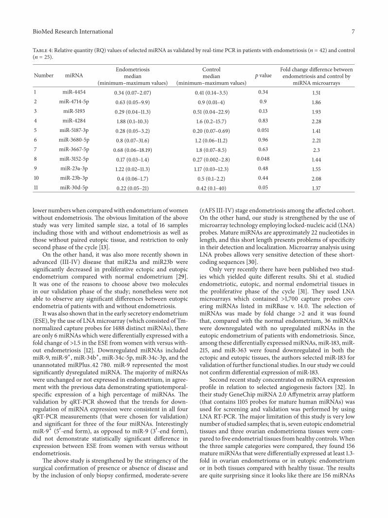