research article peripheral neuropathy and tear film...
TRANSCRIPT
Research ArticlePeripheral Neuropathy and Tear Film Dysfunctionin Type 1 Diabetes Mellitus
Stuti L. Misra,1 Dipika V. Patel,1 Charles N. J. McGhee,1 Monika Pradhan,1
Dean Kilfoyle,2 Geoffrey D. Braatvedt,3 and Jennifer P. Craig1
1 Department of Ophthalmology, New Zealand National Eye Centre, Faculty of Medical and Health Sciences,University of Auckland, Private Bag 92019, Auckland 1142, New Zealand
2Department of Neurology, Faculty of Medical and Health Sciences, University of Auckland, Private Bag 92019,Auckland 1142, New Zealand
3Department of Medicine, Faculty of Medical and Health Sciences, University of Auckland, Private Bag 92019,Auckland 1142, New Zealand
Correspondence should be addressed to Jennifer P. Craig; [email protected]
Received 4 July 2014; Accepted 23 July 2014; Published 7 August 2014
Academic Editor: Nikolaos Papanas
Copyright © 2014 Stuti L. Misra et al. This is an open access article distributed under the Creative Commons Attribution License,which permits unrestricted use, distribution, and reproduction in any medium, provided the original work is properly cited.
Purpose. To compare tear film metrics in patients with type 1 diabetes mellitus (DM) and healthy controls and investigate theassociation between peripheral neuropathy and ocular surface quality.Methods. Dry eye symptoms were quantified in 53 patientswith type 1 DM and 40 age-matched controls. Ocular examination included tear film lipid layer thickness grading, tear film stabilityand quantity measurement, and retinal photography. DM individuals additionally underwent a detailed neuropathy assessment.Results. Neither mean age nor dry eye symptom scores differed significantly between the DM and control groups (𝑃 = 0.12 and𝑃 = 0.33, resp.). Tear lipid thickness (𝑃 = 0.02), stability (𝑃 < 0.0001), and quantity (𝑃 = 0.01) were significantly lower in the DMgroup. Corneal sensitivity was also reduced in the DM group (𝑃 < 0.001) and tear film stability was inversely associated with totalneuropathy score (𝑟 = −0.29, 𝑃 = 0.03). Conclusion. The DM group exhibited significantly reduced tear film stability, secretion,and lipid layer quality relative to the age-matched control group. The negative correlation between tear film parameters and totalneuropathy score suggests that ocular surface abnormalities occur in parallel with diabetic peripheral neuropathy.
1. Introduction
While cataract and retinopathy have been extensivelyresearched in patients with diabetes mellitus (DM), only afraction of the published research has been dedicated to ocu-lar surface complications. However, dry eye symptoms andsigns of epithelial fragility, punctate keratopathy, persistentepithelial defects, and decreased corneal sensitivity are notuncommon in DM [1–3]. Compromised innervation of thecornea in patients with DM has also been described [4–6].Tear film dysfunction, characterised by impairment in tearquantity and quality, can occur in association with abnormalcorneal innervation due to the intimate, functional relation-ship between the cornea and the preocular tear film [7, 8].The resulting dry eye is a recognised cause of debilitating,
chronic ocular irritation symptoms [9]. The restrictions onlife imposed by this chronic condition can be significant and,in terms of impact on quality of life, have been equated inmore severe cases to those induced by dialysis and severeangina [10].
Tear film irregularity has been reported in DM, espe-cially in association with that of extended disease durationand severity, as defined by stage of retinopathy [2, 11]. Ithas been postulated that damage to the microvasculatureand denervation of the lacrimal gland may contribute toimpaired lacrimation in DM [2, 7, 12]. Despite this neurallink, few studies have explored whether the ocular surfacealters in association with peripheral neuropathy [7, 13, 14],although anomalous innervation of the lacrimal gland hasbeenreported in thosewithdiabetic sensoryneuropathy[2, 7].
Hindawi Publishing CorporationJournal of Diabetes ResearchVolume 2014, Article ID 848659, 6 pageshttp://dx.doi.org/10.1155/2014/848659
2 Journal of Diabetes Research
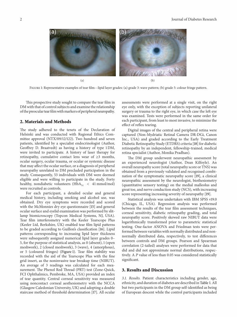
(a) (b)
Figure 1: Representative examples of tear film—lipid layer grades: (a) grade 3: wave pattern; (b) grade 5: colour fringe pattern.
This prospective study sought to compare the tear film inDMwith that of control subjects and examine the relationshipof thepreocular tearfilmwithmarkersofperipheralneuropathy.
2. Materials and Methods
The study adhered to the tenets of the Declaration ofHelsinki and was conducted with Regional Ethics Com-mittee approval (NTX/09/12/122). Two hundred and sevenpatients, identified by a specialist endocrinologist (Author,Geoffrey D. Braatvedt) as having a history of type 1DM,were invited to participate. A history of laser therapy forretinopathy, cumulative contact lens wear of ≥3 months,ocular surgery, ocular trauma, or ocular or systemic diseasethat may affect the ocular surface, or a diagnosis of peripheralneuropathy unrelated to DM precluded participation in thestudy. Consequently, 53 individuals with DM were deemedeligible and were willing to participate in the study. Fortyhealthy, nondiabetic volunteers (HbA
1c < 41mmol/mol)were recruited as controls.
For each participant, a detailed ocular and generalmedical history, including smoking and alcohol use, wasobtained. Dry eye symptoms were recorded and scoredwith the McMonnies dry eye questionnaire [15] and generalocular surface and eyelid examination was performed by slit-lamp biomicroscopy (Topcon Medical Systems, NJ, USA).Tear film interferometry with the Keeler Tearscope Plus(Keeler Ltd, Berkshire, UK) enabled tear film lipid patternsto be graded according to Guillon’s classification [16]. Lipidpatterns corresponding to increasing lipid layer thicknesswere subsequently assigned numerical lipid layer grades 0–5, for the purpose of statistical analysis, as 0 (absent), 1 (openmeshwork), 2 (closed meshwork), 3 (wave), 4 (amorphous),or 5 (coloured fringes) (Figure 1). Tear film stability wasrecorded with the aid of the Tearscope Plus with the finegrid insert, as the noninvasive tear breakup time (NIBUT).An average of 3 readings was calculated for each mea-surement. The Phenol Red Thread (PRT) test (Zone-Quick,FCI Ophthalmics, Pembroke, MA, USA) provided an indexof tear quantity. Central corneal sensitivity was measuredusing noncontact corneal aesthesiometry with the NCCA(Glasgow Caledonian University, UK) and adopting a doublestaircase method of threshold determination [17]. All ocular
assessments were performed at a single visit, on the righteye only, with the exception of subjects reporting unilateralsurgery or trauma to the right eye, in which case the left eyewas examined. Tests were performed in the same order foreach participant, from least to most invasive, to minimize theeffect of reflex tearing.
Digital images of the central and peripheral retina werecaptured (Non-Mydriatic Retinal Camera DR-DGi, CanonInc., USA) and graded according to the Early TreatmentDiabetic Retinopathy Study (ETDRS) criteria [18] for diabeticretinopathy by an independent, fellowship-trained, medicalretina specialist (Author, Monika Pradhan).
The DM group underwent neuropathic assessment byan experienced neurologist (Author, Dean Kilfoyle). Anoverall neuropathy score (total neuropathy score or TNS) wasobtained from a previously validated and recognised combi-nation of the symptomatic neuropathy score [19], a clinicalneuropathic assessment by the neurologist, biothesiometry(quantitative sensory testing) on the medial malleolus andgreat toe, and nerve conduction study (NCS), with increasingscore representing increasing severity of neuropathy [19].
Statistical analysis was undertaken with IBM SPSS v19.0(Chicago, IL, USA). Regression analysis was performedbetween the results of the tear film assessment techniques,corneal sensitivity, diabetic retinopathy grading, and totalneuropathy score. Positively skewed raw NIBUT data werelogarithmically transformed prior to parametric statisticaltesting. One-factor ANOVA and Friedman tests were per-formedbetween variableswith normally distributed andnon-normally distributed data, respectively, to test differencesbetween controls and DM groups. Pearson and Spearmancorrelation (2-tailed) analyses were performed for data thatdid and did not approximate normal distributions, respec-tively. A 𝑃 value of less than 0.05 was considered statisticallysignificant.
3. Results and Discussion
3.1. Results. Patient characteristics including gender, age,ethnicity, andduration of diabetes are described inTable 1. Allbut two participants in the DM group self-identified as beingof European descent while the control participants included
Journal of Diabetes Research 3
Table 1: Participant characteristics for the patients with type 1diabetes and control group.
Diabetes Controls
Subjects (𝑛) 53 40
M : F ratio 26 : 27 17 : 23
Age (years) 48.6 ± 11.8 44.3 ± 14.7
HbA1c (mmol/mol) 61.3 ± 12.0 35.0 ± 2.5
EthnicityEuropean 51 19
Indian 0 9
Asian (excluding Indian) 1 8
Maori 1 0
Others 0 4Mean diabetesDuration (years)
25.8 ± 11.4
<10 5
10–20 10
21–31 22
>31 16
47.5%NZEuropean, 22.5% Indian, and 20%Asian (excludingIndian). The mean age of the DM group (49 ± 12 years) didnot differ significantly from that of the control group (44 ± 15years) (𝑃 = 0.12) and no significant differences in tear filmcharacteristics were identified between the ethnic subgroups(𝑃 > 0.05).
Dry eye symptom scores and clinical findings were com-pared for the diabetes and control groups (Table 2).Themeandry eye symptom score was not statistically significantlydifferent between the diabetes and control groups (𝑃 =0.33). However, clinical tests showed significant differencesbetween the two groups, with lipid layer grading (𝑃 =0.02), NIBUT (𝑃 < 0.0001), and PRT test (𝑃 = 0.01)results all statistically, and clinically, significantly lower inthe DM group. NIBUT was observed to be lower in femalesthan in males in both the control (𝑃 = 0.06) and DM(𝑃 < 0.01) groups. The patients with DM exhibited reducedcorneal sensitivity compared to controls (𝑃 < 0.0001)(corneal sensitivity and subbasal nerve density reported indetail elsewhere) [20].The total neuropathy score (maximumpossible score 40) ranged between 0 and 21 (5.3 ± 5.1) in thepatients with diabetes.
Retinopathy was not observed in the majority of patients(60%) while 21% exhibited mild and 19% exhibited moderateDR. Regression analysis between interferometry, tear filmstability, tear quantity, diabetic retinopathy grade, and totalneuropathy score (Table 3) highlighted a positive relationshipbetween lipid layer grading and both NIBUT (𝑟 = 0.56,𝑃 < 0.01) and tear quantity (𝑟 = 0.38, 𝑃 < 0.01). Tear filmstability was noted to be inversely related to total neuropathyscore (𝑟 = −0.29, 𝑃 = 0.03). The association betweencorneal sensitivity and total neuropathy score failed to reachstatistical significance (𝑟 = 0.24, 𝑃 = 0.08).
3.2. Discussion. Comparison of the status of the tear filmbetween patients with type 1DM and age-matched healthycontrol subjects demonstrated a significantly poorer tear filmquality in diabetes. Lipid layer grade, tear film stability, andtear quantity (basal with minimal reflex tear secretion) [21]were all significantly reduced in patients with type 1DM,confirming compromised protection of the ocular surface inpatients with diabetes.
Dry eye symptoms were observed to increase in severitywith advancing age in both groups, in the current study,consistent with reports in the literature [22, 23]. Interestingly,age-matched subjects in both groups reported similar dryeye symptom severity, despite clear differences in tear quality(𝑃 < 0.0001). This lack of difference in symptoms is believedto be related to the impaired sensitivity of the ocular surfacein diabetes.
Tear film stability and lipid layer thickness have beenshown to be influenced by gender [24], with older womentending to exhibit thinner and more contaminated lipidlayers [25]. The current study also reported reduced tearfilm stability in females compared to males, both in the DMgroup (𝑃 < 0.01) and in the control group (𝑃 = 0.06). Adecrease in circulating androgens in postmenopausal womenis believed to play an important role in affecting meibomiangland function, supporting female gender as a risk factor fordry eye disease [26].
In addition to a reduction in tear film stability, severalstudies have reported a diminished tear secretion in thediabetic eye [7, 11, 27], as observed in the present research.This further degrades the quality of tear film in an alreadycompromised diabetic eye.
Reduced tear film stability has been associated withthe presence of superficial punctate keratitis [8]. Functionalabnormalities of corneal innervation may contribute to theincidence of superficial punctate keratitis in these patientsthrough its adverse effect on tear film instability [28]. Aninverse association between corneal innervation and periph-eral neuropathy has been reported previously [6, 29].The cur-rent study supports this relationship, with a modest inversecorrelation observed between NIBUT and total neuropathyscore (a measure of peripheral neuropathy) (𝑟 = −0.29, 𝑃 =0.03).
Retinopathy grade was observed to be unrelated to tearfilm stability in the current study (𝑟 = −0.03, 𝑃 = 0.82),contrary to previous observations [11, 30]. However, it shouldbe noted that, as history of laser treated retinopathy wasan exclusion criterion in the current study, patients withmore than moderate retinopathy were generally ineligible toparticipate. This restriction in the range of disease severity inthe cohort enrolled in the current study likely contributed tothe absence of a relationship in our study.
In this New Zealand-based study, the control groupcomprised patients of a variety of ethnic backgrounds reflect-ing the multiethnic population [31]. Type 1DM in NewZealand is predominantly reported in those with European(Caucasian) heritage [32] and thiswas observed in the presentcohort. Although ethnicity has previously been shown tobe a determinant of tear film stability [33], no significant

4 Journal of Diabetes Research
Table 2: Comparison of symptoms and tear characteristics for control and patient groups (mean/median), together with the significance oftheir differences (ANOVA/Friedmann). Noninvasive tear breakup time (NIBUT) values are extrapolated from logarithmically transformeddata.
Diabetes Healthy controls ANOVA/Friedmann(𝑃 values)
McMonnies questionnaire(mean ± SD) 8.8 ± 6.7 7.6 ± 4.6 0.33
Lipid layer thickness grade (median) 2 3 0.02NIBUT (s) (mean ± SD) 6.0 ± 1.9 8.2 ± 2.5 <0.0001Phenol red thread test (mm) (mean ± SD) 13.7 ± 4.7 16.3 ± 4.9 0.01Corneal sensitivity threshold (mBAR) 1.3 ± 1.3 0.2 ± 1.3 <0.001
Table 3: Correlation analysis between age, diabetes duration, McMonnies questionnaire scores (DEQ), phenol red thread test (PRTT) (mm),tear film interferometry including lipid layer grade, stability (NIBUT), diabetic retinopathy grading, and total neuropathy score (TNS) inthose with DM (𝑛 = 53).
Correlation (𝑟 values) Probability (𝑃 values)Age versus NIBUT −0.28∗ 0.05Age versus TNS 0.41∗∗ 0.00DEQ versus TNS −0.05 0.73Lipid layer thickness versus NIBUT 0.56∗∗ 0.00Lipid layer thickness versus PRTT 0.39∗∗ 0.00NIBUT versus diabetes duration −0.29∗ 0.03NIBUT versus TNS −0.29∗ 0.03NIBUT versus retinopathy grade −0.03 0.82PRTT versus TNS 0.01 0.96PRTT versus retinopathy grade −0.16 0.26∗∗Correlation is significant at the 0.01 level (2-tailed).∗Correlation is significant at the 0.05 level (2-tailed).
difference was observed between the ethnic subgroups of thecontrol subjects in the present study.
There is potential for lipid, aqueous, and mucins, themajor components of the tear film, to be adversely affected inpatients with diabetes. The meibomian and lacrimal glandsare responsible for the secretion of the lipid and aqueousportions of the preocular tear film, respectively. Meibomianglands are innervated by parasympathetic fibres with asmaller contribution from sympathetic and sensory neurons[34]. Disease or any damage to these neurons leads to dry eyein an animal model [35]. Two human studies have previouslyreported a compromised tear lipid layer in DM patients, asconfirmed in the current study [8, 36]. Clinical observationof noncontiguous or absent lipid layers is associated withsignificantly increased tear film evaporation [37], one of thekey factors in dry eye development [38].
Elevated expression of advanced glycation end productsin the lacrimal gland has been postulated as a reason forchanges in lacrimal gland function described in diabetes [12].A reduction in goblet cell numbers, compromising mucinquantity, may also contribute to the tear film instabilityobserved in diabetes [7, 39]. Goblet cell loss in those withdiabetic peripheral neuropathy and poor metabolic controlhas been previously reported [7].
The reduced tear production identified in patients withdiabetes (𝑃 = 0.01) in the current study lends support to
the concept that lacrimal gland function might be adverselyaffected by a neuropathic mechanism [40], resulting indysfunction of the ocular surface secretory glands via theirinnervation. Such dysfunction could arise from a peripheralneuropathy involving the afferent sensory nerves from theocular surface affecting corneal sensitivity and the auto-nomic (efferent) nerves responsible for innervating the tear-component secreting glands and the lacrimal andmeibomianglands [35, 40, 41]. This is supported by the current results ofreduced corneal sensitivity; however the previously reportedassociation between corneal sensitivity and peripheral neu-ropathy failed to reach statistical significance in the currentstudy (𝑟 = 0.24, 𝑃 = 0.08) [42]. Reduced tear secretion haspreviously been identified in patients with type 2 diabetesrelative to healthy controls [8, 11].
4. Conclusion
In summary, the current study confirms the underlying threatto ocular surface health in patients with type 1DM comparedto control subjects. The reduction in tear production inpatients with DM and the association between reduced tearfilm stability and diabetic peripheral polyneuropathy add cre-dence to the hypothesis that diabetic peripheral neuropathyis associated with, or directly affects, secretory lacrimal glandfunction [43].Hence, patientswithDM, particularly the older
Journal of Diabetes Research 5
female, are at a greater risk of dry eye and compromisedpreocular tear film.
Conflict of Interests
The authors declare that there is no conflict of interestsregarding the publication of this paper.
Acknowledgments
This work was supported by Save Sight Society NZ Incor-porated and New Zealand Optometric and Vision ResearchFoundation (NZOVRF). The statistical analysis consultationwas provided by Dr. Avinesh Pillai from the Department ofStatistics, University of Auckland.
References
[1] R. A. Hyndiuk, E. L. Kazarian, R. O. Schultz, and S. Seideman,“Neurotrophic corneal ulcers in diabetes mellitus,” Archives ofOphthalmology, vol. 95, no. 12, pp. 2193–2196, 1977.
[2] I. Kaiserman, N. Kaiserman, S. Nakar, and S. Vinker, “Dry eyein diabetic patients,” American Journal of Ophthalmology, vol.139, no. 3, pp. 498–503, 2005.
[3] R. O. Schultz, M. A. Peters, K. Sobocinski, K. Nassif, and K. J.Schultz, “Diabetic keratopathy as a manifestation of peripheralneuropathy,” American Journal of Ophthalmology, vol. 96, no. 3,pp. 368–371, 1983.
[4] P. Kallinikos, M. Berhanu, C. O’Donnell, A. J. M. Boulton, N.Efron, and R. A. Malik, “Corneal nerve tortuosity in diabeticpatients with neuropathy,” Investigative Ophthalmology andVisual Science, vol. 45, no. 2, pp. 418–422, 2004.
[5] R. A. Malik, P. Kallinikos, C. A. Abbott et al., “Corneal confocalmicroscopy: a non-invasive surrogate of nerve fibre damage andrepair in diabetic patients,”Diabetologia, vol. 46, no. 5, pp. 683–688, 2003.
[6] M. Tavakoli, P. Kallinikos, A. Iqbal et al., “Corneal confocalmicroscopy detects improvement in corneal nerve morphologywith an improvement in risk factors for diabetic neuropathy,”Diabetic Medicine, vol. 28, no. 10, pp. 1261–1267, 2011.
[7] M. Dogru and M. J. Goebbels, “Tear secretion and tear filmfunction in insulin dependent diabetics,” British Journal ofOphthalmology, vol. 84, article 1210, 2000.
[8] K. Inoue, S. Kato, C. Ohara, J. Numaga, S. Amano, andT. Oshika, “Ocular and systemic factors relevant to diabetickeratoepitheliopathy,” Cornea, vol. 20, no. 8, pp. 798–801, 2001.
[9] R. Herrero-Vanrell and A. Peral, “International dry eye work-shop (DEWS). Update of the disease,” Archivos de la SociedadEspanola de Oftalmologia, vol. 82, no. 12, pp. 733–734, 2007.
[10] P. Buchholz, C. S. Steeds, L. S. Stern et al., “Utility assessment tomeasure the impact of dry eye disease,”TheOcular Surface, vol.4, no. 3, pp. 155–161, 2006.
[11] J. Saito, M. Enoki, M. Hara, N. Morishige, T. Chikama, andT. Nishida, “Correlation of corneal sensation, but not of basalor reflex tear secretion, with the stage of diabetic retinopathy,”Cornea, vol. 22, no. 1, pp. 15–18, 2003.
[12] M. Alves, V. C. Calegari, D. A. Cunha, M. J. A. Saad, L. A.Velloso, and E. M. Rocha, “Increased expression of advancedglycation end-products and their receptor, and activation ofnuclear factor kappa-B in lacrimal glands of diabetic rats,”Diabetologia, vol. 48, no. 12, pp. 2675–2681, 2005.
[13] M. E. Rosenberg, T. M. T. Tervo, I. J. Immonen, L. J. Muller,C. Gronhagen-Riska, andM. H. Vesaluoma, “Corneal structureand sensitivity in type 1 diabetesmellitus,” InvestigativeOphthal-mology & Visual Science, vol. 41, no. 10, pp. 2915–2921, 2000.
[14] M. Hom and P. De Land, “Self-reported dry eyes and diabetichistory,” Optometry, vol. 77, no. 11, pp. 554–558, 2006.
[15] C. W. McMonnies and A. Ho, “Responses to a dry eye ques-tionnaire from a normal population,” Journal of the AmericanOptometric Association, vol. 58, no. 7, pp. 588–591, 1987.
[16] J. Guillon, “Non-invasive tearscope plus routine for contact lensfitting,”Contact Lens andAnterior Eye, vol. 21, no. 1, pp. S31–S40,1998.
[17] D. V. Patel, M. Tavakoli, J. P. Craig, N. Efron, and C. N. J.McGhee, “Corneal sensitivity and slit scanning in vivo confocalmicroscopy of the subbasal nerve plexus of the normal centraland peripheral human cornea,” Cornea, vol. 28, no. 7, pp. 735–740, 2009.
[18] “Grading diabetic retinopathy from stereoscopic color fundusphotographs—an extension of the modified Airlie House clas-sification. ETDRS report number 10. Early Treatment DiabeticRetinopathy Study Research Group,” Ophthalmology, vol. 98,pp. 786–806, 1991.
[19] D. R. Cornblath, V. Chaudhry, K. Carter et al., “Total neuropa-thy score: validation and reliability study,”Neurology, vol. 53, no.8, pp. 1660–1664, 1999.
[20] S. Misra, Corneal microstructure in diabetes mellitus and itsassociation with peripheral neuropathy and cardiac autonomicneuropathy [Ph.D. thesis], University of Auckland, Auckland,New Zealand, 2013.
[21] A. Tomlinson, K. J. Blades, and E. I. Pearce, “What does thephenol red thread test actually measure?” Optometry & VisionScience, vol. 78, no. 3, pp. 142–146, 2001.
[22] O. D. Schein, J.M. Tielsch, B.Munoz, K. Bandeen-Roche, and S.West, “Relation between signs and symptoms of dry eye in theelderly: a population-based perspective,” Ophthalmology, vol.104, no. 9, pp. 1395–1401, 1997.
[23] A. Tomlinson and C. Giesbrecht, “Effect of age on humantear film evaporation in normals,” Advances in ExperimentalMedicine and Biology, vol. 350, pp. 271–274, 1994.
[24] J. P. Craig and A. Tomlinson, “Age and gender effects onthe normal tear film,” Advances in Experimental Medicine andBiology, vol. 438, pp. 411–415, 1998.
[25] C. Maıssa and M. Guillon, “Tear film dynamics and lipid layercharacteristics—effect of age and gender,” Contact Lens andAnterior Eye, vol. 33, no. 4, pp. 176–182, 2010.
[26] D. A. Sullivan, B. D. Sullivan, M. D. Ullman et al., “Androgeninfluence on the meibomian gland,” Investigative Ophthalmol-ogy & Visual Science, vol. 41, no. 12, pp. 3732–3742, 2000.
[27] M. Ozdemir, M. A. Buyukbese, A. Cetinkaya, and G. Ozdemir,“Risk factors for ocular surface disorders in patients withdiabetes mellitus,” Diabetes Research and Clinical Practice, vol.59, no. 3, pp. 195–199, 2003.
[28] P. R. Herse, “A review of manifestations of diabetes mellitus inthe anterior eye and cornea,” American Journal of Optometryand Physiological Optics, vol. 65, no. 3, pp. 224–230, 1988.
[29] M. Tavakoli, C. Quattrini, C. Abbott et al., “Corneal confocalmicroscopy: A novel noninvasive test to diagnose and stratifythe severity of human diabetic neuropathy,” Diabetes Care, vol.33, no. 8, pp. 1792–1797, 2010.
[30] M. Ozdemir and H. Temizdemir, “Age- and gender-related tearfunction changes in normal population,” Eye, vol. 24, no. 1, pp.79–83, 2010.
6 Journal of Diabetes Research
[31] S. N. Zealand, Ed., 2013 Census QuickStats about Culture andIdentity, Statistics New Zealand, 2014.
[32] J. A. Willis, R. S. Scott, B. A. Darlow et al., “Incidence oftype 1 diabetes mellitus diagnosed before age 20 years inCanterbury, New Zealand over the last 30 years,” Journal ofPediatric Endocrinology and Metabolism, vol. 15, no. 5, pp. 637–643, 2002.
[33] S. Patel, S. K. Virhia, and P. Farrell, “Stability of the precornealtear film in Chinese, African, Indian, and Caucasian eyes,”Optometry and Vision Science, vol. 72, no. 12, pp. 911–915, 1995.
[34] C. W. Chung, M. Tigges, and R. A. Stone, “Peptidergicinnervation of the primate meibomian gland,” InvestigativeOphthalmology and Visual Science, vol. 37, no. 1, pp. 238–245,1996.
[35] X. J. Song, D. Li, W. Farley et al., “Neurturin-deficient micedevelop dry eye and keratoconjunctivitis sicca,” InvestigativeOphthalmology and Visual Science, vol. 44, no. 10, pp. 4223–4229, 2003.
[36] K. Inoue, K.Okugawa, S. Amano et al., “Blinking and superficialpunctate keratopathy in patients with diabetes mellitus,” Eye,vol. 19, no. 4, pp. 418–421, 2005.
[37] J. P. Craig and A. Tomlinson, “Importance of the lipid layerin human tear film stability and evaporation,” Optometry andVision Science, vol. 74, no. 1, pp. 8–13, 1997.
[38] “The definition and classification of dry eye disease: Report ofthe definition and classification subcommittee of the interna-tional Dry Eye WorkShop (2007),” The Ocular Surface, vol. 5,no. 2, pp. 75–92, 2007.
[39] S. C. G. Tseng, L. W. Hirst, and A. E. Maumenee, “Possiblemechanisms for the loss of goblet cells in mucin-deficientdisorders,” Ophthalmology, vol. 91, no. 6, pp. 545–552, 1984.
[40] P. Cousen, P. Cackett, H. Bennett, K. Swa, and B. Dhillon,“Tear production and corneal sensitivity in diabetes,” Journal ofDiabetes and its Complications, vol. 21, no. 6, pp. 371–373, 2007.
[41] N. Ishida, G. N. Rao, M. Del Cerro, and J. V. Aquavella,“Corneal nerve alterations in diabetes mellitus,” Archives ofOphthalmology, vol. 102, no. 9, pp. 1380–1384, 1984.
[42] N. Pritchard, K. Edwards, D. Vagenas et al., “Corneal sensitivityas an ophthalmic marker of diabetic neuropathy,” Optometryand Vision Science, vol. 87, no. 12, pp. 1003–1008, 2010.
[43] I. S. Zagon, M. S. Klocek, J. W. Sassani, and P. J. McLaughlin,“Dry eye reversal and corneal sensation restoration with topicalnaltrexone in diabetes mellitus,”Archives of Ophthalmology, vol.127, no. 11, pp. 1468–1473, 2009.

Submit your manuscripts athttp://www.hindawi.com
Stem CellsInternational
Hindawi Publishing Corporationhttp://www.hindawi.com Volume 2014
Hindawi Publishing Corporationhttp://www.hindawi.com Volume 2014
MEDIATORSINFLAMMATION
of
Hindawi Publishing Corporationhttp://www.hindawi.com Volume 2014
Behavioural Neurology
EndocrinologyInternational Journal of
Hindawi Publishing Corporationhttp://www.hindawi.com Volume 2014
Hindawi Publishing Corporationhttp://www.hindawi.com Volume 2014
Disease Markers
Hindawi Publishing Corporationhttp://www.hindawi.com Volume 2014
BioMed Research International
OncologyJournal of
Hindawi Publishing Corporationhttp://www.hindawi.com Volume 2014
Hindawi Publishing Corporationhttp://www.hindawi.com Volume 2014
Oxidative Medicine and Cellular Longevity
Hindawi Publishing Corporationhttp://www.hindawi.com Volume 2014
PPAR Research
The Scientific World JournalHindawi Publishing Corporation http://www.hindawi.com Volume 2014
Immunology ResearchHindawi Publishing Corporationhttp://www.hindawi.com Volume 2014
Journal of
ObesityJournal of
Hindawi Publishing Corporationhttp://www.hindawi.com Volume 2014
Hindawi Publishing Corporationhttp://www.hindawi.com Volume 2014
Computational and Mathematical Methods in Medicine
OphthalmologyJournal of
Hindawi Publishing Corporationhttp://www.hindawi.com Volume 2014
Diabetes ResearchJournal of
Hindawi Publishing Corporationhttp://www.hindawi.com Volume 2014
Hindawi Publishing Corporationhttp://www.hindawi.com Volume 2014
Research and TreatmentAIDS
Hindawi Publishing Corporationhttp://www.hindawi.com Volume 2014
Gastroenterology Research and Practice
Hindawi Publishing Corporationhttp://www.hindawi.com Volume 2014
Parkinson’s Disease
Evidence-Based Complementary and Alternative Medicine
Volume 2014Hindawi Publishing Corporationhttp://www.hindawi.com