research article influence of pvp on the morphologies of...
TRANSCRIPT
Hindawi Publishing CorporationJournal of NanomaterialsVolume 2013, Article ID 314012, 6 pageshttp://dx.doi.org/10.1155/2013/314012
Research ArticleInfluence of PVP on the Morphologies of Bi2S3 NanostructuresSynthesized by Solvothermal Method
Anukorn Phuruangrat,1 Titipun Thongtem,2 and Somchai Thongtem3,4
1 Department of Materials Science and Technology, Faculty of Science, Prince of Songkla University, Hat Yai, Songkhla 90112, Thailand2Department of Chemistry, Faculty of Science, Chiang Mai University, Chiang Mai 50200, Thailand3Department of Physics and Materials Science, Faculty of Science, Chiang Mai University, Chiang Mai 50200, Thailand4Materials Science Research Center, Faculty of Science, Chiang Mai University, Chiang Mai 50200, Thailand
Correspondence should be addressed to Anukorn Phuruangrat; [email protected] TitipunThongtem; [email protected]
Received 1 June 2013; Revised 5 November 2013; Accepted 27 November 2013
Academic Editor: Steve Acquah
Copyright © 2013 Anukorn Phuruangrat et al. This is an open access article distributed under the Creative Commons AttributionLicense, which permits unrestricted use, distribution, and reproduction in any medium, provided the original work is properlycited.
Different morphologies of Bi2S3nanostructures were synthesized by a 180∘C and 12 h solvothermal reaction of solutions
containing Bi(NO3)3⋅5H2O and thioacetamide (CH
3CSNH
2) in diethylene glycol (DEG) as a solvent. The as-synthesized Bi
2S3
products characterized by XRD, Raman spectroscopy, SEM, and TEM showed that they were well-crystallized orthorhombicBi2S3phase with morphologies of nanorod-like, sheaf-like, carnation-like, and microspherical, controlled by different contents
of polyvinylpyrrolidone (PVP) in the solutions. Based on the experimental results, a growth mechanism was also proposed anddiscussed.
1. Introduction
Presently, nanostructuredmaterials are very important to sci-ence and technology due to their novel optical, magnetic, andcatalytic properties as compared to the corresponding bulks.They are elementary materials used for the fabrication ofoptoelectronic devices. Among them, the V-VI chalcogenidegroupwith AV
2BVI3(A =As, Sb, and Bi; B = S, Se, and Te) for-
mula has a wide variety applications in television cameras forphotoconducting targets, thermoelectric devices, electronicand optoelectronic devices, and IR spectroscopy [1–8]. Bi
2S3
is a 1.3–1.7 eV direct band gap layered semiconductor withorthorhombic crystal system andD
2h16 or Pbnm space group,
which has structure similar to Sb2S3and Sb
2Se3. It has a
number of potential applications such as photovoltaic andoptoelectronic, thermoelectric cooling systems, IR devices,X-ray computed tomography, photocatalysis for hydrogenevolution, and electrochemical hydrogen storage [9–11]. It is ahighly anisotropic semiconductor with layered structure par-allel to its growth direction and has lattice parameters of 𝑎 =11.123 A, 𝑏 = 11.282 A, and 𝑐 = 3.971 A with pseudolayers ofribbon-like Bi
4S6polymers linked together by intermolecular
attraction of bismuth and sulfur atoms along the [001] direc-tion. Bond lengths within the ribbons are different due to twodifferent types of coordination exhibited by both bismuth andsulfur. Two crystallographic independent Bi atoms belong toBi2S3: Bi1 of 6-fold coordinated to three strong Bi−S bonds
and three weak ones and Bi2of 5-fold coordinated square
pyramidal sphere with three strong and two weak ones [12–15]. Morphologies of nanomaterials can play the role in thephysical and chemical properties, for instances, 148mA⋅h⋅g−1initial discharge capacity of Bi
2S3microflowers in lithium-
ion batteries [16] and good electrochemical Li+ insertionwith826mA⋅h⋅g−1 capacity of dandelion-like Bi
2S3microspheres
[9]. In addition, uniform Bi2S3nanodots showed higher
and more stable photocatalytic activity for the degradationof persistent toxic organic pollutants under UV light irra-diation than Bi
2S3nanorods [17]. Different morphologies
and structures can play the role in energy gap (𝐸𝑔) and
photoluminescence (PL) of materials [1, 2, 10, 12], includingtheir properties.
In this research, nanorods, double sheaf-like Bi2S3, carna-
tion-like Bi2S3, and microspheres of Bi
2S3nanorods were sy-

2 Journal of Nanomaterials
nthesized by a solvothermal method in solutions containingdifferent contents of PVP. A possible formation mechanismwas proposed and discussed according to the experimentalresults.
2. Experimental Procedures
All chemicals were analytical grade and were used withoutfurther purification. In a typical experimental procedure,0.0010mol Bi(NO
3)3⋅5H2O and 0.0015mol thioacetamide
(TAA, CH3CSNH
2) were dissolved in 80ml of diethylene
glycol (DEG) under vigorous stirring for 1 h and followedby the addition of 0.1, 0.5, and 1.0 g of polyvinylpyrrolidone(PVP)withmolecularmass of 40,000 and 0.5mL 37%HNO
3.
These resulting solutions were loaded into 100mL Teflon-lined stainless-steel autoclaves. The autoclaves were tightlyclosed, processed at 180∘C for 12 h in an electric oven, and leftcooling to room temperature naturally. At the conclusion ofthe process, deep gray precipitates were synthesized, filtered,washed with distilled water and absolute ethanol repeatedlyto remove undesired impurities, and dried at 60∘C for 4 h.
X-ray diffraction (XRD) patterns were obtained on aPhilips X’Pert MPD X-ray powder diffractometer (XRD)using a Cu-K
𝛼radiation at 45 kV and 35mA with a scanning
rate of 0.04 deg/s in the 2𝜃 range from 15 to 60 deg. Scanningelectron microscopic (SEM) analysis was conducted by aJEOL JSM-6335F electron microscope operating at 35.0 kV.Transmission electron microscopic (TEM) images, high-resolution TEM (HRTEM) images, and selected area electrondiffraction (SAED) patterns were performed by a JEOL JEM-2010 transmission electron microscope at an acceleratingvoltage of 200 kV. A Raman spectroscopy was operated on aHORIBA Jobin Yvon T64000 spectrometer using Ar+ laserbeam of 514.5 nmwavelength, calibrated using a silicon waferas the standard.
3. Results and Discussion
Figure 1 shows XRD patterns of Bi2S3synthesized using
different contents of PVP by a solvothermal method at 180∘Cfor 12 h. Compared to the JCPDS database no. 17-0320 [18](𝑎 = 11.1490 A, 𝑏 = 11.3040 A, and 𝑐 = 3.9810 A), allof the diffraction patterns were interpreted as orthorhombicBi2S3. No impurities of Bi
2O3, Bi, and others were detected
in these main products. The diffraction peaks were narrowand sharp, indicating that the products were of high degreeof crystallinity with their atoms residing in crystalline lattice.
Figure 2 shows Raman spectra of Bi2S3products over
the wavelength range of 100–1800 cm−1. The spectra presentfour peaks at 129, 251, 420, and 968 cm−1. Generally, thepresence of the first peak at 129 cm−1 can be attributed tosurface optical (SO) phonon mode. The second correspondsto the vibrationmode of Bi
2S3nanorods at 252 and 259 cm−1,
and Bi2S3nanoribbons and hierarchical nanostructures at
250 cm−1, specified as the vibration mode of Bi–S bonds.Thevibration at 968 cm−1 was in accordance with that of Bi
2S3
nanorods [2].
15 20 25 30 35 40 45 50 55 600
20406080
100120140160180200220240260280300320340360380
(242
)
(351
)
(431
)(0
02)
(430
)(1
41)
(041
)(2
40)
(301
)(3
31)(221
)(211
)(0
21)
(130
)(2
20)
(101
)
(120
)(0
20)
Inte
nsity
(a.u
.)
Non
0.5 g
1.0 g
2𝜃 (deg)
Figure 1: XRD patterns of Bi2S3solvothermally synthesized in the
solutions containing 0.0, 0.5, and 1.0 g PVP.
200 400 600 800 1000 1200 1400 1600 1800
90
100
110
120
Non
Inte
nsity
(a.u
.)
0.5 g
1.0 g
Wavenumber (cm−1)
Figure 2: Raman spectra of Bi2S3solvothermally synthesized in the
solutions containing 0.0, 0.5, and 1.0 g PVP.
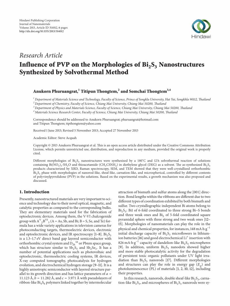
Size and structure of the products under differentreaction conditions were further investigated by electronmicroscopy (EM). Figure 3 shows typical SEM images of theas-synthesized Bi
2S3products in the solutions with different
PVP contents. SEM analysis reveals that Bi2S3synthesized
in PVP-free solution is mostly consisted of large quantity ofnanorodswith 0.5–2𝜇min length and 50 nm in diameter.Themorphologies of Bi
2S3products were varied in sequence by
adding of different contents of PVP.Themixedmorphologiesofmicrosized double sheaf-like and carnation-like Bi
2S3were
detected in 0.1 g PVP solution under solvothermal at 180∘Cfor 12 h. They became uniformly and completely carnation-like Bi
2S3microflowers with diameter in the range of 5-6𝜇m
in the solution containing 0.5 g PVP. High magnificationof the carnation-like Bi
2S3nanostructure reveals the lobe
of carnation-like Bi2S3microsized crystals consisting of
nanopetals with the shape in between rods and sheets asbuilding blocks. By increasing the PVP content to 1.0 g, theproduct appeared as 3D microsphere-like Bi
2S3. Its high
Journal of Nanomaterials 3
(a) (b)
(c) (d)
Figure 3: SEM images of Bi2S3solvothermally synthesized in the solutions containing (a–d) 0, 0.1, 0.5, and 1.0 g PVP, respectively.
magnification SEM image revealed that each microspherewas composed of a number of nanorods with 3-4 𝜇m inlength and 50 nm in diameter, growing out of Bi
2S3core to
build up 3D Bi2S3microspheres. There was some difference
between the carnation-like and microsphere-like products.The carnation-like flowers were composed of a number ofnanopetals with the shape in between rods and sheets but themicrospheres were composed of a number of nanorods withsharp tips or spears radiating from central cores. Chemicalcomposition of the as-synthesized Bi
2S3microspheres was
further analyzed by energy dispersive X-ray spectrometry(EDS), which revealed the presence of Bi and S in the as-synthesized Bi
2S3product with Bi : S atomic ratio very close
to 2 : 3.Figures 4 and 5 show TEM images of Bi
2S3synthesized
in free-PVP, 0.1 g PVP, 0.5 g PVP, and 1.0 g PVP solutions.In PVP-free solution, the product was composed of large-scale Bi
2S3nanorods with an average length of 1-2 𝜇m and
diameter of 20–50 nm. Its HRTEM image was recorded ona single orthorhombic Bi
2S3nanorod and demonstrated that
the Bi2S3nanorod grew along the [001] direction. In 0.1 g
PVP solution, TEM image indicates the transformation of thenanorods into themixture of double sheaf-like and carnation-like Bi
2S3crystals, which were composed of a number of
nanorods with the length up to a few micrometers. In 0.5 gPVP solution, the uniformly and completely carnation-likeBi2S3crystals were synthesized at the same condition as
above. In 1.0 g PVP solution, representative TEM image ofthe 3D spherical Bi
2S3of nanorods clearly confirmed the
existence of nanostructured 3Dmicrospheres with 4–6𝜇m in
diameter. It was built up from uniform nanorods with diam-eter of about 30–50 nm and length of 3 𝜇m. The crystallinestructure of the individual Bi
2S3nanorods of the 3D spherical
structure was characterized by HRTEM. A typical HRTEMimage of an individual nanorod exhibited clear lattice fringecorresponding to the (020) plane aligning parallel to the[001] growth direction. The analysis confirmed that eachnanorod was single crystal. It should be noted that the (020)planes were parallel to the individual Bi
2S3nanorod growth
direction, indicating its preferential orientation growth alongthe [001] direction, due to the exceeding surface energy overother planes [2, 3, 13]. Wang et al. calculated surface energyof the (100), (010), and (001) planes of orthorhombic Bi
2S3
structured model: 3.5878 × 10⁻⁴, 3.4964 × 10⁻⁴, and 4.2307 ×10⁻⁴ kJ/m2, respectively [12]. The results supported very well
with the preferential orientation growth along the 𝑐 axis inbuilding up of 1D nanostructured orthorhombic Bi
2S3. For
the synthesis of superparamagnetic Fe3O4and CoFe
2O4with
high saturation magnetization by solvothermal processing ofthe solution containing PVP, these products were in the shapeof microspheres [19, 20].
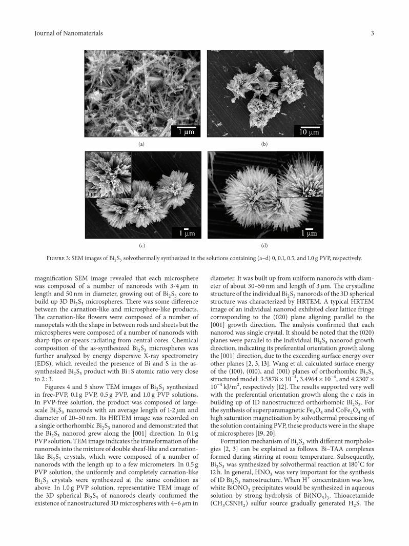
Formation mechanism of Bi2S3with different morpholo-
gies [2, 3] can be explained as follows. Bi–TAA complexesformed during stirring at room temperature. Subsequently,Bi2S3was synthesized by solvothermal reaction at 180∘C for
12 h. In general, HNO3was very important for the synthesis
of 1D Bi2S3nanostructure. When H+ concentration was low,
white BiONO3precipitates would be synthesized in aqueous
solution by strong hydrolysis of Bi(NO3)3. Thioacetamide
(CH3CSNH
2) sulfur source gradually generated H
2S. The

4 Journal of Nanomaterials
(a) (b)
(c) (d)
Figure 4: TEM and HRTEM images of Bi2S3solvothermally synthesized in the solutions containing (a–d) 0, 0.1, 0.5, and 1.0 g PVP,
respectively.
(a) (b)
Figure 5: TEM andHRTEM images of 3Dmicrospherical Bi2S3of nanorods solvothermally synthesized in the solution containing 1.0 g PVP.
Journal of Nanomaterials 5
Figure 6: Schematic illustration for the formation of Bi2S3nanos-
tructures.
concentration of S2− was controlled by the concentration ofH+ in the solution. In a strong acidic solution, the concen-tration of S2− was lower, and the generation rate of Bi
2S3was
slowed down accordingly.Thus Bi2S3molecules have enough
time to find their best sites to reside on crystalline seeds.Finally, one-dimensional product was formed by nucleationof molecules and growth of nuclei through the directionalgrowth of the crystal. It should be noted that Bi
2S3has a
lamellar structure with linked Bi2S3units, which formed
infinite chains parallel to the [001] direction.This anisotropicstructure suggested that Bi
2S3has a strong tendency toward
1D growth along the [001] direction, leading to the formationof 1D nanostructure [4, 21].
In general, controlling of different shapes and sizes ofmaterials was influenced by the presence of surfactant andpolymer.The evolution ofmorphologies of the as-synthesizedBi2S3products from nanorods in the PVP-free solution to
mixed double sheaf-like and carnation-like Bi2S3, micro-
sized carnation-like Bi2S3, and microspheres of nanorods
in the solutions containing 0.1, 0.5, and 1.0 g of PVPsurfactant under solvothermal reaction at 180∘C for 12 h canbe explained by a splitting mechanism, shown in Figure 6.Once Bi
2S3nuclei are formed just after the supersaturation
process in PVP-free solution by solvothermal reaction athigh temperature, Bi
2S3nuclei grew along the [001] direction
to form nanorods, due to the strong tendency toward 1Dgrowth along the [001] direction. In 0.1 g PVP solution, PVPmacromolecules were preferentially adsorbed on the (010)planes of Bi
2S3nanoparticles with subsequent promoting of
the growth along the [001] direction to form nanorods ata faster rate, even at the same solvothermal condition of180∘C and 12 h. PVP surfactant also promoted the splittingprocess at both ends of nanorods. Thus these nanorodsfurther formed double sheaf-like structure by the splittingmechanism in combination with the growth process. Uponincreasing the content of PVP from 0.1 g to 0.5 and 1.0 g at180∘C and 12 h solvothermal condition, splitting and growingof the nanorods were strengthened. Thus these productsfurther developed into more complete carnation-like flowersand microspheres of nanospears radiating from central coresin sequence [22].
4. Conclusions
In this research, different morphologies of bismuth sulfidewere synthesized in solutions containing different contents
of PVP by the solvothermal reaction. The morphology evo-lution of Bi
2S3developed by 180∘C and 12 h solvothermal
reaction controlled by the contents of PVP in the solutions:nanorods, mixture of double sheaf-like and carnation-likeBi2S3, carnation-like Bi
2S3, and microspheres of nanorods in
the PVP-free, 0.1, 0.5, and 1.0 g PVP solutions, respectively.The formation of different morphologies can be explained bya splitting mechanism.
Acknowledgments
The authors wish to thank the Thailand’s Office of theHigher Education Commission for providing financial sup-port through theNational ResearchUniversity (NRU)Projectfor Chiang Mai University, and the National Nanotechnol-ogy Center (NANOTEC), National Science and Technol-ogy Development Agency, for providing financial supportthrough the Project P-10-11345.
References
[1] W.-H. Li, “Synthesis and characterization of bismuth sulfidenanowires through microwave solvothermal technique,” Mate-rials Letters, vol. 62, no. 2, pp. 243–245, 2008.
[2] T.Thongtem, C. Pilapong, J. Kavinchan, A. Phuruangrat, and S.Thongtem, “Microwave-assisted hydrothermal synthesis ofBi2S3nanorods in flower-shaped bundles,” Journal of Alloys and
Compounds, vol. 500, no. 2, pp. 195–199, 2010.[3] T. Thongtem, A. Phuruangrat, S. Wannapop, and S. Thongtem,
“Characterization of Bi2S3with different morphologies synthe-
sized using microwave radiation,”Materials Letters, vol. 64, no.2, pp. 122–124, 2010.
[4] J. Lu, Q. Han, X. Yang, L. Lu, and X.Wang, “Preparation of Bi2S3
nanorods via a hydrothermal approach,” Materials Letters, vol.61, no. 16, pp. 3425–3428, 2007.
[5] C. J. Tang, G. Z. Wang, H. Q. Wang, Y. X. Zhang, and G. H. Li,“Facile synthesis of Bi
2S3nanowire arrays,” Materials Letters,
vol. 62, no. 21-22, pp. 3663–3665, 2008.[6] X. Zhu, J. Ma, Y. Wang et al., “Morphology-controlled synthe-
sis and characterization of bismuth sulfide crystallites via a hy-drothermal method,” Ceramics International, vol. 34, no. 1, pp.249–251, 2008.
[7] Z. Chen and M. Cao, “Synthesis, characterization, and hydr-ophobic properties of Bi
2S3hierarchical nanostructures,”Mate-
rials Research Bulletin, vol. 46, no. 4, pp. 555–562, 2011.[8] S.-H. Yu, J. Yang, Y.-S. Wu, Z.-H. Han, Y. Xie, and Y.-T. Qian,
“Hydrothermal preparation and characterization of rod-likeultrafine powders of bismuth sulfide,” Materials Research Bul-letin, vol. 33, no. 11, pp. 1661–1666, 1998.
[9] Z. Zhang, C. Zhou, H. Lu, M. Jia, Y. Lai, and J. Li, “Facile synt-hesis of dandelion-like Bi
2S3microspheres and their electro-
chemical properties for lithium-ion batteries,”Materials Letters,vol. 91, pp. 100–102, 2013.
[10] H.Kim,C. Jin, S. Park,W. I. Lee, I. J. Chin, andC. Lee, “Structureand optical properties of Bi
2S3and Bi
2O3nanostructures synth-
esized via thermal evaporation and thermal oxidation routes,”Chemical Engineering Journal, vol. 215-216, pp. 151–156, 2013.
[11] A. Abdi, A. Denoyelle, N. Commenges-Bernole, and M. Trari,“Photocatalytic hydrogen evolution on new mesoporous mate-rial Bi
2S3/Y-zeolite,” International Journal of Hydrogen Energy,
vol. 38, pp. 2070–2078, 2013.

6 Journal of Nanomaterials
[12] Y. Wang, J. Chen, P. Wang, L. Chen, Y.-B. Chen, and L.-M. Wu,“Syntheses, growth mechanism, and optical properties of [001]growing Bi
2S3nanorods,” Journal of Physical Chemistry C, vol.
113, no. 36, pp. 16009–16014, 2009.[13] Y. Yu andW.-T. Sun, “Uniform Bi
2S3nanowires: structure, gro-
wth, and field-effect transistors,” Materials Letters, vol. 63, no.22, pp. 1917–1920, 2009.
[14] C. An, S. Wang, and Y. Liu, “Controlled creation of self-supp-orted patterns of radially aligned one-dimensional Bi
2S3nanos-
tructures,” Materials Letters, vol. 61, no. 11-12, pp. 2284–2287,2007.
[15] A. Phuruangrat, T. Thongtem, and S. Thongtem, “Characteri-zation of Bi
2S3nanorods and nano-structured flowers prepared
by a hydrothermal method,”Materials Letters, vol. 63, no. 17, pp.1496–1498, 2009.
[16] H. Zhou, S. Xiong, L. Wei, B. Xi, Y. Zhu, and Y. Qian, “Acetyl-acetone-directed controllable synthesis of Bi
2S3nanostructures
with tunable morphology,” Crystal Growth and Design, vol. 9,no. 9, pp. 3862–3867, 2009.
[17] T.Wu,X. Zhou,H. Zhang, andX. Zhong, “Bi2S3nanostructures:
a new photocatalyst,” Nano Research, vol. 3, no. 5, pp. 379–386,2010.
[18] Powder Diffraction File, JCPDS-ICDD, Newtown Square, Pa,USA, 2001.
[19] H. Yuan, Y. Wang, S.-M. Zhou, and S. Lou, “Fabrication of sup-erparamagnetic Fe
3O4hollowmicrospheres with a high satura-
tion magnetization,” Chemical Engineering Journal, vol. 175, no.1, pp. 555–560, 2011.
[20] H. L. Yuan, Y.Q.Wang, S.M. Zhou et al., “Low-temperature pre-paration of superparamagnetic CoFe
2O4microspheres with
high saturation magnetization,”Nanoscale Research Letters, vol.5, no. 11, pp. 1817–1821, 2010.
[21] L. Dong, Y. Chu, and W. Zhang, “A very simple and low costroute to Bi
2S3nanorods bundles and dandelion-like nanostruc-
tures,”Materials Letters, vol. 62, no. 27, pp. 4269–4272, 2008.[22] G.-Y. Chen, B. Dneg, G.-B. Cai et al., “The fractal splitting gro-
wth of Sb2S3and Sb
2Se3hierarchical nanostructures,” Journal of
Physical Chemistry C, vol. 112, no. 3, pp. 672–679, 2008.

Submit your manuscripts athttp://www.hindawi.com
ScientificaHindawi Publishing Corporationhttp://www.hindawi.com Volume 2014
CorrosionInternational Journal of
Hindawi Publishing Corporationhttp://www.hindawi.com Volume 2014
Polymer ScienceInternational Journal of
Hindawi Publishing Corporationhttp://www.hindawi.com Volume 2014
Hindawi Publishing Corporationhttp://www.hindawi.com Volume 2014
CeramicsJournal of
Hindawi Publishing Corporationhttp://www.hindawi.com Volume 2014
CompositesJournal of
NanoparticlesJournal of
Hindawi Publishing Corporationhttp://www.hindawi.com Volume 2014
Hindawi Publishing Corporationhttp://www.hindawi.com Volume 2014
International Journal of
Biomaterials
Hindawi Publishing Corporationhttp://www.hindawi.com Volume 2014
NanoscienceJournal of
TextilesHindawi Publishing Corporation http://www.hindawi.com Volume 2014
Journal of
NanotechnologyHindawi Publishing Corporationhttp://www.hindawi.com Volume 2014
Journal of
CrystallographyJournal of
Hindawi Publishing Corporationhttp://www.hindawi.com Volume 2014
The Scientific World JournalHindawi Publishing Corporation http://www.hindawi.com Volume 2014
Hindawi Publishing Corporationhttp://www.hindawi.com Volume 2014
CoatingsJournal of
Advances in
Materials Science and EngineeringHindawi Publishing Corporationhttp://www.hindawi.com Volume 2014
Smart Materials Research
Hindawi Publishing Corporationhttp://www.hindawi.com Volume 2014
Hindawi Publishing Corporationhttp://www.hindawi.com Volume 2014
MetallurgyJournal of
Hindawi Publishing Corporationhttp://www.hindawi.com Volume 2014
BioMed Research International
MaterialsJournal of
Hindawi Publishing Corporationhttp://www.hindawi.com Volume 2014
Nano
materials
Hindawi Publishing Corporationhttp://www.hindawi.com Volume 2014
Journal ofNanomaterials