research article immunomorphologic manifestations in...
TRANSCRIPT
Hindawi Publishing CorporationClinical and Developmental ImmunologyVolume 2013, Article ID 342686, 5 pageshttp://dx.doi.org/10.1155/2013/342686
Research ArticleImmunomorphologic Manifestations in Mice Liver Infected withInfluenza A/H5N1, A/Goose/Krasnoozerskoye/627/05 Strain
Oxana V. Potapova, Tatyana V. Sharkova,Vyacheslav A. Shkurupiy, and Alexander M. Shestopalov
FSBI Research Center of Clinical and Experimental Medicine, SB RAMS, Timakova Street 2, Novosibirsk 630117, Russia
Correspondence should be addressed to Tatyana V. Sharkova; [email protected]
Received 1 August 2013; Accepted 1 December 2013
Academic Editor: Basak Kayhan
Copyright © 2013 Oxana V. Potapova et al. This is an open access article distributed under the Creative Commons AttributionLicense, which permits unrestricted use, distribution, and reproduction in any medium, provided the original work is properlycited.
Highly pathogenic avian influenza H5N1 (HPAI H5N1) viruses can infect mammals, including humans, causing severe systemicdisease with the inhibition of the immune system and a high mortality rate. In conditions of lymphoid tissue depletion,the liver plays an important role in host defence against viruses. The changes in mice liver infected with HPAI H5N1 virusA/goose/Krasnoozerskoye/627/05 have been studied. It has been shown that the virus persistence in the liver leads to the expressionof proinflammatory cytokines (TNF-𝛼, IL-6) and intracellular proteases (lysozyme, cathepsin D, and myeloperoxidase) by Kupffercells. Defective antiviral response exacerbates destructive processes in the liver accelerating the development of liver failure.
1. Introduction
Influenza takes a special place in the human infectiouspathology because it has no equivalent in prevalence andincidence of diseases. The causative agent of the highlypathogenic avian influenza A/H5N1 (HPAI H5N1) is ofparticular interest. Its characteristic features are wide geo-graphical distribution and high pathogenicity to humanscausing the severe disease with high mortality in the absenceof specific immunity in the human population [1–3]. Thereis a constant threat of a pandemic, due to the constantevolution of influenza viruses, the high population density inareas of active circulation of HPAI H5N1, its ability to directtransmission from birds to humans, and the possibility ofreassortment with viruses adapted in the human population[4–6].
An infectious disease caused by HPAI H5N1 virusesoccurs with damage to many organs and systems of theorganism and severe symptoms of intoxication [7, 8]. Whenstudying the distribution of HPAI H5N1 viruses in tissues ofinternal organs of infected animals in a number of studies, itwas shown that they are able to replicate not only in the lung,but also in extrapulmonary organs including liver, kidney,spleen, and brain [9–11]. The incubation period typical for
HPAI H5N1 is too brief for the occurrence of primaryhumoral response [12], and the initial period of the diseaseoccurs with the inhibition of the interferon response andreduction of interferon in the blood [10]. At the same time,many researchers have reported significant depletion of lym-phoid tissue as a result of massive apoptosis of lymphocytesunder the infection [13, 14]. In this connection, a special roleacquires nonspecific protectionmechanisms of the organism,particularly cells of the mononuclear phagocyte system, asignificant pool of which is concentrated in the lungs and liver[12]. Macrophages can phagocytize viruses, destroying them[12, 15]; however, in cases of infection with highly pathogenicviruses, including HPAI H5N1, their successful replicationoccurs in macrophages with the development of cytopathiceffect expressed in many organs, which often results in fataloutcome [16].
Because of pneumotropic influenza viruses, most studiesare devoted to the investigation of morphological manifesta-tions of HPAI H5N1 in the lungs [1, 2, 8, 16, 17]. However, theliver being the central organ of detoxification and immunity isan important barrier to the spread of viruses in the organism.Kupffer cells, which are the resident macrophages of the liver,in addition to phagocytic function, have a high secretory
2 Clinical and Developmental Immunology
activity. Among the products of secretion themost importantare inflammatory (IL-1, IL-6, TNF-𝛼, IL-8, IL-12) and anti-inflammatory (IL-10, TGF) cytokines [18].
Due to the developed lysosomal apparatus, macrophagesare showing microbicidal activity by the secretion of lyso-somal proteases. In the conditions of viral infections themost important are proteolytic enzymes from the group ofendopeptidase (lysozyme, cathepsin D, and myeloperoxi-dase), asking for intracellular disintegration of the proteinstructures of viruses [18, 19].
Thus, the high pathogenicity of HPAI H5N1 in mam-mals is associated with the features of immune response.Structural and functional state of the liver cells play theimportant role in this process. Aforesaid the purpose ofthis research was to study the structural and functionalchanges in the cells of the liver (Kupffer cells, hepatocytes,and endothelial cells) of mice infected with HPAI H5N1 virusA/goose/Krasnoozerskoye/627/05.
2. Materials and Methods
The study has been conducted on 240 6–8-week-old outbredwhite male mice from the laboratory animal nursery of FBRISRC VBVector. Animals were housed in standard conditionswith free access to food and water. Period of animal adap-tation to housing conditions before the experiment was twoweeks. The investigation was conducted in accordance withtheDeclaration ofHelsinki international principles.Themicewere taken out of the experiment through the dislocation ofthe cervical spine vertebrae under ether narcosis.
A/goose/Krasnoozerskoye/627/05 (H5N1) influenzavirus (hereinafter referred to as A/H5N1 virus), isolatedfrom the lung of a goose which died in September 2005during avian influenza epizootic in Krasnoozerskoe village,Novosibirsk Region, Russia, was used as infectious agent.Mice were intranasally infected with 10 MLD
50of A/H5N1.
Experimental works with studied HPAI H5N1 virusstrain have been performed in accordance with sanitaryregulations on safe work with microorganisms of III-IVpathogenicity group and helminthes. The study has beenconducted in three experiments on 80 mice in each—first group (control) consisted of intact animals (20 mice)and the second group consisted of 60 mice infected withA/goose/Krasnoozerskoye/627/05 (H5N1) influenza virus.
Samples of liver of animals infected with A/H5N1 wereobtained 1, 3, 6, and 10 days after infection, 10 animals ineach experimental period. Control animals were withdrawnfrom the experiment on the 1st and 10th day. Specimens werefixed in 10% formalin, dehydrated in alcohols of increasingconcentrations, and embedded in paraffin pouring mixture“HISTAMIX” (“BioVitrum,” Russia). Sections of 3 micronthickness were prepared on a rotary microtome HM355S(“Microm,” Germany) and stained with hematoxylin andeosin by standard procedure and by immunohistochemical(IHC) method.
Immunohistochemical (IHC) analysis was performed byusing indirect streptavidin-peroxidase method with specificprimary antibodies against influenza A antigen (Inf A/FITC
(“Abcam”)), TNF-𝛼 (“DBS”), and IL-6 (“Novocastra”) andlysosomal enzymes (Cathepsin D (“DBS”), Myeloperoxidase(“DBS”), and Lysozyme (“DBS”)). For IHC studies sectionswere dewaxed and rehydrated. After antigen unmasking ina microwave oven at 700W power for 20–25 minutes andwashing with distilled water, phosphate buffer, endogenousperoxidase was blocked within 5 minutes. Exposure time tothe primary antibodies was 30–45 minutes at 37∘C. Sectionswere incubated with streptavidin-peroxidase complex, DAB-substrate and further counterstained with Mayer’s hema-toxylin. To visualize the antibodies “NovoLink” detectionsystem (“Novocastra”) was used.
Morphometric study of tissue structural elements wasconducted using closed test system consisting of 100 points,square 3.64 × 10⁵ 𝜇m2. There were registered volume density(𝑉V) of destructive changes (as the sum of dystrophy andnecrosis) and inflammatory infiltrates in the liver and thenumerical density (𝑁ai) of cells expressing studied IHCmarkers [20]. Statistical analysis of the results was per-formed using the statistical analysis packageMicrosoft OfficeExcel 2007 and standard software package STATISTICA v.6.The arithmetic mean value (𝑀) and standard error of themean (𝑚) were determined. To identify the probability ofsignificance of differences of compared average values weused Student’s 𝑡-test. Differences were considered statisticallysignificant at the 5% significance level (𝑃 < 0.05).
3. Results and Discussion
In experimental infection of mice with influenza virusA/H5N1 A/goose/Krasnoozerskoye/627/05 we observed ahigh mortality rate of 75%. The death of animals was regis-tered in 7–11 days after infection. The maximum number ofdead animals was recorded on day 8 of the experiment.
In previous virological studies, it has been shown thatthe selected isolate of HPAI H5N1 A/goose/Krasnooz-erskoye/627/05 is highly pathogenic formice and is capable ofreplicating in many organs, including the liver, without prioradaptation [10].
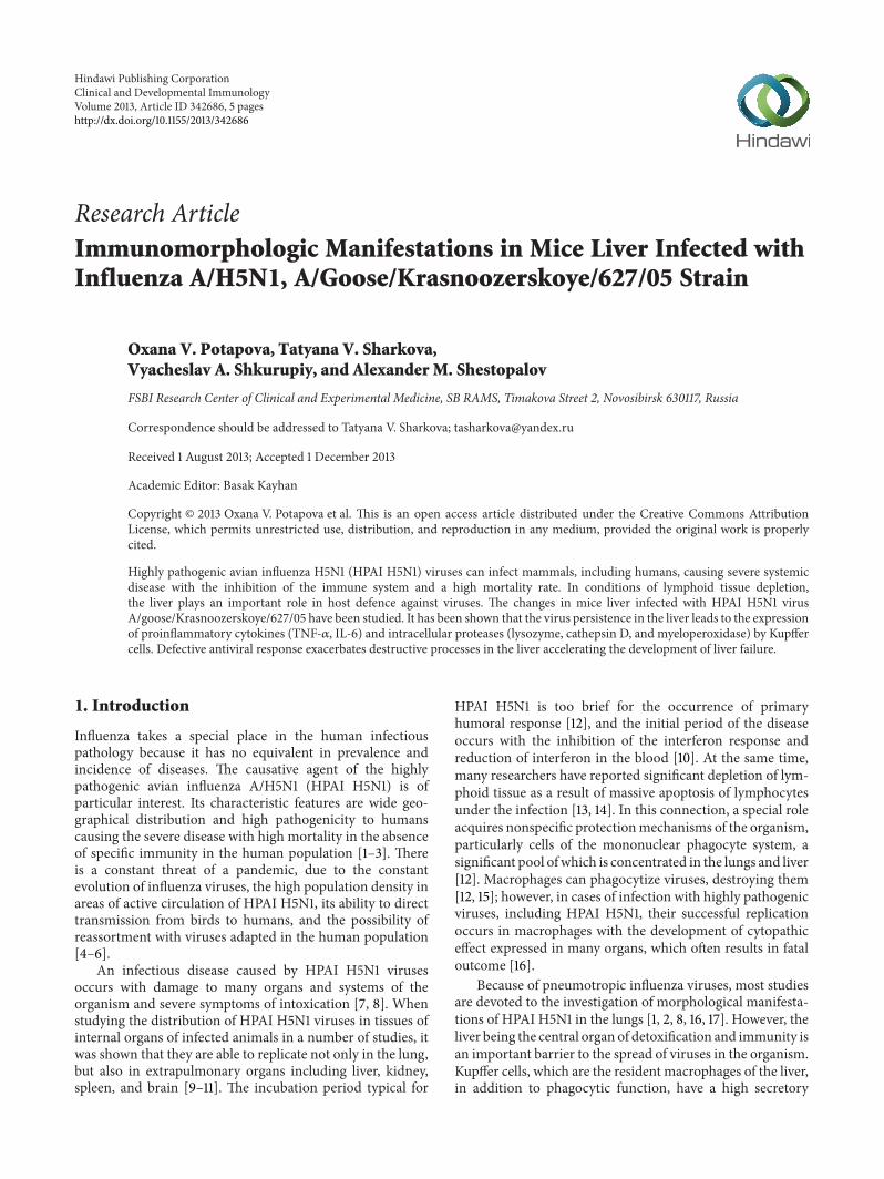
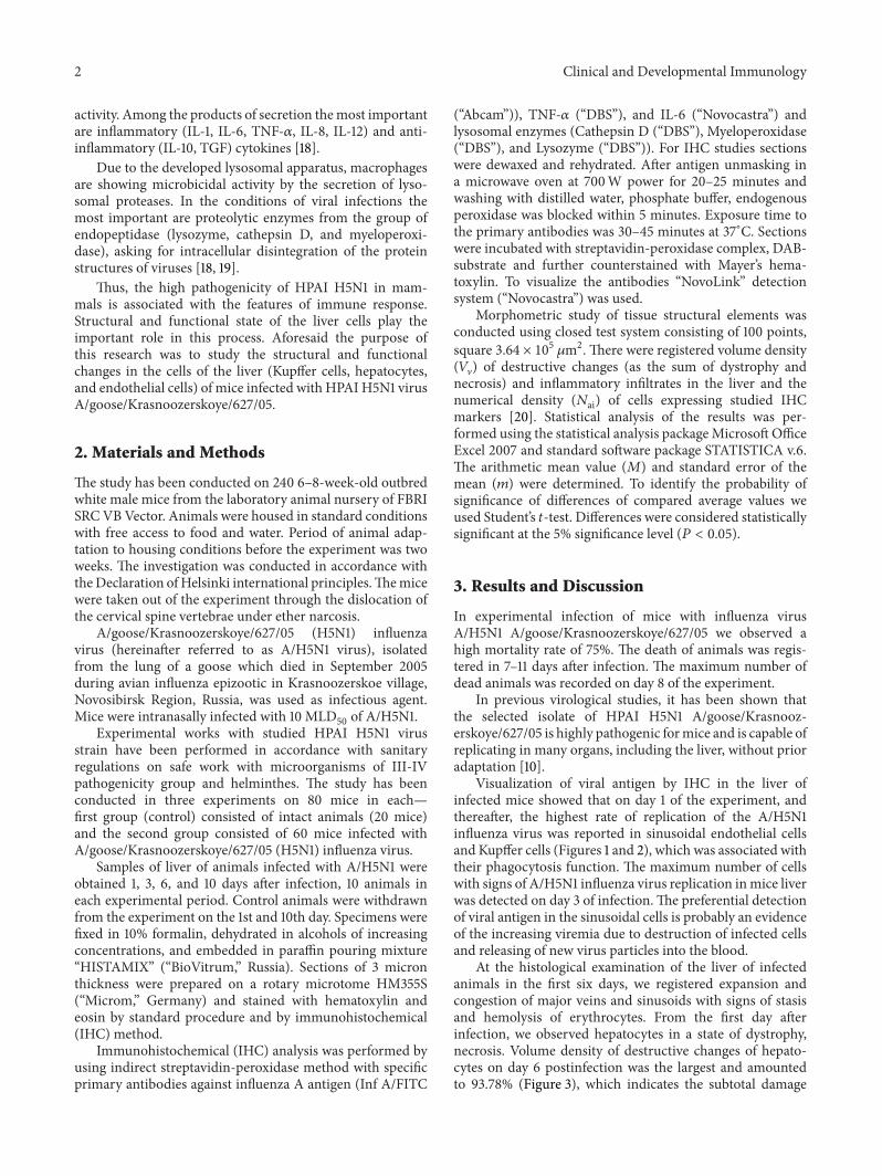
Visualization of viral antigen by IHC in the liver ofinfected mice showed that on day 1 of the experiment, andthereafter, the highest rate of replication of the A/H5N1influenza virus was reported in sinusoidal endothelial cellsand Kupffer cells (Figures 1 and 2), which was associated withtheir phagocytosis function. The maximum number of cellswith signs of A/H5N1 influenza virus replication inmice liverwas detected on day 3 of infection.The preferential detectionof viral antigen in the sinusoidal cells is probably an evidenceof the increasing viremia due to destruction of infected cellsand releasing of new virus particles into the blood.
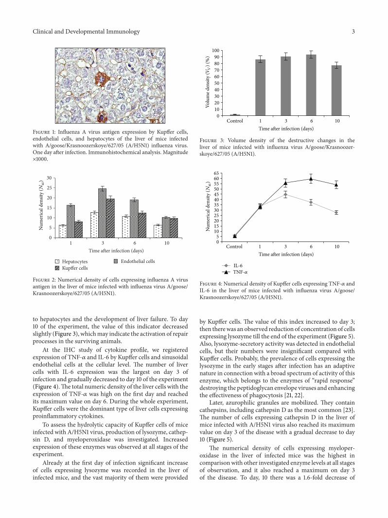
At the histological examination of the liver of infectedanimals in the first six days, we registered expansion andcongestion of major veins and sinusoids with signs of stasisand hemolysis of erythrocytes. From the first day afterinfection, we observed hepatocytes in a state of dystrophy,necrosis. Volume density of destructive changes of hepato-cytes on day 6 postinfection was the largest and amountedto 93.78% (Figure 3), which indicates the subtotal damage
Clinical and Developmental Immunology 3
Figure 1: Influenza A virus antigen expression by Kupffer cells,endothelial cells, and hepatocytes of the liver of mice infectedwith A/goose/Krasnoozerskoye/627/05 (A/H5N1) influenza virus.One day after infection. Immunohistochemical analysis. Magnitude×1000.
0
5
10
15
20
25
30
1 3 6 10Time after infection (days)
Hepatocytes Endothelial cellsKupffer cells
Num
eric
al d
ensit
y (N
ai)
Figure 2: Numerical density of cells expressing influenza A virusantigen in the liver of mice infected with influenza virus A/goose/Krasnoozerskoye/627/05 (A/H5N1).
to hepatocytes and the development of liver failure. To day10 of the experiment, the value of this indicator decreasedslightly (Figure 3), whichmay indicate the activation of repairprocesses in the surviving animals.
At the IHC study of cytokine profile, we registeredexpression of TNF-𝛼 and IL-6 by Kupffer cells and sinusoidalendothelial cells at the cellular level. The number of livercells with IL-6 expression was the largest on day 3 ofinfection and gradually decreased to day 10 of the experiment(Figure 4).The total numeric density of the liver cells with theexpression of TNF-𝛼 was high on the first day and reachedits maximum value on day 6. During the whole experiment,Kupffer cells were the dominant type of liver cells expressingproinflammatory cytokines.
To assess the hydrolytic capacity of Kupffer cells of miceinfected with A/H5N1 virus, production of lysozyme, cathep-sin D, and myeloperoxidase was investigated. Increasedexpression of these enzymes was observed at all stages of theexperiment.
Already at the first day of infection significant increaseof cells expressing lysozyme was recorded in the liver ofinfected mice, and the vast majority of them were provided
0102030405060708090
100
Control 1 3 6 10Time after infection (days)
Volu
me d
ensit
y (V
V) (%
)
Figure 3: Volume density of the destructive changes in theliver of mice infected with influenza virus A/goose/Krasnoozer-skoye/627/05 (A/H5N1).
05
101520253035404550556065
Control 1 3 6 10Time after infection (days)
IL-6
Num
eric
al d
ensit
y (N
ai)
TNF-𝛼
Figure 4: Numerical density of Kupffer cells expressing TNF-𝛼 andIL-6 in the liver of mice infected with influenza virus A/goose/Krasnoozerskoye/627/05 (A/H5N1).
by Kupffer cells. The value of this index increased to day 3;then therewas an observed reduction of concentration of cellsexpressing lysozyme till the end of the experiment (Figure 5).Also, lysozyme-secretory activity was detected in endothelialcells, but their numbers were insignificant compared withKupffer cells. Probably, the prevalence of cells expressing thelysozyme in the early stages after infection has an adaptivenature in connection with a broad spectrum of activity of thisenzyme, which belongs to the enzymes of “rapid response”destroying the peptidoglycan envelope viruses and enhancingthe effectiveness of phagocytosis [21, 22].
Later, azurophilic granules are mobilized. They containcathepsins, including cathepsin D as the most common [23].The number of cells expressing cathepsin D in the liver ofmice infected with A/H5N1 virus also reached its maximumvalue on day 3 of the disease with a gradual decrease to day10 (Figure 5).
The numerical density of cells expressing myeloper-oxidase in the liver of infected mice was the highest incomparisonwith other investigated enzyme levels at all stagesof observation, and it also reached a maximum on day 3of the disease. To day, 10 there was a 1.6-fold decrease of
4 Clinical and Developmental Immunology
01020304050607080
Control 1 3 6 10Time after infection (days)
Lysozyme Cathepsin DMyeloperoxidase
Num
eric
al d
ensit
y (N
ai)
Figure 5: Numerical density of Kupffer cells expressing lysozyme,cathepsin D, and myeloperoxidase in the liver of mice infected withinfluenza virus A/goose/Krasnoozerskoye/627/05 (A/H5N1).
Figure 6: Myeloperoxidase expression by Kupffer cells of the liverof mice infected with A/goose/Krasnoozerskoye/627/05 (A/H5N1)influenza virus. Three days after infection. Immunohistochemicalanalysis. Magnitude ×630.
this parameter (Figures 5 and 6). Myeloperoxidase oxidizescofactors, translating them into an active form, thus provid-ing a powerful microbicidal effect [24]. However, the outputof myeloperoxidase in the extracellular space by exocytosisor phagocytic destruction can cause pathological action ofthe enzyme under the conditions for its development, forexample, low pH values during ischemia.The resulting strongoxidants cause tissue damage of the organism [25].
Analysis of the results suggests that in conditions ofdepletion of lymphoid tissue caused by HPAI H5N1, a specialrole in protecting the organism from infection belongs to theliver as a key organ of the nonspecific protection.
High HPAI H5N1 virus tropism for vascular endothelialcells of all organs promotes early viremia and generalizationof infection. A/H5N1 virus persistence in cells of differenthistogenesis is associated with a significant activation of cell-mediated immunity, resulting in an increased productionof proinflammatory cytokines (TNF-𝛼 and IL-6) that, onthe one hand, is a protective mechanism consisting in therecruitment of immune cells into the focus infection andin the activation of regenerative processes in the organs viagrowth factors. On the other hand, an excessive accumulation
of inflammatory cytokines facilitates the initiation of degra-dation processes [12].
A/H5N1 virus infection ofmicewas accompanied by earlyactivation of the expression of myeloperoxidase, lysozyme,and cathepsin D by Kupffer cells. Intracellular proteases arethe most important factors of nonspecific protection thatprovide the intracellular disintegration of the virus proteins.But, apparently, these mechanisms are ineffective for HPAIH5N1 viruses.
High hydrolytic capacity of cells, being an early nonspe-cific protection factor, at the same time, can be a triggerpoint to initiate lysis of host cells (secondary alteration) withthe development of spread destructive complications in theorgans in case of phagolysosome membrane labilization afterabsorption of viral particles.
Thus, defective antiviral response that triggers a cas-cade of cytokine-mediated responses, only exacerbates thedestructive processes in the liver of experimental animals,which together with insufficient oxygen supply caused bydamage of lungs lead to irreversible structural and functionalchanges and the early development of acute liver failure.
Conflict of Interests
The authors declare that there is no conflict of interestsregarding the publication of this paper.
References
[1] K. F. To, P. K. S. Chan, K. F. Chan et al., “Pathology of fatalhuman infection associated with avian influenza AH5N1 virus,”Journal of Medical Virology, vol. 63, no. 3, pp. 242–246, 2001.
[2] F. Pei, J. Zheng, Z.-F. Gao et al., “Lung pathology and patho-genesis of severe acute respiratory syndrome: a report of six fullautopsies,”Zhonghua Bing Li Xue ZaZhi, vol. 34, no. 10, pp. 656–660, 2005.
[3] Y. Suzuki, “Sialobiology of influenza molecular mechanismof host range variation of influenza viruses,” Biological &Pharmaceutical Bulletin, vol. 28, no. 3, pp. 399–408, 2005.
[4] R. G. Webster, M. Peiris, H. Chen, and Y. Guan, “H5N1outbreaks and enzootic influenza,” Emerging Infectious Diseases,vol. 12, no. 1, pp. 3–8, 2006.
[5] N. Sirinonthanawech, M. Uiprasertkul, O. Suptawiwat, andP. Auewarakul, “Viral load of the highly pathogenic avianinfluenza H5N1 virus in infected human tissues,” Journal ofMedical Virology, vol. 83, no. 8, pp. 1418–1423, 2011.
[6] http://www.who.int/influenza/human animal interface/ENGIP 20130705CumulativeNumberH5N1cases 2.pdf.
[7] M. Uiprasertkul, P. Puthavathana, K. Sangsiriwut et al.,“Influenza A H5N1 replication sites in humans,” EmergingInfectious Diseases, vol. 11, no. 7, pp. 1036–1041, 2005.
[8] T. Xu, J. Qiao, L. Zhao et al., “Acute respiratory distresssyndrome induced by avian influenza A (H5N1) virus in mice,”American Journal of Respiratory and Critical Care Medicine, vol.174, no. 9, pp. 1011–1017, 2006.
[9] H. Nishimura, S. Itamura, T. Iwasaki, T. Kurata, andM. Tashiro,“Characterization of human influenza A (H5N1) virus infectionin mice: neuro-, pneumo- and adipotropic infection,” Journal ofGeneral Virology, vol. 81, no. 10, pp. 2503–2510, 2000.
Clinical and Developmental Immunology 5
[10] A. M. Shestopalov, K. A. Sharshov, A. V. Zaykovskaya et al.,“Studying the pathogenicity of avian influenza virus subtypeH5N1 strains from the Russian Federation using mouse model,”Bulletin of Experimental Biology and Medicine, vol. 146, no. 3,pp. 341–343, 2008.
[11] A.-E. Tolnay, C. R. Baskin, T. M. Tumpey et al., “Extrapul-monary tissue responses in cynomolgus macaques (Macacafascicularis) infected with highly pathogenic avian influenza A(H5N1) virus,” Archives of Virology, vol. 155, no. 6, pp. 905–914,2010.
[12] I. Roitt, D. K. Male, and J. Brostoff, Immunology, ElsevierScience, Amsterdam, The Netherlands, 2001.
[13] T. M. Tumpey, X. Lu, T. Morken, S. R. Zaki, and J. M. Katz,“Depletion of lymphocytes and diminished cytokine produc-tion in mice infected with a highly virulent influenza A (H5N1)virus isolated from humans,” Journal of Virology, vol. 74, no. 13,pp. 6105–6116, 2000.
[14] M. Uiprasertkul, R. Kitphati, P. Puthavathana et al., “Apoptosisand pathogenesis of avian influenzaA (H5N1) virus in humans,”Emerging Infectious Diseases, vol. 13, no. 5, pp. 708–712, 2007.
[15] A. F. Valledor, M. Comalada, L. F. Santamarıa-Babi, J. Lloberas,and A. Celada, “Macrophage proinflammatory activation anddeactivation. A question of balance,” Advances in Immunology,vol. 108, pp. 1–20, 2010.
[16] M. Peiris, “Pathogenesis of avian flu H5N1 and SARS,” NovartisFoundation Symposium, vol. 279, pp. 56–60, 2006.
[17] O. V. Potapova, V. A. Shkurupiy, T. V. Sharkova, A. V. Troitskiy,N. G. Lusgina, and A. M. Shestopalov, “Preventive efficacy ofoxidized dextran and pathomorphological processes in mouselungs in avian influenza A/H5N1,” Bulletin of ExperimentalBiology and Medicine, vol. 150, no. 6, pp. 707–710, 2011.
[18] L. A. Possamai, C. G. Antoniades, Q. M. Anstee et al., “Role ofmonocytes andmacrophages in experimental and human acuteliver failure,” World Journal of Gastroenterology, vol. 16, no. 15,pp. 1811–1819, 2010.
[19] T. Burster, T. Giffon, M. E. Dahl et al., “Influenza A virus ele-vates active cathepsin B in primary murine DC,” InternationalImmunology, vol. 19, no. 5, pp. 645–655, 2007.
[20] E. R. Weibel, “Morphometry: stereological theory and practicalmethods,” inModels of Lung Disease. Microscopy and StructuralMethods, pp. 199–252, Marcel Dekker, New York, NY, USA,1990.
[21] Y. Miyake, H. Kaise, K.-I. Isono, H. Koseki, K. Kohno, and M.Tanaka, “Protective role of macrophages in noninflammatorylung injury caused by selective ablation of alveolar epithelialtype II cells,” Journal of Immunology, vol. 178, no. 8, pp. 5001–5009, 2007.
[22] D. P. Granados, P.-L. Tanguay, M.-P. Hardy et al., “ER stressaffects processing of MHC class I-associated peptides,” BMCImmunology, vol. 10, article 10, 2009.
[23] M. A. Bewley, T. K. Pham, H. M. Marriott et al., “Proteomicevaluation and validation of cathepsin D regulated proteins inmacrophages exposed to Streptococcus pneumoniae,”Molecular& Cellular Proteomics, vol. 10, no. 6, 2011.
[24] R. C. Allen and J. T. Stephens Jr., “Myeloperoxidase selectivelybinds and selectively kills microbes,” Infection and Immunity,vol. 79, no. 1, pp. 474–485, 2011.
[25] I. Hendrikje Buss, R. Senthilmohan, B. A. Darlow, N.Mogridge,A. J. Kettle, and C. C. Winterbourn, “3-chlorotyrosine as amarker of protein damage by myeloperoxidase in tracheal aspi-rates from preterm infants: association with adverse respiratoryoutcome,” Pediatric Research, vol. 53, no. 3, pp. 455–462, 2003.

Submit your manuscripts athttp://www.hindawi.com
Stem CellsInternational
Hindawi Publishing Corporationhttp://www.hindawi.com Volume 2014
Hindawi Publishing Corporationhttp://www.hindawi.com Volume 2014
MEDIATORSINFLAMMATION
of
Hindawi Publishing Corporationhttp://www.hindawi.com Volume 2014
Behavioural Neurology
EndocrinologyInternational Journal of
Hindawi Publishing Corporationhttp://www.hindawi.com Volume 2014
Hindawi Publishing Corporationhttp://www.hindawi.com Volume 2014
Disease Markers
Hindawi Publishing Corporationhttp://www.hindawi.com Volume 2014
BioMed Research International
OncologyJournal of
Hindawi Publishing Corporationhttp://www.hindawi.com Volume 2014
Hindawi Publishing Corporationhttp://www.hindawi.com Volume 2014
Oxidative Medicine and Cellular Longevity
Hindawi Publishing Corporationhttp://www.hindawi.com Volume 2014
PPAR Research
The Scientific World JournalHindawi Publishing Corporation http://www.hindawi.com Volume 2014
Immunology ResearchHindawi Publishing Corporationhttp://www.hindawi.com Volume 2014
Journal of
ObesityJournal of
Hindawi Publishing Corporationhttp://www.hindawi.com Volume 2014
Hindawi Publishing Corporationhttp://www.hindawi.com Volume 2014
Computational and Mathematical Methods in Medicine
OphthalmologyJournal of
Hindawi Publishing Corporationhttp://www.hindawi.com Volume 2014
Diabetes ResearchJournal of
Hindawi Publishing Corporationhttp://www.hindawi.com Volume 2014
Hindawi Publishing Corporationhttp://www.hindawi.com Volume 2014
Research and TreatmentAIDS
Hindawi Publishing Corporationhttp://www.hindawi.com Volume 2014
Gastroenterology Research and Practice
Hindawi Publishing Corporationhttp://www.hindawi.com Volume 2014
Parkinson’s Disease
Evidence-Based Complementary and Alternative Medicine
Volume 2014Hindawi Publishing Corporationhttp://www.hindawi.com