research article - irjponline.comirjponline.com/admin/php/uploads/3228_pdf.pdf · g.v radha et al....
TRANSCRIPT
G.VRadhaetal.Int.Res.J.Pharm.2019,10(2)
94
INTERNATIONAL RESEARCH JOURNAL OF PHARMACY www.irjponline.com
ISSN2230–8407
ResearchArticleDESIGN AND EVALUATION OF TOPICAL VAGINAL PRONIOSOMAL FORMULATIONS OF TENOFOVIR DISOPROXIL FUMARATE FOR HIV PREVENTION G.V Radha *, Vudimudi Vinisha, Jampala Rajkumar, Ankita Ghosh Department of Pharmaceutics, Gitam Institute of Pharmacy, Gitam (Deemed to be University), Rushikonda, Visakhapatnam-530045, Andhra Pradesh, India *Corresponding Author Email: [email protected] Article Received on: 09/11/18 Approved for publication: 18/01/19 DOI: 10.7897/2230-8407.100250 ABSTRACT The aim of the present study was to design and evaluate topical proniosomal vaginal gels and proniosomal gel suppositories of Tenofovir Disoproxil Fumarate (TDF) for the treatment of HIV prevention. Evaluation of formulating proniosomal gels was performed such as morphology, pH, entrapment efficiency and In-Vitro diffusion release formulation and also evaluated for EX-Vivo drug permeation and drug retention studies. The optimised formulation FE2 from Enriched lecithin (FE2) showed a high release rate of 92.2% than F2 (soya lecithin) showed 73%. The %EE had also showed higher for FE2 formulation followed by SEM analysis, which showed good vesicles. Ex-vivo studies had been conducted from goat vaginal tissue for 24hrs, FE2 gel showed 82.2% release where as 75% found for F2. Proniosomal suppository was prepared from the optimized formulation of proniosomal gel and characterized the average M.P for Suppositories are found 37.2ºC and average weight of provision suppository found within limits 0.8623. Proniosomal suppository in-vitro dissolution studies showed the controlled release rate when compared to proniosomal formulations. Proniosomal suppository formulation in-order to overcome drug leakage and stability problems of provisional gel TDF. KEYWORDS: Proniosomal gel, suppository, controlled release, non-ionic surfactants, coacervation phase separation, Tenofovir Disoproxil Fumarate (TDF). INTRODUCTION Tremendous research and efforts have been taken over the past decade to develop the vaginal products for HIV prevention1. For the various purposes various dosage forms have been studied, most commonly used are creams, gels, suppositories, forms vaginal rings inserts, ointments etc. gels and suppositories are widely utilised for vaginal application from a microbicides product development point of view and consider factors affecting patient compliance and acceptability, within this body of work gel and suppositories were studied for their potential as vaginal microbicides dosage forms. Attempts have been made previously to develop a topical microbicide for rectal or vaginal administration in different dosage forms, one of the drug candidates used in these studies2. Tenofovir disoproxil fumarate (TDF) belongs to a class of antiretroviral drugs known as nucleotide analogue reverse transcriptase inhibitors (nRTIs), which block reverse transcriptase, an enzyme crucial to viral production in HIV-infected individuals. It is frequently prescribed not only for its efficacy but also for its declined side effect profile compared with other nucleoside analogues. Proniosomes are microscopic lamellar structures. They mix with a non-ionic wetter of the chemical group or dialkyl polyglycerol ether category and sterol followed by association in liquid media3.The chemical agent molecule direct themselves such the deliquescent ends of the non-ionic chemical agent orient outward, whereas the hydrophobic ends area unit within the other way to make the bilayer. Like liposomes, proniosomes also are fictitious of a bilayer4. That the bilayer is created of non-ionic surface active agents. On the idea of methodology of preparation proniosomes area unit unilamellar or multi-lamellar5.The niosome is made of a surfactant bilayer with its hydrophilic ends uncovered on the outside and inside of the vesicles while the hydrophobic chains face each other within the bilayer. Hence the
vesicle holds hydrophilic drugs within the space enclosed in the vesicle and the hydrophobic drugs are surrounded within the bilayer6, studied aims to discover the potential of non-ionic surfactant based proniosomal gel (PNG) in improving the topical delivery of tazarotene by in vitro and in vivo studies. In vaginal drug delivery, the physiological conditions imposed by the protective mechanisms of the vagina often lead to the limited contact time of administered drugs with vaginal mucosa and a short duration of therapeutic efficacy, making a frequent dosing regimen necessary7. The objective of this project was to develop a safe, effective, and acceptable intra vaginal dosage form for HIV prophylaxis. In this study we prepared proniosomal gel, proniosomal suppositories containing TDF and evaluating for best formulation for efficient drug release. TDF successfully developed by span 20,40,60,80 grades and also used enriched lecithin and soya lecithin to evaluate release characteristics. A comparative in-vitro dissolution study had been conducted between proniosomes suppository, proniosomal gel formulations. MATERIALS AND METHODS Tenofovir Disoproxil Fumarate from (Mylan Pharmaceuticals Private Limited, Bangalore), Cholesterol (Finar chemicals Ltd, India),Span20,40,60,80(Molychem,India)Tween 20,Tween 80, PEG 6000, PEG 600 (Loba Chemie Pvt.Ltd,India) Lecithin (Otto Chemie Pvt. Ltd,India). PREPARATION OF TDF PRONIOSOMAL GEL TDF proniosomal gel was prepared by coacervation phase separation methodology. Exactly weighed amounts of surfactants, lecithin, cholesterol and drug were taken in clean and dry wide mouthed glass ampoule of 5.0 millilitre unit capacity and ethanol 2.5 ml was added to it. once warming, all the
G.VRadhaetal.Int.Res.J.Pharm.2019,10(2)
95
ingredients were mixed well with a glass rod the open end of the glass bottle was covered with lid to prevent the loss from it and warm over water bath at 60-70oC for about 5 minutes till the chemical agent mixture was dissolved fully8. Then the 1.6 ml of PH 4.4 phosphate buffer was added and warm over water bath until transparent solution was formed that was converted into
proniosomal gel on cooling. The gel therefore obtained was preserved within the same glass bottle in dark conditions for characterization. Formulation composition showed in table no.1. Proniosomal suppository composition had shown in table no.2. Drug suppository composition showed in table 3.
Table 1: Composition of proniosomal gel formulations
FORMULATION FE1 FE2 FE3 FE4 F1 F2 F3 F4
Drug(mg) 10 10 10 10 10 10 10 10 Soya lecithin/enriched lecithin(mg) 450 450 450 450 450 450 450 450
Surfactants(mg) (S20) 450
(S40)
450 (S60) 450
(S80) 450
(S20) 450
(S40)
450 (S60) 450
(S80) 450
Cholesterol(mg) 100 100 100 100 100 100 100 100 Ethanol(ml) 2.5 2.5 2.5 2.5 2.5 2.5 2.5 2.5
Buffer ph4.4(ml) 1.6 1.6 1.6 1.6 1.6 1.6 1.6 1.6
Table 2: Proniosomal suppository composition
INGREDIENTS WEIGHT(1g) Drug 40mg
Span 40 (optimized formula) 316mg PEG 6000:PEG 600 158:158mg
Cholesterol 24mg Lecithin 316mg
DRUG SUPPOSITORY COMPOSITION
Table 3: Drug suppository composition
INGREDIENTS WEIGHT(1g) DRUG 40mg
PEG6000:PEG600 324:324mg CHARACTERIZATION OF PRONIOSOMAL GEL FORMULATION OPTICAL MICROSCOPY The vesicle formation by the particular procedure was confirmed by optical microscopy in 100x resolution. Procedure gel before hydration clear liquid crystalline state was observed than upon hydration niosomal suspension made was placed over a glass slide and glued over by drying at space temperature, the dry skinny film of niosomal suspension determined for the formation of vesicles9.The photomicrograph of the preparation additionally obtained from the magnifier by employing a digital SLR camera. SCANNING ELECTRON MICROSCOPY AND SIZE ANALYSIS (SEM) Particle size of proniosomes may be a issue of prime importance. The surface morphology and size distribution of proniosomes were studied by SEM. A double aspect tape that was mounted on metallic element stubs and therefore the proniosomal powder was unfolded on it10. The metallic element stub was placed in chamber of scanning microscope (XL thirty ESEM with EDAX, Philips, Netherlands). The morphological characterization of the sample was ascertained employing a aerosolized secondary lepton detector (Work pressure of zero.8 torr, acceleration voltage-30.00KV) XL thirty, (Philips, Neherlan) FTIR-SPECTROSCOPY Fourier transform infrared spectroscopy (FT-IR) could be a easy is technique for the detection of modification within the excipient-drug mixture. The disappearance of absorption peak intensity combined with the appearance of new peak provides a clear proof for interactions between drug and excipient. FTIR spectra of drug and polymers were mixed together and were obtained by the conventional KBr disc/pellet method11. The
sample was grounded gently with anhydrous KBr and compressed to form a pellet. The scanning range was in between 400 and 4000 cm-1. ENCAPSULATION EFFICIENCY Encapsulation efficiency (EE %) Weighed amount, 0.2 g, of each formula was mixed with 10ml pH 4.4 phosphate buffers in glass tube; the aqueous suspension was sonicated for 30 min and then exposed to centrifugation at 9000 rpm for 45 min at 4oC to separate the TDF proniosomes. The supernatant was recovered and assayed by spectrophotometric UV analysis (Schimadzu spectrophotometer. Model UV-1601, Japan) at ʎmax 270 nm. The percentage of drug encapsulation efficiency calculated by following formula12.
Formula for %EE %Encapsulation efficiency= {1 – (unencapsulated drug/Total drug)} x
100 IN VITRO DIFFUSION STUDIES In vitro release studies on the proniosomal gel were performed by using Franz diffusion cell. The volume of the receptor compartment was 15 ml. Donor compartment with an area of 1.41cm2 was exposed to the receptor compartment. Cellophane membrane was soaked in a phosphate buffer pH 4.4 for 1 hour before carrying the experiment. The dialysis cellophane membrane (MMCO14KDC) was mounted between the donor and receptor compartment. A Weighed quantity of proniosomal gel was placed on one side of the dialysis membrane13. The receptor medium was phosphate buffer pH 4.4. The receptor compartment was enclosed by a vessel to take care of the temperature at 37±1ºc. The heat was provided by using a hot plate with a magnetic stirrer. The receptor fluid was stirred by a magnetic bead fitted to a magnetic stirrer. At every sampling intermission, 3ml samples
G.VRadhaetal.Int.Res.J.Pharm.2019,10(2)
96
were withdrawn and were replaced by equal volumes of fresh receptor fluid. EX-VIVO SKIN PERMEATION AND RETENTION STUDIES Isolated goat vaginal mucosa was collected from a local slaughterhouse, immediately after the animal was slaughtered, unwanted fat-containing tissues were removed away by using a razor blade. The tissue so prepared was kept in an aluminium foil at 4oC and used within two days. On the day of the experiment, goat vaginal mucosa was cut into 5-cm×5- cm pieces and mounted on a simulated vaginal system with the mucosal side up. The proniosomal gel formula 4mg for F2 and 7mg for FE 2, were mounted on the mucosal membrane on the determined area (1 cm x 2 cm), the goat vaginal tissue was transferred to the pH4.4 phosphate buffer solution. Where it was kept for about 1hour. Then skin was kept between donor and receptor compartment of Franz diffusion cell for carrying permeation study14 using pH 4.4 Phosphate buffer at temperature 37.5±10 0C which maintains 100 rpm. The heat was provided employing a regulator hot plate with a magnetic stirrer. The receptor fluid was stirred by a magnetic bead placed on a magnetic stirrer. At every sampling time, 3ml of samples were withdrawn were analysed by using ultraviolet light spectrophoto metrically. TISSUE DRUG RETENTION STUDY The drug retained in skin is measured after permeation experiments, for which the skin was removed from the diffusion cells after completion of experiments. The surface of skin specimens was washed 10 times with 1 ml distilled water and dried with filter paper. The effective surface area of the skin was separated and minced with a surgical sterile scalpel then finally homogenized in a vial filled with 40ml methanol by using homogenizer (D-LAB D-500 Homogenizer India) at 4000 rpm for 5min in an ice bath15. The tissue suspension was centrifuged (Refrigerated Centrifuge REMI Mumbai, India) at 9000 rpm for 45 min and then the supernatant was filtered. Further, the supernatant tissue suspension was extracted with methanol and filtered. The supernatant from receptor solution and tissue suspension as well as washing solution were assayed for TDF content by double beam UV‐visible spectrophotometer using phosphate buffer 4.4 as blank at 270 nm. CHARACTERIZATION OF DRUG SUPPOSITORY AND PRONIOSOMAL SUPPOSITORY UNIFORMITY OF THE WEIGHT Weigh 20 suppositories individually then together and calculate the average weight according to the following equation16
Average weight = Total weight of the 20 suppositories/20 There must be not more than 2 suppositories differ from the average weight by more than 5% and no suppository differs from the average weight by more than 10% MELTING POINT The ascending melting point method was used to determine the melting point of suppositories. Capillary tubes,10 cm in length , sealed at one end, were filled with the formulation to about 1cm height, then was dipped in gradually heated electro-thermal thermometer17. DETERMINATION OF PH OF SUPPOSITORIES The suppository was digested in warm water then filtered and the pH of the filtrate was measured by suitable pH meter18.
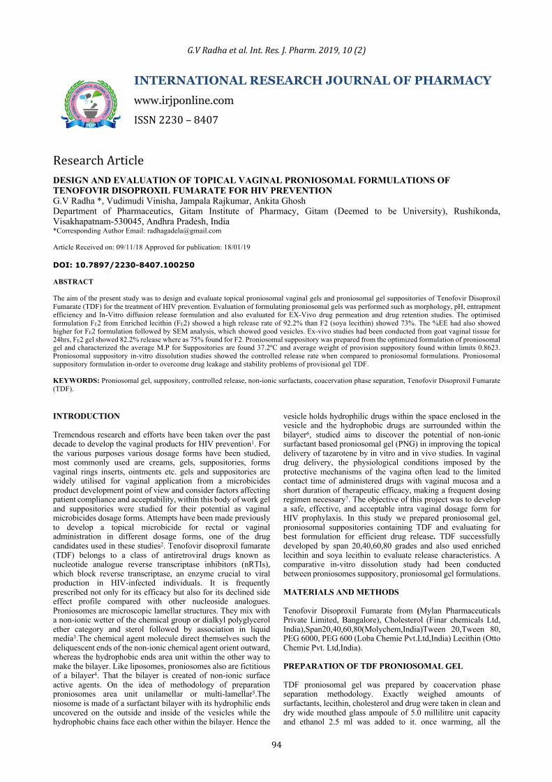
OPTICAL MICROSCOPY The vesicle formation by the particular procedure was confirmed by optical microscopy in 100x resolution. Procedure suppositories upon hydration was placed over a glass slide and observed for vesicles. The photomicrograph of the preparation additionally obtained from the magnifier by employing a digital SLR camera. COMPARATIVE IN-VITRO RELEASE STUDY The release of TDF gel, TDF proniosomal suppository, TDF drug suppository was determined using membrane diffusion technique19 In brief, 1ml of simulated vaginal fluid was added to proniosomal gel containing 40 mg TDF placed in a glass cylinder of diameter 2.5 cm enclosed at its lower end with a soaked cellulose dialysis membrane (Spectra pore dialysis membrane 2000–15 000Mw cut-off). The cylinder was rotated at a speed of 50 rpm with its lower end immersed in the dissolution flask of a USP dissolution tester (electro lab dissolution tester). The dissolution flask contained phosphate buffer of pH4..4 maintained at a temperature of 37 ºC20. A schematic illustration of the experiment setup is shown in Figure 1. At pre-determined time intervals, samples from the release medium were withdrawn and assayed spectrophoto metrically at 270 nm for drug content. Each withdrawn sample was replaced by an equal volume of simulated vaginal fluid. Experiment setup was showed in figure 1.
Fig 1: In-vitro release experiment setup
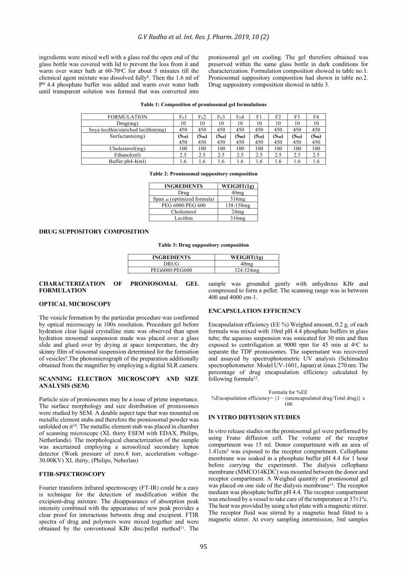
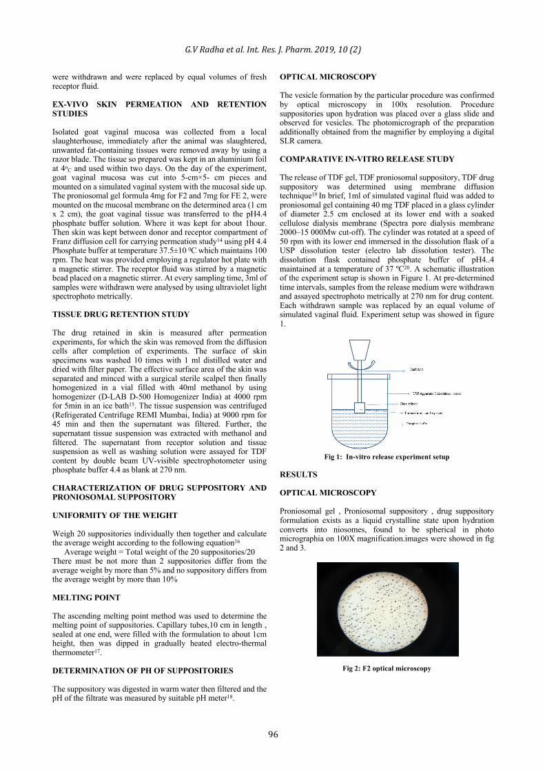
RESULTS OPTICAL MICROSCOPY Proniosomal gel , Proniosomal suppository , drug suppository formulation exists as a liquid crystalline state upon hydration converts into niosomes, found to be spherical in photo micrographia on 100X magnification.images were showed in fig 2 and 3.
Fig 2: F2 optical microscopy
G.VRadhaetal.Int.Res.J.Pharm.2019,10(2)
97
Fig 3: FE2 optical microscopy ENCAPSULATION EFFICIENCY A weighed amount of proniosomal gel formulation was taken in a centrifugal tube, to this 10ml of phosphate buffer pH 4.4 added to this. Then it is kept for ultra-centrifugation about an RPM of 9000 for 45 minutes at 4oC. Later a clear fraction and cloudy fraction can be clearly observed in the centrifugal tube, 1ml of clear fraction is used for calculation of free drug in the formulation. Then to the cloudy solution add 1:1 ratio of propanolol and ethyl alcohol, which makes vesicles get braked and encapsulated drug can be estimated. Encapsulation efficiency data showed in table 4.
Table 4: Encapsulation efficiency data
Formulation % Encapsulation efficiency FE1 72.1 FE2 70.6 FE3 70.3 FE4 32.4 F1 74.4 F2 73.7 F3 66.9 F4 33.9
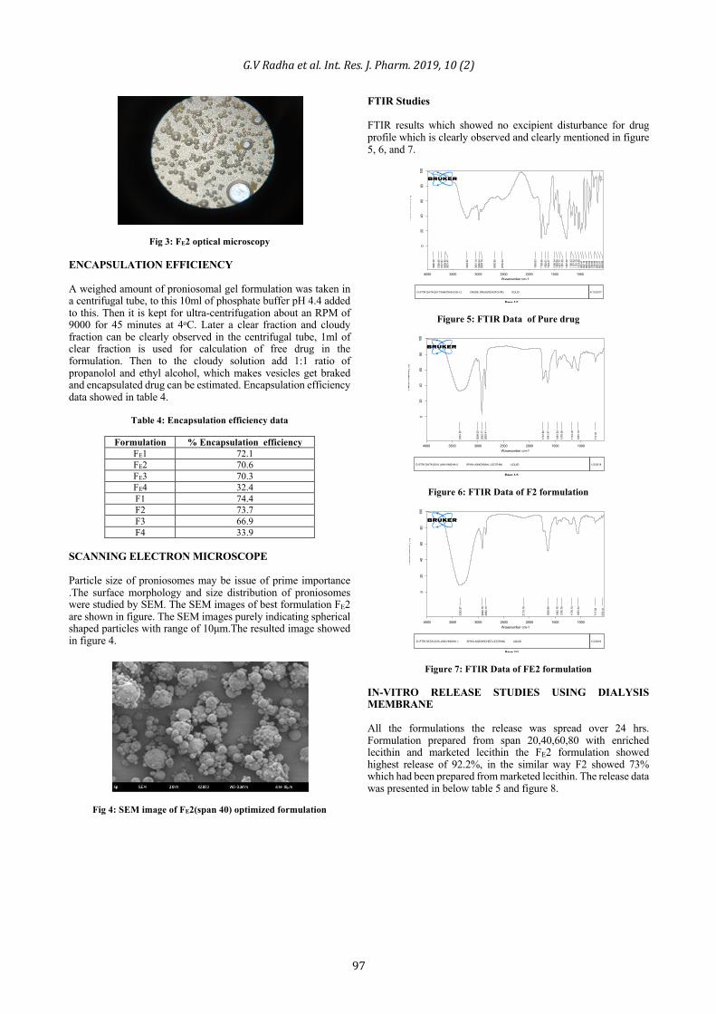
SCANNING ELECTRON MICROSCOPE Particle size of proniosomes may be issue of prime importance .The surface morphology and size distribution of proniosomes were studied by SEM. The SEM images of best formulation FE2 are shown in figure. The SEM images purely indicating spherical shaped particles with range of 10μm.The resulted image showed in figure 4.
Fig 4: SEM image of FE2(span 40) optimized formulation
FTIR Studies FTIR results which showed no excipient disturbance for drug profile which is clearly observed and clearly mentioned in figure 5, 6, and 7.
Figure 5: FTIR Data of Pure drug
Figure 6: FTIR Data of F2 formulation
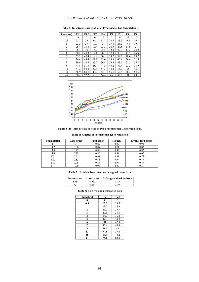
Figure 7: FTIR Data of FE2 formulation IN-VITRO RELEASE STUDIES USING DIALYSIS MEMBRANE All the formulations the release was spread over 24 hrs. Formulation prepared from span 20,40,60,80 with enriched lecithin and marketed lecithin the FE2 formulation showed highest release of 92.2%, in the similar way F2 showed 73% which had been prepared from marketed lecithin. The release data was presented in below table 5 and figure 8.
D:\FTIR DATA\2017\ANKITAGHOSH.2 CRUDE DRUG(TENOFOVIR) SOLID 4/13/2017
3880
.36
3780
.95
3721
.91
3658
.80
3631
.47
3224
.52
3051
.14
2986
.64
2938
.78
2682
.85
2530
.24
1883
.63
1758
.96
1682
.01
1624
.37
1506
.34
1469
.96
1421
.17
1381
.50
1267
.00
1182
.43
1157
.42
1101
.10
1033
.89
985.
4195
2.16
895.
6983
0.09
788.
9672
8.89
699.
4365
3.61
599.
89
1000150020002500300035004000Wavenumber cm-1
020
4060
8010
0
Tran
smitt
ance
[%]
Page 1/1
D:\FTIR DATA\2018 JAN\VINISHA.0 SPAN 40(NORMAL LECITHIN) LIQUID 1/2/2018
3351
.25
3009
.03
2922
.77
2853
.41
1737
.60
1641
.57
1461
.32
1376
.06
1174
.18
1051
.19
717.
07
1000150020002500300035004000Wavenumber cm-1
020
4060
8010
0
Tran
smitt
ance
[%]
Page 1/1
D:\FTIR DATA\2018 JAN\VINISHA.1 SPAN 40(ENRICHED LECITHIN) LIQUID 1/2/2018
3352
.97
2920
.79
2852
.10
2119
.79
1639
.99
1462
.76
1376
.79
1178
.74
1053
.34
717.
56
576.
41
1000150020002500300035004000Wavenumber cm-1
020
4060
8010
0
Tran
smitt
ance
[%]
Page 1/1
G.VRadhaetal.Int.Res.J.Pharm.2019,10(2)
98
Table 5: In-Vitro release profiles of Proniosomal Gel formulations
Time(hrs) FE1 FE2 FE3 FE4 F1 F2 F3 F4 0 0 0 0 0 0 0 0 0
0.5 21.7 26 9.3 18.7 23.4 21.2 6.2 16.1 1 22.2 27 10.9 21 27.4 23.2 9.8 19.3 2 25.4 35.8 12.4 23.3 28.9 26.5 11.6 21 3 28.7 39 14.2 25.5 32.4 27.2 13.2 24.2 4 30.2 40.3 17.1 28.1 37.9 35.8 15.7 26.3 5 33.2 45.4 19.8 30.1 38.2 39.3 18.6 29.7 6 36.4 49.4 21.2 33.6 38.6 40.8 20.4 32.5 7 39.6 50.6 25.7 36.4 39.6 45.4 21.2 35.8 8 41.8 51.3 28.4 41.5 40.2 47.4 23.2 38.9
12 47.2 60.2 32.3 54.3 48.2 53.2 26 46.1 18 56.9 80.8 40.2 62.1 55.1 62.6 38.3 54.5 24 68.4 92.2 53.3 66.4 66 82.5 48 60.1
Figure 8: In-Vitro release profiles of Drug Proniosomal Gel formulations.
Table 6: Kinetics of Proniosomal gel Formulation
Formulation Zero order First order Higuchi n value for peppas F1 0.81 0.89 0.92 0.91 F2 0.86 0.89 0.71 0.83 F3 0.77 0.88 0.92 0.83 F4 0.78 0.86 0.94 0.65
FE1 0.95 0.94 0.81 0.75 FE2 0.83 0.88 0.94 0.87 FE3 0.78 0.86 0.94 0.87 FE4 0.90 0.92 0.97 0.30
Table 7: Ex-Vivo drug retention in vaginal tissue data
Formulation Absorbance %Drug retained in tissue
Fe2 0.332 18.5 F2 0.214 23.9
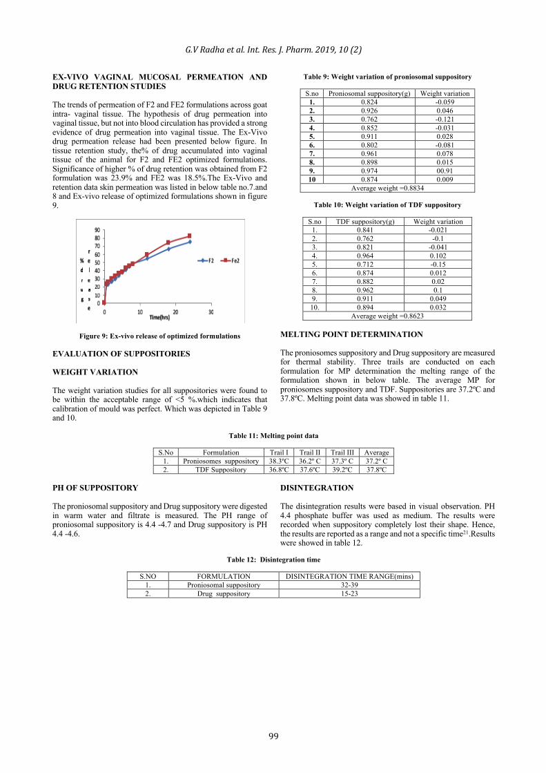
Table 8: Ex-Vivo skin permeation data
Time(hrs) F2 Fe2
0 0 0 0.5 22.7 23.5 1 23.2 25.3 2 26.7 29.7 3 29.6 33.1 4 34.2 36.4 5 37.8 38.3 6 41 42.8 7 45.4 45.4 8 48.9 48
12 54.8 59.4 18 66.6 74.2 24 75.1 82.4
G.VRadhaetal.Int.Res.J.Pharm.2019,10(2)
99
EX-VIVO VAGINAL MUCOSAL PERMEATION AND DRUG RETENTION STUDIES The trends of permeation of F2 and FE2 formulations across goat intra- vaginal tissue. The hypothesis of drug permeation into vaginal tissue, but not into blood circulation has provided a strong evidence of drug permeation into vaginal tissue. The Ex-Vivo drug permeation release had been presented below figure. In tissue retention study, the% of drug accumulated into vaginal tissue of the animal for F2 and FE2 optimized formulations. Significance of higher % of drug retention was obtained from F2 formulation was 23.9% and FE2 was 18.5%.The Ex-Vivo and retention data skin permeation was listed in below table no.7.and 8 and Ex-vivo release of optimized formulations shown in figure 9.
Figure 9: Ex-vivo release of optimized formulations EVALUATION OF SUPPOSITORIES WEIGHT VARIATION The weight variation studies for all suppositories were found to be within the acceptable range of <5 %.which indicates that calibration of mould was perfect. Which was depicted in Table 9 and 10.
Table 9: Weight variation of proniosomal suppository
S.no Proniosomal suppository(g) Weight variation 1. 0.824 -0.059 2. 0.926 0.046 3. 0.762 -0.121 4. 0.852 -0.031 5. 0.911 0.028 6. 0.802 -0.081 7. 0.961 0.078 8. 0.898 0.015 9. 0.974 00.91 10 0.874 0.009
Average weight =0.8834
Table 10: Weight variation of TDF suppository
S.no TDF suppository(g) Weight variation 1. 0.841 -0.021 2. 0.762 -0.1 3. 0.821 -0.041 4. 0.964 0.102 5. 0.712 -0.15 6. 0.874 0.012 7. 0.882 0.02 8. 0.962 0.1 9. 0.911 0.049
10. 0.894 0.032 Average weight =0.8623
MELTING POINT DETERMINATION The proniosomes suppository and Drug suppository are measured for thermal stability. Three trails are conducted on each formulation for MP determination the melting range of the formulation shown in below table. The average MP for proniosomes suppository and TDF. Suppositories are 37.2ºC and 37.8ºC. Melting point data was showed in table 11.
Table 11: Melting point data
S.No Formulation Trail Ι Trail ΙΙ Trail ΙΙΙ Average
1. Proniosomes suppository 38.3ºC 36.2º C 37.3º C 37.2º C 2. TDF Suppository 36.8ºC 37.6ºC 39.2ºC 37.8ºC
PH OF SUPPOSITORY The proniosomal suppository and Drug suppository were digested in warm water and filtrate is measured. The PH range of proniosomal suppository is 4.4 -4.7 and Drug suppository is PH 4.4 -4.6.
DISINTEGRATION The disintegration results were based in visual observation. PH 4.4 phosphate buffer was used as medium. The results were recorded when suppository completely lost their shape. Hence, the results are reported as a range and not a specific time21.Results were showed in table 12.
Table 12: Disintegration time
S.NO FORMULATION DISINTEGRATION TIME RANGE(mins)
1. Proniosomal suppository 32-39 2. Drug suppository 15-23
G.VRadhaetal.Int.Res.J.Pharm.2019,10(2)
100
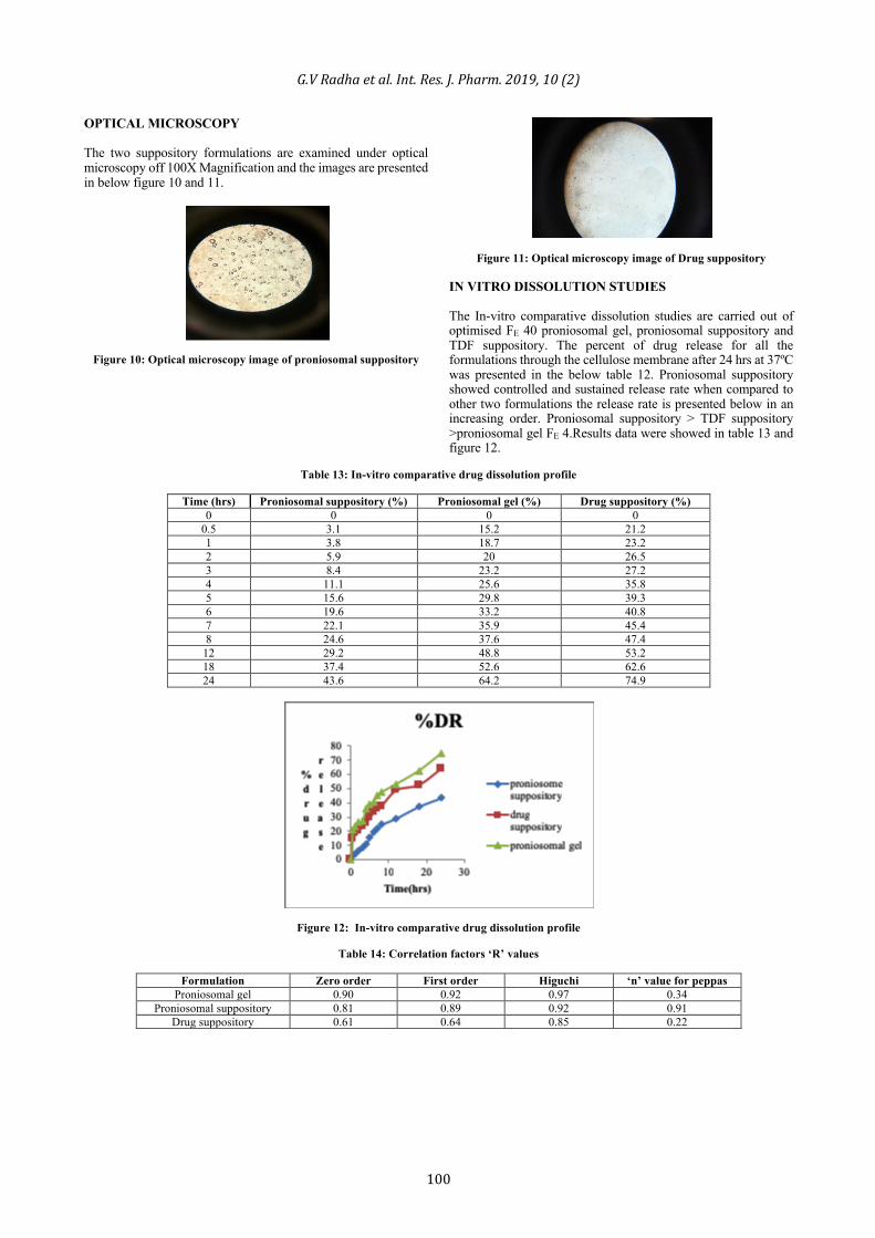
OPTICAL MICROSCOPY The two suppository formulations are examined under optical microscopy off 100X Magnification and the images are presented in below figure 10 and 11.
Figure 10: Optical microscopy image of proniosomal suppository
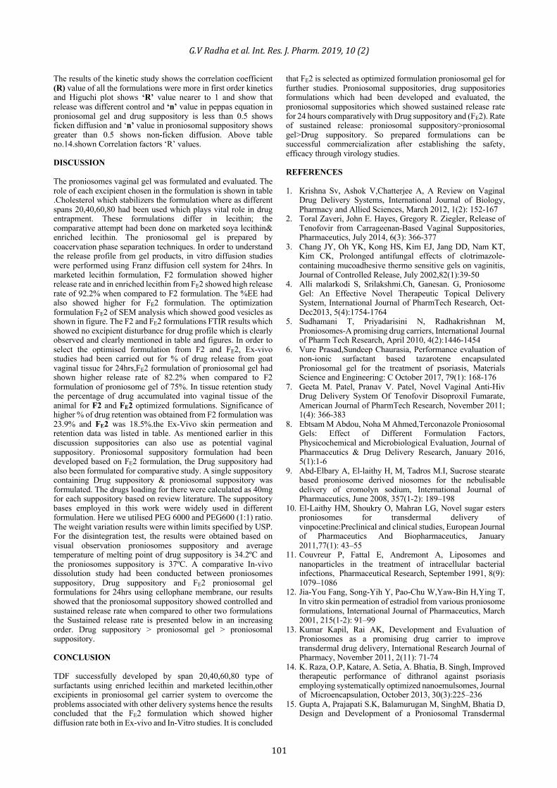
Figure 11: Optical microscopy image of Drug suppository IN VITRO DISSOLUTION STUDIES The In-vitro comparative dissolution studies are carried out of optimised FE 40 proniosomal gel, proniosomal suppository and TDF suppository. The percent of drug release for all the formulations through the cellulose membrane after 24 hrs at 37ºC was presented in the below table 12. Proniosomal suppository showed controlled and sustained release rate when compared to other two formulations the release rate is presented below in an increasing order. Proniosomal suppository > TDF suppository >proniosomal gel FE 4.Results data were showed in table 13 and figure 12.
Table 13: In-vitro comparative drug dissolution profile
Time (hrs) Proniosomal suppository (%) Proniosomal gel (%) Drug suppository (%)
0 0 0 0 0.5 3.1 15.2 21.2 1 3.8 18.7 23.2 2 5.9 20 26.5 3 8.4 23.2 27.2 4 11.1 25.6 35.8 5 15.6 29.8 39.3 6 19.6 33.2 40.8 7 22.1 35.9 45.4 8 24.6 37.6 47.4
12 29.2 48.8 53.2 18 37.4 52.6 62.6 24 43.6 64.2 74.9
Figure 12: In-vitro comparative drug dissolution profile
Table 14: Correlation factors ‘R’ values
Formulation Zero order First order Higuchi ‘n’ value for peppas Proniosomal gel 0.90 0.92 0.97 0.34
Proniosomal suppository 0.81 0.89 0.92 0.91 Drug suppository 0.61 0.64 0.85 0.22
G.VRadhaetal.Int.Res.J.Pharm.2019,10(2)
101
The results of the kinetic study shows the correlation coefficient (R) value of all the formulations were more in first order kinetics and Higuchi plot shows ‘R’ value nearer to 1 and show that release was different control and ‘n’ value in peppas equation in proniosomal gel and drug suppository is less than 0.5 shows ficken diffusion and ‘n’ value in proniosomal suppository shows greater than 0.5 shows non-ficken diffusion. Above table no.14.shown Correlation factors ‘R’ values. DISCUSSION The proniosomes vaginal gel was formulated and evaluated. The role of each excipient chosen in the formulation is shown in table .Cholesterol which stabilizers the formulation where as different spans 20,40,60,80 had been used which plays vital role in drug entrapment. These formulations differ in lecithin; the comparative attempt had been done on marketed soya lecithin& enriched lecithin. The proniosomal gel is prepared by coacervation phase separation techniques. In order to understand the release profile from gel products, in vitro diffusion studies were performed using Franz diffusion cell system for 24hrs. In marketed lecithin formulation, F2 formulation showed higher release rate and in enriched lecithin from FE2 showed high release rate of 92.2% when compared to F2 formulation. The %EE had also showed higher for FE2 formulation. The optimization formulation FE2 of SEM analysis which showed good vesicles as shown in figure. The F2 and FE2 formulations FTIR results which showed no excipient disturbance for drug profile which is clearly observed and clearly mentioned in table and figures. In order to select the optimised formulation from F2 and FE2, Ex-vivo studies had been carried out for % of drug release from goat vaginal tissue for 24hrs,FE2 formulation of proniosomal gel had shown higher release rate of 82.2% when compared to F2 formulation of proniosome gel of 75%. In tissue retention study the percentage of drug accumulated into vaginal tissue of the animal for F2 and FE2 optimized formulations. Significance of higher % of drug retention was obtained from F2 formulation was 23.9% and FE2 was 18.5%.the Ex-Vivo skin permeation and retention data was listed in table. As mentioned earlier in this discussion suppositories can also use as potential vaginal suppository. Proniosomal suppository formulation had been developed based on FE2 formulation, the Drug suppository had also been formulated for comparative study. A single suppository containing Drug suppository & proniosomal suppository was formulated. The drugs loading for there were calculated as 40mg for each suppository based on review literature. The suppository bases employed in this work were widely used in different formulation. Here we utilised PEG 6000 and PEG600 (1:1) ratio. The weight variation results were within limits specified by USP. For the disintegration test, the results were obtained based on visual observation proniosomes suppository and average temperature of melting point of drug suppository is 34.2ºC and the proniosomes suppository is 37ºC. A comparative In-vivo dissolution study had been conducted between proniosomes suppository, Drug suppository and FE2 proniosomal gel formulations for 24hrs using cellophane membrane, our results showed that the proniosomal suppository showed controlled and sustained release rate when compared to other two formulations the Sustained release rate is presented below in an increasing order. Drug suppository > proniosomal gel > proniosomal suppository. CONCLUSION TDF successfully developed by span 20,40,60,80 type of surfactants using enriched lecithin and marketed lecithin,other excipients in proniosomal gel carrier system to overcome the problems associated with other delivery systems hence the results concluded that the FE2 formulation which showed higher diffusion rate both in Ex-vivo and In-Vitro studies. It is concluded
that FE2 is selected as optimized formulation proniosomal gel for further studies. Proniosomal suppositories, drug suppositories formulations which had been developed and evaluated, the proniosomal suppositories which showed sustained release rate for 24 hours comparatively with Drug suppository and (FE2). Rate of sustained release: proniosomal suppository>proniosomal gel>Drug suppository. So prepared formulations can be successful commercialization after establishing the safety, efficacy through virology studies. REFERENCES 1. Krishna Sv, Ashok V,Chatterjee A, A Review on Vaginal
Drug Delivery Systems, International Journal of Biology, Pharmacy and Allied Sciences, March 2012, 1(2): 152-167
2. Toral Zaveri, John E. Hayes, Gregory R. Ziegler, Release of Tenofovir from Carrageenan-Based Vaginal Suppositories, Pharmaceutics, July 2014, 6(3): 366-377
3. Chang JY, Oh YK, Kong HS, Kim EJ, Jang DD, Nam KT, Kim CK, Prolonged antifungal effects of clotrimazole-containing mucoadhesive thermo sensitive gels on vaginitis, Journal of Controlled Release, July 2002,82(1):39-50
4. Alli malarkodi S, Srilakshmi.Ch, Ganesan. G, Proniosome Gel: An Effective Novel Therapeutic Topical Delivery System, International Journal of PharmTech Research, Oct-Dec2013, 5(4):1754-1764
5. Sudhamani T, Priyadarisini N, Radhakrishnan M, Proniosomes-A promising drug carriers, International Journal of Pharm Tech Research, April 2010, 4(2):1446-1454
6. Vure Prasad,Sundeep Chaurasia, Performance evaluation of non-ionic surfactant based tazarotene encapsulated Proniosomal gel for the treatment of psoriasis, Materials Science and Engineering: C October 2017, 79(1): 168-176
7. Geeta M. Patel, Pranav V. Patel, Novel Vaginal Anti-Hiv Drug Delivery System Of Tenofovir Disoproxil Fumarate, American Journal of PharmTech Research, November 2011; 1(4): 366-383
8. Ebtsam M Abdou, Noha M Ahmed,Terconazole Proniosomal Gels: Effect of Different Formulation Factors, Physicochemical and Microbiological Evaluation, Journal of Pharmaceutics & Drug Delivery Research, January 2016, 5(1):1-6
9. Abd-Elbary A, El-laithy H, M, Tadros M.I, Sucrose stearate based proniosome derived niosomes for the nebulisable delivery of cromolyn sodium, International Journal of Pharmaceutics, June 2008, 357(1-2): 189–198
10. El-Laithy HM, Shoukry O, Mahran LG, Novel sugar esters proniosomes for transdermal delivery of vinpocetine:Preclinical and clinical studies, European Journal of Pharmaceutics And Biopharmaceutics, January 2011,77(1): 43–55
11. Couvreur P, Fattal E, Andremont A, Liposomes and nanoparticles in the treatment of intracellular bacterial infections, Pharmaceutical Research, September 1991, 8(9): 1079–1086
12. Jia-You Fang, Song-Yih Y, Pao-Chu W,Yaw-Bin H,Ying T, In vitro skin permeation of estradiol from various proniosome formulations, International Journal of Pharmaceutics, March 2001, 215(1-2): 91–99
13. Kumar Kapil, Rai AK, Development and Evaluation of Proniosomes as a promising drug carrier to improve transdermal drug delivery, International Research Journal of Pharmacy, November 2011, 2(11): 71-74
14. K. Raza, O.P, Katare, A. Setia, A. Bhatia, B. Singh, Improved therapeutic performance of dithranol against psoriasis employing systematically optimized nanoemulsomes, Journal of Microencapsulation, October 2013, 30(3):225–236
15. Gupta A, Prajapati S.K, Balamurugan M, SinghM, Bhatia D, Design and Development of a Proniosomal Transdermal
G.VRadhaetal.Int.Res.J.Pharm.2019,10(2)
102
Drug Delivery System for Captopril. Tropical Journal of Pharmaceutical Research, June 2007, 6(2): 687-693
16. Pushkar Baviskar, Shivkumar Jaiswal, Sayyad Sadique, Amol Landged, Formulation and Evaluation of Lornoxicam Suppositories, The Pharma Innovation, July 2013, 2(7):20-28
17. El-majri M. Sharma RK, Formulation and Evaluation of Piroxicam Suppository, International Journal of Drug Delivery, 2010, 1 (2): 108-112
18. Gowthamarajan K,Venketeshwaran G, Suresh B, Formulation and Evaluation properties of Meloxicam Solid dispersion incorporated Suppositories, Indian Journal of Pharmaceutical Sciences, Nov-Dec 2002, 64(6): 525-528
19. M.Glavas-Dodov K,Goracinova, K. Mladenovska E. Fredro-Kumbaradzi, Release profile of lidocaine HCl from topical liposomal gel formulation, International Journal of Pharmaceutics, February2002, (242): 381-384
20. Saleem MA, Taher M, Sanaullah S, Formulation and evaluation of Tramadol Hydrochloride Rectal Suppositories, International Journal of Pharmaceutical Sciences. Sep-Oct2008, 2(5): 640-644
21. Sah LM, Saini RT, Formulation development and release studies of indomethacin suppositories, Indian journal of pharmaceutical sciences, August 2008, 70(4): 498-501
Cite this article as: G.V Radha et al. Design and evaluation of topical vaginal proniosomal formulations of tenofovir disoproxil fumarate for HIV prevention. Int. Res. J. Pharm. 2019;10(2):94-102 http://dx.doi.org/10.7897/2230-8407.100250
Source of support: Nil, Conflict of interest: None Declared
Disclaimer:IRJPissolelyownedbyMokshaPublishingHouse-Anon-profitpublishinghouse,dedicatedtopublishqualityresearch,whileeveryefforthasbeentakentoverifytheaccuracyofthecontentpublishedinourJournal.IRJPcannotacceptanyresponsibilityorliabilityforthesitecontentandarticlespublished.TheviewsexpressedinarticlesbyourcontributingauthorsarenotnecessarilythoseofIRJPeditororeditorialboardmembers.